Chapter 5
Embryology and Anatomy of the Eyelid
EDWARD H. BEDROSSIAN, JR.
Main Menu   Table Of Contents

Search

EMBRYOLOGY
SURFACE TOPOGRAPHY
BASIC EYELID ANATOMY
SPECIAL CONSIDERATIONS
VASCULAR AND LYMPHATIC SUPPLY TO THE EYELIDS
NERVE SUPPLY TO THE EYELIDS
REFERENCES

In this chapter, eyelid anatomy is discussed in a layered structural fashion. A brief discussion of the embryology and surface topography is reviewed, and the anatomy of the eyelid is then discussed—beginning with the skin anteriorly and proceeding to the conjunctiva posteriorly. Because the dynamics of eyelid function are associated with the globe and with the surrounding orbital bones, the chapter continues with a discussion of the suspensory ligaments and the eyelid margin. The chapter concludes with a discussion of the vascular supply and the nerve supply to the eyelids. Clinical correlation is provided where applicable.
Back to Top
EMBRYOLOGY

EARLY DEVELOPMENT OF THE EMBRYO

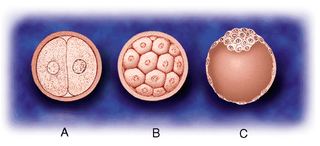
A general understanding of the development of the embryo facilitates an understanding of the development of the eyelids. The fertilized ovum is the original cell, which undergoes several divisions to form a solid clump of cells called a morula. The morula then enlarges and forms a central cavity, to become known as a blastula or blastodermic vesicle (Fig. 1). The outer wall of the blastula forms the placenta. The blastodermic vesicle embeds itself in the uterine mucosa, enlarges, and forms two cavities—each with a single layer of cuboidal epithelium. The upper cavity is the amnion, the lower is the archenteron or yolk sac. The embryo develops from the embryonic plate at the area of contact between the two. At this stage, the embryonic plate consists of two layers: an outer or dorsal ectoderm (epithelial lining of the amnion) and an inner or ventral entoderm (epithelial lining of the yolk sac). A central depression (the primitive streak) followed by a groove develops on the ectodermal surface of the embryonic plate. A middle layer (intraembryonic mesoderm) appears, thus dividing the embryonic plate into three germinal layers from which all tissues of the body develop. The neural groove closes to form the neural canal (24th day). The surface ectoderm within the neural canal is known as the neuroectoderm.1–3

Fig. 1. Early cell division: (A) fertilized ovum, (B) morula (C) blastula.

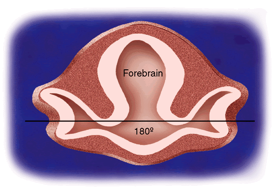
At the anterior end of the embryonic plate, three headfolds form: the forebrain, the midbrain, and the hindbrain. At the front of the forebrain, a small depression is noted on each side of the midline. This depression is the anlage of the eye and is called the optic pit. Toward the end of the third week of gestation, the optic pit deepens until it contacts the surface ectoderm and becomes the primary optic vesicle. At this stage, the eyes are at the side of the head at an angle of 180° (Fig. 2). The embryo consists of a head with the three divisions of the brain, a segmented body with spinal canal, and a tailfold. That portion of the mesoderm closest to the spinal canal is known as paraxial mesoderm. The primary optic vesicle then undergoes invagination to form the secondary optic vesicle or the optic cup. This starts at 2.5 weeks' gestation and is complete by 1 month.3–5

Fig. 2. Fetal optic axis. At 3 to 4 weeks' gestation, the primary optic vesicles are at 180°.

SEGMENTATION

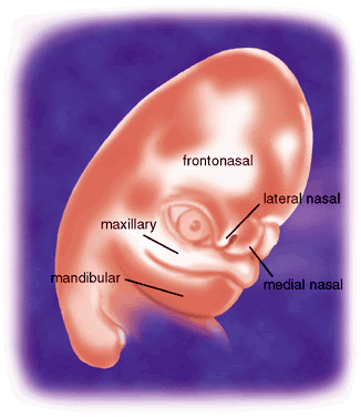
Humans are segmented beings. Segmentation begins simultaneously with closure of the neural groove (24th day). Segmentation extends posteriorly and anteriorly to the head region, where segmentation is lost. Instead, five branchial or pharyngeal arches form. At 5 weeks' gestation, a sheet of immature mesoderm originates from the first branchial arch (“mandibular arch”). The first branchial arch consists of a small dorsal portion known as the maxillary process, which extends forward beneath the eye to develop into the lower eyelid. Cephalad to the first branchial arch, mesenchymal proliferation creates other folds that form other facial processes: the frontonasal, medial nasal, lateral nasal, and mandibular processes (Fig. 3).1–8

Fig. 3. The facial processes.

EYELID FORMATION

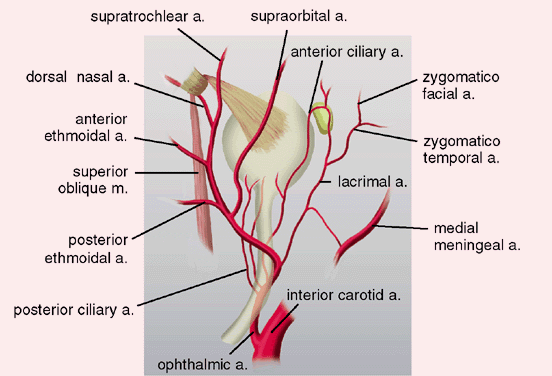
The maxillary process lies in apposition to the paraxial mesoderm of the eye and the nasal process. Between them is a groove of thickened ectoderm, which becomes buried as the maxillary process overgrows the nasal process. This occurs at 5 weeks' gestation and is the first indication of the nasolacrimal duct. The maxillary process extends superiorly to form the lower eyelid at 6 weeks' gestation. In doing so, the inner end folds in a layer of ectodermal tissue that is continuous with the nasolacrimal duct at its lower end. Before the lower lids are apparent, the upper lids are formed by an extension of orbital or paraxial mesoderm known as the frontonasal process. Thus, the upper lid arises from the frontonasal process slightly before the lower lid arises from the maxillary process. The inner end of the upper lid also folds in a portion of ectodermal plate, which forms the upper canaliculus. This unites with the lower canaliculus at the upper end of the nasolacrimal duct.6–8

LID FUSION

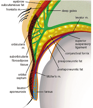
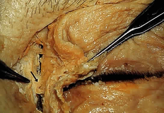
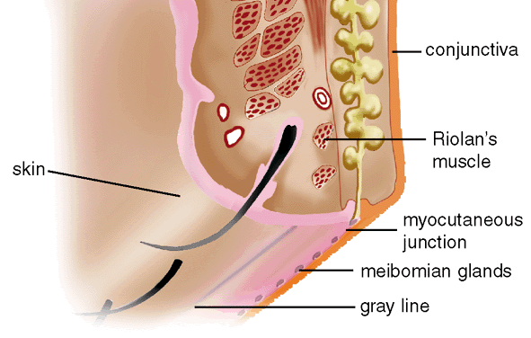
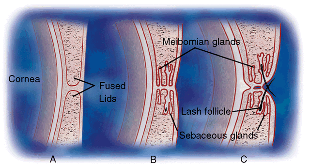
At 1.5 months' gestation, the lateral canthus is formed by the union of the upper and lower eyelid folds. The two lids come together and temporarily fuse from within outward at 8 weeks' gestation (Fig. 4A). Closure is complete at 10 weeks' gestation. Desmosomal adhesions between the lid margins isolate the eye from the amniotic fluid.1 All lid structures are formed during this period of adhesion (Fig. 4B). Riolan's muscle can be identified by the end of the third month. Hair bulbs of eyelashes appear first in the upper lids, then in the lower lids in an anteroposterior direction.2 Development is slightly more advanced in the upper lid, compared with the lower. At the beginning of the fourth month, the meibomian glands begin to appear. The posterior half of the lid shows a greater condensation of basal lamina and collagen fibers, indicating the tarsal plate region. The meibomian glands grow into the tarsal plates—first in the upper then in the lower lids. The apocrine Moll's glands also appear around the cilia follicles during the fourth month, followed shortly by the sebaceous glands of Zeis.3,6

Fig. 4. Eyelid development. (A) Eyelid fusion (8 to 10 weeks' gestation); (B) development of margin structures (3 to 4 months' gestation); eyelid dysjunction (5 to 6 months' gestation).

At 2.5 months' gestation, the levator palpebrae superioris develops. It separates from the superior rectus muscle at the fourth month of gestation. Clinically, failure of separation of these muscles would result in congenital ptosis.

LID DYSJUNCTION

At the end of the fifth month (weeks 21 to 26 gestation), the epithelial adhesions between the lids begin to break down (see Fig. 4C). This process is usually completed by the sixth month but may persist until shortly before birth. Holocrine production of lipids from the meibomian glands, keratinization of the lid margin, and pull of the developing eyelid retractors are responsible for the dysjunction.7,8

ABNORMALITIES OF EYELID DEVELOPMENT

When normal progression of lid development is interrupted, a spectrum of congenital anomalies occurs.8,9

Cryptophthalmia (ablepharon)—absence of the eyelid—is due to failure of the eyelid folds to embryologically develop. The cornea undergoes metaplasia and is covered with skin, which passes continuously from the forehead to the cheek. Most are sporadic but a recessive inheritance pattern has been suggested.10 Cryptophthalmia occurs bilaterally twice as often as unilaterally and is slightly more common in males. It can be associated with ear and nose malformations, cleft lip and palate, hypertelorism, laryngeal atresia, lacrimal duct defects, renal anomalies, syndactyly, and meningoencephalocele.11

Microblepharia is an incomplete lid development wherein the eyeball is not covered, resulting in congenital lagophthalmos.

Coloboma of the eyelid is a defect in the lid margin, with absence of lashes and glands. The most common site is in the upper lid at the junction of the inner third and the outer two thirds. In the lower eyelid, it is more frequently seen at the junction of the outer third and the inner two thirds. These abnormalities may be due to the formation and pressure of amniotic bands or from failure of the eyelids to fuse during embryonic life. This may be associated with cleft palate, mandibulofacial dysotosis, limbal dermoids, lipodermoids, iris colobomas, or brow colobomas.9

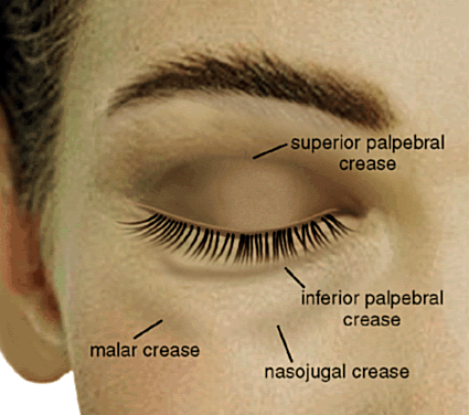
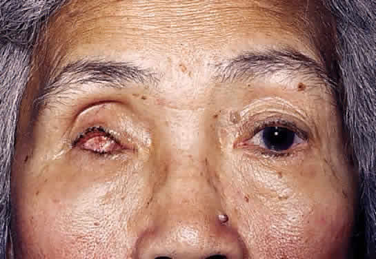
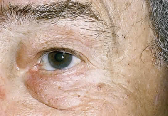
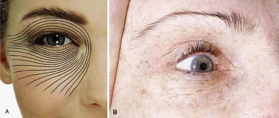
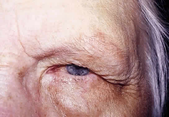
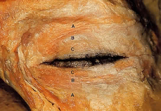
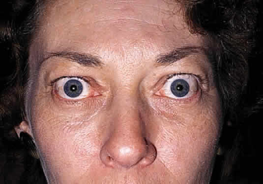
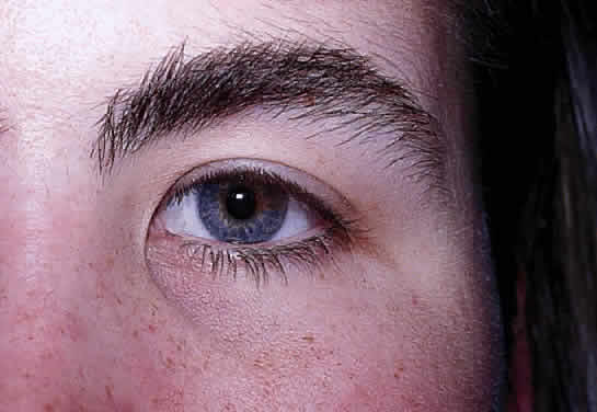
Epicanthus is a fold of skin that extends from the side of the nose to the upper lid and partially hides the inner canthus. This condition is normally present in the embryo and early infancy. Frequently, it is associated with ptosis, although it may occur independently. Epicanthus is usually bilateral but may be asymmetric or unilateral. There are four known types, depending on the origin of the upper end of the fold. In epicanthus superciliaris, the fold arises in the region of the eyebrow and extends over the lacrimal sac. Epicanthus palpebralis (Fig. 5) has an epicanthal fold arising from the upper tarsal area, extending toward the lower margin of the orbit. The fold is equally distributed in the upper and lower lids. In epicanthus tarsalis (Fig. 6), the epicanthal fold arises from the tarsal fold and ends close to the inner canthus. Thus, the fold is more prominent in the upper lid. It is a normal variation of the Asian eyelid. In epicanthus inversus, (Fig. 7) the fold arises in the lower lid and extends upward, partially covering the inner canthus, usually terminating in the upper lid.

Fig. 5. Epicanthus palpebralis (also known as simple epicanthus). The amount of tissue above is the same amount as below the canthal angle.

Fig. 6. Epicanthus tarsalis. The epicanthal fold is more prominant in the upper eyelid.

Fig. 7. Blepharophimosis syndrome. The common triad of ptosis, horizontal shortening of the palpebral fissures, and epicanthus inversus (fold more prominent in lower lid) is seen.

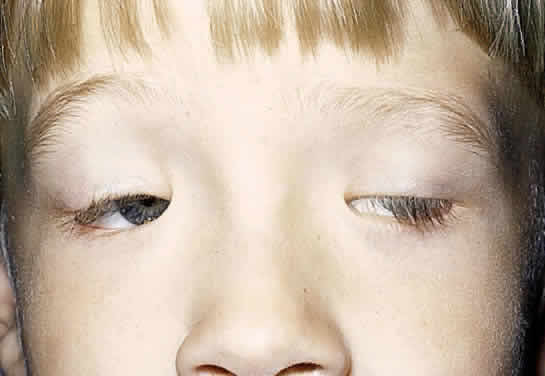
Congenital ptosis is most commonly seen because of the absence or fibrotic nature of the levator palpebrae superioris. It may be unilateral or bilateral and associated with weakness of the superior rectus muscle from which the levator is derived. In the “jaw winking” phenomena, ptosis appears when the patient chews. This is due to an abnormal association of the nerve to the levator muscle and the nerve to the external pterygoid muscle.

Many abnormalities exist of eyelid margin differentiation. Distichiasis is a condition in which aberrant lashes develop at or near the orifices of the meibomian glands. Ankyloblepharon is an abnormal fusion of the lid margins from incomplete separation of the lid folds. The fusion may be complete but is more common at the inner canthus. A variant is “ankyloblepharon filiform adnatum,” in which the lid margins are connected by multiple fine bands. Blepharophimosis is a horizontal narrowing of the palpebral aperture. The triad of blepharophimosis syndrome includes ptosis, horizontal shortening of the palpebral fissures, and epicanthus inversus (Fig. 7). Telecanthus, lower eyelid entropion or ectropion, hypoplasia of the superior orbital rims, a poorly developed nasal bridge, and hypertelorism can also be seen with this condition.

Epiblepharon is an additional fold of skin running horizontally below the lower eyelid margin. It often is associated with loss of the eyelid crease and may be partly due to a lack of deep anchoring of the superficial skin to the orbicularis oculi muscle. The weight of the skin fold may rotate the lower lid margin inward, creating an entropion. With growth of the face and nasal bridge, epiblepharon usually diminishes. It is seen more commonly in Asian eyelids.

Euryblepharon is a rounding or almond-shaped deformity of the lower lid at the lateral canthal angle. It may be associated with an inferiorly displaced lateral canthal tendon.

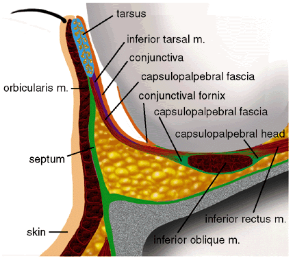
Congenital entropion is characterized by an inward turning of the eyelid margin due to hypertrophy of the orbicularis muscle, defects in lower lid retractor, or tarsal abnormalities.12

Congenital ectropion is characterized by eversion of the eyelid margin. It is rarely seen in the upper lid. In the lower lid, it is usually seen with Down's syndrome or blepharophimosis syndrome.

Back to Top
SURFACE TOPOGRAPHY
Except for prepuce and that of the labia minor, the skin of the eyelids is the thinnest in the body. It is pliable and contains relatively little adipose tissue. It is subject to unusual amounts of stress and relaxation with each blink. Such movement predisposes the paraorbital skin to several natural and dynamic topographic landmarks (Fig. 8).13–15 The upper lid displays a prominent horizontal crease, the superior palpebral crease. It marks the upper border of the tarsus, is located about 10 mm above the lid margin in white women and 7 to 8 mm above the lid margin in men. It is accented when the lid is raised. It denotes the dividing point between loosely adherent preseptal skin and that from more adherent pretarsal skin. In the Asian eyelid, the superior palpebral crease is absent or is displaced inferiorally.

Fig. 8. Surface topography.

The lower lid displays three creases. The inferior palpebral crease is less noticeable than the superior palpebral crease and marks the lower border of the lower tarsus. It courses from about 5 mm below the lower lid margin medially to about 7 mm laterally. The nasojugal crease is located below the medial aspect of the inferior palpebral crease and extends infralaterally at 45°. The malar crease originates lateral to and below the lateral canthus. It courses inferomedially until it meets the nasojugal fold 15 mm below the center of the lower eyelid margin.

The palpebral fissure is the opening between the eyelid margins. The vertical palpebral fissure is normally 9 to 10 mm, whereas the horizontal fissure measures 28 to 30 mm. The upper eyelid rests at the upper limbus in the child and about 1 to 2 mm below the upper limbus in the adult. The upper and lower lids rest over the anterior surface of the globe, allowing about 20% of its surface to be exposed in the normal palpebral fissure. The lower eyelid is generally found at the level of the lower limbus. The upper eyelid is slightly curved, with the highest point nasal to the pupil. The lowest point of the lower eyelid is slightly temporal to the pupil. The point where the upper eyelid and the lower eyelid meet medially is known as the medial commissure. The point where the upper and lower eyelid meet laterally is known as the lateral commissure. The medial canthus and the lateral canthus are the angles formed at the medial commissure and at the lateral commissure, respectively. The lateral canthal angle is normally more acute than the slightly rounded medial canthal angle. The lateral commissure rests on the globe, whereas the medial commissure is anteriorly displaced by the caruncle and plica semilunaris.

The superior orbital sulcus is that area between the upper eyelid crease and the superior orbital margin. With age or after enucleation, a concavity (Fig. 9) of the superior sulcus can be seen because of lack of supporting tissue. Convexity (Fig. 10) of the superior sulcus is seen with herniation of orbital fat.

Fig. 9. Concavity of superior orbital sulcus after enucleation of the right eye.

Fig. 10. Convexity of superior sulcus from herniated orbital fat.

Langer lines (Fig. 11) are present in the upper and lower lids and in the canthal areas. These natural static skin lines are formed by collagenous, reticular, and elastic fibers in the reticular layer of the dermis. Gravitational lines (Fig. 12) are formed with age by progressive thinning of relaxed skin. In contrast, dynamic lines (Fig. 13) result from repetitious relaxation and contraction of paraorbital muscles. Horizontal forehead furrows show the lateral extent of the frontalis muscle used to correct chronic physiologic ptosis. The action of the procerus and corrugator superciliaris muscles create the “frown lines” between the eyebrows.

Fig. 11. Langer lines (A) as they appear in a diagram and (B) clinically. These static skin lines are formed by collagenous, reticular, and elastic fibers in the reticular dermis.

Fig. 12. Gravitational lines formed by progressive thinning of relaxed skin.

Fig. 13. Dynamic lines. Overaction of the procerus and corrugator superciliaris muscle create these frowns line in this patient with ocular phemigoid.

The skin of the eyelids is thin. The transition to the thicker skin of the eyebrow above and to the skin of the malar region below is abrupt. In the preorbital and preseptal skin, subcutaneous fat is sparse. Fat is absent in the pretarsal skin. Pretarsal skin is firmly adherent to the underlying tarsus because of attachments of the levator aponeurosis.16 Clinically, edema collects under the loose preorbital and preseptal skin, leaving an identifiable border at the pretarsal skin where there are denser subcutaneous fibroadipose attachments.

Back to Top
BASIC EYELID ANATOMY

PROTRACTORS

The orbicularis oculi muscle is a thin sheet of concentrically arranged muscle fibers covering the eyelids and periorbital region (Fig. 14). It is oval, with the long axis being horizontal, corresponding to the palpebral opening. Contraction of the orbicularis oculi muscle results in the protraction or closure of the eyelid. The orbicularis is firmly attached to the underlying lateral palpebral raphe, the medial canthal region, the insertions of the upper and lower eyelid retractors, the supraorbital ridge, the naso-orbital valley and the malar crease. Jones14,17 divided the orbicularis oculi muscle into three regions: orbital, preseptal, and pretarsal.

Fig. 14. Orbicularis oculi muscle, cadaver dissection. (A) Orbital portion; (B) preseptal portion; (C) pretarsal portion.

The broad orbital orbicularis portion extends superiorly to the eyebrow, where it interdigitates with the frontalis and the corrugator superciliaris muscles. Medially, it extends from the supraorbital notch in a curvilinear fashion over the side of the nose, inferiorly to the infraorbital foramen. It continues along the infraorbital margin. Laterally, it extends to the temporalis muscle. These thick course fibers play an important role in voluntary lid closure (winking) and forced eyelid closure.

The preseptal orbicularis overlies the orbital septum. In between is a fibroadipose layer, which is continuous superiorly with the eyebrow fat pad. If this postorbicularis layer contains a significant amount of fat, the ptosis surgeon may misinterpret it as being preaponeurotic fat. Laterally, the preseptal orbicularis muscle inserts directly onto Whitnall's lateral orbital tubercle 3 to 4 mm deep to the lateral palpebral raphe. Because of the fibrous component of this lateral attachment, Jones misleadingly termed this the lateral canthal tendon rather than the lateral canthal ligament.14,17 Medially, the insertions of the preseptal orbicularis are complicated by the lacrimal sac, its fascia, and the lacrimal crests. The medial origin of the preseptal orbicularis is thought to arise from two heads—the deep and the superficial heads. The deep head or Jones muscle17,18 is adherent to the lacrimal sac and lacrimal fascia, whereas the anterior or superficial head arises from the anterior rim of the medial canthal ligament (Fig. 15). Functionally, the preseptal fibers contribute to voluntary lid closure (winking) and involuntary lid closure (blinking).

Fig. 15. Medial attachments of the orbicularis oculi muscle.

The pretarsal orbicularis is firmly adherent to the underlying tarsus and to the superficial insertion of the levator aponeurosis at the superior tarsal border. The medial origin of the pretarsal orbicularis is thought to arise from two heads—the deep and the superficial heads. The deep head, Horner's tensor tarsi muscle,19,20 arises from 4 mm behind the posterior lacrimal crest and from the lacrimal fascia to insert medially on the tarsi of the upper and lower eyelids. Its contraction pulls the eyelid medially and posteriorly, allowing the eyelids to follow and cover the convex globe. In addition, its lateral contraction on the lacrimal diaphragm creates a negative pressure in the lacrimal sac that draws tears from the canaliculi. The superficial head of the pretarsal orbicularis inserts on the anterior lacrimal crest and anterior limb of the medial canthal ligament. Superficial fibers also surround the canaliculi. Contraction shortens the canaliculi, forcing lacrimal fluid into the lacrimal sac. Functionally, involuntary lid closure (blinking) is primarily the responsibility of the smaller pretarsal orbicularis fibers.

Medially, both Horner's and Jones muscles are essential for proper functioning of the lacrimal pump. The deep fibers medially and posteriorly are responsible for proper eyelid-to-globe apposition. Laterally, the orbital and preseptal fibers fuse over the zygoma to form the lateral palpebral raphe. The deep fibers of the pretarsal orbicularis join the inferior and superior crux of the lateral canthal ligament, which inserts 3 to 4 mm posterior to the lateral orbital rim on Whitnall's tubercle.21 It is these latter posteriorly oriented attachments that are responsible for proper eyelid-to-globe apposition and not the more superficial fibers.

ORBITAL SEPTUM

Upper Eyelid

Historically, the orbital septum has been considered as a discreet, well-defined structure arising from the arcus marginalis. The arcus marginalis is a discrete line seen at the supraorbital rim, representing the condensation of the periosteum of the forehead with the periorbita of the orbit. Alternatively, the orbital septum may be considered the deep fascia of the overlying orbicularis oculi muscle.14 Others describe a loose fibrous connective tissue anterior to the orbital septum and refer to it as the ‘suborbicularis fascia.’22 The orbital septum (Fig. 16) actually is a multilayered structure.23 Fibrous septa within the submuscular fibroadipose tissue become contiguous with more compact lamellae of the orbital septum, imparting a multilayered quality to the orbital septum. Fat within the fibroadipose layer anterior to the orbital septum may be mistaken for the preaponeurotic fat pad during eyelid surgery.24

Fig. 16. With the skin and orbicularis oculi muscle incised and retracted, the multilayered orbital septum is being cut in this cadaver.

The orbital septum and the levator aponeurosis (Fig. 17) join 2 to 5 mm above the superior tarsal border. The orbital septum directly adjoins the posterior epimysium of the orbicularis for about 1 to 5 mm before joining the levator aponeurosis. The cross-sectional thickness of the orbital septum through its parallel lamella varies but is less than 1 mm. It is thickest at the arcus marginalis laterally and is thinnest in the lower lid medially. Medially, posterior orbital septal lamella are carried posteriorly by the pretarsal orbicularis muscle, which inserts on the posterior lacrimal crest. Superficial orbital septal fibers, as described by Whitnall,14 appear to cross and adhere to the lacrimal fascia before reaching the anterior lacrimal crest. At the lateral canthus, the orbital septum is also split; deep fibers insert at Whitnall's tubercle, whereas superficial fibers join at the lateral canthal raphe, just deep to the orbicularis muscle.23

Fig. 17. Cross-section of the upper eyelid.

Variation in septal strength is seen between individuals and varies with age. Clinically, weakness of the septum explains the medial upper lid bulge (bourrelet senile) seen in older patients and due to herniation of the medial fat pad through an attenuated septum (see Fig. 10).25 It also may explain the high incidence of orbital invasion of basal cell carcinoma in the medial canthal region.26

Lower Eyelid

In the lower eyelid, the orbital septum (Fig. 18) arises from the inferior orbital rim as a condensation of the periosteum and the periorbita. It continues anteriorly and superiorly to a point 4 to 5 mm below the inferior tarsus, where it joins with the lower eyelid retractors and as a single structure inserts on the lower border of the inferior tarsus.23,27 Medially, the orbital septum splits and is carried posteriorly by the pretarsal orbicularis muscle (Horner's muscle) and attaches to the posterior lacrimal crest. Laterally, the orbital septum also splits and is carried deep by the insertion of the orbicularis.

Fig. 18. Cross-section of the lower eyelid.

The orbital septa of the upper and lower eyelids form an anatomic barrier between the preseptal and orbital structures. Infectious processes anterior to the septa are considered to be more benign than those posterior to the septa. Functionally, the suborbicularis oculi fibroadipose layer and the multilayered orbital septum change with movement, enhancing eyelid and eyebrow mobility.14,23

FAT PADS

Fat within the orbit and adnexa serve as a protective cushion within which the eyeball moves. Fat within the muscle cone is termed central or conal. Fat outside the muscle cone is termed peripheral or extraconal.

Upper Eyelid

In the upper eyelid, there are two specific fat pads: the medial fat pad and the central fat pad (Figs. 19 and 20). The whitish medial fat pad is more fibrous, whereas the larger central fat pad is more yellow because of a decreased amount of fibrous tissue. The larger central fat pad is termed the preaponeurotic fat pad. The trochlea separates the two fat pads.28 Clinically, the preaponeurotic fat pad lies directly on the surface of the muscular portion of the levator and serves as an important surgical landmark to the levator aponeurosis immediately beneath it. The medial fat pad often herniates through a weakened orbital septum, creating a bulge beneath the trochlea. The preaponeurotic fat is surrounded by a thin translucent connective tissue capsule. Fine connective tissue septa extend anteriorly from the capsule of the preaponeurotic fat to the orbital septum and posteriorly to the levator aponeurosis.23 Clinically, these connections are easily broken by blunt dissection, exposing an encapsulated preanoneurotic fat pad. Connective tissue septa also exists within the preaponeurotic fat, dividing it into lobules. Although the preaponeurotic fat pad is contiguous with deeper orbital fat, these connections are more rudimentary than the medial and lower eyelid fat pads. The preaponeurotic fat pad is less vascular than the other fat pads. Conversely, the medial fat pad is more vascular because of the location of the palpebral arterial arcade, which serpiginously courses through this pad.29,30 The medial fat pad is separated from the preaponeurotic fat pad by delicate fibrous septal attachments to the trochlea.

Fig. 19. Fat pads of the upper and lower eyelids.

Fig. 20. Cadaver dissection, eyelid fat pads.

Lateral to the preaponeurotic fat pad lies the lacrimal gland, which is typically pinker in appearance, firmer in texture, and distinctly more vascular than the preaponeurotic fat pad.31 During removal of orbital fat, the eyelid surgeon is careful to avoid injury to the laterally located lacrimal gland.

Lower Eyelid

In the lower eyelid, there exists a smaller temporal fat pad and a larger medial fat pad (see Figs. 19 and 20). The temporal fat pad lies inferior to the lateral canthus. It is separated from the larger medial fat pad by a fibrous extension from the periorbita and orbital septum infralaterally, joining with the capsulopalpebral fascia and Lockwood's ligament.28,32

The medial pad extends from the fascial band to the medial canthal area. It is a single pad anteriorly but posteriorly is divided by the origin of the inferior oblique muscle (Fig. 21). With this subdivision of the larger medial fat pad of the lower lid, some surgeons consider the lower lid to have three fat pads.15,32

Fig. 21. Fat pads of the lower lid. This cadaver dissection demonstrates the posterior division of the large medial fat pad by the inferior oblique muscle (pointer).

The lower eyelid fat pads are in direct communication with the deeper extraconal fat of the orbit. Clinically, this is important during lower eyelid surgery because excessive traction may be transmitted deeper into the orbit, resulting in intraoperative or postoperative orbital hemorrhage.29,33 During transconjunctival lower eyelid blepharoplasty, the lateral eyelid fat pad tends to be more fibrotic and prolapses less easily. Care is also taken to avoid the inferior oblique muscle, which originates just lateral to the ostium of the nasolacrimal canal.

RETRACTORS

Each eyelid contains two retractors, which open the palpebral fissures. In the upper lid, they are the levator palpebrae superioris muscle and the sympathetically innervated muscle of Müller. In the lower lid, they are the capsulopalpebral fascia and the sympathetically innervated inferior tarsal muscle.

Upper Eyelid

In the upper lid, the major retractor is the levator palpebrae superioris. It arises from the same superior mesenchyme as the superior rectus muscle. At the orbital apex, the levator originates from the lesser wing of the sphenoid bone, superolateral to the optic foramen. Immediately below originates the superior rectus muscle arising from the annulus of Zinn.14,34 As the triangular levator muscle courses anteriorly in the orbit from its origin, it is composed of striated muscle. The average length of the muscular portion of the levator is 36 mm.28,34

At the level of the globe, the levator muscle fans out and thins as the whitish gray superior transverse ligament of Whitnall or Whitnall's ligament. It represents a transition zone where the horizontal levator muscle becomes more fibrous, forming the more vertical levator aponeurosis. The aponeurosis measures 18 mm in width. Whitnall's ligament is located 14 to 20 mm above the superior border of the tarsus. The medial attachment of Whitnall's ligament is to the fascia of the trochlea, whereas the lateral attachment of Whitnall's ligament is at the frontozygomatic suture (Fig. 22).

Fig. 22. Whitnall's ligament. Its medial attachment is to the fascia of the trochlea. The major lateral attachment is to the frontozygomatic suture, with minor attachments to the lateral orbital tubercle.

Anteriorly, the aponeurosis expands horizontally to insert onto the medial and lateral retinacula as the “horns” of the levator. The medial horn of the levator attaches to the medial canthal ligament. Its attachment is looser and more ill-defined than the lateral attachment.24,35 The lateral horn of the levators splits the lacrimal gland into the larger orbital lobe and the smaller palpebral lobe (Fig. 23). It then attaches to the lateral orbital tubercle by the lateral canthal tendon and may provide suspensory support for the gland.24,25 The levator palpebral aponeurosis continues anteriorly to a point 2 to 5 mm above the superior tarsal border, where it joins with fibers of the multilayered orbital septum (see Fig. 17). The thin fibrous connections between these two structures is somewhat rounded and convex. Below this point, at the level of the superior tarsal border, the fused lamellae of the orbital septum and the levator aponeurosis send connective tissue attachments to secondarily insert onto the overlying orbicularis oculi muscle and subcutaneous tissue.23 These attachments result in a sharp upper eyelid crease. Variations in these attachments result in variations in the location of the upper eyelid crease. The levator aponeurosis then sends connective tissue attachments, which insert primarily on the anterior inferior third of the superior tarsus, with the strongest attachments 3 mm from the lid margin.25,34 It is these tarsal attachments that are more important for proper upper eyelid function.24

Fig. 23. The lateral horn of levator aponeurosis. With the lateral wall and roof of the orbit removed, the levator aponeurosis (wide arrow) is seen dividing the lacrimal gland into the larger orbital lobe (thin arrow) and smaller palpebral lobe (pointer).

The levator muscle and the superior rectus muscle are joined along their medial borders by fibrous attachments that become stronger anteriorly. A dense intermuscular fascia connects the undersurface of the levator aponeurosis with the superior surface of the superior rectus muscle just posterior to the level of Whitnall's ligament. From the anterior surface of this intermuscular membrane, the superior conjunctival fornix suspensory ligament (see Fig. 17) arises.36,37 Anterior to this point, Müller's muscle takes its origin from the underside of the levator muscle 22 mm above the superior tarsal border.28 The motor innervation of the levator muscle is the superior division of the oculomotor nerve (cranial nerve III). After entering the orbit through the supraorbital fissure and through the annulus of Zinn, the superior division of the oculomotor nerve then passes around the medial border of the superior rectus muscle or directly through the superior rectus muscle to enter the undersurface of the levator muscle at its posterior third and anterior two thirds junction.

The other retractor of the upper eyelid is Müller's muscle, also known as the superior tarsal muscle (see Fig. 17; Fig. 24). Müller's muscle is a smooth muscle arising from the underside of the levator muscle, just below the level of Whitnall's ligament, 22 mm above the superior tarsal border. It inserts on the superior tarsal border. Superiorly, Müller's muscle is loosely adherent to the conjunctiva but becomes more adherent near the tarsus. Sympathetic nerve fibers innervating the muscle are thought to enter by the peripheral arterial arcade and other small arteries.24,28 Clinically, increased sympathetic stimulation (as seen in Graves' disease) is thought to be a factor in thyroid eyelid retraction (Fig. 25).

Fig. 24. Müller's muscle. A 10-mm strip of Müller's muscle is preserved in this cadaver to demonstrate its origin from the underside of the reflected levator muscle (thick arrow) and its insertion onto the superior edge of the tarsus. The tarsus is seen with the vertically oriented meibomian glands (thin arrow).

Fig. 25. Thyroid lid retraction. Sympathetic stimulation of Müller's muscle and the inferior tarsal muscle is a factor in this patient with Graves' disease.

Lower Eyelid

Posterior to the globe, a fibrous extension arises from the inferior rectus muscle. These extensions are collectively termed the capsulopalpebral head of the inferior rectus muscle. The capsulopalpebral head splits to surround the inferior oblique muscle. The external portion is termed the capsulopalpebral fascia, whereas the inner counterpart that contains smooth muscle is termed the inferior tarsal muscle. The two layers fuse anterior to the inferior oblique muscle to form a dense fibrous structure termed Lockwood's suspensory ligament of the globe.27,36 This ligament suspends the globe position within the orbit, even if all bone inferior to its attachments at the medial and lateral orbital walls are removed. The outer fibers of the capsulopalpebral fascia fuse with the inner fibers of the inferior orbital septum 4 to 5 mm below the inferior tarsus and together advance as a single layer to insert on the inferior border of the inferior tarsus (see Fig. 18).27,38 Although more rudimentary in their development, the capsulopalpebral fascia and the inferior tarsal muscle are analogous to the levator aponeurosis and the superior tarsal muscle of the upper eyelid.

The capsulopalpebral fascia has no inherent innervation but its action mirrors the the action of the inferior rectus muscle, which is innervated by the inferior division of the oculomotor nerve. Depression of the lower eyelid in downgaze is less than the movement of the globe in downgaze. Magnetic resonance imaging studies39 have shown that this discrepancy is due to stretching of the capsulopalpebral head posterior to the inferior oblique muscle, whereas the length of the anterior portion of the capsulopalpebral fascia (between Lockwood's ligament and the tarsus) remain constant.

In the lower eyelid, the more rudimentary inferior tarsal muscle arises from the inner surface of the capsulopalpebral head as it splits to surround the inferior oblique muscle. The inferior tarsal muscle consists of numerous discontinuous smooth muscle bundles and becomes totally fibrous as the inferior tarsus is approached.27 The inferior tarsal muscle is adherent to both the overlying capsulopalpebral fascia and to the underlying conjunctiva. The adherence of the capsulopalpebral fascia to the inferior tarsal muscle is stronger than the adherence of the inferior tarsal muscle to the conjunctiva. Thus, the capsulopalpebral fascia and the inferior tarsal muscle are often grouped together and termed “the lower lid retractors,” which during lower eyelid surgery are treated as one anatomic unit.

The inferior tarsal muscle is sympathetically innervated. Clinically, in Horners' syndrome, the atonic muscle may allow the lower eyelid to elevate as much as 1 mm. Conversely, in thyroid eye disease, the lower eyelid may retract from increased sympathetic tone.

TARSAL PLATES

The tarsal plates are thickened fibrous connective tissue that provide structural support to the eyelids. Medially and laterally, the tarsal plates are connected to the bony orbital margins by ligamentous fibrous tissue (Fig. 26).40

Fig. 26. The upper and lower tarsal plates.

Upper Eyelid

The superior tarsal plate is 10 mm in vertical height and 25 to 30 mm in horizontal dimension. The border nearest the eyelid margin is flat, whereas the antimarginal border is curved with its largest vertical dimension centrally. From their central portion, each tarsal plate tapers toward the canthal ligaments. Within each superior tarsus lies 30 to 40 vertically oriented sebaceous secreting glands, the meibomian glands, which produce the outer lipid portion of the tear film. The orifices of the meibomian glands are located at the eyelid margin just anterior to the mucocutaneous junction.

Lower Eyelid

The inferior tarsal plate is 3 to 5 mm in vertical height and measures 25 to 30 mm horizontally.41 The lower tarsus also tapers medially and laterally. Within each lower tarsus, there are 20 to 30 vertically oriented meibomian glands whose orifices are located anterior to the mucocutaneous junction of the lower eyelid.

The tarsi span the anterior orbital opening while articulating with the globe. With eyes closed, the lower eyelid covers a smaller area of the globe surface than does its upper eyelid counterpart. The posterior surface of both the tarsal plates is covered by conjunctiva.

CONJUNCTIVA

Embryologically, the conjunctiva represents the differentiated inner portion of the skin fold that forms the eyelid. It is composed of nonkeratinized stratified squamous epithelium with goblet cells. The goblet cells produce mucous for the inner layer of the tear film. The conjunctiva has two components: the bulbar conjunctiva, which lines the globe extending to the corneal limbus, and the palpebral conjunctiva, which lines the eyelid. These two conjunctival components meet at the conjunctival fornices. The superior conjunctival fornix is stabilized by the suspensory ligament of the superior fornix, which arises from the conjoining of the levator and the superior rectus muscles.37 The inferior conjunctival fornix is stabilized by the inferior suspensory ligament, which arises from the inferior rectus extensions that sheath the inferior oblique muscle. The superior fornix is located 13 mm from the open eyelid margin and 25 mm from the closed eyelid margin. The inferior fornix, however, is located 9 to 10 mm from the open or closed lower eyelid margin because the lower eyelid does not elevate significantly with eyelid closure.

The bulbar conjunctiva is loosely attached to the globe, except at the limbus where it is tightly bound at the episclera. Similarly, the palpebral conjunctiva is loosely adherent, except at the tarsus and at the superior tarsal muscle where it is tightly adherent. In the lower eyelid, the conjunctiva is adherent to the lower tarsus but can be elevated from the lower eyelid retractors without difficulty. The conjunctival fornices are also stabilized by attachments medially and laterally to the canthal tendon.14,42

In addition to goblet cells, the conjunctiva also contains the accessory lacrimal glands.43 The glands of Wolfring are located along the antimarginal border of the tarsal plate, whereas Krause's glands are found in the conjunctival fornices. These glands are known as the basic tear secretors because they provide the middle aqueous layer of the tear film at a constant basal rate. The main lacrimal gland, however, is a reflex secretor, providing additional aqueous fluid for the middle layer of the tear film on a reflex basis (ocular irritation or emotion). The orifices of the lacrimal gland ducts are located 4 to 5 mm superior and lateral to the upper edge of the upper tarsus.

Medially, the conjunctiva forms the semilunar fold, a vestige of the nictitating membrane of some animals. The caruncle is a small collection of transitional tissue at the medial commissure that has multiple sebaceous glands.

Back to Top
SPECIAL CONSIDERATIONS

SUSPENSORY SYSTEM OF THE EYELIDS

Five anatomic structures provide support and mechanical function to the eyelid and eyeball: Whitnall's ligament, Lockwood's ligament, the lateral canthal ligament, the medial canthal ligament, and the eyelid margin.

Whitnall's Ligament

The superior transverse ligament of Whitnall provides the dual function of acting as the main suspensory ligament of the upper eyelid and as a check ligament for the levator aponeurosis and levator muscle.24,44,45 It also gives superior conjunctival fornix suspension.34,37 The suspensory role and fulcrum effect of Whitnall's ligament is assisted by the curvature of the globe, which also provides a fulcrum and support for the levator.34 Whitnall's ligament is suspended from the periorbita of the orbital roof, extending medially from the trochlea across the horizontal dimension of the orbit to the frontozygomatic suture, 10 mm superior to the lateral orbital tubercle (Whitnall's orbital tubercle). Laterally, it sends weaker attachments to Whitnall's tubercle but the most significant lateral attachment site of Whitnall's ligament is at the frontozygomatic suture (see Fig. 22). Whitnall's ligament is found 15 to 20 mm superior to the superior border of the tarsus as a white, shiny, glistening structure where the levator muscle becomes an aponeurosis.

Clinically, this is an important structure encountered frequently during eyelid surgery.

Lockwood's Ligament

Lockwood's ligament in the lower eyelid is more rudimentary than Whitnall's ligament in the upper eyelid but it acts as a suspensory hammock for the globe.36 It also serves as an anchor for the inferior conjunctival fornix. Lockwood's ligament is a condensation of the capsulopalpebral fascia anterior to the inferior oblique muscle.27 It is composed of thickened Tenon's capsule, intramuscular septa, check ligaments, fibers from the inferior rectus sheath, and lower lid retractors. Medially, it attaches to the medial canthal ligament and laterally to the lateral canthal ligament.

Lateral Canthal Ligament

Often called the lateral canthal tendon, the lateral canthal ligament is comprised of a superior crux from the superior tarsus and an inferior crux from the inferior tarsus. The superior and inferior crux of the lateral canthal tendons fuse at the lateral border of the tarsal plates to join the lateral retinaculum, a condensation of several anatomic structures that inserts onto the lateral orbital tubercle of Whitnall. This tubercle is located 2 to 4 mm posterior to the lateral orbital rim, 10 to 12 mm inferior to the frontozygomatic suture, and at the level of the lateral commissure.40 The lateral retinaculum consists of fibers from the lateral horn of the levator aponeurosis, Lockwood's ligament, check ligaments of the lateral rectus muscle, fibers of the suspensory ligaments of the lacrimal gland, and some deep fibers of the pretarsal orbicularis oculi muscle. In the space between the lateral retinaculum and the more anteriorally placed orbital septum is sometimes found a small fat pad, Eisler's pocket.22

The lateral canthal ligament is a clinically important anatomic structure. The inferior and superior crux of the lateral canthal ligament must be released from the tarsal plates during a lateral canthotomy or cantholysis to decompress the orbit and to lower intraorbital pressure.

Medial Canthal Ligament

The medial canthal ligament, often called the medial canthal tendon, provides support to the eyelids and aids in the function of the lacrimal pump.18 The medial canthal ligament has two components—an anterior limb and a posterior limb. The anterior limb (Fig. 27) is a broad fibrous structure that attaches the eyelids to the frontal process of the maxillary bone and to the anterior lacrimal crest. It gives origin to the superficial head of the pretarsal orbicularis oculi muscle. The posterior limb of the medial canthal ligament inserts on the posterior lacrimal crest and the lacrimal fascia. It is weaker than the anterior limb but with the deep heads of the pretarsal and preseptal orbicularis muscle draws the medial portion of the eyelids posteriorly to follow the vector forces necessary for good medial apposition of the eyelids to the globe.31

Fig. 27. Anterior limb of medial canthal ligament. In this cadaver dissection, the anterior limb of the medial canthal ligament (arrow) is seen originating from the frontal process of the maxillary bone (pointer). Forceps reflect the medial aspect of the eyelid.

Eyelid Margin

The eyelid margins are divided by the lacrimal puncta into a medial lacrimal portion and a lateral palpebral portion. At the lid margin near the medial edge of the tarsus, the lacrimal papilla is seen as the fibrous ring surrounding the lacrimal punctum. The medial lacrimal portion is rounded and without lashes. It carries the horizontal portion of the canaliculi 1 to 2 mm from the marginal surface. The upper punctum is located at the junction of the medial 1/6 and the lateral 5/6 of each eyelid. The lower punctum is located slightly more laterally than the upper punctum. The longer lower canaliculus (10 mm) is directed into the lacrimal lake lateral to the plica semilunaris, whereas the shorter superior canaliculus (8 mm) is directed between the plica semilunaris and the caruncle. The canaliculi are surrounded by thick pretarsal orbicularis oculi muscle fibers. Posterior and horizontal contraction slightly inverts the medial eyelid margin.

The lateral portion of the lid margin has a more distinct or sharp border, which acts like a “squeegee” or “windshield wiper” to assist in moving the tear film toward the punctum.

At the level of the tarsus, both the upper and lower eyelids consist of four anatomic structures: the skin, the orbicularis oculi muscle, the tarsus, and the conjunctiva. At the eyelid margin, the eyelids are arbitrarily divided into an anterior lamella, consisting of the skin and orbicularis oculi muscle, and a posterior lamella, consisting of the tarsus and conjunctiva. This distinction is only approximate because fibrous elements from the tarsus extend anteriorly, hair follicles may extend posteriorly to imbed in the tarsus, and fine interdigitations of the eyelid retractors may be found. The skin of the anterior lamella contains 100 to 120 cilia in the upper eyelid and 50 to 75 in the lower eyelid. Each ciliary follicle also contains about two sebaceous glands (glands of Zeis). Near the cilia lie the sweat glands of the eyelid (Moll's glands), which empty into the adjacent follicles. A strip of pretarsal orbicularis oculi muscle, isolated from the remainder of the orbicularis muscle by the eyelash follicles, is known as the ciliary bundle of Riolan or Riolan's muscle. This strip of muscle deep within the eyelid skin creates an optical (Tyndall) effect known as the gray line (Fig. 28).46 The mucocutaneous junction at the eyelid margin is found posterior to the gray line. In the posterior lamella, the orifices of the meibomian glands are seen exiting the tarsus. Division of the eyelid margin into an anterior and posterior lamella has practical implications when considering eyelid malpositions and eyelid reconstruction.

Fig. 28. Lid margin.

Back to Top
VASCULAR AND LYMPHATIC SUPPLY TO THE EYELIDS

ARTERIAL SYSTEM

The eyelids receive a rich and anastomotic supply of blood from the internal carotid system (the deep or intraorbital system) and the external carotid system (the superficial or facial system).

Deep Arterial System

At the orbital apex, the ophthalmic artery—a branch of the internal carotid artery—lies lateral to the optic nerve. As the ophthalmic artery passes over the optic nerve and supramedially within the orbit, four terminal branches pierce the orbital septum to supply the upper eyelid (Fig. 29). The four named branches are the lacrimal artery, the supraorbital artery, the supratrochlear (frontal) artery, and the dorsal nasal artery.

Fig. 29. Deep arterial circulation of the eyelid.

The lacrimal artery is the most temporal branch and runs forward along the upper border of the lateral rectus muscle with the lacrimal nerve to supply blood to the lacrimal gland, the conjunctiva, and the lateral aspect of the upper eyelids.47 The lacrimal artery terminates as the lateral palpebral artery.

The supraorbital artery arises from the ophthalmic artery as it courses over the optic nerve. The supraorbital artery travels forward in the orbit between the levator muscle and the periorbita of the orbital roof. It accompanies the supraorbital nerve through the supraorbital foramen to supply blood to the upper eyelid, scalp, forehead, levator muscle, periorbita, and diploe of the frontal bone.

The supratrochlear (frontal) artery accompanies the supratrochlear nerve to supply arterial blood to the skin of the superior medial aspect of the orbit, the forehead, and the scalp.

The ophthalmic artery continues dorsally and nasally within the orbit to terminate as the fourth branch, which pierces the orbital septum and is known as the dorsal nasal artery. It supplies the skin of the bridge of the nose and the lacrimal sac. It terminates as the medial palpebral artery.

The medial palpebral artery and the lateral palpebral artery richly anastomose to form two well-defined arterial arcades in each upper eyelid—the marginal palpebral arcade and the peripheral palpebral arcade. The marginal palpebral arcade lies on the anterior tarsal surface 2 to 3 mm from the eyelid margin. The peripheral palpebral arcade parallels superior to the superior border of the tarsus, posterior to the levator aponeurosis, and anterior to Müller's muscle (Fig. 30). Medially vascular arcades have a serpiginous course through the medial fat pads of the upper eyelid, which may result in bleeding if incised during eyelid surgery.30 The deep peripheral palpebral arcade supplies the superior conjunctival fornix and anastomoses with the anterior ciliary arteries near the corneal scleral limbus.

Fig. 30. Peripheral arcade of the upper lid. With the levator reflected in the forceps, the peripheral arterial arcade (thin arrow) is seen superior to the tarsus (thick arrow).

In the lower eyelid, a well-developed marginal arcade is found anterior to the tarsus, 2 to 3 mm from the eyelid margin. The peripheral arcade is less well developed.14,48 These arcades arise in the lower lid, as in the upper lid, from the medial palpebral branches and from the lateral palpebral branches. They also receive lateral anastomoses from the zygomatico-orbital branch of the superficial temporal artery.

Superficial Arterial System

The facial system (Fig. 31) is derived from the external carotid artery and gives off three branches that eventually supply the eyelid—the facial artery, the superficial temporal artery, and the infraorbital artery.

Fig. 31. Superficial arterial supply.

The facial artery crosses over the mandible anterior to the masseter muscle and courses diagonally to the nasolabial fold. It courses between the levator labii superioris muscle and levator alae nasi muscle to become the angular artery. The angular artery lies within the orbicularis oculi muscle 6 to 8 mm medial to the medial canthus and 5 mm anterior to the lacrimal sac. The angular artery perforates the orbital septum above the medial canthal ligament to anastomose with the dorsal nasal branch of the ophthalmic artery.

The superficial temporal artery is a terminal branch of the external carotid, which courses posterior to the angle of the jaw and in front of the ear. It lies beneath the skin but superficial to the fascia of the temporalis muscle.28 The superficial temporal artery gives off three branches to supply the eyelids—the frontal branch, which courses upward across the temple to supply the frontalis muscle of the forehead and the orbicularis oculi muscle anastomosing with the lacrimal and supraorbital arteries; the zygomatico-orbital branch, which courses along the upper border of the zygoma supplying the upper eyelid and anterior orbit; and the transverse facial branch, which runs below the zygoma to supply the malar region and the lateral aspect of the lower eyelid to anastomoses with the lacrimal and infraorbital arteries.

The infraorbital artery enters the orbit through the pterygopalatine fossa and passes through the posterior end of the infraorbital fissure, through the infraorbital canal, and exits the orbit through the infraorbital foramen to give a rich contribution of arterial blood to the lower eyelid. The infraorbital artery is a branch of the internal maxillary artery.

VENOUS SYSTEM

Like the arterial system, the venous system has both a superficial and a deep distribution. The superficial system consists of the angular vein, the anterior facial vein, and the superficial temporal vein. Branches of the deep venous system involving the eyelids consist of the superior ophthalmic vein and the inferior ophthalmic vein.

Superficial Venous System

The angular vein is formed by the junction of the superficial frontal vein from the forehead and the deep supraorbital vein from the orbit. The angular vein is located about 8 mm medial to the inner canthus and lies lateral to its artery deep in the skin (Fig. 32). Venous blood from the angular vein has a dual drainage: posteriorly into the deep venous system by the superior ophthalmic vein or superficially and inferiorly into the anterior facial vein. The anterior facial vein crosses the mandible and joins the posterior facial vein to form the common facial vein, which then drains into the internal jugular vein. Superiorly and laterally, venous blood from the forehead, eyebrow, and eyelid drain from the supraorbital vein into the superficial temporal vein, then pass in front of the ear to drain into the external jugular vein.14,28,49

Fig. 32. Superficial venous system.

Deep Venous System

The supraorbital vein drains the forehead, eyebrow, and upper eyelid and runs horizontally deep to the orbicularis oculi muscle to course medially into the frontal vein and then into the superior ophthalmic vein. The superior ophthalmic vein is formed at the supranasal aspect of the orbit by the union of the angular and supraorbital veins. It travels posteriolaterally into the orbit, penetrates the muscle cone in the mid-orbit, and receives venous drainage from the superior vortex veins of the globe. It then leaves the orbit near the annulus of Zinn by the superior orbital fissure to enter the cavernous sinus. When present, the inferior ophthalmic vein begins as a plexus near the anterior aspect of the orbital floor. It receives venous blood from the lower eyelid, lacrimal sac, inferior rectus muscle, inferior oblique, and the two inferior vortex veins. It courses posteriorly to divide into two branches: a smaller branch to the pterygoid plexus by the inferior orbital fissure and a larger branch into the superior ophthalmic vein to enter the cavernous sinus. Occasionally, a larger branch may drain directly into the cavernous sinus.

Other branches of the deep orbital system include the central retinal vein, the anterior ciliary veins, and the cavernous sinuses.

LYMPHATIC SYSTEM TO THE EYELIDS

The lymphatic system of the eyelids has two divisions: a superficial system and a deep system.14,49 The superficial system drains the skin and orbicularis oculi muscle, whereas the deep system drains the tarsi and the conjunctiva. Most of the upper eyelid,lateral third of the lower eyelid, and lateral canthus drain into the preauricular and deep parotid nodes (Fig. 33). They eventually empty into the deep cervical nodes near the internal jugular vein. The medial portion of the upper eyelids, the medial canthus and the medial two thirds of the lower lid and conjunctiva drain into the submandibular nodes by channels following the angular and facial veins. Lymph drainage eventually empties into the internal jugular vein.

Fig. 33. Facial lymphatic system.

Back to Top
NERVE SUPPLY TO THE EYELIDS
Of the twelve cranial nerves (CN), three involve the eyelids: the oculomotor nerve (CN III), the trigeminal (CN V), and the facial nerve (CN VII). In addition, the eyelids receive sympathetic innervation.

Oculomotor Nerve

The oculomotor nerve (CN III) emerges from the base of the midbrain medial to the cerebral peduncles and passes between the superior cerebellar artery and the posterior cerebral artery. It then passes into the interpeduncular fossa and courses medial to the tentorial notch to lie inferior to the optic tract. It enters the middle cranial fossa to pass lateral to the anterior clinoid process and pierces the dura to lie in the lateral wall of the cavernous sinus. Here it divides into the superior and inferior divisions before entering the orbit through the superior orbital fissure. Both divisions enter the orbit within the annulus of Zinn.42,50

The superior division of the oculomotor nerve courses 1.5 cm anterior to the annulus of Zinn, where it innervates the superior rectus muscle. The nerve courses medial to the the superior rectus muscle, where it continues to innervate the levator palpebral superioris muscle. Infrequently, the nerve passes directly through the superior rectus muscle to innervate the levator muscle.

The inferior division of the oculomotor nerve courses beneath the optic nerve and sends branches to the medial rectus, the inferior rectus muscles, and the inferior oblique muscles. The inferior division courses along the lateral side of the inferior rectus muscle, giving off a vertically directed parasympathetic twig to the ciliary ganglion. The large terminal branch of the inferior division continues anteriorly to enter the posterior edge of the inferior oblique muscle, just as the inferior oblique muscle crosses under the inferior rectus muscle.

Trigeminal Nerve

The trigeminal nerve (CN V), as the name implies, has three divisions: ophthalmic, maxillary, and mandibular. The trigeminal nerve is the major sensory nerve of the eyelids and face. It arises on the side of the pons from three roots: sensory, motor, and mesencephalic. All roots meet at the semilunar or gasserian ganglion, which sits in a notch on the upper border of the petrous bone. The three divisions of the trigeminal nerve then proceed forward from the ganglion. Sensory supply to the eyelids (Fig. 34) is exclusively from the first two divisions, which in the middle cranial fossa pierce the dura to enter the cavernous sinus. The third or mandibular division is mainly motor for the muscles of mastication. Additionally, sensory fibers from the mandibular division give sensation to the jaw and to the lower lip.

Fig. 34. Sensory innervation. Sensation to the upper lids is provided by the first division of cranial nerve V, whereas the lower lid is by the second division.

The first or ophthalmic division (CN V1) of the trigeminal nerve runs forward in the middle cranial fossa to enter the lateral wall of the cavernous sinus. It then enters the orbit through the supraorbital foramen, where it subdivides into three branches: the lacrimal, the frontal, and the nasociliary nerves. The lacrimal nerve enters the orbit above the annulus of Zinn. The lacrimal nerve gives sensory innervation to the lacrimal gland, the lateral aspects of the eyelid, and the lateral forehead. It courses anteriorly in the orbit along the lateral orbital wall between the superior rectus muscle and the lateral rectus muscle to enter the posterior aspect of the lacrimal gland.47 Here it divides into a superior and inferior division; the superior division sends sensory fibers to the lacrimal gland and skin as the lateral palpebral nerve. The inferior division also innervates the gland and anastomoses with the fibers from the zygomaticotemporal branch of the maxillary division of CN V2, which carries parasympathetic secretory fibers to the lacrimal gland from CN VII.42

The frontal nerve also enters the orbit above the annulus of Zinn. It is the largest subdivision of the ophthalmic nerve and courses anteriorly in the orbit superior to the levator muscle and inferior to the periosteum of the orbit to terminate as two branches: the supraorbital nerve and the supratrochlear nerve. The supraorbital nerve leaves the orbit through the supraorbital foramen or supraorbital notch to innervate the skin of the upper eyelid, forehead, and scalp. The supratrochlear nerve passes above the trochlea and emerges at the superior nasal aspect of the orbit through an opening in the orbital septum. It gives sensory innervation to the medial commissure, the lacrimal drainage structures, the skin of the root of the nose, and the middle of the forehead.50–52 The supratrochlear nerve also sends an anastomotic branch to the infratrochlear twig of the nasociliary nerve.

The nasociliary nerve enters the orbit laterally within the annulus of Zinn. It passes over the optic nerve below the superior rectus muscle to run along the medial wall of the orbit between the superior oblique muscle and the medial rectus muscle. It then gives off several branches: (1) the long sensory root of the ciliary ganglion; (2) the long ciliary nerves, which pass anteriorly into the suprachoroidal space to supply sensory fibers to the iris, ciliary body, and cornea; (3) the infratrochlear nerve, which is sensory to the medial canthus, conjunctiva, lacrimal sac, canaliculi, and caruncle; and (4) the posterior ethmoidal nerve which is sensory to the ethmoidal air cells and sphenoid sinus.14,51 Clinically, patients with herpes zoster ophthalmicus who have skin lesions and associated kerato-uveitis (Hutchinson's sign) have involvement of the nasociliary subdivision of CN V1 (Fig. 35).

Fig. 35. Herpes zoster ophthalmicus. Demonstrating distribution of the ophthalmic division of cranial nerve V (V1).

The second or maxillary division (CN V2) proceeds forward from the gasserian ganglion to enter the most inferior aspect of the cavernous sinus. From here, it leaves the middle cranial fossa through the foramen rotundum and crosses the pterygopalatine (sphenomaxillary or sphenopalatine) fossa. It enters the orbit through the inferior orbital fissure, where it becomes the infraorbital nerve. After passing through the infraorbital canal in the orbital floor, it emerges through the infraorbital foramen 4 to 8 mm below the infraorbital rim. It innervates the skin and conjunctiva of the lower eyelid, the ala of the nose, the superior lip, and the medial and lateral canthi.42,50,51 Clinically, this nerve can be involved in orbital floor fractures.

Before its entrance into the infraorbital canal, the maxillary nerve gives off a branch known as the zygomatic nerve. The zygomatic nerve has two subdivisions: the zygomaticotemporal nerve, which communicates with the lacrimal nerve, and the zygomaticofacial nerve, which gives sensation to the cheek.

Facial Nerve

The facial nerve (CN VII) emerges ventrally from the brainstem at the pons-medullary junction near the cerebellum. The facial nerve contains both motor and parasympathetic secretomotor elements. The nerve enters the temporal bone at the internal auditory meatus with the sensory intermedius nerve and the acoustic nerve (CN VIII). CN VII nerve fibers reach the geniculate ganglion, where they take one of two pathways. One pathway is that of the motor fibers. They exit the facial canal of the temporal bone to emerge from the stylomastoid foramen and pass anteriorly through the parotid gland. Within the gland, the nerve divides into five branches (Fig. 36): temporal, zygomatic, buccal, mandibular, and cervical. These serve as the muscles of facial expression. The orbicularis oculi muscle and the procerus and corrugator muscles are served by the temporal, zygomatic, and buccal divisions of the facial nerve.32,49

Fig. 36. In this cadaver, the branches of cranial nerve VII to the muscles of facial expression are seen against the probe. The main trunk is seen at the tip of the pointer.

The other pathway is that of the parasympathetic secretomotor fibers. Preganglionic parasympathetic sensory fibers of the facial nerve pass through the geniculate ganglion without synapse and course into the middle cranial fossa as the great superficial petrosal nerve. They join with the great deep petrosal sympathetic nerve from the internal carotid plexus to form the vidian nerve. Fibers then pass into the sphenopalatine (pterygopalatine) ganglion and synapse. Postganglionic parasympathetic fibers are sent with the zygomaticotemporal (CN V2) and the lacrimal nerve (CN V1) to give parasympathetic secretory function to the lacrimal gland.

SYMPATHETIC SUPPLY TO THE EYELIDS

The sympathetic supply to the superior and inferior tarsal muscles is controversial.53,54 Sympathetic nerves to the orbit provide vasoconstriction, smooth muscle function, hidrosis, pupillary dilation, and pilomotor and sweat gland function of the skin of the face. The sympathetic nerves arise from the carotid plexus and enter the cavernous sinus sheathing the intracavernous carotid artery. Within the cavernous sinus, sympathetic fibers join the nerve branches and arteries entering the orbit. Sympathetic innervation may reach the tarsal muscles through a combination of roots: with the (1) levator muscle, (2) marginal vascular arcades, (3) perivascular plexus of arterioles of muscles, (4) motor nerves of the ocular motor muscles, or (5) sensory nerves.

Clinically, interruption of the sympathetic nerve fibers may result in Horner's syndrome, with vascular dilation, ptosis, anhidrosis, miosis, and heterochromia.

Back to Top
REFERENCES

1. Mann I: Development of the Human Eye. Philadelphia, Grune & Stratton, 1964

2. Cook C, Ozanics V, Jakobiec F: Prenatal development of the eye and its adnexa. In Tasman W, Jaeger E (eds): Foundations of Clinical Ophthalmology, p 1. Vol 1, 2nd ed. Philadelphia, Lippincott-Raven, 1996

3. O'Rahilly R: The early development of the eye in staged human embryos. Contrib Embryol Carnegie Inst 38:1, 1966

4. Bartelmez GW: The formation of neural crest from the primary optic vessicle in man. Contrib Embryol Carnegie Inst 35:603, 1954

5. Ehler N, Matthiesen ME, Andersen H: The prenatal growth of the human eye. Acta Ophthalmol 46:329, 1968

6. Andersen H, Ehler N, Matthiessen ME: Histochemistry and development of the human eyelids. Acta Ophthalmol 43:642, 1965

7. Andersen H, Ehler N, Matthiessen ME, Claesson MH: Histochemistry and development of the human eyelids. Acta Ophthalmol 45:288, 1967

8. Foster J, Katowitz J: Embryology and anomalies of the eyelids, orbit and lacrimal system in surgeryof the eyelids, orbita and lacrimal system. Am Acad Ophthalmol 8:100, 1993

9. Crawford JS: Congenital eyelid anomalies in children. J Pediatr Ophthalmol Strabismus 4:140, 1984

10. Thomas TT, Frias J, Felix V et al: Isolated and syndromic cryptophthalmos. Am J Med Genet 25:85, 1986

11. Gunduz R, Gunalp I: Congenital symblepharon (abortive cryptophthalmos) associated with meningoencephalocele. Ophthalmic Plast Reconstr Surg 2:139, 1997

12. Biglan A, Buerger G: Congenital horizontal tarsal kink. Am J Ophthalmol 89:522, 1980

13. Bedrossian E Jr: Removal of small skin lesions around the eye. In Reinecke R (ed): Ophthalmology Annual, p 223. Norwalk, CT, Appleton-Century-Crofts, 1986

14. Whitnall SE: The Anatomy of the Human Orbit and Accessory Organs. Special ed. London, Oxford Medical, 1985

15. Kikkawa D, Lemke B: Orbital and eyelid anatomy. In Dortzbach, R (ed): Ophthalmic Plastic Surgery: Prevention and Management of Complications, p 1. New York, Raven, 1994

16. Collin J, Beard C, Wood I: Experimental and clinical cata on insertion of the levator palpebrae superioris muscle. Am J Ophthalmol 85:792, 1978

17. Jones L: An anatomical approach to problems of the eyelids and lacrimal apparatus. Arch Ophthalmol 66:111, 1961

18. Jones L: Anatomy of the tear system. Int Ophthalmol Clin 13:2, 1973

19. Ahl N, Hill J: Horner's muscle and the lacrimal system. Arch Ophthamol 100:488, 1982

20. Horner W: Description of a small muscle at the internal commissure of the eyelids. Phila J Med Physical Sci 8:70, 1824

21. Gioia V, Linberg J, McCormick S: The anatomy of the lateral canthal tendon. Arch Ophthalmol 105:529, 1987

22. Putterman A, Urist M: Surgical anatomy of the orbital septum. Ann Ophthalmol 6:290, 1974

23. Meyer D, Linberg J, Wobig J et al: Anatomy of the orbital septum and associated eyelid connective tissues: Implications for ptosis surgery. Ophthalmic Plast Reconstr Surg 7:104, 1991

24. Anderson R, Beard C: The levator aponeurosis. Arch Ophthalmol 95:1437, 1977

25. Whitnall SE: The levator palpebrae superioris muscle: The attachments and relations of its aponeruosis. Ophthalmoscope 12:258, 1914

26. Anderson R: Moh's micrographic technique (editorial). Arch Ophthalmol 104:818, 1986

27. Hawes M, Dortzbach R: The microscopic anatomy of the lower eyelid retractors. Arch Ophthalmol 100:1313, 1982

28. Lemke B, Della Rocca R: Surgery of the Eyelids and Orbit, an Anatomical Approach. East Norwalk, CT, Appleton & Lange, 1990

29. Sutcliffe T, Baylis H, Fett D: Bleeding in cosmetic blepharoplasty: An anatomic approach. Ophthalmic Plast Reconstr Surg 1:107, 1985

30. Tucker S, Linberg J: Vascular anatomy of the eyelid. In: Hawes M (ed): Twenty-fourth Annual Scientific Symposium, p 35. Chicago, American Society of Ophthalmic Plastic and Reconstructive Surgery, 1993

31. Dutton J: Atlas of Clinical and Surgical Orbital Anatomy. Philadelphia, WB Saunders, 1994

32. Sullivan J, Bearch C: Anatomy of the eyelids, orbit and lacrimal system. In Stewart W (ed): Surgery of the Eyelids, Orbit and Lacrimal System, p 84. American Academy of Ophthalmology, Ophthalmology Monograph, 1993

33. Callahan M: Prevention of blindness after blepharoplasty. Ophthalmology 90:1047, 1983

34. Lemke B, Stasior O, Rosen P: The surgical relations of the levator palpebrae superioris muscle. Ophthalmic Plastic Reconstr Surg 4:25, 1988

35. Shore J, McCord C: Anatomic changes in involutional blepharoptosis. Am J Ophthalmol 98:21, 1984

36. Lockwood CB: The anatomy of the muscles, ligaments and fascia of the orbit, including an account of the capusle of Tenon, the check ligaments of the recti, and of the suspensory ligament of the eye. J Anat Physiol 20:1, 1886

37. Shovlin J, Lemke B, Dortzbach R: The anatomy of the suspensory apparatus of the conjunctival fornix. In Fleming JC (ed): 25th Annual Scientific Symposium, p 96. San Francisco: American Society of Ophthalmic Plastic and Reconstructive Surgery, 1994

38. Jones LT: The anatomy of the lower eyelid. Am J Ophthalmol 49:29, 1960

39. Goldberg R, Lufkin R, Farahani K et al: Physiology of the lower eyelid retractors: Tight linkage of the anterior capsulopalpebral fascia demonstrated using dynamic ultrafine surface coil MRI. Ophthalmic Plast Reconstr Surg 2:87, 1994

40. Whitnall S: On a tubercle on the malar bone and on the lateral attachment of the tarsal plates. J Anat Physiol 45:426, 1911

41. Wesley R, McCord C, Jones N: Height of the tarsus of the lower eyelid. Am J Ophthalmol 90:102, 1980

42. Lemke B: Anatomy of the ocular adnexa and orbit. Ophthalmic Plast Reconstr Surg 31, 1987

43. Jones L: The lacrimal secretory system and its treatment. Am J Ophthalmol 62:47, 1966

44. Whitnall S: On a ligament acting as a check to the action of the levator palpebrae superioris muscle. J Anat Physiol 45:131, 1911

45. Anderson R, Dixon R: The role of Whitnall's ligament in ptosis surgery. Arch Ophthalmol 97:705, 1979

46. Wulc A, Dryden R, Khatchaturian T: Where is the grayline? Arch Ophthalmol 105:1092, 1987

47. Bedrossian E Jr: The lacrimal system. In Tasman W, Jaeger E (eds): Duane's Foundation of Clinical Ophthalmology, p 8. Vol 30. Philadelphia, Lippincott-Raven, 1996

48. Zide B, Jelks G: Surgical Anatomy of the Orbit. New York: Raven Press, 1985

49. Bodian M: Anatomy of the ocular adnexa. In Hornblass A (ed): Tumors of the Ocular Adnexa and Orbit, p 17. St. Louis: Mosby, 1979

50. Bedrossian EH: The Eye, p 41. Springfield, IL, Charles C. Thomas, 1958

51. Bruce R: Regional anesthetic techniques. In Bruce R, McGoldrick K, Oppenheimer P (eds): Anesthesia for Ophthalmology, p 27. Birmingham, Aesculapius, 1982

52. Vestal R, Rathbun J, Seiff S: Anatomy of the terminal nerves in the upper eyelid. Ophthalmic Plast Reconstr Surg 10:1, 1994

53. Manson P, Lazarus R, Morgan R et al: Pathways of sympathetic innervation to the superior and inferior ( Müller's) tarsal muscles. Plast Reconstr Surg 78:33, 1986

54. Collin J, Beard C, Wood I: Terminal course of nerve supply to Müller's muscle in the rhesus monkey and its clinical significance. Am J Ophthalmol 78:33, 1979

Back to Top