|
|
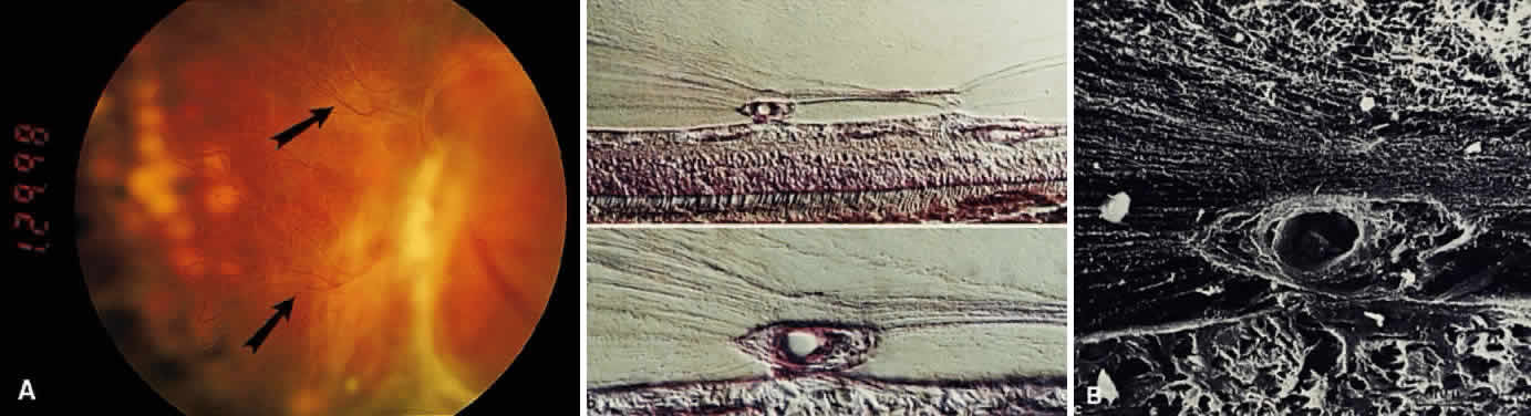
| Fig. 25. Proliferative diabetic vitreoretinopathy. A: Fundus photograph of the left eye in a patient with proliferative diabetic vitreoretinopathy. Neovascularization is present in a fibrous stalk that arises from the optic disc. The new vessels (arrows) are in sharp focus, whereas the surrounding retina is out of focus. This difference is because the vessels are not in the same focal plane as the retina and optic disc. Because the new vessels proliferated out into the peripapillary posterior vitreous cortex, they are anterior to the retinal plane. B: Histopathology of retinal neovascularization in a patient with proliferative diabetic vitreo-retinopathy demonstrates that the new vessels grow out of the retinal plane into the overlying posterior vitreous cortex. Prominent vitreous fibers insert into the new vessels and can transmit tractional forces induced by the diabetic vitreopathic changes shown in Figure 24. Such traction can be important in the pathogenesis of vitreous hemorrhage and traction retinal detachment. (From Faulborn J, Bowald S. Microproliferations in proliferative diabetic retinopathy and their relation to the vitreous. Graef Arch Clin Exp Ophthalmol 223:130, 1985, with permission) |