|
|
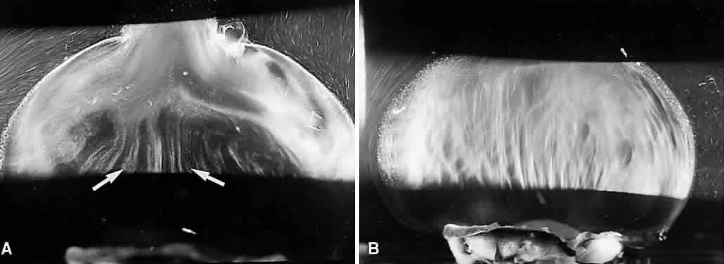
| Fig. 24. Diabetic vitreopathy. A: Right eye of a 9-year-old girl with a 5-year history of type 1 diabetes shows extrusion of central vitreous through the posterior vitreous cortex into the retrocortical (preretinal) space. The subcortical vitreous appears dense and scatters light intensely. Centrally, there are vitreous fibers (arrows) with an anteroposterior orientation and adjacent areas of liquefaction. B: Central vitreous in the left eye of the patient in (A) shows prominent fibers that resemble those seen in nondiabetic adults (see Fig. 11). (Fom Sebag J. Abnormalities of human vitreous structure in diabetes. Graef Arch Clin Exp Ophthalmol 231:257, 1993, with permission) |