|
|
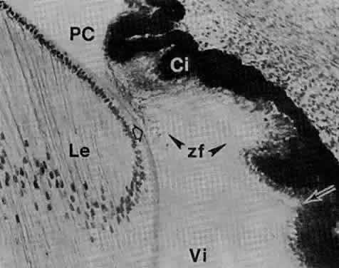
| Fig. 37. Commencement of tertiary vitreous, or zonular fibers in an 85-mm fetus (fourth month) (× 160). Newly formed zonula fibers (zf) span the space between the inner, nonpigmented ciliary epithelium (Ci) and the lens capsule (hollow arrow). The zonular fibers have crossed the faisceau isthmique (or marginal bundle of Drualt) at right angles (see Figs. 21, 22, and 24). The faisceau isthmique has retreated posteriorly with the forward growth of the anterior portion of the optic cup and its anterior anchorage at this age is in the posterior portion of the developing pars plicata (arrow). This attachment will be displaced further posteriorly with growth of the eyeball. It is the fetal origin of the vitreous base. PC, posterior chamber; Vi, vitreous; Le, lens. |