|
|
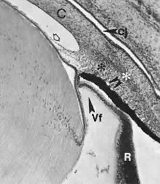
| Fig. 21. Section through the anterior portion of the eye of a fetus of 54 mm (about 10.5 weeks). The rim of the optic cup extends anteriorly beyond the lens equator. Small vessels indent the outer (basal) surface of the pigment epithelium (arrowheads). The inner, nonpigmented wall of the optic cup is smooth. Dense arrays of vitreous fibers (Vf) attached to its inner surface form a faint condensation from the lens equator region to near the margin of the cup. This is the “faiseau isthmique” or marginal bundle of Druault. There is a narrow space between the tip of the optic cup and the artifactually detached lens epithelium. Pupillary membrane is indicated by hollow arrow. cj, conjunctival sac; C, cornea; R, retina. Anlage of ciliary muscle is marked by asterisks. |