Chapter 111
Pediatric Ocular Trauma
GARY L. ROGERS, DON L. BREMER, MARY LOU MCGREGOR and CHRISTINA N. NYE
Main Menu   Table Of Contents

Search

PRINCIPLES OF EMERGENCY OCULAR TRAUMA
REPAIR OF LACERATED GLOBE
EYELID LACERATION
EYELID MARGIN
BLOWOUT FRACTURE
HYPHEMA
OCULAR MANIFESTATIONS OF CHILD ABUSE
AMBLYOPIA
PREVENTION
REFERENCES

PRINCIPLES OF EMERGENCY OCULAR TRAUMA
Ocular trauma frequent causes emergency department visits at pediatric hospitals. The extent of trauma may range from simple superficial injuries to devastating penetrating injuries of the eyelids, lacrimal system, and globe. Regardless of the severity, it is often difficult to examine a child because of his or her discomfort, inability to understand, and inability to communicate. These problems all result in the label “poor cooperation.” Suspicion of the level of the injury is raised after taking a detailed history of the events surrounding the injury. A full medical and ocular history should be taken. A complete and accurate assessment of the injury must be made with confidence. If this is not possible in the emergency department, the child must be sedated or taken to the operating room for an examination under anesthesia. Never neglect the uninjured eye. Findings of the “normal” eye may have an impact on management of the injured eye.

INCIDENT HISTORY

First, a complete account of the accident should be obtained. In some situations parents or baby-sitters have not witnessed the accident and may be unable to give an exact accounting. The child should be questioned and may be able to provide valuable information regarding the accident. Isolating the child from the parents may reveal a more accurate history of the events. From the history, the nature and extent of the injury can be determined, which aids in how extensive an evaluation is needed. Knowing the type of trauma may alert one to look for secondary ocular effects, such as fat emboli from a crush injury. Injuries caused by large blunt objects may produce contusions to the periorbital region and would favor contrecoup retinal problems and blowout fractures of the orbit along with rim fractures. Missiles or sharp objects would raise the suspicion of a perforated globe or retained foreign bodies in soft tissues or the orbit.

Because of the potential for general anesthesia, pertinent past surgical history of the patient and drug allergies are important to obtain along with current immunizations. Although the incidence of tetanus is rare, in the United States booster doses of tetanus toxoid should be received every 5 years to maintain adequate immunity and should be administered in “dirty” cases.1 A dose of 0.5 ml of tetanus toxoid is appropriate for immunized children. If the child has not been immunized, the tetanus immunization series should be initiated with a 0.5-ml dose of diphtheria-pertussis-tetanus vaccine.

MEDICAL HISTORY

Even in the pediatric age group, a complete medical history is necessary. Although the incidence of transmissible disease is low, the potential exists and one should not hesitate to obtain permission for testing for human immunodeficiency virus and hepatitis. Medical conditions should be considered with particular attention to hematologic and bleeding disorders. Knowing the sickle cell status in black patients is important before general anesthesia is administered.

EXAMINATION

Examination should be as extensive as the injury permits. If there are other injuries that must be addressed, the examination and treatment may be modified as determined by the severity and extent of those injuries. Recording visual acuity should be attempted on all patients with ocular and periocular trauma. Various methods of evaluating the vision include Snellen letters, random Es, Allen pictures, finger counting, and light perception with or without projection. One must consider the child's age and the severity of the injury when determining the appropriate visual acuity test. The severity of ocular injury can be assessed by its affect on vision. Initial visual acuity is known to be a prognostic indicator of final outcome.

External

The external examination should consist of looking for puncture wounds of the lids and brow, which could be through and through lacerations or punctures that could involve the globe. If there is ever any question of an open globe, a shield should be placed on the eye until the child is placed under general anesthesia for thorough evaluation and possible repair. Lacerations of the eyelid margin must be attended to with particular attention to the nasolacrimal system and its integrity. Palpation of the orbital rim helps to rule out fractures with possible displacement.

Ocular motility, ductions, and versions must be evaluated because of the possibility of muscle entrapment, laceration, or possible paralysis.

When there has been ocular trauma, one should always ask about diplopia. A small child often does not understand the meaning of double vision, and the clinician needs to explain this problem in very simple terms.

Interior Examination

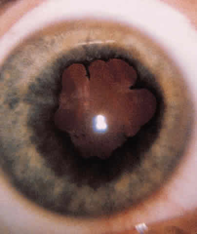
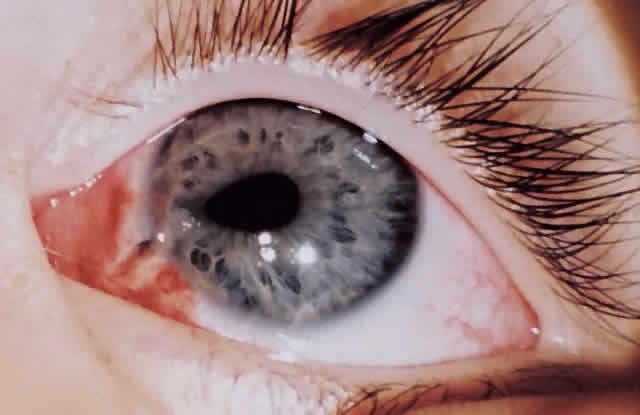
ANTERIOR SEGMENT. A slit-lamp or penlight examination should assess the clarity of the cornea, the anterior chamber depth, distortion of the pupil, and the presence of a hyphema (Fig. 1).

Fig. 1. A 3-year-old child with a “peaked” pupil indicating a full-thickness corneal laceration requiring surgical intervention.

PUPILS. The size and shape of the pupil can aid in evaluation of an open globe. The reaction of the pupil, depth of the anterior chamber, consensual pupillary response, and afferent defect are critical in the initial evaluation.

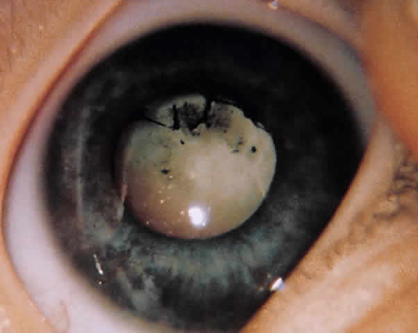
RETINA. If possible, the retina must be evaluated for tears, hemorrhage, perforation, and retained foreign bodies. No retinal examination or manipulation of the globe should be attempted if there is a concern about a lacerated globe. The nature of the injury would also direct the need for appropriate diagnostic tests: roentgenography, computed tomography (CT), or ultrasonography.

If the retina is unable to be visualized and there is no obvious penetrating injury to the globe, B-scan ultrasonography can help in assessing the status of the retina. This plays a role in patients with hyphemas and vitreous hemorrhages. If a retained foreign body is suggested, plain films or CTs are helpful in localizing metallic foreign bodies. Magnetic resonance imaging is preferred when localizing glass but should never be ordered if one suspects a metallic foreign body.

PLAN OF ACTION

After all information has been gathered, a definitive plan of action can be considered. If surgery is needed for a rupture, repair must be made as soon as possible. Correction of external problems involving the eyelids and lacrimal system can be delayed for 24 to 48 hours without increased risk of infection.

After a thorough history and examination have been performed, it is important to document all this information to help in the management of the patient and also be available if any legal action regarding the injury is taken.

Back to Top
REPAIR OF LACERATED GLOBE
A perforated globe is a true ophthalmic emergency. Missile injuries from children playing with BB guns or sharp projectiles are the most common cause for lacerated globes. Any eye suspected of being lacerated should be treated as such until it can be proven that the globe is intact.

HISTORY

A detailed history of the trauma may indicate the nature and extent of the injury. The initial questioning should be directed toward the child, with the parents present to corroborate the story. Children may not readily volunteer the exact cause of the accident for fear of parental reprisal. In this case, it is helpful to repeat the questions to the child in a nonthreatening manner away from the parents. This gives the child an opportunity to provide more accurate details about the accident. The history should always probe for the possibility of an intraocular foreign body with the appropriate ancillary test of roentgenography, CT, or ultrasonography. Plain films are of little value except as a screening device. It is important to note whether eyeglasses or contacts were worn at the time of the injury.

It is especially important to know the visual status of both eyes before the injury and whether there is a history of amblyopia or previous ocular surgery. Any medications the child is taking or any hematologic or clotting problems should also be ascertained.2

EXAMINATION

In our experience, most ruptured globes in children that involve sclera are evident because of severe chemosis and poor vision. These ruptures are often associated with a protruding iris and flat anterior chamber. There may be difficulty in diagnosing a small corneal perforation, which can be self-sealing. As in any ocular injury, the best corrected vision should be determined either with lenses or pin hole testing. Noting whether the pupil is round, eccentric, miotic, or mydriatic or has an altered reaction to light may lead to the realization of a ruptured globe, even if the puncture wound is small.

Motility examination, manipulation of the lids, and intraocular pressure measurements should be avoided in a patient suspected of having a perforation. Shields should be immediately placed on the eye to minimize the possibility of extruding intraocular contents.

Slit-lamp examination can help determine the status of lacerations and hemorrhages of the conjunctiva, corneal defects, foreign bodies, and depth of the anterior chamber. The Seidel test for evaluation of a corneal wound leak is important in determining whether the wound has sealed itself or whether aqueous is being emitted.

Posterior segment examination should be avoided in cases of ruptured globes to prevent possible extrusion of intraocular contents. However, it may be necessary to look for choroidal and retinal ruptures or vitreous foreign bodies.3

PREOPERATIVE PLAN

Depending on the age and cooperation level of the child, the examiner selects the approach needed to correct the problem. Partial thickness lacerations or small self-sealing corneal lacerations may be managed conservatively with patching, a contact lens, or cyanoacrylate adhesives. These solutions avoid the use of general anesthetics and possible corneal irregularities, which may lead to amblyogenic factors, depending on the age of the child. Admission to the hospital with close observation and antibiotic administration is ideal.

One must consider when faced with the self-sealing wound that a wound leak may develop when the edema from the cornea subsides. The ocular status could also be compromised by the activity of the child.

However, after a surgical decision has been made, it is best to proceed immediately with the repair to avoid possible endophthalmitis. A shield should be worn at all times by the patient to prevent additional complications to the eye. It is also prudent to wait until the laceration is repaired before adding medications to the eye, either in the form of antibiotics or mydriatics, which could prove toxic to the retina.

Every open globe should be treated as a potentially infected case until cultures, which should be taken at the time of surgery, are proven negative and no clinical signs of infection are present. Ideally, antibiotics should not be given until cultures are taken. If there is a delay in taking the child to the operating room, one must use clinical judgment. Broad-spectrum parenteral antibiotics should be initiated either before or during surgery as a precaution against endophthalmitis. Intravenous antibiotics should be continued for 2 to 3 days and then oral antibiotics continued for another 7 days. Cefazolin at a dosage of 50 to 100 mg/kg/day intravenously divided every 8 hours would provide broad spectrum coverage until culture results are available. There are many other choices for broadspectrum coverage. With time and availability of culture results, an antibiotic can be selectively targeted to the specific pathogen found.

OPERATIVE PLAN

The extent of the wound should first be explored so the surgeon understands the complete limits of the laceration. The operating microscope has facilitated this end and when possible it should be used at the time of surgery. The goal is a watertight globe with the lacerated tissues restored to as closely as possible to the original anatomy to help regain normal function.4

We have found that most prolapsed tissue can be repositioned within the first 48 hours. Repositioning of uveal tissue is contraindicated when tissues appear necrotic or infected; when this occurs they should be excised. Although a theoretical risk exists, it has not been documented that the risk of infection is greater if prolonged exposed tissue is repositioned so long as there is no evidence of necrosis or infection.5

Wound closure should begin by identifying landmarks that can be approximated. Lacerations through the sclera and cornea should first be closed at the anatomic limbus, thus securing normal apposition of the wound and preventing sliding and malapposition of the anatomy. Exposed uveal tissue can be repositioned with an iris or cyclodialysis spatula along with the use of viscoelastics. A close-as-you-go technique for long scleral-corneal lacerations is acceptable as well as halving techniques, in which you approximate the midline of the wound and approximate the two halves. This helps prevent “dog-ears” at the end of the wound.

Nonabsorbable suture (8-0 silk or 9-0 nylon) is preferable at the limbus. The suture of choice for corneal wounds is 10-0 nylon. The knots should be buried when possible. It is preferable to use longer suture bites at the limbus with smaller suture bites closer to the center of the cornea. This aids in preserving normal corneal architecture and prevents severe flattening, which can occur with longer central suture bites.

Vitreous prolapse through a scleral or corneal wound can occur. It is best excised at the lips of the wound using cellulose sponges and scissors. Tugging of the vitreous should be kept to a minimum to prevent retinal complications. As the vitreous is removed, the uveal structure can be repositioned. The use of a viscoelastic may assist in hydraulically repositioning uveal tissue.

Two suture techniques can be used to close the corneal wound. The round circular bite is probably the best but is the hardest to achieve. It is recommended that the box suture be used, which is a through-and-through suture with even bites on either side of the wound. A running suture technique should be avoided in these situations because it may distort the ends of the wounds.

Determining the status of the lens is important. If the lens capsule has been lacerated, the surgeon must decide whether to remove the lens now or later. Removal of the lens can only be attempted after watertight closure has been achieved. A severe corneal laceration may prevent good visualization or stabilization of the wound. Under such circumstances, it may be advisable to wait until the wound has had a chance to heal and can withstand the intraocular pressure generated during lens aspiration. Once the lens appears cataractous, it is best to remove it as early as possible because of the difficulties associated with a hard calcified lens that occur when cortex is exposed to aqueous, as well as the associated risk of amblyopia. Fibrinous precipitation can occur on the anterior lens capsule and masquerade as a cataract. This can be difficult to differentiate from a lens capsule rupture. A lens should only be removed at the time of the initial wound repair if there is evidence of anterior capsule rupture. It is best to wait until a cataract is definitely confirmed in the postoperative period (Fig. 2). Children older than 1 year who have a cataract and corneal laceration that precludes fitting with a contact lens are candidates for intraocular lenses. Only a surgeon with experience in treating pediatric cataracts with intraocular lenses should consider this. A safer situation occurs when the child is older than 3 years of age and will cooperate with a slit-lamp examination. If a lens needs to be removed, it is best to leave the posterior capsule intact for a secondary posterior chamber intraocular lens. If a cataract is mild and there is no capsule rupture, it is best to wait to perform the cataract extraction with intraocular lens insertion until a later date. This allows the posttraumatic inflammation to quiet as well as to decrease the risk of postoperative complications. Extreme care must be taken when removing a traumatic cataract in that there may be an associated zonular weakness increasing the possibility of nuclear loss into the vitreous.

Fig. 2. A 5-year-old child with a traumatic cataract with posterior synechia that developed 3 months after blunt injury to the eye.

Cefazolin (0.2 ml of 50-mg/ml concentration) should be injected under the conjunctiva at the end of the surgical procedure. Aminoglycosides should be avoided because of the risk of retinal toxicity.6 Subconjunctival injection of corticosteroids should be avoided until the surgeon is certain that no infection is present. Atropine ointment (0.5% for children younger than age 1 year and 1% for children older than 1 year) should be placed to ensure maximal cycloplegia.

POSTOPERATIVE MANAGEMENT

Control of postoperative inflammation with restoration of vision in the pediatric age range is critical. Many successful procedures have been performed but have resulted in poor vision because of irregular astigmatism and secondary amblyopia. In visually immature children (younger than 8 years old), plans must be made for visual rehabilitation as soon as the situation permits. This includes the use of contact lenses, patching of the normal eye, and a possible keratoplasty procedure to clear the pupillary space.7 Patching the injured eye for more than 24 hours following injury should also be avoided to prevent the risk of iatrogenic amblyopia. Corticosteroids, both topical and systemic, can be used early in the course of treatment. Treating severe inflammation and returning the child to a comfortable condition is paramount.

Children seem to heal much sooner than adults. It has been our experience that corneal sutures can be removed as early as 2 to 4 weeks after trauma. One should monitor signs of healing, such as suture loosening and early vascularization along the corneal wound. Clearing the visual axis and correcting any abnormal refractive errors are essential for a good visual outcome.

Back to Top
EYELID LACERATION
Laceration of the eyelids may or may not involve the lacrimal drainage system. The clinician should assume that if a laceration involves the medial canthal area, the lacrimal system is involved. Repair in the operating room with the opportunity to address the lacrimal system should be arranged.

If one is certain that the lacrimal system is not involved, the physician must use judgment to determine whether the child could tolerate repair in the emergency department. If anesthesia is required, it is wise to remember that a delay of even 48 to 72 hours is well tolerated. The eyelid has excellent blood supply, and this delay does not compromise the healing process. Therefore, if the child has eaten recently, one must not declare the case an emergency. It is best to wait until the conditions are more favorable. The child is placed on antibiotics and the repair is scheduled.

The first step is to clean the wound well. This should be done with minimal disruption to the surrounding tissue. When there are a large number of foreign particles, one should first try to irrigate the material from the wound. The next step should be the removal of individual particles with the use of fine forceps. Scrubbing tends to destroy the tissue and make repair more difficult.

It is usually not necessary to debride or “freshen” the wound edges. The more tissue left to close the wound, the more favorable the results. In a situation in which tissue has already been lost due to the injury, one should not consider primary grafts unless the extent of the injury threatens the integrity of the cornea. Major reconstruction should be delayed for 6 to 9 months.

Back to Top
EYELID MARGIN
An assessment of the laceration as well as a complete eye examination should be done. Concomitant ocular injuries are common. The laceration should be described according to its length, level or depth of involvement, loss of tissue, and mechanism of action (e.g., dog bite, knife wound). This aids in the plan of treatment. A barbed object, such as a fishhook, must be pushed forward and not pulled out. After the barb of the hook enters through the skin, wire cutters can be used to remove the barb and the remainder of the hook can be pulled backward through the entrance site.

The best approach is to close layer by layer, which helps maintain function and promote good cosmesis. Extruding fat should be excised because it increases the risk of infection. The orbital septum need not be closed so long as care is taken to close the other layers.

With a lid laceration involving the margin, the first step is to align the margin to avoid notching. With the use of 5-0 or 6-0 chromic catgut, the first suture is placed through the tarsal plate and as closely as possible to the lid margin (Fig. 3A). The sutures should remain intratarsal and one should avoid full-thickness bites, which can cause corneal abrasion during the healing process. This suture is left untied to allow access to the remainder of the wound. The next step is to close the length of the tarsus with interrupted sutures (see Fig. 3B).

Fig. 3. A. Interrupted sutures are placed intratarsally. B. Silk suture is placed at the gray line. C. Intratarsal sutures are tied. D. Silk suture is used to align the margin anterior to the gray line.

Next, the lid margin is aligned, and a 6-0 silk suture is placed at the level of the gray line into the tarsus on either side of the wound (see Fig. 3B). Now the physician can return to the first tarsal suture and tie it. The silk suture is tied loosely and left long to avoid corneal contact. A 6-0 Vicryl suture is also acceptable in children and may eliminate having to remove the suture at a later date.

Next, another silk suture at the level of the lash line is placed. This is also left long. These long ends can be incorporated into a future skin suture or taped to the cheek with a Steri-Strip.

The orbicularis muscle is closed with interrupted absorbable 6-0 suture, such as Vicryl. The skin can then be closed with interrupted 6-0 fast-absorbing catgut suture (see Fig. 3D). If absorbable sutures are not used on the skin, sutures need to be removed in 3 to 5 days and the lid margin sutures should remain for 10 to 14 days.

If a dressing is used, Telfa or petrolatum gauze is recommended because other dressings adhere to the incision. Cold compresses may help reduce swelling. Postoperative orders should include frequent visual acuity checks, especially when the septum has been disrupted. Lid edema and orbital swelling must be monitored because these may be early signs of retrobulbar blood accumulation. Formation of an expanding hematoma may compromise the blood supply to the optic nerve.

If vision begins to lessen, the wound should be opened immediately at the bedside with a cantholysis to relieve pressure. Reducing ocular pressure with hyperosmotics and antiglaucoma medications may help improve perfusion and restore vision. After vision has been stabilized, CT can localize a large hematoma, which may require evacuation.

Antibiotic drops or ointment is all that is needed unless the wound is considered contaminated. Oral antibiotics are indicated in contaminated cases, including dog bites, concomitant sinus fractures, or presence of considerable foreign material.

When a lid laceration involves the canalicular system, it is best to address the canalicular laceration first and then close the lid laceration as described previously. Canalicular injuries are very common in children. There has always been controversy surrounding the indications for repair and the method used to repair these lacerations. It has been proven that both canaliculi are not necessary for adequate lacrimal drainage.8 Most surgeons have their own preference for repairing a single lacerated canaliculus. We believe that children obtain best cosmesis and function if the canaliculus is repaired at the time of the injury. The system must be repaired if both canaliculi or the common canaliculus is involved in the injury. The three important steps in repairing a canalicular laceration involve (1) temporary placement of a well-tolerated stent in the lacrimal system9, (2) mucosal anastomosis with an 8-0 absorbable suture using microscopic or loop magnification, and (3) a layered closure of the remainder of the laceration. Many modifications of canalicular laceration repair have occurred over the past 30 to 40 years. The reasons for so many changes include the introduction of magnifying devices such as loupes and the operating microscope, the development of new stent materials, and other technologic advances.10 Our preference is for the Crawford tube system, which employs silicone as the stent material. The probes are olive tipped for easy retrieval with the accompanying hook11 (Fig. 4). If the laceration involves the margin medial to the puncta, one should assume that the canalicular system is involved and further evaluation and repair should be undertaken in the operating room. If it is unclear whether the laceration is through the canalicular system, the system can be probed with a standard metal nasolacrimal duct probe. This can be performed in the emergency room with sedation in most cases. If one can see the distal end of the lacerated canalicular system, one should pass the probe the entire length of the system. The undamaged canaliculus should then be intubated with a Crawford tube.

Fig. 4. Syringe with irrigating tip, lacrimal probe, punctal dilator, and a Crawford silicone tube on olive-tipped probes.

A thorough understanding of the canthal anatomy is essential if the distal end of the lacerated canaliculus is not readily identified. The inferior and superior lacrimal canaliculi extend vertically for 1.5 to 2 mm from the puncta to the ampulla. At that point, they turn medially for 10 mm. The lateral portions of the canaliculi lie very superficially beneath the conjunctiva 2 mm inferior and parallel to the lid margin. The medial portions of the canaliculi pass more deeply to penetrate Horner muscle in the posterior reflection of the medial canthal ligament. There is some variation at that point. The canaliculi can either join medial to the lacrimal sac and form a common canaliculus or can enter the sac separately but close together. The diameter of a normal canaliculus is 0.5 to 1 mm in a normal nonstressed condition.12

If the laceration is close to the punctum, the distal end of the lacerated canaliculus is near the lid margin and easy to locate. If the laceration is closer to the lacrimal sac, the surgeon must explore the laceration more deeply for the distal end of the cut canaliculus. A helpful step is to place the probe through the punctum and gently reapproximate the tissues in their normal anatomic positions, looking where the probe points delineates the opening into the remainder of the canalicular system.

Injecting a viscoelastic material or a corticosteroid suspension through the uninvolved canaliculus may help to expose the distal cut end. During this maneuver, the lacrimal system is occluded past the sac to force the material to reflux through the lacerated canaliculi. Methylene blue or other dyes are not recommended because they stain tissues, making it difficult to identify the lacerated canalicular opening. After the distal aspect of the lacerated canaliculi is found, the next step is to insert the Crawford tube.

Once the Crawford tube has been passed, it is retrieved beneath the inferior turbinate. The other end of the Crawford tube is then passed through the uninvolved canaliculus. After the tubing has been placed, the anastomosis of the canalicular system is continued by placing two or three interrupted 8-0 Vicryl sutures to reapproximate the two cut ends of the canaliculus. The lid laceration can then be closed.13 The metal probes are cut free of the silicone tubing. The silicone tubing is then tied in a knot and excess tubing is removed. A small knot facilitates later removal. The silicone knot is placed under the inferior turbinate with bayonet forceps. The silicone tube in the interpalpebral fissure is then adjusted to ensure that there is not excessive tension on the punctum that may cause erosion. Too much tension on the canalicular system can cause the silicone tube to go through the medial canthal area like a cheese cutter. Excessive laxity enables a child to pull the tube, creating a big loop in the medial canthal area that potentially can lead to corneal trauma. Other complications of a lax tube include conjunctivitis, granuloma formation, chronic nosebleeds, and epiphora.14

Postoperatively, the child should be placed on antibiotics because the nasal passages are contaminated and are continuous with the eyelid laceration. Oral antibiotics for 5 days as well as antibiotic drops are recommended. Amoxicillin can be used in the range of 20 to 40 mg/kg/day; cefaclor can be given at 40 mg/kg/day, twice daily or every 8 hours. The clinician should check the position of the silicone tube in 1 week and again in 1 month. There are many opinions as to how long the tubing should be left in place. We have found that the children heal well and do not develop recurrent nasolacrimal duct blockages if the tube is left in position for 3 to 4 months.15 It has been proven that friction of the silicone tube does not prevent epithelial healing nor does it promote damage to the uninjured portion of the lacrimal system.16 At that time, the tube is removed and the lacrimal system is irrigated to ensure patency. This can usually be done successfully in the office by cutting the tube at the medial canthus and pulling it through the canaliculus that had not been injured.

Back to Top
BLOWOUT FRACTURE
Blowout fractures are orbital floor fractures that are not continuous with the inferior rim of the orbit. The term blowout fracture was developed by Smith and Regan to describe a fracture of the orbital floor and often the medial wall without any associated fracture of the orbital rim.17 Their studies in cadavers demonstrated it was the compression of the orbital contents that caused the floor fracture or blowout of the thinned bone.

Objects larger in diameter than the orbital rim usually cause blowout fractures. Smaller objects usually cause penetrating injuries to the eye, whereas larger objects, such as baseballs, compress the eye and the other orbital contents, resulting in a blowout fracture.

Orbital tissue may or may not herniate through the fracture site. Orbital fat with or without extraocular muscles may become entrapped. CT with coronal reconstruction is sensitive in detecting small fractures. Plain films are of little value. Initial CT may also predict final clinical outcome, such as significant enophthalmos or diplopia.18 Often patients are asymptomatic except for eyelid emphysema. Antibiotics are recommended to prevent sinusitis and secondary orbital cellulitis. Cephalexin (25 to 50 mg/kg/day every 6 hours) or ampicillin (50 mg/kg/day every 6 hours) is used for prophylaxis in these cases.

Typically, a patient with a blowout fracture presents with severe eyelid ecchymosis and edema. There may also be eyelid emphysema. Because the infraorbital nerve is often involved, one should check sensation over the cheek and also along the gum line due to involvement of alveolar branches. Often edema increases over the first 24 hours. Therefore, a full eye examination should be obtained when the patient is seen initially. Remember that a patient may not complain of double vision because the eyelid is swollen shut and he or she is only using one eye. It is only after the edema resolves that double vision may be appreciated. Any child with a blowout fracture should be advised not to blow his or her nose, in that this may force air into the orbit and possibly damage the optic nerve.

There are essentially two indications for surgically repairing an orbital floor blowout fracture. One is diplopia (in an important gaze direction) due to entrapment of a muscle in the fracture site. The second is clinically significant enophthalmos.19 Ophthalmologists are best qualified to manage blowout fractures because they are knowledgeable about orbital anatomy and are the only ones capable of identifying and treating ocular complications. Other disciplines should be consulted if there is extensive bony damage.

Not all diplopia after a blowout fracture results from entrapment of the inferior rectus muscle. Isolated medial wall fractures or medial wall fractures in combination with floor fractures are often responsible for horizontal diplopia resulting from medial rectus problems.20 Frequently, orbital hemorrhage and edema in the immediate posttraumatic period limit ocular motility. Such diplopia starts to improve within 1 week and resolves without any ocular sequelae. More rarely, paresis of either superior or inferior rectus muscle directly from the trauma causes vertical deviation and limitation of ocular movement. In either case, there will be a negative forced duction test and a slow saccadic velocity in the field of action for the involved muscle. The key to suspecting paralysis of the inferior rectus muscle is a hypertropia on the side of the blowout fracture.

Clinically significant enophthalmos typically occurs with a fracture involving over 50% of the orbital floor. It usually measures 3 mm or more and causes a prominent superior sulcus and pseudoptosis, producing the appearance of a sunken eye. There may even be downward displacement of the globe.

If clear evidence of a large fracture with muscle entrapment exists, immediate repair may be indicated. However, in other cases, it is appropriate to repair the fracture within 7 to 14 days of the injury. If possible, wait at least 7 days after the trauma for edema and inflammation to resolve. Not only will it make the surgery easier by improving exposure, but it may alter the degree of enophthalmos and change the surgical procedure. Waiting beyond 14 days can complicate the situation because extensive fibrotic scar tissue may form, sealing orbital tissue to fracture site. This makes elevating the orbital soft tissues out of the fracture site difficult.

The time-honored approach of freeing the orbital tissues from the fracture site and re-establishing the floor of the orbit with Silastic or Teflon sheeting has proven effective in most cases. However, a couple factors should be considered preoperatively that may alter the procedure. First, if the fracture is extensive, there may not be enough base to hold the implant material in place. In that situation, a titanium mesh plate can be used and fixed to the orbital floor at the rim with internal screws.

Second, enophthalmos is usually due to volume expansion of the orbit. In these situations, elevating the orbital tissues out of the sinus is not be adequate. Soft tissue volume augmentation will be needed. This can be determined preoperatively with three-dimensional CT to calculate the volume of each orbit. The volume of the normal orbit can be subtracted from the volume of the injured orbit to determine the amount of bony volume expansion. This number can be used to determine the volume of soft tissue augmentation needed.

If volume augmentation is needed, several options exist. Autogenous material such as ear and nasal septum cartilage, split-thickness cranial bone, and bone from the rib or iliac crest can be used. Synthetics such as Silastic blocks, porous polyethylene, and hydroxyapatite can also be used. The autogenous implants have less migration and extrusion than the synthetic implants, but their variable rate of absorption can lead to overcorrection or undercorrection.

The operation can be performed through an inferior fornix or subciliary incision. Before making the incision, a 4-0 silk bridle suture is placed beneath the inferior rectus. This can later be used to pull the inferior rectus muscle out of the fracture site if it is entrapped.

With a subciliary incision, an incision is made 2 to 3 mm below the eyelashes to avoid both the hair follicles and blood supply. The skin is undermined over the orbicularis muscles down to the inferior orbital rim. It is easiest to dissect between the skin and the orbicularis. With blunt dissection, the orbicularis muscle is split parallel to the orbital rim to the periosteum.

With either incision, once the periosteum is reached, it is opened along the inferior orbital rim with a no. 15 blade. A periosteal elevator is used to elevate the periosteum. With the use of a malleable retractor placed between the periosteum and orbital floor to hold the orbital contents back for exposure, the periosteum is elevated more posteriorly until the fracture site is reached. At the fracture site, the malleable retractor and periosteal elevator are used to free the soft tissues from the fracture. Pulling on the 4-0 silk bridle suture at this point may aid in extracting the tissue trapped in the fracture site.

After the full extent of the fracture site has been freed from orbital tissue, a piece of Silastic or Teflon sheeting is shaped to completely cover the fracture site. It is placed over the fracture under the periosteum. It is left to lie there and not sutured in place. The periosteum at the orbital rim is closed with absorbable sutures such as 5-0 Vicryl. The orbicularis muscle is closed with a 6-0 Vicryl suture, and the skin is closed with a 6-0 fast absorbing gut suture. If an inferior fornix incision is used, the conjunctiva can be closed with 8-0 Vicryl suture or it can be left unsutured.

The fracture usually occurs where the infraorbital nerve is transversing through it. When freeing the soft tissue from the fracture site, one should be on the alert for the nerve and preserve it. Because the nerve can be injured during the dissection, one must document the presence or absence of infraorbital sensation before surgery.

Postoperatively, there should be immediate improvement, but the final result will take a month or more to see. In terms of the motility, seeing single in the primary position and reading position is considered the primary goal to be achieved by surgery. Despite achieving this, frequently there remain limits to elevation in upgaze, causing diplopia in this gaze direction. Inasmuch as diplopia in upgaze is usually not a significant problem to the patient, no additional surgery is needed.

In terms of diplopia, large fractures do best with early repair and are associated with a lower incidence of long-standing diplopia then smaller fractures. This presumably results from soft tissue scarring induced by the sharp edges of bone around the small fracture site.

If the patient is still experiencing diplopia in the primary position after early surgical repair of a blowout fracture, or if the patient is not managed until late after injury, reoperating on the fracture site will be of little value. The restriction is best handled by extraocular muscle surgery.

When enophthalmos is adequately corrected, a prominent superior sulcus and pseudoptosis usually diminish significantly. If they do not, blepharoplasty, perhaps even involving the noninjured eye, must be considered to achieve symmetry in the eyelid appearance.

With any synthetic implants, there is a small but increased risk for migration. Posterior migration can cause damage to the optic nerve. Migration anteriorly can lead to extrusion. To prevent infection of the implant in the immediate postoperative period, a 10-day course of a broad-spectrum oral antibiotic is given.

Back to Top
HYPHEMA
Hyphema can be managed conservatively in most cases. This consists of avoidance of excess activity (no running, jumping, or rough play). The once common practice of extreme restriction of activity including strict bed rest and bilateral patching has never been proven to improve prognosis. It is reasonable to admit the child to the hospital during the first 5 days to ensure daily followup as well as compliance with prescription of limited activity. Various topically applied medications, such as cycloplegics for traumatic iritis,21 miotics to increase iris surface area for clot reabsorption,22 and corticosteroid eye drops to lessen occurrence of rebleeding, have been recommended in the literature.23 Systemically, oral corticosteroids 24,25 and aminocaproic acid (Amicar)25,26 have been used to decrease the incidence of a second hemorrhage. The recommended dosage for aminocaproic acid is 100 mg/kg every 6 to 8 hours, with a maximum dose of 30 g/day. It is rare to use aminocaproic acid in children younger than 8 years of age because it can cause severe nausea. This drug is also extremely expensive, and this often prevents patients from having access to this drug. In our experience, a long-lasting topically applied cycloplegic such as atropine 1% and a topical corticosteroid are usually sufficient to allow clearing of the clot, prevent rebleeding, and provide patient comfort. In large hyphemas, an oral corticosteroid should also be considered. We recommend a dose of 1 or 2 mg/kg for 3 days. Patients with sickle cell disease present a unique challenge in treating hyphema. They are at increased risk of rebleeding as well as glaucoma. Any patient at risk for sickle cell disease should have a screen performed at the time of presentation.

Surgery for hyphema in children is rare unless blood fills the entire anterior chamber, which is referred to as an “eight ball” hyphema. However, this finding by itself is not an indication for surgery. There should be persistent elevated intraocular pressure despite maximal medical therapy (which places the optic nerve at risk for damage), detection of corneal blood staining, or unresolved hyphema of 9 days' duration that can cause peripheral anterior synechiae.27

Controversy remains regarding the criteria for surgical intervention.28 We believe the optic nerve is at risk for injury if intraocular pressure cannot be reduced to below 40 mm Hg after 3 days. Therefore, if maximal medical management that includes acetazolamide (Diamox), 10 to 20 mg/kg/day, and timolol maleate (Timoptic), 0.25% to 0.50% twice daily, does not achieve this, evacuation should be considered. Newer antiglaucoma medications should be avoided in children and in inflamed eyes until their safety in these situations has been documented. Children have demonstrated that their nerves can tolerate intraocular pressure in the mid 30s for 6 to 7 days without damage. During this time, the hyphema may resolve and the pressure return to a more normal level. After 6 to 7 days, if the pressure remains elevated in the high 20s to 30s one should reconsider surgery.

Patients with sickle cell hemoglobinopathies can have significant intraocular pressure elevation with relatively small hyphemas.29 The optic nerve in these patients is less tolerant of elevated intraocular pressure than the optic nerve in patients with normal red blood cells. In addition, central retinal artery occlusion has been reported by both Michelson and Pfaffenbach30 and Radius and Finkelstein31 in patients with sickle cell hemoglobinopathies and may be exacerbated by systemic hypotensive agents. Acetazolamide should not be used in patients with sickle cell disease because it may cause a sickle cell crisis. Early anterior chamber paracentesis needs to be considered in these patients.

Corneal blood staining typically occurs in patients with total hyphemas and is rarely if ever seen in hyphemas occupying less than 50% of the anterior chamber.32 At the first sign of corneal blood staining, the hyphema should be evacuated. This usually occurs 4 to 6 days after onset of the total hyphema but can occur at an accelerated rate with increased intraocular pressure.

Preoperatively, the patient with increased intraocular pressure can be treated with mannitol. In older children and adults, mannitol can be given 30 minutes before the operation. In very young children, one may have to wait to give the mannitol until an intravenous line can be started in the operating room. Up to 0.25 g/kg can be used safely in children. The maximal dose is rarely needed to bring down intraocular pressure.

The procedure can be performed with either a limbal-based or a fornix-based conjunctival flap. After the surgical limbus is cleaned of connective tissue and hemostasis is obtained, an incision appropriate to the vitrectomy hand-piece size is made into the anterior chamber. If the hyphema is not clotted, the blood can be evacuated with only irrigation and aspiration. If the hyphema is clotted, the cutting mode needs to be used.

With suction and cutting speed on low, the surgeon should start nibbling away at the clot right at the incision, keeping the tip of the instrument as much in view as possible. The cutting part should not be directed anteriorly toward the cornea or posteriorly toward the iris and lens where structures can be injured.

As the clot is removed, it becomes easier to keep the tip in site and visualize the iris and maybe even the lens. The goal of surgery is not complete removal of the clot. All one needs to do is remove a significant portion of the center of the clot. One should not pass the tip of the probe into the angle. With removal of a large portion of the clot, the remainder of the clot can retract, pulling the clot out of the anterior chamber angle and restoring filtration. After the clot has been appropriately removed, the incision is closed in the standard manner.

If a cataract is encountered at the time of surgery, one can either proceed with cataract extraction at that time or can close the incision after evacuating the clot and perform surgery at another time. Unless the capsule is ruptured and the lens cortex fluffy, returning to remove the cataract at a later date is probably the better part of valor. Trying to remove the cataract at the initial procedure is hindered because visualization is impaired by the presence of the hyphema. In addition, the extra manipulation is not beneficial to the ruptured blood vessel that caused the hemorrhage to begin with.

Before a lensectomy is done, one must be sure a cataract is present. A fibrin deposit on the anterior capsule can mimic a cataract. This deposit need not be treated and absorbs in 1 to 2 weeks.

After the hyphema has been evacuated, the patient is out of immediate danger from blood staining and glaucoma unless rebleeding occurs. Approximately 60% of patients with a total hyphema achieve a good visual result. Final visual outcome frequently depends on the damage done by the original injury rather than the complications of the hyphema. The chief causes of poor vision are usually macular problems or traumatic cataract23 (Fig. 5). In the long term, patients need to be followed for the possible development of chronic open-angle glaucoma, cataract, and retinal damage. An examination with the patient under anesthesia may be required to obtain a thorough view of the peripheral retina. Until an extensive retinal examination is performed, acute care of a child with a hyphema is not complete. Any child who has sustained an ocular injury severe enough to result in a hyphema requires annual examinations because of the high incidence of late cataract and angle recession glaucoma. A child may not recognize the decreased vision associated with these problems and may present only after the visual loss is severe.

Fig. 5. A dense cataract 2 years following a hyphema in a 7-year-old girl. It was extracted with placement of an all polymethylmethacrylate intraocular lens with uncorrected visual acuity of 20/25.

Back to Top
OCULAR MANIFESTATIONS OF CHILD ABUSE
Eye findings are very common in physically abused children. It is very rare that these findings necessitate surgery; however, we include ocular manifestations of child abuse in this section because it is important for the ophthalmologist to maintain a high index of suspicion for child abuse in the traumatized eye. In most states, it is law that all physicians, including hospital interns and residents, report suspected cases of child abuse to the children's services board or protective agency. Initially, the report can be made verbally, but the agency may request further documentation in addition to the history and physical findings that generated the suspicion. Further, the law specifies that photographs and additional procedures, such as roentgenograms, may be obtained to substantiate a report of suspected abuse. No consent is necessary for these procedures. Once child abuse is suspected, it is best to take a multidisciplinary approach and get the pediatrician and social worker involved inthe care of the child.

Eye findings are very common in abused children. Separate reports have estimated the incidence of ocular pathology in abused children at approximately 40%.33,34 This indicates that it would be good practice to have all abuse victims thoroughly evaluated by an ophthalmologist. The following findings are commonly seen in abused children: periorbital injuries, conjunctival lacerations, subconjunctival hemorrhage, orbital ecchymosis, conjunctivitis, burns, hyphema, cataract, subluxation of the lens, retinal detachment, vitreous hemorrhage, and retinal hemorrhages.

The investigating clinician must obtain a history of the traumatized eye and correlate it with the findings. Often, the injury is out of proportion to the trauma described. Eisenbrey reported in 1979 that 64% of abused children referred for eye examinations who were younger than 3 years of age had retinal hemorrhages. Only 4% of children who sustained head injury due to other causes, such as motor vehicle accidents, had retinal hemorrhages on examination. He concluded that retinal hemorrhages without external evidence of trauma in children younger than 3 years of age may be pathognomonic for child abuse.35

Child abuse in the form of shaken baby syndrome results in a higher rate of retinal hemorrhages (50% to 80%) than does other forms of abuse (5% to 25%).33–36 The hemorrhages are noted to be bilateral in between 60% and 90% of these cases.37 They usually involve the posterior pole but may extend to the periphery. They are most commonly seen in the nerve fiber layer and the ganglion cell layer, but other layers can be involved. Vitreous hemorrhage is also commonly seen. Because the incidence of retinal hemorrhage in shaken baby syndrome is so high, one must look for confirmatory evidence of abuse when this is seen.

Child abuse rarely results in the need for ocular surgical intervention except in a nonclearing vitreous hemorrhage. Because these young children are sensitive to amblyogenic factors, if a hemorrhage remains unresolved for several months, evacuation may be considered. Regardless of the need for surgery, the ophthalmologist must provide followup on all ocular injuries until resolution and keep the pediatrician informed of potential sequelae.

All these injuries can result in permanent visual loss. Cortical visual impairment may result from brain injury. If one sees an older child with unexplained visual loss, previous child abuse must be a consideration. Ophthalmologists must get involved in screening children who are suspected of being abused. They should be keenly aware of the ocular manifestations of nonaccidental injuries.

Back to Top
AMBLYOPIA
One of the most important aspects in obtaining a good visual outcome in children who have sustained an ocular injury is the management of amblyopia. Any child under 8 years of age risks developing amblyopia if the injury obscures the visual axis even for a relatively short time. The younger the child, the greater the risk of amblyopia. The longer the visual axis is obscured, the more profound the amblyopia. A cataract that is adversely affecting the vision should be removed as soon as the eye is quiet and an intraocular lens should be placed as described earlier in this chapter. If there is an associated corneal scar, the fitting of a contact lens should be done immediately. If the contact lens does not provide adequate vision, a penetrating keratoplasty should be performed as early as possible. A vitreous hemorrhage should be cleared without delay by a vitreoretinal specialist. Amblyopia management should be undertaken as soon as the injured eye has potential for good vision. This involves patching the sound eye and close followup. The child's parents should be educated in the importance of treating the amblyopia. Any child who has poor vision in an eye following trauma requires protective polycarbonate lenses to prevent injury to the sound eye.
Back to Top
PREVENTION
More than 41,000 sports-related and recreational eye injuries were treated in hospital emergency departments in 1993.38 Most of these (71%) occurred in individuals under 25 years of age. About 6% occurred in children under 5 years of age. Children and adolescents are particularly vulnerable to ocular injuries because they are likely to be involved in contact sports and have a tendency for rough play.39–41 In 1996, a Joint Statement of the American Academy of Pediatrics and the American Academy of Ophthalmology was published recommending interventions regarding appropriate types of protective lenses and frames for specific sports.42 Prevention of eye injuries is of utmost importance and is the combined responsibility of pediatricians, ophthalmologists, optometrists, coaches, parents, and teachers.
Back to Top
REFERENCES

1. Recommendation of the Immunization Practices Advisory Committee (ACIP): Diphtheria, tetanus and pertussis: Guidelines for vaccine prophylaxis and other preventive measures. MMWR 34:405–426, 1985

2. Barr CC (1983): Prognostic factors in corneoscleral lacerations. Arch Ophthalmology 101:919–924

3. Hamill MB, Thompson WS: The evaluation and management of corneal lacerations. Retina 10(Suppl 1):51–75, 1990

4. Kenyon KR: Anterior segment reconstruction after trauma. In: Brightbill FS (ed): Corneal Surgery: Theory, Technique and Tissue, pp 410–415. St Louis: CV Mosby, 1986

5. Shingleton BJ, Hersh PS, Kenyon KR (eds): Eye Trauma. St Louis, Mosby-Year Book, 1991

6. Campochiaro PA, Lim JI: Aminoglycoside toxicity in the treatment of endophthalmitis. The Aminoglycoside Toxicity Study Group. Arch Ophthalmol 112:48–53, 1994

7. Rowsay JJ, Hays JC: Refractive reconstruction for acute eye injuries. Ophthalmic Surg 15:569–574, 1984

8. Linberg JV, Moore CA: Symptoms of canalicular obstruction. Ophthalmology 95:1077–1079, 1988

9. Quickert MH, Dryden R: Probes for intubation in lacrimal drainage. Trans Am Acad Ophthalmol Otolaryngol 74:431–433, 1970

10. Pashby RC, Rathbun JE: Silicone tube intubation of the lacrimal drainage system. Arch Ophthalmol 97:1318–1322, 1979

11. Crawford JS, Pashby RC: Lacrimal system disorders. Int Ophthalmol Clin 24:39–53, 1984

12. Reifler DM: Management of Canalicular Laceration. Surv Ophthalmol 36:113–132, 1991

13. Lauring L: Silicone intubation of the lacrimal system: Pitfalls, problems, and complications. Ann Ophthalmol 8:489–498, 1976

14. Anderson RL, Edwards JJ: Indications, complications and results with silicone stents. Ophthalmology 86:1474–1487, 1979

15. Dortzbach RK, France TD, Kushner BJ et al: Silicone intubation for obstruction of the nasolacrimal duct in children. Am J Ophthalmol 94:585–590, 1982

16. Snead JW, Rathbun JE, Crawford JB: Effects of the silicone tube on the canaliculus: An animal experiment. Ophthalmology 87:1031–1036, 1980

17. Smith B, Regan WF Jr: Blow-out fracture of the orbit: Mechanism and correction of internal orbital fracture. Am J Ophthalmol 44:733, 1957

18. Gilbard SM, Mafee MF, Lagouros PA et al: Orbital blowout fractures: The prognostic significance of computed tomography. Ophthalmology 92:1523–1528, 1985

19. Pfeiffer RL: Traumatic enophthalmos. Arch Ophthalmol 30:718, 1943

20. Darr JL, Passmore JW: Management of traumatic hyphema. Am J Ophthalmol 63:134–136, 1967

21. Gilbert HD, Jensen AD: Atropine in the treatment of traumatic hyphema. Ann Ophthalmol 5:1297–1300, 1973

22. Loring MJ: Traumatic hyphema. Am J Ophthalmol 46: 873, 1958

23. Kushner AG: Traumatic hyphema. Surv Ophthalmol 4:19, 1959

24. Rynne MV, Romano PE: Systemic corticosteroids in the treatment of traumatic hyphema. J Pediatr Ophthalmol Strabismus 17:141–143, 1980

25. McGetrick JJ, Jampol LM, Goldberg MF et al: Aminocaproic acid decreases secondary hemorrhage after traumatic hyphema. Arch Ophthalmol 101:1031–1033. 1983

26. Crouch ER Jr, Frenkel M: Aminocaproic acid in the treatment of traumatic hyphema. Am J Ophthalmol 81:355–360, 1976

27. Read JE: Traumatic hyphema: Surgical vs medical management. Ann Ophthalmol 7:659–670, 1975

28. Read JE, Crouch ER Jr: Trauma: Ruptures and bleeding. In: Tasman W, Jaeger EA (eds): Duane's Clinical Ophthalmology, chap 61. Vol 4. Philadelphia: JB Lippincott, 1992

29. Goldberg M: The diagnosis and treatment of sickled erythrocytes in human hyphemas. Trans Am Ophthalmol Soc 76:481–501, 1978

30. Michelson PE, Pfaffenbach D: Retinal arterial occlusion following ocular trauma in youths with sickle-trait hemoglobinopathy. Am J Ophthalmol 74:494–497, 1972

31. Radius RL, Finkelstein D: Central retinal artery occlusion (reversible) in sickle trait with glaucoma. Br J Ophthalmol 60:428–430, 1976

32. Brodrick JD: Corneal blood staining after hyphaema. Br J Ophthalmol 56:589–593, 1972

33. Friendly DS: Ocular manifestations of physical child abuse. Trans Am Acad Ophthalmol Otolaryngol 75:318–332, 1971

34. Jensen AD, Smith RE, Olson MI: Ocular clues to child abuse. Child Abuse Negl 9:207, 1985

35. Eisenbrey AB: Retinal hemorrhage in the battered child. Child Brain 5:40–44, 1979

36. Johnson CT, Showers J: Injury variables in child abuse. Child Abuse Negl 9:207–215, 1985

37. Levin AV: Ocular manifestations of child abuse. Ophthalmol Clin North Am 3:249–264, 1990

38. Prevent Blindness America (formerly National Society to Prevent Blindness): 1993 Sports Recreational Eye Injuries. Schaumburg, IL: Prevent Blindness America, 1994

39. Nelson LB, Wilson TW, Jeffers JB: Eye injuries in childhood: Demography, etiology, and prevention. Pediatrics 84:438–441, 1989

40. Grin TR, Nelson LB, Jeffers JB: Eye injuries in childhood. Pediatrics 80:13–37, 1987

41. Jeffers JB: An on-going tragedy: Pediatric sports-related eye injuries. Semin Ophthalmol 5:216–223, 1990

42. American Academy of Ophthalmology: Policy Statement—Protective Eyewear for Young Athletes. A Joint Statement of the American Academy of Pediatrics and the American Academy of Ophthalmology. Ophthalmology 103:1325–1327, 1996

Back to Top