Chapter 110
Ophthalmic Management of Craniofacial Syndromes
JAMES A. KATOWITZ, RICHARD W. HERTLE and JEFFREY B. GOLDSTEIN
Main Menu   Table Of Contents

Search

SPECIFIC SYNDROMES
MANAGEMENT OF CRANIOFACIAL DEFORMITIES
OPHTHALMIC SURGICAL REPAIR
GENETICS
REFERENCES

Congenital and developmental anomalies of the face and cranium often present challenging diagnostic and reconstructive problems that are best handled by a team of specialists. A craniofacial team should include representatives from plastic surgery, ophthalmology, oral surgery, orthodontics, otolaryngology, neurosurgery, genetics, psychiatry, social work, and anesthesia. Each team member contributes specialized skills that enhance the diagnostic evaluation and clinical management of these patients. The relationship between the craniofacial team and each patient and family may continue for years, particularly when the patient requires multiple surgical procedures. Long-range planning must be an integral part of the team's activities. The entire family also may be involved in the evaluation of potential hereditary diagnoses and genetic counseling.

The ophthalmologist has a unique role as both a diagnostician and surgeon. Since orbital structures represent a bridge between the face and cranium, ocular and adnexal abnormalities are frequent components of craniofacial anomalies. Subspecialists in oculoplastics, strabismus, and vision development assist in the evaluation of visual, orbital, adnexal, and motility problems, which are often present in these patients. The ophthalmologist's responsibilities are, first, to preserve visual and protective adnexal functions and, second, to improve cosmesis. When a major deformity is recognized, interaction of the ophthalmologist and other team members is required to formulate an appropriate treatment plan.

The pediatrician or obstetrician usually is the first to recognize craniofacial abnormalities. In subtle deformities, however, the parents may be the first to recognize that something is different. The patient then is referred to the craniofacial team for more extensive evaluation. In the initial consultation, the ophthalmologist evaluates the patient for potential vision-threatening disorders and documents structural defects of the globe, as well as any orbital or adnexal deformities. Each detected anomaly is assigned a priority treatment based on its functional or cosmetic nature.

If there are no disorders that need ophthalmic treatment, then further ophthalmic evaluation depends on the timing of craniofacial surgery. If no immediate surgery is required, then the patient is seen again in 4 to 6 months as part of the craniofacial team follow-up survey. If the patient requires craniofacial surgery and early ophthalmic intervention also is necessary, ophthalmic procedures may be performed under the same anesthetic. If there are no postoperative ocular complications resulting from the craniofacial surgery, then routine follow-up is suggested 6 to 8 weeks later.

The ophthalmic surgeon's primary responsibility involves reconstruction of the lids, canthi, nasolacrimal system, ocular surface, globe, extraocular muscles, and orbital walls. Midfacial advancement, orbital realignment, cranial vault reshaping, and major cleft repair usually are performed by other team members.

Back to Top
SPECIFIC SYNDROMES
The number of published classification systems describing craniofacial anomalies attests to the variety, overlapping features, and variable severity with which craniofacial patients may present.1–13 Deformities of the head can be divided into abnormalities of the cranium and abnormalities of the face. The term craniofacial deformity is preferred when both the face and the cranium are involved.

The most commonly encountered ophthalmic disorders belong to the craniofacial dysplasias. The craniosynostoses include Apert's syndrome, Crouzon's syndrome, Pfeiffer's syndrome, and other isolated suture abnormalities such as plagiocephaly. The clefting syndromes include cleft lip and palate, oblique facial clefts, and mandibulofacial deformities (Goldenhar's syndrome, Treacher Collins syndrome, and facial microsomia).

Hypertelorism may present as an isolated anomaly but frequently is a component of syndromes affecting the midface such as frontonasal dysplasia or the craniosynostoses.

CRANIOSYNOSTOSES

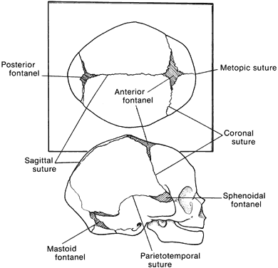
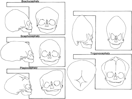
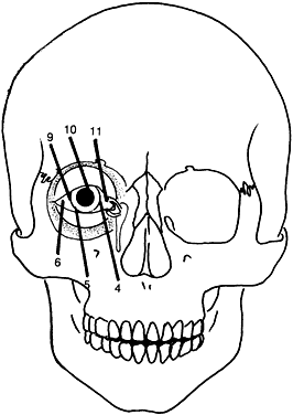
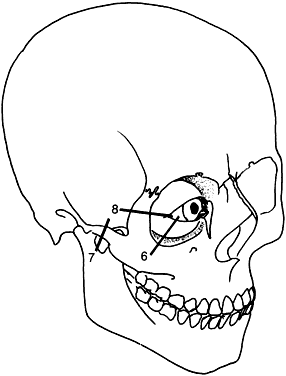
Craniosynostosis (craniostenosis) is the premature closure of one or more cranial sutures.14–21 The dysmorphologic makeup observed in craniostenosis is a direct result of premature suture closure and, chronologically, is the initial manifestation. The diagnosis usually is obvious. The malformation varies, depending on the site and the number of sutures involved in the synostosis and on subsequent alterations of growth. In fetuses of 14 weeks' gestation, that is, about 7.5 cm crown-to-rump length, most of the sutures are formed.22,23 The metopic suture (between the two frontal bones) closes before birth.23 The posterior fontanel normally closes by 2 months of age, the anterolateral suture between 3 and 6 months, and the anterior and posterolateral sutures during the first year of life.23 The sagittal suture closes by 2 years, and the coronal suture closes by 3 years (Fig. 1). The cause of premature suture closure is not known. Either an abnormal fusion of bone centers occurs, or there is malfunction of the underlying dura mater, which normally ensures suture patency.22–26 Subsequent cranial and facial growth are influenced by any prematurely closed suture. Virchow demonstrated in 1851 that, after premature suture closure, growth proceeds parallel to the suture and is inhibited perpendicular to it (Virchow's law).27 Closure of the sagittal suture results in an elongated skull (scaphocephaly). Single coronal, sagittal, metopic, or lambdoidal suture closure leads to a variety of abnormal shapes such as brachycephaly, scaphocephaly, trigonocephaly, or plagiocephaly (Fig. 2). Classification of craniosynostosis has been divided into three main groups: (1) craniofacial dysostosis (Crouzon's syndrome), (2) acrocephalosyndactyly (Apert's syndrome), and (3) simple or single craniosynostosis (plagiocephaly). Classification systems can be confusing because of the overlapping clinical and genetic manifestations.3–5 Although a functional classification based on pathomorphogenic features can be used, diagnosis by syndrome has been the traditional method of classification.7 The most commonly encountered craniofacial synostoses are Crouzon's and Apert's syndromes.

Fig. 1. Position of sutures in a normal full-term infant.

Fig. 2. Skull shapes resulting from premature suture closure.

Crouzon's Syndrome

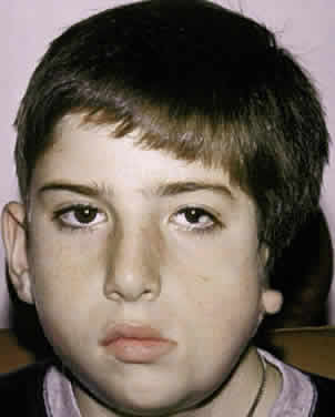
Crouzon's syndrome originally was described in 1912 in a mother and her daughter under the name of a hereditary craniofacial dysostosis.20 It is characterized by craniostenosis of the acrobrachycephalic or oxycephalic type with associated hypoplasia of the maxilla and exophthalmos. A progressive craniosynostosis often is observed at birth and usually is completed by 2 to 3 years of age. Although there is variable presentation with multiple suture involvement and no characteristic calvarial shape, 96% to 98% of patients show radiographic evidence of coronal and sagittal sutural fusion. This pattern of fusion leads to a more predictable skull shape, which features a shortened calvarium, steep forehead, and flattened occiput6,15–17,19,22,28,29 (Fig. 3). Neurologic complications of early suture closure occur commonly and include headaches (29% to 50%), increased intracranial pressure, mental retardation (13%), and seizures (11.5%).17,19 The most characteristic facial feature is midface retrusion, hypoplasia, and shallow orbits.29,30 Brachycephaly, in combination with hypoplasia of the frontal, sphenoid, ethmoid, and maxillary bones, is mostly responsible for these features.

Fig. 3. Patient with typical craniofacial features of Crouzon's syndrome.

Hypertelorism and exophthalmos are universal features of all variants of Crouzon's syndrome. The exophthalmos is secondary to midfacial hypoplasia, resulting in shallow, widespread orbits. One theory states that premature suture closure results in increased intracranial pressure and preferential brain growth in the anterior and middle cranial fossae.24 Another, based on examination of affected fetuses and animal studies, suggests that cranial base changes with resultant stresses initiate osteogenic overstimulation and aberrant bony growth in the calvarium and face.13

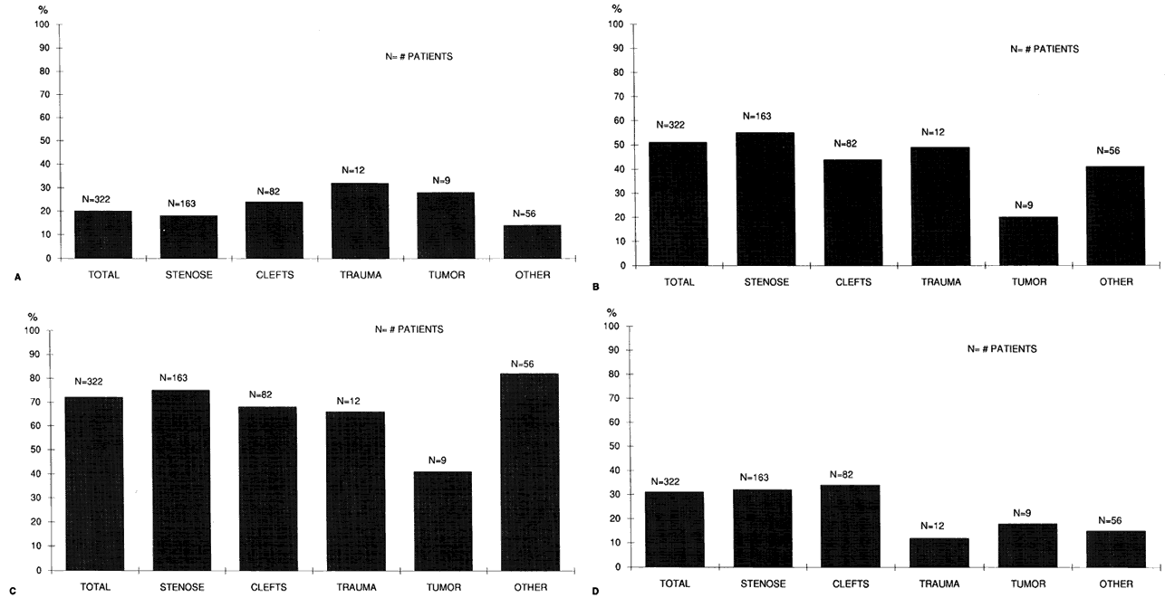
Crouzon's syndrome can affect all ocular structures. Shallow orbits predispose to proptosis and exposure of the cornea and conjunctiva.16,22,31,32 Optic nerve atrophy and edema are vision-threatening complications and have been reported in as many as 80% of patients with this syndrome.33 However, in a series of 25 patients with Crouzon's syndrome, only 1 had optic nerve disease.32 The most plausible current explanation for optic nerve atrophy is that it is the result of a primary disturbance (hypoplasia or dysplasia) rather than the result of secondary factors (compression atrophy or chronic edema).32,33 Early surgical intervention has resulted in decreased morbidity, although the mechanisms involved in secondary optic nerve problems continue to occur.

Other structural abnormalities of the globe reported in patients with Crouzon's syndrome include aniridia, anisocoria, corectopia, blue sclera, cataract, ectopia lentis, glaucoma, chorioretinal colobomas, megalocornea, and microcornea.3,13,16,17,22,28,31

Strabismus is present in as many as 92% of patients with Crouzon's syndrome.32,34–39 V-pattern exotropia is the most common misalignment.32,34,39 The V-pattern and hypertropia on adduction usually are secondary to overaction of the inferior oblique muscles, which occurs as a result of an absent or ineffective ipsilateral superior oblique muscle.36–39 Morax has postulated that the widened, shallow orbits and anomalous muscle insertions change the actions of the muscles.37–39 The superior oblique seems to be sagittalized and less effective.33–39 In a series of 25 patients with Crouzon's syndrome, 48% had exotropia, 32% had esotropia, and 68% were hypertropic in some position of gaze.32

Patients with Crouzon's syndrome have large degrees of compound astigmatism, and many also are anisometropic.16,32 Adnexal abnormalities include ptosis, canthal dystopia, nasolacrimal duct obstruction, entropion, and ectropion.31,32,40–44

The advances made in the surgical treatment of Crouzon's syndrome have changed the pattern and prevalence of visual loss. Structural abnormalities of the globe (e.g., optic atrophy, corneal opacification), which previously were the main causes of vision loss, are less common. The main reason for vision loss is amblyopia. The cause of the amblyopia usually is multifactorial, with contributions from strabismus, uncorrected refractive errors, anisometropia, and ptosis.32

Apert's Syndrome

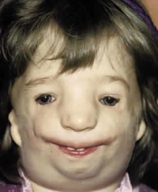
In 1896, Apert described this syndrome in a 15-month-old girl.45 He suggested the term acrocephalosyndactyly in a report of nine cases in 1906.46,47 The classification of this syndrome has since been modified and can be confused with other craniofacial syndromes with syndactyly48 (Table 1). This syndrome occurs with a frequency of 1:160,000 births with an autosomal dominant mode of inheritance. Spontaneous mutations have been associated with increased paternal age.46


TABLE 1. Craniosynostosis Syndromes Associated


SyndactylyAssociated Syndrome
ACS, type IApert's syndrome
ACS, type IIVogt's ACS
ACS, type IIISaethre-Chotzen syndrome
ACS, type IVWaardenburg-type ACS
ACS, type VPfeiffer's syndrome
ACPS, type INoack's syndrome
ACPS, type IICarpenter's syndrome
ACPS, type IIISakati-Nyhan-Tiscale syndrome

ACS, acrocephalosyndactyly; ACPS, acrocephalopolysyndactyly.
(McKusick VA: Mendellian Inheritance in Man: Catalogs of Autosomal Dominant, Autosomal Recessive, and X-Linked Phenotypes. Baltimore, The Johns Hopkins Press, 1974)


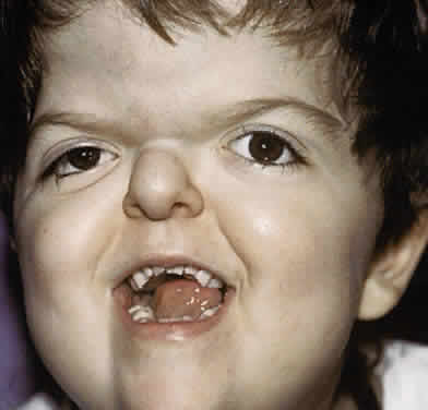
Clinically, the cranium, face, and extremities are involved. Although the cranial features vary, the coronal suture always is affected. The skull is oxycephalic with a flat occiput and steep forehead. The face has a hypoplastic midfacial region with a “parrot-beak” nose. Other features include low-set ears, hypertelorism, and exorbitism.45–47 More prominent features of Apert's syndrome are associated palatal and dental abnormalities, including a high-arched palate, clefting of the soft palate or a bifid uvula, a V-shaped maxillary arch with malocclusion, dental crowding, and supranumerary teeth.47,49 Ear anomalies include pinna defects, conductive hearing loss, and congenital otosclerosis45–47 (Fig. 4). Hydrocephalus and mental retardation also occur in these patients.50

Fig. 4. Patient with typical craniofacial features of Apert's syndrome.

Ophthalmic manifestations are similar to those patients described with Crouzon's syndrome.31,32 Hypertelorism and proptosis are less severe than in Crouzon's syndrome. Structural abnormalities of the globe also occur in this syndrome and include keratoconus, congenital glaucoma, ectopia lentis, cataracts, exposure keratitis, and optic atrophy.30,31,44,45 Strabismus is common, occurring in 70% to 100% of patients.32,36,39 Exotropia with a V-pattern predominates, although V-pattern esotropia is common. Patients also can have nystagmus, dissociated vertical deviations, and incomitant vertical deviations. The structure of the extraocular muscles is abnormal both microscopically and grossly.38 Anomalous or missing muscles, especially the superior rectus and superior oblique, occur commonly in these patients.31,33,36–39,51 Refractive errors, in particular, high degrees of oblique astigmatism (from 60% to 100% of patients), are common, as is anisometropia.31,32,34

Adnexal abnormalities include ptosis, nasolacrimal duct obstruction, canthal dystopia, entropion, and ectropion.31,32,41–44 The high prevalence of visual loss (up to 57% of eyes) in this population of patients results from amblyopia for the same multifactorial reasons as in Crouzon's syndrome.32

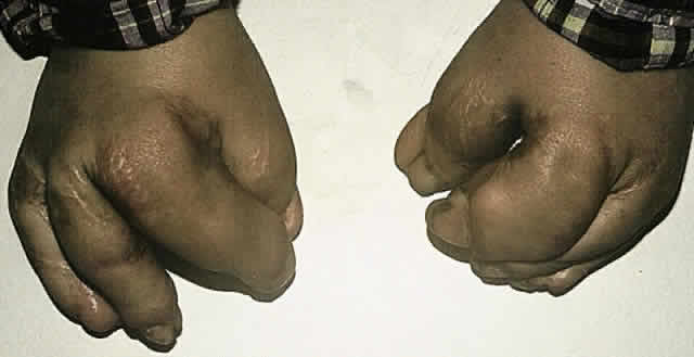
The major associated systemic malformation is syndactyly of the hands and feet. This is most commonly a middigital hand mass involving the second, third, and fourth digits (Fig. 5). Other bony abnormalities include aplasia or ankylosis of the elbow, shoulder, or hip and cervical stenosis.13,46,47

Fig. 5. Middigital hand mass typical of the syndactyly present in Apert's syndrome.

Other systemic associations include congenital heart disease, tracheoesophageal fistulas, hydrocephalus, and pyloric stenosis.13,46,47,51

Other Craniostenosis Syndromes

A variety of other craniostenoses (Table 2) share common features with Crouzon's and Apert's syndromes but occur less frequently. Pfeiffer's syndrome is autosomal dominant and has craniosynostosis with oxycephaly, maxillary hypoplasia, hypertelorism, proptosis, depressed nasal bridge, beaked nose, highly arched palate, crowded teeth, hydrocephalus, seizures, and, occasionally, mental retardation.13,52 Ophthalmic features are similar to Apert's and Crouzon's syndromes. Distinguishing features include broad, long thumbs and great toes.


TABLE 2. Clinical Characteristics of Craniofacial Synostoses


SynostosisClinical Manifestations
Crouzon's syndromeCoronal synostosis, shallow orbits, proptosis, hypertelorism, exotropia, exposure keratitis, luxation of globe, autosomal dominant
Apert's syndromeCoronal synostosis, hypertelorism shalloworbits, strabismus, proptosis, syndactyly, autosomal dominant
Pfeiffer's syndromeCoronal synostosis, hypertelorism, proptosis, strabismus, broad fingers and toes, auto-somal dominant
Saethre-Chotzen syndromeCoronal synostosis, hypertelorism, ptosis, strabismus, nasolacrimal duct obstruction,optic atrophy, syndactyly, autosomal dominant
Carpenter's syndromeAll sutures synostotic, mental deficiency, short stature, obesity, syndactyly, hypertelorism, epithelial folds, medial canthal dystopia, corneal opacities, autosomal recessive
Kleeblattschädel anomalyVariable: proptosis, hypertelorism, exposurekeratitis, iris coloboma, orbital hypoplasia, nasolacrimal duct obstruction (cloverleaf skull)
Christian syndromeAll sutures synostotic, hypertelorism, ophthalmoplegia, lateral canthal dystopia, microcephaly, cleft palate, arthrogryposis, autosomal recessive
Lowry's syndromeCoronal synostosis, proptosis, strabismus, cleft palate, simian creases, autosomal recessive
Gorlin-Chaudhry-Moss syndromeCoronal synostosis, hypertrichosis, lid colobomas, ptosis, microphthalmia, patent ductus arteriosus, autosomal recessive.
Baller-Gerold syndromeCoronal synostosis, hypertelorism, epicanthal folds, radial aplasia, absent carpal bones, autosomal recessive


Saethre-Chotzen syndrome is characterized by a more variable cranial vault with coronal synostosis, plagiocephaly, scaphocephaly, and oxycephaly. Other features include low-set frontal hairline, facial asymmetry, variable brachydactyly and syndactyly, low-set ears, hearing loss, highly arched palate, and dental abnormalities. Intelligence usually is normal, but mental retardation has been reported.11,13,22

Ophthalmic manifestations of Saethre-Chotzen syndrome differ slightly from Apert's or Crouzon's syndrome because of less severe maxillary and midfacial hypoplasia. Hypertelorism and exorbitism are less common. Ptosis is much more common in this syndrome, as well as strabismus and nasolacrimal dysfunction. Amblyopia and optic atrophy again contribute to visual loss in these patients.32

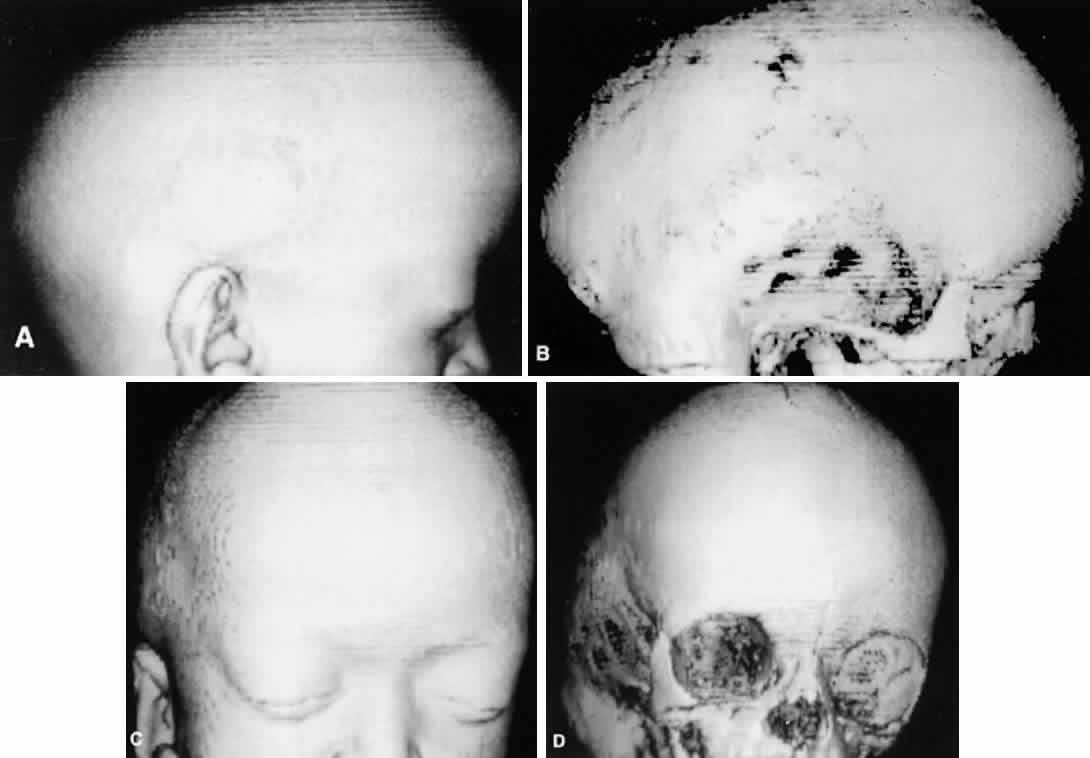
Patients with plagiocephaly exhibit a cranial asymmetry caused by a premature closure of the coronal sutures in one half of the skull. The deformities may be subtle. The skull is flattened in the frontal region on the affected side, often with a compensatory expansion on the opposite side. There is retrusion of the orbital arch on the affected side with a raised eyebrow. The maxillary bone and ear may be affected, with abnormal dental apposition and a downward and forward displaced ear.

Up to 68% of patients with plagiocephaly have ophthalmic involvement. This includes strabismus, cranial nerve palsies, optic neuropathy, ptosis, canthal dystopia, and nasolacrimal duct dysfunction.52,53 Although esotropia is common in these patients, an unusually high prevalence of clinical underaction of the superior oblique muscle ipsilateral to the stenotic suture is present. This manifests with a head tilt and hypertropia on the affected side, which increases on contralateral gaze and ipsilateral head tilt. Often there is underaction of the ipsilateral superior oblique and overaction of the ipsilateral inferior oblique muscle.54 This is postulated to result from a “sagittalization” of the superior oblique because of the orbital deformity.39,44,54 This condition is treated in the same manner as a superior oblique palsy. If the superior oblique muscle is found to be redundant or “loose,” a “tuck” of the tendon is necessary.

Carpenter's syndrome, first described in 1901, is an autosomal recessive craniosynostosis involving many sutures with preaxial polysyndactyly of the feet and variable brachydactyly and syndactyly of the hands. Mental retardation is uniformly present. Congenital heart disease, short stature, obesity, and lower limb abnormalities are other systemic associations. Hypertelorism, epicanthal folds, and telecanthus are reported ocular abnormalities.19,55–57 Other less common craniostenosis syndromes are described in Table 2.

CLEFTING SYNDROMES

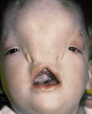
The most familiar form of clefting syndromes is isolated cleft lip and palate syndromes. These clefts occur early in development before the 17-mm crown-to-rump length (17 to 20 weeks' of gestation).58 Interruptions in fusion of the developing facial processes during embryonic development possibly are responsible for these clefting syndromes.58,59 There are five facial processes: a frontonasal, two maxillary, and two mandibular. There may be insufficient outgrowth of the facial processes, poor cellular adhesion resulting in an abnormal epithelial plate, or abnormal cell degeneration of the epithelial plate. The defects of bone, musculature, and soft tissues result from poor fusion of the facial swellings. Failure of mesodermal penetrators with subsequent epithelial dehiscence in the developing facial process as it reaches the midline also is a postulated mechanism of soft tissue defects.7,9,13,58,59

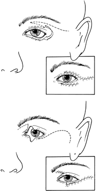
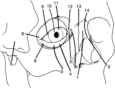
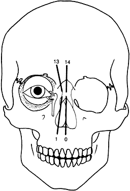
Tessier has revolutionized the classification of clinical clefting syndromes.59 This system assigns each major cranial, orbital, maxillary, and mandibular cleft a number from 0 to 14 in a clocklike circle, beginning at 0 in the lower facial midline and running clockwise on the right side of the face and counterclockwise of the left, and ending at 14 in the upper facial midline (Fig. 6). This system represents an architectural system rather than a true pathognomonic description of specific clinical syndromes.

Fig. 6. Tessier system of cleft classification shown here for the right eye.

Heredity appears to play a limited role in the etiology of clefting syndromes, except for Treacher Collins syndrome (mandibulofacial dysostosis). Some factors seem to be implicated in the development of craniofacial clefts and include radiation, influenza A2 virus, toxoplasmosis, chemotherapeutic agents, anticonvulsant agents, steroids, and tranquilizers.53

The amniotic band syndrome is a related, nonhereditary syndrome that can lead to facial clefts, probably secondary to pressure necrosis. Amniotic bands result from a rupture in the amniotic membrane with formation of tissue cords that can wrap themselves around limbs and digits and, if swallowed, can tighten across or around the face, cranium, or mandible distorting or “cutting” the developing craniofacial structures.1,5,53

Fries and Katowitz suggest that orbital clefts can be grouped into four regional categories that emphasize the associated ophthalmic abnormalities53:

  1. Median facial clefts (Tessier cleft numbers 0, 1, 13, 14), which primarily present with hypertelorism
  2. Medial canthal and nasolacrimal system clefts (Tessier cleft numbers 2, 3, 4, 11, 12, 13), which primarily present with medial canthal dystopia and nasolacrimal duct abnormalities
  3. Central eyelid clefts (Tessier cleft numbers 4, 5, 6, 9, 10, 11), which primarily present with colobomatous defects
  4. Lateral facial clefts (Tessier cleft numbers 6, 7, 8), which primarily present with lateral canthal dystopia

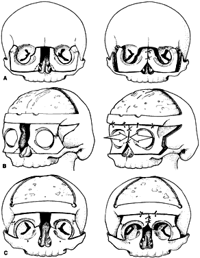
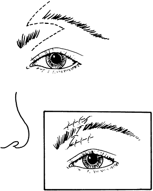
Although these clefts may occur as independent malformations, combinations of clefts frequently occur. Commonly observed pairings include 0–14, 4–10, and the 5–9 clefts (see Fig. 6). Area 1 (midline) clefts usually have orbital hypertelorism as the major ophthalmic manifestation. This also includes a broadened nasal root; bifid anterior cranium and midline facial cleft involving the nose, lip, and sometimes the palate; a lack of formation of the nasal tip; and a widow's peak (Fig. 7). Area 2 (medial canthal/nasolacrimal) clefts produce variable degrees of hypertelorism, medial canthal dystopia, nasolacrimal abnormalities, lid colobomas, and occasional displaced eyebrows and frontal and forehead dysplasia (Fig. 8). Area 3 (eyelid) clefts (see Fig. 8) produce defects that involve the lid and orbit, and, depending on the severity, can be associated with significant structural consequences. In addition to the eyelid deformities, underlying soft tissue and bony absence may permit the orbital contents to be prolapsed into the maxillary sinus. The globe usually is microphthalmic, and true anophthalmos can be present. The lateral lower lid may be affected to varying degrees, often without an oral component. The skin defect is mild and associated with malar hypoplasia and a pseudocoloboma. This lid deformity is manifested by a bowing of the lateral lid structures, usually associated with lateral canthal dystopia, and contrasts with a true coloboma, which has an actual defect in the lid margin (see Fig. 8). Area 4 (lateral canthal clefts) are rarely isolated and more often are associated with distinguishing features categorizing them into a syndrome such as Treacher Collins syndrome, Goldenhar's syndrome, or facial microsomia. When they are isolated, they can be responsible for lateral canthal dystopia; ear deformities; auricular tags; and maxillary, mandibular, parotid, tongue, and soft palate hypoplasia (Fig. 9).

Fig. 7. Area 1 (midline) clefts.

Fig. 8. Area 2 (medial canthal/nasolacrimal) clefts and area 3 (eyelid) clefts.

Fig. 9. Area 4 (lateral canthal) clefts.

Goldenhar's Syndrome (Oculoaurovertebral Dysplasia)

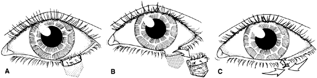
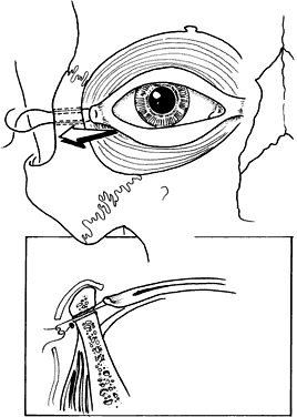
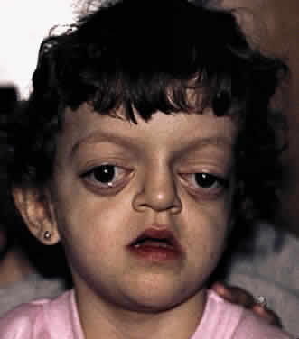
Goldenhar's syndrome first was described by Van Duyse in 1882, with additional cases and differentiation from the other first and second branchial arch syndromes reported by Goldenhar in 1952.13,31,53,60,61 The clinical features include corneoscleral dermoids, subconjunctival anterior dermoids or lipodermoids, upper eyelid colobomas, unilateral aplasia or hypoplasia of the mandibular ramus, small size or abnormal shape of one or both ears, preauricular skin tags, and vertebral anomalies13 (Fig. 10).

Fig. 10. Patient with typical craniofacial features of Goldenhar's syndrome.

Ocular abnormalities have been well documented, and epibulbar choristomas are the most consistent finding (32% to 92%).53,60 Lid colobomas frequently are lateral and appear in association with epibulbar dermoids, but they also can occur nasally.60,61 Ptosis, nasolacrimal duct dysfunction, iris colobomas, and lid skin tags are less frequent but also have been reported.60,61 Strabismus is noted in 10% to 19% of patients and includes esotropia, exotropia, Duane's syndrome, and sixth nerve palsy.31,53,60,61 Microphthalmos with associated intraocular malformations can occur in severe cases.53,60,61

Associated systemic features include microcephaly, hydrocephalus, plagiocephaly, intracranial dermoids, congenital heart disease, and kidney malformations.8

The etiology of Goldenhar's syndrome is unknown, but the clinical findings suggest aberrant development of the first and second branchial arches similar to Treacher Collins syndrome (see later) and the facial microsomias.23–25,58 An early cell induction abnormality originated by some environmental, vascular, or topographic insult also has been postulated.53 An animal model of stapedial artery hemorrhage with resultant maxillary and mandibular hypoplasia has been proposed to explain some forms of the first and second branchial arch syndromes.62

Treacher Collins Syndrome

Treacher Collins syndrome is the only clefting syndrome with a definite hereditary pattern. It has autosomal dominant transmission with variable penetrance. Treacher Collins syndrome also is known as mandibulofacial dysostosis or zygoauromandibular dysplasia.63–67 This bilateral anomaly represents a cluster of malformations produced by zygomatic temporoaural and mandibular dysplasia. Zwahlen in 1944 and immediately afterward Franceschetti and Zwahlen (1944) confirmed and elaborated on the original reports of Treacher and Collins.63–67

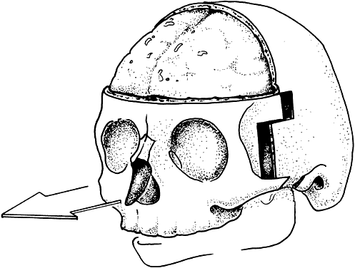
Mandibulofacial dysostosis is characterized by retrusion of the malar region, obliteration of the frontonasal angle, and a receding chin. Protrusion of the nose and maxilla may produce an enophthalmic appearance. The inferior-lateral angle of the orbit is defective, and the superolateral orbit is displaced caudally, giving the orbit an egg-shaped appearance. The orbital contents appear displaced into the deficiency created by the malar hypoplasia63–67 (Fig. 11).

Fig. 11. Patient with severe form of Treacher Collins syndrome.

Ocular abnormalities include pseudocolobomas and true colobomas of the lids, especially inferolaterally; canthal dystopia; orbital lipodermoids; limbal dermoids; and, occasionally, microphthalmos and anophthalmos. Strabismus is less common than in the syndromes described earlier but has been reported in several forms including esotropia, exotropia, Duane's syndrome, and cranial nerve palsies.31,63–67 Less frequently, nasolacrimal dysfunction and structural abnormalities of the globe can be present.

An infinite variety of ear abnormalities can be present. Respiration can be affected by choanal atresia and by lingual obstruction from mandibular retrusion.13,63

Facial Microsomia

Facial microsomia is one of the more frequent craniofacial malformations occurring with an estimated frequency of 1:4000 live births.68,69 Only rarely have familial cases been reported. A male predominance has been suggested, which was confirmed by our report of 49 patients with facial microsomias, 73% of whom were male.70 The deformity varies in extent and degree, at times showing a predominance of ear or jaw findings. A characteristic finding is hypoplasia of the mandible on the affected side or sides. The ear deformity ranges from complete absence to a normal ear with additional “ear tags.” The muscles of mastication, subcutaneous tissue, and facial muscles often are hypoplastic. The temporal bone, middle ear, mastoid process, base of the skull, and craniofacial skeleton may be involved and hypoplastic (Fig. 12). A classification scheme has been proposed.67 The hemifacial form accounts for 90% to 94% of facial microsomias (Table 3).

Fig. 12. Patient with hemifacial form of microsomia.


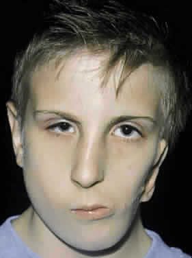
TABLE 3. Classification of Hemifacial Microsomia


TypeClinical Manifestations
IComplete facial skeleton, infact intramuscular joint, asymmetric mandible
IIAplasia of the condylar head, hypoplastic ascending ramus, ear deformities
IIIType II defects and hypoplastic zygomatic arch, rudimentary glenoid fossa
IVType III defects and hypoplastic lateral and inferior orbital rims
VType IV defects and orbital hypoplasia and dystopia, flattened temporal fossa


Ocular findings in patients with facial microsomia are varied. In a study of 49 patients, ocular or adnexal findings were present in 67% of patients. Sixteen percent of patients had visual loss, all secondary to unilateral amblyopia.70 This resulted from various combinations of uncorrected refractive errors (27%), anisometropia (8%), strabismus (22%), nystagmus (2 of 49 patients), and ptosis (12%). Forty-one percent of patients had associated lid or adnexal abnormalities, most commonly dacryostenosis and ptosis. As in other first and second branchial arch syndromes, incomitant strabismus was common. Of 11 patients with strabismus, 3 had Duane's syndrome, 2 had sixth nerve palsies, and 1 had bilateral superior oblique palsies.70

The absence of more severe systemic features (vertebral, renal, cerebral, multiple cranial nerves) justifies the separation of facial microsomias from Treacher Collins and Goldenhar's syndromes. The etiology may be explained by the animal model of stapedial artery hemorrhage discussed earlier.53,62,63,70

HYPERTELORISM

Converse and colleagues stressed the importance of differentiating between hypertelorism and teleorbitism.3 They defined teleorbitism as a congenital condition in which the interorbital distance increases from the apex to the orbital rim (true lateralization of the orbits)1,3,13 (Fig. 13). Hypertelorism, however, shows no true lateralization of the orbits but rather an increase only in the interorbital width. Hypertelorism is differentiated from telecanthus, which refers to a lateral displacement of the soft tissue. Telecanthus can occur without underlying hypertelorism, whereas hypertelorism always has associated telecanthus. Sometimes, a low nasal bridge may be another source of a false impression of hypertelorism. Measurement of the inner canthal distance and outer canthal distance may be used. Hypertelorism is present when the inner canthal distance is increased and the outer canthal distance is normal. Teleorbitism exists when both distances are increased.

Fig. 13. Patient with hypertelorism associated with frontonasal dysplasia and encephalocele.

Hypertelorism and telecanthus may present as isolated anomalies but more often are associated with other malformations.1,3,13,71 Teleorbitism is seen most commonly in patients with craniofacial synostosis.72 Other causes of teleorbitism include encephaloceles, pneumatoceles, meningoencephaloceles, and frontonasal dysplasia.72 In teleorbitism, the orbital volumes may be symmetric or asymmetric, and it is more commonly associated with a syndrome.

Greig's syndrome, or primary orbital hypertelorism in combination with other skeletal abnormalities, was the first form of primary hypertelorism to be described.13 This is a rare syndrome (1:100,000). Cohen's syndrome, or craniofrontonasal dysplasia, is more common. Teleorbitism, internasal dysplasia, and maxillary arching are the main characteristics and may be associated with premature coronal synostosis inherited as an autosomal dominant trait.12,13,71

Teleorbitism and hypertelorism often are associated with other anomalies. This includes the brain (agenesis of the corpus callosum, V-shaped separation of the lateral ventricles), the forehead (widow's peak, meningoencephaloceles), the eyebrow (dystopia, distortion), the eye (anophthalmia, microphthalmia, colobomata, strabismus, ptosis, refractive errors, and amblyopia), and the nose (internasal or nasal dysplasia).4,26,31,42,44,53,73

Back to Top
MANAGEMENT OF CRANIOFACIAL DEFORMITIES
Patients with craniofacial anomalies benefit from a team approach to management.73,74 This concept is not confined to the surgical treatment alone but begins immediately after birth as a continuous dialogue with all disciplines involved, as described earlier.

Once a specific diagnosis has been made, the decision of whether or when to operate and the selection of appropriate surgical techniques require an understanding of the relation between treatment and growth in the affected region.75–79 Timing and type of surgical intervention depend on three factors: (1) the growth potential inherent in the type of malformation, (2) the morphologic repercussions determined by the severity of the malformation combined with the surgical intervention, and (3) the effectiveness of extrinsic forces (e.g., dentofacial orthopedics) or intrinsic surgical stimuli (e.g., muscle transfer procedures).75–79

In most cases, intervention is staged, with one or more procedures performed in infancy and future procedures delayed until final craniofacial growth has been completed. The early procedures are performed to allow growth and function to be uninterrupted in major organ systems (brain, eyes, and nasopharynx).75–79

Team management begins after each member has evaluated the patient and family. A diagnosis is determined by characterizing the craniofacial deformity, ocular and adnexal anomalies, systemic disorders, and familial occurrence. Other testing may include chromosomal analysis, skeletal surveys, magnetic resonance imaging of the brain, computerized tomography with three-dimensional reconstruction of the skull and face, cephalometry, and dental panorex radiography. The use of ancillary testing is on a case-by-case basis. If the surgical teams are familiar with the deformity, then radiography is not routinely performed. If neurologic impairment is diagnosed clinically or the deformity is not characteristic, then imaging is obtained.

The special needs of these patients and their families extend beyond medical and surgical treatment. Special education, speech therapy, hearing and vision impairment, feeding difficulties, physical rehabilitation, travel, and the resultant financial stresses are overwhelming for many families. Once the medical and surgical problems are addressed, team members from psychiatry, psychology, and social work continue intimate involvement to assist the family with these problems. This involvement is initiated at the time of the family's first visit with the craniofacial team.

Overall, craniofacial surgical complication rates up to 16.5% have been reported in several series totaling 1793 patients.80–83 Central nervous system complications include meningitis, dural tears with cerebrospinal fluid leaks, and subdural or intracerebral hematomas. Complications of coronal incisions include sensory disturbance, wide scars, hair loss, and skin necrosis. Death, severe blood loss, velopharyngeal incompetence, bone graft resorption or infection, and relapse of condition all have been reported.80–83

Ocular and orbital complications occur with sufficient frequency so that ophthalmologists participating in the care of these patients must be familiar with them.33,80,84 Blindness can occur and probably is secondary to vascular insult from traction on the ophthalmic artery and its branches in the orbit. The alert craniofacial surgeon can recognize this by monitoring the pupil size intraoperatively. If dilation is recognized, then traction must be released. If the pupil remains dilated then corrective actions are undertaken, which may include removing bone grafts from the orbit or incision and decompression of the periorbita. Immediate ophthalmic consultation is obtained. Corneal abrasions (instrument or suture trauma), rupture of the globe (compression injury), orbital dystopia (unilateral shifts, trauma, or tumor resection), enophthalmos (trauma or resection of tumor), extraocular muscle dysfunction (entrapment, contusion, nerve damage), blepharoptosis (orbital advancement, trauma), epiphora (congenital or traumatic nasolacrimal duct obstruction), canthal dystopia (coronal flap incision, orbital bone shifts), distortion of the palpebral fissures (maxillary advancement), and transorbital constriction (hypertelorism correction) all have been reported.31–33,42–44,51,60,61,69,70,73,80,85–87

SPECIFIC MANAGEMENT

The management of ocular and adnexal problems in patients with craniofacial syndromes challenges the ophthalmologist because of the diversity and severity of problems that can be encountered. These problems can be directly related to the syndrome or result from either craniofacial or ophthalmic surgery. In addition, management of these problems often is further complicated because they occur during the critical period of visual development. Specific steps in management are best understood in terms of initial evaluation and problems resulting from the craniofacial surgery or the syndrome itself.

The craniofacial surgeon is primarily interested in changing periorbital bony contour and position.75–79,81,82 Periosteal elevation and dissection of the periorbita from the orbital wall usually are required and are most often accomplished using a bicoronal approach with an anterior craniotomy. After mobilizing the orbital contents, all four orbital walls can be fractured 15 mm anterior to the orbital apex as needed for particular syndrome repairs, allowing the orbit to be shifted in any desired direction (Figs. 14 and 15).

Fig. 14. Original Tessier mobilization of midface and orbits.

Fig. 15. Three different orbital medialization procedures used for the correction of hypertelorism.

The most obvious structures affected by these maneuvers include the medial and lateral canthal tendons, the nasolacrimal apparatus, the upper levator complex, and the inferior lid retractors. Remarkably, the intraorbital contents, including extraocular muscles, globe, and the nerve supply, usually are unaffected. Although the trochlea is disinserted with periosteal stripping, it spontaneously reattaches with preservation of preoperative function. Whereas the posterior orbit, with its important vascular and nervous structures, is not mobilized, extensive manipulation of the orbital contents may result in injury to these tissues.

The initial ophthalmic evaluation should be accomplished as soon as possible after the diagnosis of any craniofacial abnormality is made, preferably before any surgical intervention. This evaluation is easy to overlook because of the child's more serious deformities, but this prevents delay in treatment and diagnosis of critical ophthalmic disorders. A systematic evaluation of the visual system, ocular abnormalities, and adnexal structure and function allows the generation of a problem list for complete ophthalmic functional and cosmetic treatment. Use of a preprinted examination sheet provides a record of important baseline information (Fig. 16).

Fig. 16. Craniofacial examination recording form.

Vision

In general, visual function should be evaluated first. In infancy and early childhood, acuity testing with fixation preference technique is used. For better amblyopia detection, other useful acuity tests are the 10-prism diopter base-down test and grating acuity measured by Teller visual acuity cards.85 When the child can cooperate with subjective testing, usually at about 3 years of age, recognition acuities such as crowded HOTV, Allen cards, and Snellen optotypes can be obtained. If below-normal function is suspected, tests of visual function should include a visual-evoked response and an electroretinogram.

Globe

Abnormalities can be diagnosed with routine examination techniques. Young infants may be swaddled and positioned upright in the slit lamp or supine for a hand-held portable slit lamp. Pupillary examination and anterior segment examinations are performed in standard fashion. Intraocular pressure measurements may be necessary and can be performed by hand-held applanation or indentation devices on some infants while they are feeding or nursing and on older children at the slit lamp. Examination under anesthesia must be considered if these office efforts are unsatisfactory and there is clinical suspicion of glaucoma.

Motility

Examination includes sensory testing of fusional status and measurements of versions and ocular alignment in five (straight, right, left, and up, and down) positions of gaze, right and left head tilt, and near. The presence or absence of Bell's phenomenon should be noted. If warranted, forced duction and forced generation testing can be added. A convenient recording system for these measurements is illustrated by the diagram shown on the craniofacial evaluation sheet (Fig. 17).

Fig. 17. Three-dimensional computed tomography of a patient with Crouzon's syndrome with both lateral (A and B) and anterior (C and D) reconstruction.

Adnexal Examination

Adnexal examination includes evaluation of the position and shape of the lids, palpebral fissures, and canthi. The eyelids should be evaluated for trichiasis, entropion, ectropion, colobomas, distichiasis, ptosis, and epicanthal folds. Vertical and horizontal fissure length, upper and lower lid crease position, and marginal reflex distance are important points of information in the evaluation of ptosis in these patients. Levator muscle excursion from downgaze to upgaze should be measured in millimeters. If ptosis is present, the response of Müller's muscle to 2.5% phenylephrine hydrochloride (Neo-Synephrine) is recorded. Upward or downward displacement of the canthi from a horizontal line “drawn” through both medial canthi and extending to each lateral rim (dystopia) also should be noted and recorded in millimeters. The normal lateral canthus usually is 1 to 2 mm above the horizontal level of the medial canthal tendons.

Telecanthus and Hypertelorism

Telecanthus is measured by the intercanthal distance, which is the soft tissue distance between the medial canthal angles of each eyelid fissure. In infants, this generally is less than 20 mm; in older children, less than 24 mm; and in adults, less than 30 mm. Hypertelorism is technically measured by the distance between the bony medial orbital walls. This usually parallels the measurements for the overlying telecanthus. Although it is possible to have isolated telecanthus, this is unusual in craniofacial syndromes. A good clinical guide to the degree of telecanthus is the ratio of the intercanthal distance to the interpupillary distance. The normal ratio is approximately 50%. Values larger than this represent telecanthus and a possible underlying hypertelorism. This measurement system may be inaccurate in the presence of large degrees of strabismus if the strabismic angle is ignored.

Tear System Analysis

In tear system analysis, the presence of bony defects or extra tissue is noted. Dye disappearance testing is performed in patients with epiphora to evaluate tear outflow. If additional information about the nasolacrimal system is needed, irrigation, dacryocystography, and lacrimal scintigraphy can be used.40,88

Proptosis

Measurements can be accomplished with the Luedde or Hertel exophthalmometer. The Luedde exophthalmometer is more useful in children, since it is less threatening than the Hertel instrument. Proptosis is one of the clinical findings in many syndromes such as Crouzon's and Apert's or may be seen in the immediate postoperative period as a result of orbital dissection. If the lateral orbital rims, which are used as reference points for measurements with both the Hertel and Luedde exophthalmometers, are asymmetric or hypoplastic, these measurements will provide less than accurate comparisons between the two eyes. Such asymmetries should be noted.

Cyclopegic Refraction and Intraocular Examination

Retinoscopy is accomplished 30 to 60 minutes after using 0.5% cyclogyl in infants younger than 1 year of age and 1% cyclogyl in infants and children older than 1 year of age. Direct and indirect ophthalmoscopic examination is performed. This is important, since many patients with craniofacial abnormalities have structural problems of the globe, anisometropia, and amblyogenic refractive errors.

Additional Testing

Additional testing may include formal visual fields and assessment of color vision. In many cases, radiographic investigations are necessary. Computed tomography with three-dimensional reconstruction is useful for evaluating bony intraorbital and intracranial abnormalities (see Fig. 17). Specific orbital soft tissue and intracranial deformities may be more thoroughly examined with magnetic resonance imaging.

Follow-Up Evaluation

Follow-up evaluation is performed in the first 2 months after craniofacial surgery for evaluation of any changes secondary to the surgery. If indicated by the initial examination results or by a postoperative complication, other follow-up evaluations are performed. If no significant ocular abnormalities exist, then the patients are examined as part of their routine evaluation in the craniofacial clinic.

Craniofacial abnormalities spare no part of the visual system. The globe, lids, extraocular muscles, adnexa, optic nerves, and central visual pathways may be affected individually or in numerous combinations. Secondary visual loss from amblyopia is a constant threat to vision in this population of patients because of the immaturity of the visual system. Because of the advances made in the early diagnosis and treatment of many craniofacial disorders, amblyopia is the most constant and prevalent cause of sustained visual loss in this population of patients.31,70

Back to Top
OPHTHALMIC SURGICAL REPAIR
The variety and types of surgical repairs for the ophthalmic surgical problems generally follow the basic tenets that govern their use in noncraniofacial patients. Critical areas in these patients include globe abnormalities (conjunctival and corneal exposure, glaucoma, cataract), eyelid deformities, ectropion, canthal deformities, ptosis, nasolacrimal abnormalities, colobomas, amblyopia, and motility problems. The indications and contraindications for treatment are reviewed, including a description of certain unique aspects of these procedures, expected results, and complications. Ophthalmic surgery texts and other chapters in these volumes provide detailed explanations of the more common related surgical procedures referred to in the following discussion.

GLOBE

More common abnormalities of the globe include exposure conjunctivitis and keratitis. Conjunctival prolapse and keratinization may occur from prolonged exposure common in many craniofacial syndromes. Corneal scarring, thinning, or infectious keratitis also are potential sequelae from exposure or irritation from trichiasis, distichiasis, entropion, ectropion, and lagophthalmos after ptosis repair. Treatment is indicated if signs and symptoms of inflammation appear. These include mild conjunctivitis, punctate keratitis, and nocturnal lagophthalmos. The definitive treatment for severe exophthalmos is orbital advancement (Fig. 18), which can be carried out in the first few weeks of life, if necessary. Except in the most severe cases, however, this usually is not required, and exposure usually can be managed by frequent and careful lubrication with eye drops or ointment. In more severe circumstances, a protective moist chamber using periorbital clear plastic and frequent ointment instillation, or even tarsorrhaphy, may be necessary. In such situations, the cornea must be evaluated on a daily basis for evidence of corneal breakdown, infection, thinning, or scarring.

Fig. 18. Superior view demonstrating correction of proptosis by orbital advancement with bone grafts.

Glaucoma is in the differential diagnosis and occurs frequently enough that this diagnosis should be considered in patients with corneal clouding, enlargement, tearing, and photophobia. If necessary, intraocular pressure measurements are obtained under anesthesia.14,76,89

Other structural problems of the globe are managed in standard fashion. Glaucoma, as well as congenital or infantile cataracts, are managed surgically and optically in the same way as the child without a craniofacial problem. The treatment of these problems should be prompt and not automatically delayed until after the patient has had craniofacial surgery.

LIDS AND LASHES

Lid and lash malformations occur frequently and include (1) ptosis, (2) colobomas, (3) entropion, (4) ectropion, and (5) trichiasis and distichiasis.

Ptosis

TREATMENT INDICATIONS. Early surgical treatment is initiated only when occlusion of the visual axis is significant. This usually is of most concern in unilateral ptosis, when the threat of amblyopia is the greatest. In most instances, amblyopia prevention can be successfully instituted by patching the normal eye a few hours a day. Attention to the child's effort to use the brows combined with a posterior head tilt are valuable clinical signs that the child is attempting to use the affected eye, but this does not rule out amblyopia. Absence of these signs in a child with unilateral ptosis indicates poor vision in the eye. If possible, use of preverbal objective assessment of vision should be accomplished. This includes methods such as the Teller Acuity Cards and the 10-prism diopter base-down test. These measures are obtained as a baseline value and repeated on subsequent visits to follow the effectiveness of amblyopia treatment.

Craniofacial surgery usually has little effect on the level of a ptotic lid. In some cases, the level of the lid is lowered after surgery, but in most cases it is unaffected. Definitive surgery usually is postponed until 1 year of age, if possible, and the choice of procedure depends on the amount of levator function present.

CONTRAINDICATIONS. Contraindications include lid retraction, proptosis with exposure keratoconjunctivitis, poor extraocular motility, and poor function of cranial nerves V, VII, or both.

TREATMENT. In later infancy and childhood, when definitive surgery is accomplished, minimal to no levator function usually requires a suspension procedure with autogenous or irradiated donor fascia lata.90 Moderate to good levator function can be treated with a levator resection or, less frequently, with a conjunctivotarsomüllerectomy (Fasanella-Servat procedure) if there is a good response to stimulation of Müller's muscle with 10% phenylephrine ophthalmic drops (2.5% for infants).

In the early months of life, if there is severe unilateral ptosis, a levator resection is not practical. In such circumstances, a nonabsorbable material (e.g., Silastic or Supramid) may be used as a temporizing frontalis suspension. In later infancy and childhood, when definitive surgery is accomplished, minimal to no levator function usually requires a suspension procedure with fascia lata, whereas moderate to good levator function can be treated with a levator resection or less frequently with a conjunctivotarsomüllerectomy, if there is a good response to stimulation of Müller's muscle with 2.5% phenylephrine hydrochloride drops.

RESULTS. In most instances, the results of ptosis procedures in patients with craniofacial disorders are similar to those observed in patients with other forms of congenital ptosis. With moderate to good levator function, resection is successful approximately 80% of the time with one procedure. The conjunctivotarsomüllerectomy success rate approaches 90% if performed in patients with a good response to 2.5% phenylephrine hydrochloride. Frontalis suspension usually gives improvement and has less risk for exposure than maximal levator resection. A satisfactory contour with use of the brow is observed in 60% to 70% of all cases, but the severity of the problems in certain conditions, such as those associated with severe blepharophimosis, renders the results much less successful. In any event, whereas frontalis suspension is not a particularly physiologic method for producing a normal eyelid, it is the only alternative to provide adequate lid elevation. Maximal levator resections, especially if Whitnall's ligament is used for suspension, also can be problematic, since there is much greater lagophthalmos with attendant difficulties in controlling exposure. A good level and contour in severe ptosis may be achieved, but then it may be found that a recession of the levator is required to control exposure. This also is a problem in circumstances in which there is poor elevation of the globe.

COMPLICATIONS. Complications include undercorrection and overcorrection, excessive hemorrhage and scarring, misplaced or slippage of sutures, lid lag and lagophthalmos leading to exposure keratitis, peaking of the lid, and infection.

Lid Colobomas

INDICATIONS. The appropriate treatment of lid colobomas depends on the size, configuration, and location of the defect. Upper lid colobomas are the greatest threat for exposure keratoconjunctivitis. Small defects in the nasal portion of the lower lid associated with lacrimal anomalies need more aggressive treatment than small notches in the outer lid as, for example, those associated with Treacher Collins syndrome.

CONTRAINDICATIONS. Contraindications include age younger than 6 months (unless severe exposure develops), proptosis, and severe underlying bony deficiency.

TREATMENT. Sliding or rotational flaps are preferred treatment techniques, since lid sharing procedures risk amblyopia. Two techniques include pentagonal repair with or without cantholysis and a Tenzel myococutaneous flap.

Pentagonal Repair. With pentagonal repair, the coloboma is converted into a pentagonal defect and repaired as though it were a lid margin laceration. This can be used to correct up to 50% of lid margin defects if combined with a canthotomy and cantholysis (Fig. 19, top).

Fig. 19. Myocutaneous flap incision for upper and lower eyelid reconstruction. (Top) Lateral lower lid coloboma correction. (Bottom) Middle lid coloboma correction.

Semicircle Flap. A semicircle flap is a myocutaneous flap designed at the lateral canthus and cheek. The size of the flap is determined by the size of the coloboma (see Fig. 19, bottom). The size of the flap can range from small (Tenzel) to a large cheek flap (Mustarde). A large defect may require a tarsoconjunctival graft, which can be obtained from the opposite lid as free graft, or a sliding flap from the lower lid. If such a procedure needs to be performed, amblyopia in the operated eye will need to be treated after separation of the flap at 7 to 10 days.

RESULTS. Sliding flaps are effective in covering the defect and improving cosmesis. Lid movement may not be restored to normal, leaving the patient with variable degrees of ptosis, lagophthalmos, or lid lag, depending on the amount of the original defect.

COMPLICATIONS. Horizontal shortening may produce lid malpositions, including ptosis, entropion, or ectropion. The tethering effect of a repair may be reduced by a careful advancement of the levator muscle during the coloboma repair. Most often, however, it is best to defer definitive ptosis surgery until adequate time has passed for healing and swelling to reduce.

Entropion

Entropion may occur as a congenital problem associated with many craniofacial syndromes, or it may result from disinsertion of the lower eyelid retractors.41–44 This condition often coexists with epiblepharon.

TREATMENT INDICATIONS. Entropion in Asian children is common and resolves in most cases without treatment. The clinical course often is asymptomatic with spontaneous resolution. Surgery is indicated in cases that persist or in which corneal integrity is threatened.

CONTRAINDICATIONS. If reconstructive orbital surgery is anticipated, it is best to delay surgery until after this is completed.

TREATMENT APPROACH. A tuck or advancement of the inferior lid retractors is the recommended procedure. A horizontal incision is made 2 to 3 mm below the lower eyelid margin for 30% to 80% of the horizontal length, depending on the horizontal extent of the malposition. The incision is dissected to the lower tarsal border, excising excess preseptal orbicularis oculi muscle and a strip of skin. The lower lid retractors are identified and advanced on the tarsus with interrupted 5-0 vicryl sutures, which rotates the margin. The skin then is closed with a running 6-0 fast-absorbing plain gut suture with supporting interrupted 7-0 dexon sutures.

RESULTS. This procedure prevents trichiasis and corneal damage and improves the appearance of the lower lid.

COMPLICATIONS. Damage to the inferior oblique origin and penetration of the inferior sclera, which are in close proximity to the lower lid retractors, recurrence of entropion, and secondary ectropion can occur.

Ectropion

Lateral lower lid ectropion may be observed with canthal dystopia that accompanies many syndromes.31,32,44 This is much less common than entropion and usually is the result of vertical skin deficiency.

INDICATIONS. Lacrimal outflow dysfunction, corneal exposure, and cosmetic deformity indicate the need for treatment.

CONTRAINDICATIONS. Anticipated major craniofacial reconstruction is a contraindication to treatment.

TREATMENT. Tightening of the horizontal lid length by canthal tendon plication or tarsal shortening may be required.

RESULTS. Effective restoration of normal lid position and function is achieved with these procedures.

COMPLICATIONS. Complications include overcorrection, undercorrection, and lacrimal system damage.

Trichiasis or Distichiasis

TREATMENT INDICATIONS. Treatment indications encompass corneal irritation, abrasion, or ulceration.

CONTRAINDICATIONS. Major craniofacial reconstruction, particularly around the face and orbits contraindicate treatment.

TREATMENT. If the trichiasis is secondary to a primary lid abnormality, surgical correction of the lid position resolves the trichiasis. Primary trichiasis is managed conservatively first, and this may include epilation, soft contact lenses, or electrolysis. A new lash will regrow in 6 weeks, and soft contact lenses usually are not useful for long-term treatment. Electrolysis is valuable if only a few lashes are affected. If more than a few lashes are involved, a more complex surgical approach is recommended.

Distichiasis has a higher recurrence rate after conservative management and often needs surgical intervention. Management of this entity is difficult and often unsuccessful. If it is localized, then a wedge resection with primary closure may be effective. Cryotherapy can be used after splitting the posterior lamella and applying a double “freeze-thaw” to this layer only. The technique of treatment used most often includes splitting the lid and excising the posterior lamella, including the abnormal lashes and metaplastic meibomian glands. The excised area then can be replaced with full-thickness mucous membrane grafts. This is best done with the operating microscope because all offending lashes must be removed, and suturing of the graft with a small-caliber suture requires adequate visualization (Fig. 20).

Fig. 20. Margin and posterior lamellar pentagonal wedge resection for the treatment of distichiasis. A. Lid margin line of resection. B. Resected margin and posterior lamella. C. Reapproximation of lid margin.

RESULTS. Although cryotherapy can have a good success rate when there are only few offending lashes, electrolysis works as effectively with less risk of complications. For distichiasis or severe trichiasis, the long-term results often are poor and also may involve significant cosmetic deformity.

COMPLICATIONS. Cryotherapy may cause a complete loss of lashes, depigmentation, cicatricial changes in the lid margins, edema and bullae formation, infection, and secondary entropion and ectropion. Posterior lamella treatments for distichiasis may cause lid distortion, entropion, and trichiasis.

CANTHAL DYSTOPIA

Treatment Indications

Treatment indications include lid malfunction and deformity that can produce epiphora, chronic keratoconjunctivitis, or exposure problems.

Contraindications

No underlying bony support, future underlying bony reconstruction, and severe mental retardation where cosmetic improvement is of questionable value.

Treatment

This deformity usually is corrected at the time of craniofacial repair. Medial canthal dystopia often is amenable to repair during major craniofacial reconstruction because of the excellent exposure. Lateral canthal dystopia also can be repaired at this time, but other associated deformities such as maxillary hypoplasia may need to be repaired in staged procedures. Nonfunctional lid and canthal deformities often are managed surgically at a later time.

MEDIAL CANTHOPEXY. Medial canthopexy can be achieved in several ways, depending on the severity of the problem. For mild forms, the canthal tendon can be tucked or advanced with sutures placed into the periosteum. A 4-0 vicryl suture on a curved P2 needle is used. An alternative technique is use of a titanium anchor system, which is placed into a predrilled hole posterior to the lacrimal sac.91,92 The canthal tendon can be attached to this point, pulling the lid into proper position against the globe.

For more severe cases, transnasal wiring is most effective. A hole is created with a trocar posterior to the site of insertion of the canthal tendon. Care must be taken to protect the lacrimal sac. More importantly, a malleable retractor must be placed in front of the opposite globe to avoid perforation while creating the bony opening. The hole must be of a diameter sufficiently wide to allow the tendon to be inserted so as to pull the lid in apposition to the globe. After identification of the tendon, it is secured with a 30-gauge stainless steel suture, leaving both ends long. Then, parallel to and just below the bridge of the nose, a Wright needle is passed from the opposite side through the hole made by the trocar. Use of an air drill facilitates creation of this pathway. The sutures are threaded into the eye of the needle and then withdrawn. The suture ends are brought through the opposite canthal tendon if there is a bilateral problem such as telecanthus. For unilateral problems, two holes can be made on the opposite side above the canthal tendon and lacrimal sac and the suture ends brought through with the needle as a guide. They are then twisted to the appropriate degree of tension to pull the abnormal canthus into the desired position. The ends are cut, and the skin incision closed in layers with absorbable sutures (Fig. 21).

Fig. 21. Transnasal wiring technique for medial canthopexy. A. Isolation of tendon and nasal osteotomies. B. Cannulation of osteotomies with a 30-gauge wire. C. Positioning of medial canthal tendon.

LATERAL CANTHOPEXY. Abnormal insertion of the lateral canthal tendon may be caused by deficiency of the tendon itself or by a faulty position of an otherwise normal tendon. In severe cases, two small holes are burred in the lateral wall of the orbit, directly behind the rim. The lateral canthal tendon is secured with a 30-gauge stainless steel wire or a heavy suture such as a monofilament 3-0 nylon, and both ends are left long. Hollow needles passed through the holes can guide the ends of the tendon suture. The needles are withdrawn as soon as the suture ends become visible at the other end of the needle. A firm knot is tied. The restoration of the contour in the lateral canthal area depends on the close approximation of the skin to the underlying skeleton. This may be achieved by fixation of the dermis to the periosteum of the lateral orbital rim using one or two sutures. In mild cases, the lid can be brought into position with a tarsal strip type procedure (Fig. 22).

Fig. 22. Lateral canthopexy through lateral orbital rim.

Results

Restoring normal relations between the two canthi is basic in patients with craniosynostoses. In patients with clefting syndromes, this may prove more difficult, particularly when there are severe bony deficiencies (craniofacial clefts, Goldenhar's syndrome, Treacher Collins syndrome). Although canthal repositioning may be effective in restoring some lid function, lacrimal dysfunction often is the result of primary disorders of the nasolacrimal system and requires additional procedures.

Complications

Complications are infrequent and usually are mechanical, including canthal drift flattening of the nasal root sulcus and epicanthus, asymmetry; suturing the lid to the anterior reflection of the medial canthal tendon or outside of the lateral orbital rim, resulting in poor apposition to the globe; entropion; ectropion; trichiasis; injury of the upper lacrimal system; and, rarely, flap infection, hemorrhage, and necrosis.

TELECANTHUS AND ASSOCIATED LID AND BROW DEFORMITIES

Eyebrow anomalies and other lid anomalies of shape, size, and structure can be associated with telecanthus. Telecanthus is associated with craniofacial synostoses, encephaloceles, and clefting syndromes, whereas other severe structural abnormalities are associated with anophthalmos, cryptophthalmos, and microphthalmos.

Treatment Indications

Treatment indications include exorbitism, occlusion of the visual axis, interference with normal lacrimal function, and cosmetic deformity.

Contraindications

Poor prognosis for patient survival is a contraindication to treatment. If there is no functional disability and the deformity is largely a cosmetic problem, deferral of surgery until complete craniofacial growth occurs is preferred.

Treatment

Eyebrow repair is directed toward reorientation of distorted parts, restoration of continuity, and reconstruction of defects. This is accomplished with Z-plasties, H-plasties, and hair from the other eyebrow or scalp (Fig. 23).

Fig. 23. Z-plasty eyebrow reconstruction.

Surgical correction of telecanthus consists of separate approaches to three soft tissue elements within the medial canthus responsible for the anomaly. These include (1) skin and underlying fascia, (2) subcutaneous tissue and muscle, and (3) the medial canthal tendon. The skin is most commonly repaired with a Y-to-V flap or Roveda procedure (Fig. 24). The subcutaneous tissue and muscle are excised under direct visualization after inserting lacrimal probes in the canalicular system. Medial canthal tendon shortening procedures include tucks, resection, or transnasal wiring in more severe cases, as described previously. Tucking or resection of the tendon is indicated only in mild forms of telecanthus.

Fig. 24. Various epiblepharon repair procedures for telecanthus.

Results

Eyebrow repair is best if sliding flaps or the other eyebrow are used. Telecanthus repair provides symmetric and more normal-appearing medial canthal areas.

Complications

Asymmetry, nasolacrimal damage, lid margin malposition may be complications.

NASOLACRIMAL SYSTEM

Nasolacrimal dysfunction occurs in association with many craniofacial syndromes.

Indications

Acute treatment is required for dacryocystitis and amniocele. In patients with these problems, careful office probing with a No. 000 or 0000 probe is accomplished to open the system if prior massage is unsuccessful. In most instances, however, conservative management is followed if possible until the patient is 1 year of age. Major surgical correction of nasolacrimal dysfunction should be deferred until all bony reconstruction in the midfacial region has been completed. Because of this, on occasion, definitive surgical treatment such as dacryocystorhinostomy may be deferred until the teenage years. Since a well-functioning system may be disrupted during craniofacial surgery leading to postoperative epiphora, prophylactic intubation of the nasolacrimal system can be considered, although this is not performed routinely. In those instances in which there is a high likelihood of nasolacrimal duct dysfunction, preoperative intubation may save the system. In general, treatment guidelines are similar to noncraniofacial conditions (covered elsewhere in these volumes).

Contraindications

Anticipated major craniofacial reconstruction is a contraindication.

Treatment

Probing and irrigation with infracture of the inferior turbinate is performed initially; if this is unsuccessful, it can be followed by Silastic intubation of the entire nasolacrimal system. Silastic intubation can be performed in several ways. The traditional bicanalicular intubation can be performed with Crawford tubes or with a Ritleng system, which uses a prolene leader to the Silastic tubing. The suture and tubing are introduced through a grooved metallic probe.93 The suture is retrieved from the inferior meatus and the probe withdrawn. The suture then is slid out from the groove and the attached tubing pulled through the canaliculus and duct. This is repeated for the opposing canaliculus for bincanalicular intubation. The Ritleng system obviates the need to retrieve a metallic probe from the inferior meatus, minimizing trauma to the nasal passageway. Monocanalicular intubation is another option, especially useful in cases of canalicular agenesis where only one canaliculus is present.94 The success rate of monocanalicular intubation has been reported to be 79% in one study; however, this is not as high a success rate as demonstrated by most bicanalicular studies.95

Balloon dilation of the nasolacrimal duct has been suggested (1) for patients who have failed previous probing, (2) for patients who present at a later age of onset, or (3) as an adjunct or alternative to Silastic tubing intubation. The balloon catheter, similar to classic balloon angioplasty, introduces a balloon on a flexible wire to the nasolacrimal duct. The balloon then is inflated with saline to a prescribed pressure for a prescribed period of time. Preliminary studies report success rates as high as 95%, but more controlled studies without use of systemic steroids or antibiotics are needed.96 Use as a primary procedure is in effect the same as initial probing, which generally has a an even higher success rate.

If the above efforts fail in the face of a lower system obstruction, then a dacryocystorhinostomy may be necessary. This may be accomplished by an external approach or by an endoscopic approach, which avoids an external scar but may have a lower success rate than an external approach.97,98 The endoscopic approach may include use of the carbon dioxide or KTP laser, which introduces a higher cost and does not appear to contribute to an increased success rate.97 The KTP laser also can be used to lyse soft tissue scarring and reopen the ostium in the case of failed dacryocystorhinostomy. Again, however, if dacryocystorhinostomy, canaliculodacryocystorhinostomy, or conjunctivodacryocystorhinostomy are needed, they should be deferred until all bony work has been completed in the midface region. Whereas craniofacial patients are more challenging, these techniques are essentially similar in concept to the routine. Detailed descriptions of these techniques are provided elsewhere.

Results

Satisfactory results are obtained with successful probing and irrigation. If this fails, most patients do well with Silastic intubation. In some craniofacial syndromes, the puncta may be displaced far laterally, making Silastic intubation a potential problem because of potential corneal irritation from the loop of tubing. The need for more extensive lacrimal surgery decreases the chances for long-term success. Dacryocystorhinostomy and conjunctivodacryocystorhinostomy may be more difficult in patients with major craniofacial malformations; however, but can be successfully achieved in these patients with perseverance.99 Patients and families should understand that multiple procedures may be required to establish and maintain tear drainage. Abnormal structures and unusual anatomic relations provide a challenge and make it difficult to describe a “standard” approach. This is essentially true after orbital shifts or onlay bone grafts are placed in this region. Creating an ostium through a bone graft is a major challenge. The ophthalmic surgeon also must understand the previous craniofacial procedures and underlying disease to avoid central nervous system complications such as cerebrospinal fluid leaks, meningitis, and damage to the brain. This is especially important when operating on patients with frontonasal dysplasia.

Complications

Slippage and loss of Silastic tubing and restenosis after Silastic removal are contraindications. Dacryocystorhinostomy and conjunctivodacryocystorhinostomy can result in hemorrhage and infection. Reoperation procedures can fail secondary to scarring from previous craniofacial and oculoplastic procedures.

MOTILITY

Ocular motor disturbances occur in most patients with craniofacial syndromes. Depending on the syndrome, the reported incidence can be as high as 100%, but an overall estimate for all craniofacial patients is 70% to 75%. All types of motility disturbances have been reported, with horizontal deviations the most common and exotropia slightly more common than esotropia. Vertical deviations and A- or V-patterns may occur alone or in combination with the horizontal deviation.100,101 Dissociated vertical deviation, overacting oblique muscles, absent muscles (predominantly the superior oblique and superior rectus), and anomalous muscles are largely responsible for these patterns and vertical deviations. Nystagmus and other forms of strabismus (cranial nerve palsies, Duane's syndrome, and Brown's syndrome) also occur with a higher frequency in this population of patients. Unlike noncraniofacial strabismus, infranuclear, muscular, and orbital maldevelopment contribute to the eventual development of strabismus.36–38 Management of these disorders begins with a routine motility evaluation. This includes evaluating for accommodative, refractive, distance-near relations, and amblyopia.

Treatment Indications

In general, strabismus is treated in the same manner as in noncraniofacial patients. Spectacle and appropriate amblyopia treatment should be carried out before surgical intervention. Since early ocular alignment is beneficial in the treatment of strabismus in infancy and childhood, our approach to the timing of surgical treatment is independent of the craniofacial surgery and is governed by the diagnosis.

Contraindications

It has been shown that in most cases, craniofacial surgery does not usually affect the type or degree of strabismus.36,37,80,87 We do not perform strabismus surgery if there is a variable and inconsistent deviation, associated neurologic abnormalities that may be contributing to the deviation, or a suspected cranial nerve palsy, unless stable alignment during 6 months of observation has been accomplished. A blind eye with a sensory deviation is not operated on until noticeably disturbing to the patient or family.

Treatment

Surgical techniques for routine strabismus problems are equally effective in craniofacial patients. Because of associated extraocular muscle anomalies, it is our preference to use the fornix incision, from which we can easily explore all muscle insertions to observe and record the anomalous anatomy. Two incisions are placed in the inferotemporal and superonasal quadrants. From these incisions, all the muscles can be explored without disturbing the limbal conjunctiva and intermuscular membranes (Fig. 25). Occasionally, an absent or anomalous muscle may change the surgical plan. We also prefer large recessions (14 mm) of the inferior oblique rather than myectomy. This allows sufficient correction of vertical deviation in adduction and of the V-pattern while preserving some elevation function. When the patients are older, we prefer to use the adjustable suture technique.

Fig. 25. Superior nasal (N) and inferior temporal (T) fornix incisions for exploration of superior rectus (SR), superior oblique (SO), medial rectus (MR), and inferior rectus (IR), inferior oblique (IO), lateral rectus (LR), respectively.

Results

In most common forms of esotropia and exotropia, there is a 60% to 70% success rate for alignment of the visual axis in the primary position with one procedure.80,87 Many patients require multiple procedures, and comitancy is extremely difficult to obtain, especially in patients with absent and anomalous muscles.

Complications

The most frequent complications are overcorrection and undercorrection. They occur more frequently in these patients because of their anomalous anatomy, which predisposes them to incomitancy. The lack of comitancy and high prevalence of amblyopia and refractive errors make it difficult for these patients to attain binocularity. Without developing some degree of sensory fusion, these patients are predisposed to recurrence of strabismus. More serious complications, such as infections, scleral perforations, slipped muscles, and retinal detachment, are observed no more frequently than in patients with noncraniofacial strabismus.

AMBLYOPIA

As treatment techniques have changed, the pattern and prevalence of visual loss in patients with major craniofacial abnormalities also have changed. In a recent review of 322 patients with major craniofacial abnormalities presenting to the Craniofacial Clinic at The Children's Hospital of Philadelphia between 1979 and 1991, we report that visual loss (defined as vision less than 20/40 Snellen optotype or clinical equivalent) occurred in 20% of all eyes.87 Ninety percent of the visual loss was secondary to amblyopia, whereas 10% was secondary to structural abnormalities of the globe or visual pathways.87 This amblyopia was multifactorial with contributions from strabismus, refractive errors, anisometropia, and deprivation (corneal opacities, ptosis, cataracts) (Fig. 26).

Fig. 26. Percentage of 322 patients with craniostenoses (stenose), clefting syndromes (clefts), traumatic deformities (trauma), tumors of the orbit (tumors), and other isolated anomalies (other) with (A) abnormal vision, (B) strabismus. (C) significant refractive errors (greater than + 2.00 or -1.50 diopter sphere or 1.50 diopter cylinder), and (D) amblyopia (visual loss not attributable to organic lesions in combination with known amblyogenic factors, e.g., strabismus, anisometropia, high refractive errors, ptosis).

Treatment of amblyopia in these patients often presents unique challenges. There are many demands placed on these children and their families. The child usually has other disabilities such as hearing loss, poor dentition and speech, developmental delay, mental retardation, or learning disabilities. Whereas all of these problems are addressed through the team approach to treatment, from the family's perspective, the surgical treatment of the child's cosmetic deformities may take precedence over what is perceived as less important treatment, such as patching for amblyopia. Orthodontic devices, educational activities, emotional adjustment, and physical rehabilitation require time and energy that also can potentially detract from amblyopia therapy. Even in severe craniofacial deformities, amblyopia is a preventable form of vision loss. Extra effort is needed in detecting the presence of the multiple factors often responsible for visual loss. Careful attention to amblyopia management, including occlusion, refraction, spectacle wear, and timely correction of strabismus, is critical to maximize the visual potential of these patients.

Back to Top
GENETICS
Both genetic and environmental factors contribute to craniosynostosis. These malformations initially were classified on a clinical basis, and those systems continue to dominate much of the clinical literature. Several reports clarify the underlying mutations in many of these syndromes, allowing the complexity of the relation between mutation and resultant phenotype to be viewed more clearly. Abnormal mechanical factors (external pressure or underlying deficiency in brain growth) may be a predisposing cause in some cases. In others, a family history or associated anomalies suggest a genetically determined condition. Now, over 100 syndromes associated with craniostenosis have been delineated, and most of the common ones exhibit dominant inheritance.

The clinical observation that many craniostenosis are accompanied by limb abnormalities suggests that aspects of craniofacial and limb development use common molecular pathways, an idea supported by experimental evidence. This insight has been an important contributor to recent success in the identification of genes mutated in craniosynostosis. These discoveries in the genetics of craniosynostosis are the foundation for a new classification that relies more on the genotype rather than the phenotype. There are currently 64 different mutations of six genes described in over 400 patients that cause craniosynostosis. These range from unique missense mutations in the MSX2 (muscle segment homeobox 2) and FBN1 (fibrillin) genes described in single families, to 46 mutations in seven phenotypes for FGFR2 (fibroblast growth factor receptor 2).

The fibroblast growth factor receptor (FGFR) complex is a ubiquitous regulator of development and adult tissue homeostasis that bridges the pericellular matrix and the intracellular environment. Diverse members of the fibroblast growth factor polypeptide family, the FGFR tyrosine kinase (FGFRTK) family, and the FGFR heparin sulfate proteoglycan (FGFRHS) family combine to result in active and specific FGFR signal transduction complexes. Many human craniosynostosis syndromes are caused by mutations in the extracellular domain of receptors for fibroblast growth factors, which result in constitutive receptor activation. Fibroblast growth factor signaling pathways are involved in craniofacial development and suggest that some human malformation pedigrees or sporadic craniosynostosis may be caused by mutations that deregulate expression of the fibroblast growth factor ligands. In the more common syndromes of Crouzon, Pfeiffer, Jackson-Weiss, and Apert, mutations were found in the gene coding for FGFR2. Less frequently, mutations are observed in FGFR1 and FGFR3 in some cases of Crouzon's and Pfeiffer's syndromes. The mutations identified in FGFR2 are located in exons 5 and 7 of the gene that codes for immunoglobulin (Ig)-like chain III and the region linking Ig II and Ig III of the receptor. These domains of the receptor are important for ligand binding. Numerous mutations in these two exons have been shown to cause various craniosynostotic syndromes. Apart from Apert's syndrome, identical mutations are found in the clinically distinct syndromes of Crouzon, Pfeiffer, and Jackson-Weiss. Furthermore, the same gene defect can result in a highly variable phenotype, even within one family. Therefore, the clinically distinct craniosynostotic syndromes are extremes of a spectrum of craniofacial abnormalities and not nosologic entities. Dominant mutations in three fibroblast growth factor receptor genes (FGFR1–3) cause Crouzon's, Jackson-Weiss, Pfeiffer's, and Apert's syndromes. In mouse embryos, the muscle segment homeobox genes, Msx-1 and Msx-2, are expressed during critical stages of neural tube, neural crest, and craniofacial development, suggesting that these genes play important roles in organogenesis and cell differentiation. A mutation in the homeotic gene MSX2 was the first genetic defect identified in an autosomal dominant primary craniosynostosis (i.e., in craniosynostosis type 2 [Boston type]).

In Saethre-Chotzen syndrome, the gene coding for transcription factor TWIST is mutated. The disease genes identified in craniosynostotic syndromes either regulate transcription or are required for signal transduction and play a central role in the development of the calvarial sutures. More than 35 different TWIST mutations are known in the literature. The most common phenotypic features, present in more than a third of patients with TWIST mutations, are coronal synostosis, brachycephaly, low frontal hairline, facial asymmetry, ptosis, hypertelorism, broad great toes, and clinodactyly. Significant intrafamilial and interfamilial phenotypic variability is present for either TWIST mutations or FGFR mutations. The overlap in clinical features and the presence, in the same genes, of mutations for more than one craniosynostotic condition—such as Saethre-Chotzen, Crouzon's, and Pfeiffer's syndromes—support the hypothesis that TWIST and FGFR are components of the same molecular pathway involved in the modulation of craniofacial and limb development in humans. The TWIST gene is the most recent addition to list of mutations causing craniosynostosis. Experimental and clinical observations suggest that the human MSX2 and FGFR mutations involve gain of function, whereas the TWIST mutations are largely loss of function (Table 4). It has been demonstrated that MSX2 and FGFR2, as well as transforming growth factor B 1–3, are expressed in rat suture development. Although the mechanism of the TWIST and FGFR2 link up in suture genesis and maintenance is not known, TWIST is a critical gene for mesodermal induction and later in myogenesis. It is clear that further developments in the genetics of these disorders will result in profound changes in classification of craniofacial syndromes.


TABLE 4. Proposed Mechanisms of Dominance in Craniosynostosis Mutations


MechanismExample
HaploinsufficiencyTWIST
Structural disruptionFBN1
Reduced ligand dissociationDNA Binding MSX2 and FGFR2
Covalent cross-link of cysteineFGFR2
Transmembrane hydrogen bondingFGFR2 and FGFR3

TWIST, TWIST gene; FBN1, fibrillin 1 gene; DNA, deoxyribonucleic acid; MSX, muscle segment homeobox gene; FGFR, fibroblast growth factor receptor gene.


Back to Top
REFERENCES

1. Bergsma D: Birth Defects: Atlas and Compendium. Baltimore, Williams & Wilkins, 1973

2. Cohen MM Jr: An etiologic and nosologic overview of craniosynostosis syndromes. Birth Defects 11:137, 1975

3. Converse JM, McCarthy JG, Wood-Smith D: Clinical aspects of craniofacial dysostosis. In Converse JM, McCarthy JG, Wood-Smith D (eds): Symposium on Diagnosis and Treatment of Craniofacial Anomalies, p 241. St Louis, CV Mosby, 1979

4. Converse JM, McCarthy JG, Wood-Smith D (eds): Symposium on Diagnosis and Treatment of Craniofacial Anomalies. St Louis, CV Mosby, 1979

5. Berliner M, Gartner S: Hypertelorism. Arch Ophthalmol 24:691, 1940

6. Hermann J, Pallister PD, Opitz JM: Craniosynostosis and craniosynostosis syndromes. Rocky Mt Med J 66:45, 1969

7. van der Meulen JCH, Mazzola R, Vermeij-Keers C et al: A morphogenetic classification of craniofacial malformations. Plast Reconstr Surg 71:560, 1983

8. Vermeij-Keers CHR, Mazzola RF, van der Meulen JC, Strickler M: Cerebrocraniofacial and craniofacial malformations: An embryological analysis. Cleft Palate J 20: 128, 1983

9. Tessier P: Anatomical classification of facial, cranio-facial and latero-facial clefts. J Maxillofac Surg 4:69, 1976

10. Whitaker LA, Pashayan H, Reichman J: A proposed new classification of craniofacial anomalies. Cleft Palate J 18: 161, 1981

11. Kernahan DA, Stark RB: A new classification for cleft lip and cleft palate. Plast Reconstr Surg 51:578, 1958

12. Fairman D, Horrax G: Classification of craniostenosis. J Neurosurg 6:307, 1949

13. van der Meulen J, Mazzola R, Stricker M, Rapheal B: Classification of craniofacial malformations. In Stricker M, van der Meulen J, Rapheal B, Mazzola R (eds): Craniofacial Malformations, pp 149–309. New York, Churchill Livingstone, 1989

14. Bertelson TI: The premature synostosis of the cranial sutures. Arch Ophthalmol (Suppl) 5:47, 1958

15. Marhac D, Renier D: Craniosynostosis. World J Surg 13: 358, 1989

16. Parks MM, Costenbader FO: Craniofacial dysostosis (Crouzon's disease). Am J Ophthalmol 33:77, 1950

17. Anderson FM, Geiger L: Craniosynostosis: A survey of 204 cases. J Neurosurg 22:229, 1965

18. Bertelsen TI: The premature synostosis of the cranial sutures. Acta Ophthalmol 51(suppl):1, 1958

19. Anderson H, Gomes SP: Craniosynostosis: Review of the literature and indications for surgery. Acta Paediatr Scand 57:47, 1968

20. Crouzon O: Dysostoses craniofaciale hereditaire. Bull Mem Soc Med Hôp Paris 33:545, 1912

21. Shillito J Jr, Matson DD: Craniosynostosis: A review of 519 surgical patients. Pediatrics 41:829, 1968

22. David JD, Poswillo D, Simpson D: The Craniosynostoses: Causes, Natural History and Management. New York, Springer, 1982

23. Jones KL, Cohen MM Jr: The Crouzon syndrome. J Med Genet 10:398, 1973

24. Moss ML: The pathogenesis of premature cranial synostosis in man. Acta Anat 37:351, 1959

25. Stewart RE, Dixon G, Cohen A: The pathogenesis of premature craniosynostosis in acrocephalosyndactyly (Apert's syndrome): A reconsideration. Plast Reconstr Surg 59: 699, 1977

26. Friede H: Normal development and growth of the human neurocranium and cranial base. Scand J Plast Reconstr Surg 15:163, 1981

27. Virchow R: Uber den crestinismus, namentlich in franken, und uber pathologische Schadelformen. Verh Phys Med Ges Warsburg 2:230, 1851

28. Krieborg S: Crouzon syndrome. A clinical and roentgencephalometric study. Scand J Plast Reconstr Surg 18(suppl):1, 1981

29. Atkinson FRB: Hereditary cranio-facial dysostosis or Crouzon's disease. Med Press 195:118, 1937

30. Kreiborg S: Crouzon syndrome: A clinical and roentgencephalometric study. Scand J Plast Reconstr Surg 18(suppl):28, 1981

31. Miller MT: Ocular abnormalities in craniofacial malformations. Int Ophthalmol Clin 24:143, 1984

32. Hertle RW, Quinn GE, Minguini N, Katowitz JA: Visual loss in patients with craniofacial synostosis. J Pediatr Ophthalmol Strabismus 28:344, 1991

33. van Balen AThM: Ophthalmological aspects. In Stricker M, Van der Meulen J, Rapheal B, Mazzola R, (eds): Craniofacial Malformations, pp 323–331. New York, Churchill Livingstone, 1989

34. Morax S, Beaumont C: Relationship between motor defects of the eyes and cranio-orbito facial malformations. Transactions of the 4th International Symposium on Orbital Disorders, Amsterdam, September 1–4, 1981

35. Morax S: Oculo-motor disorders in craniofacial malformations (abstract). Presented at Symposium on Present Status of Cranio-Facial Surgery, Rome, Italy, March 9–12, 1982

36. Diamond GR, Katowitz JA, Whitaker LA et al: Ocular alignment after craniofacial reconstruction. Am J Ophthalmol 90:248, 1980

37. Diamond GR, Whitaker LA: Ocular motility in craniofacial reconstruction. Plast Reconstr Surg 73:31, 1984

38. Diamond GR, Katowitz JA, Whitaker LA et al: Variations in extraocular muscle and structure in craniofacial dysostosis. Am J Ophthalmol 90:416, 1980

39. Morax S: Oculo-motor disorders in craniofacial malformation. In Caronni EP (ed): Craniofacial Surgury, pp 97–107. Boston, Little, Brown, 1985

40. Whitaker L, Katowitz J: Facial anomalies involving the nasolacrimal apparatus. In Tessier P, Callahan A, Mustarde J et al (eds): Symposium on Plastic Surgery in the Orbital Region. St Louis, CV Mosby, 1974

41. Whitaker L, Katowitz J: Canthal and nasolacrimal apparatus problems. In Tessier P, Callahan A (eds): Symposium on Pediatric Surgery. St Louis, CV Mosby, 1980

42. Whitaker LA, Katowitz JA, Randall P: The nasolacrimal apparatus in congenital facial anomalies. J Maxillo Fac Surg 2:59, 1974

43. Francois J, Bacskulin J: External congenital fistula of the lacrimal sac. Ophthalmologica 159:249, 1969

44. Whitaker LA, Katowitz JA, Jacobs WE: Ocular adnexal problems in craniofacial deformities. J Maxillofac Surg 7:55, 1979

45. Apert E: De l'acrocephalosyndactylie. Bull Soc Med 23:1310, 1906

46. Buchanan RC: Acrocephalosyndactyly or Apert's syndrome. Br J Plast Surg 21:406, 1968

47. Blank CE: Apert's syndrome (a type of acrocephalosyndactyly): Observations on a British series of 39 cases. Ann Hum Genet 24:151, 1960

48. McKusick VA: Mendelian Inheritance in Man: Catalogs of Autosomal Dominant, Autosomal Recessive and X-Linked Phenotypes. Baltimore, The Johns Hopkins Press, 1974

49. Cohen MM Jr, Gorlin RJ, Bergman MD et al: Facial variability in Apert type acrocephalo-syndactyly. Birth Defects 7:262, 1971

50. Hogan GR, Bauman ML: Hydrocephalus in Apert's syndrome. J Pediatr 79:782, 1971

51. Cuttone JM, Brazis PT, Miller MT et al: Absence of the superior rectus muscle in Apert's syndrome. J Pediatr 56: 661, 1981

52. Naveh Y, Freedman A: Pfeiffer's syndrome: Report of a family and review of the literature. J Med Genet 13:227, 1976

53. Fries PD, Katowitz JA: Congenital craniofacial anomalies of ophthalmic importance. Surv Ophthalmol 35:87, 1990

54. Bagolini B, Campos EC, Chiesi C: Plagiocephaly causing superior oblique deficiency and ocular torticollis. Arch Ophthalmol 100:1093, 1982

55. DerKaloustian VM, Sinno AA, Nassar SI: Acrocephalopolysyndactyly type II (Carpenter's syndrome). Am J Dis Child 124:716, 1972

56. Eaton AP, Sommer A, Kontres SB et al: Carpenter syndrome: Acrocephalopolysyndactyly type II. Birth Defects 10:249, 1974

57. Temtamy SA: Carpenter's syndrome: Acrocephalopolysyndactyly: An autosomal recessive syndrome. J Pediatr 69: 111, 1966

58. Stark RB: The pathogenesis of harelip and cleft palate. Plast Reconstr Surg 13:20, 1954

59. David JS, Ritchie HP: Classification of congenital clefts of the lip and palate. JAMA 70:1323, 1922

60. Baum JL, Feingold M: Ocular aspects of Goldenhar's syndrome. Am J Ophthalmol 75:250, 1973

61. Mansour AM, Wang F, Henkind P et al: Ocular findings in facioauriculovertebral sequence (Goldenhar-Gorlin syndrome). Am J Ophthalmol 100:555, 1985

62. Caldarelli DD, Hutchinson JG Jr, Pruzansky S et al: A comparison of microtia and temporal bone anomalies in hemifacial microsomia and mandibulofical dysostosis. Cleft Palate J 17:103, 1980

63. Franceschetti A, Klein D: The mandibulofacial dysostosis: A new hereditary syndrome. Acta Ophthalmol 27:144, 1949

64. Hurwitz P: Mandibulofacial dysostosis. Arch Ophthalmol 51:69, 1954

65. McKenzie J, Craig J: Mandibulofacial dysostosis (Treacher Collins syndrome). Arch Dis Child 30:391, 1955

66. Poswillo D: The pathogenesis of the Treacher Collins syndrome. Br J Oral Surg 13:1,1975

67. Rogers BO: Berry-Treacher Collin's syndrome: A review of 200 cases. Br J Plast Surg 17:109, 1964

68. David DJ, Mahatumarat C, Cooler RD: Hemifacial microsomia: A multisystem classification. Plast Reconstr Surg 80:525, 1987

69. Feingold M, Gellis SS: Ocular abnormalities associated with first and second arch syndromes. Surv Ophthalmol 14:30, 1968

70. Hertle RW, Quinn GE, Katowitz JA: Ocular and adnexal findings in patients with facial microsomias. Ophthalmology 99:144, 1992

71. Costaras M, Pruzansky S: Hypertelorism: Pathogenetic mechanisms. J Craniofac Genet Dev Biol 2: 19, 1982

72. Mazzola RD: Congenital malformations in the frontonasal area: Their pathogenesis and classification. Clin Plast Surg 3:573, 1976

73. Marsh JL, Vannier MW: Comprehensive Care of Craniofacial Deformities, pp 146–188. St Louis, CV Mosby, 1985

74. Barden RC, Ford ME, Crenshaw JA et al: Effects of craniofacial deformity in infancy on the quality of mother-infant interactions. Child Dev 60:829, 1989

75. David CJ, Sheen R: Surgical correction of Crouzon syndrome. Plast Reconstr Surg 85:334, 1990

76. Jackson IT, Munro IR, Salyer KE, Whitaker LA: Atlas of Craniomaxillofacial Surgery. St Louis, CV Mosby, 1982

77. Laurent JP, Balasubramanian C, Screnci MK et al: Early surgical management of coronal synostosis. Clin Plast Surg 17:183, 1990

78. McCarthy JG, Coccaro PJ, Epstein F, Converse JM: Early skeletal release in the infant with craniofacial dysostosis. Plast Reconstr Surg 62:335, 1978

79. van der Meulen JC, Vaandrager JM: Surgery related to the correction of hypertelorism. Plast Reconstr Surg 71:6, 1983

80. Diamond GR, Katowitz JA, Whitaker LA et al: Ocular and adnexal complication of unilateral orbital advancement for plagiocephaly. Arch Ophthalmol 105:381, 1987

81. Muhlbauer W, Anderl H, Heeckt P et al: Early operation in craniofacial dysostosis. World J Surg 13:366, 1989

82. McCarthy JG, Cutting CB: The timing of surgical intervention in craniofacial anomalies. Clin Plast Surg 17: 161, 1990

83. Andre M: Prognostic et resultats a long terme des operations sur les craniostenoses prematurees.Lyon Med 227: 1081, 1972

84. Preston KL, McDonald M, Sebris SL et al: Validation of the acuity card procedure for assessment of infants with ocular disorders. Ophthalmology 94:644, 1987

85. Whitaker LA, Munro IA, Salyer KE et al: Combined report of problems and complications in 793 craniofacial operations. Plast Reconstr Surg 64: 198, 1979

86. Marsh JL: Blepharocanthal deformities in patients following craniofacial surgery. Plast Reconstr Surg 61:842, 1978

87. Hertle RW, Quinn GE, Katowitz JA et al: Visual abnormalities and strabismus in a craniofacial clinic. Poster 134. Presented at the Annual Meeting of the American Academy of Ophthalmology, 1990

88. Katowitz JA: Lacrimal drainage surgery. In Duane TD (ed): Duane's Clinical Ophthalmology, vol 5, pp 1–31. Philadelphia, Harper & Row, 1983

89. Crawford JS: Intubation of obstruction in the lacrimal system. Can J Ophthalmol 12:289, 1977

90. Esmaeli B, Chung H, Pashby RC: Long-term results of frontalis suspension using irradiated, banked fascia lata. Ophthalmic Plast Reconstr Surg 14:159, 1998

91. Okazaki M, Akizuki T, Ohmori K: Medial canthoplasty with the Mitek Anchor System. Ann Plast Surg 38:124, 1997

92. Antonyshyn OM, Weinberg MJ, Dagum AB: Use of a new anchoring device for tendon reinsertion in medial canthopexy. Plastic Reconstr Surg 98:520, 1996

93. Pe MR, Langford JD, Linberg JV, Schwartz TL, Sondhi N: Ritleng intubation system for treatment of congenital nasolacrimal duct obstruction. Arch Ophthal 116:387, 1998

94. Mauriello JA, Abdelsalam A: Use of a modified monocanalicular silicone stent in 33 eyelids. Ophthalmic Surg Lasers 27:929, 1996

95. Kaufman LM, Guay-Bhatia LA: Monocanalicular intubation with Monoka tubes for the treatment of congenital nasolacrimal duct obstruction. Ophthalmology 105:336, 1998

96. Becker BB, Berry FD, Koller H: Balloon catheter dilatation for treatment of congenital nasolacrimal duct obstruction. Am J Ophthal 121:304, 1996

97. Kong YT, Kim TI, Kong BW: A report of 131 cases of endoscopic laser lacrimal surgery. Ophthalmology 101: 1793, 1994

98. Gonnering RS, Lyon DB, Fischer JC: Endoscopic laser assisted lacrimal surgery. Am J Ophthal 111:152, 1991

99. Heher KL, Johnson MA, Katowitz JA: Pediatric Conjunctival Dacryocystorhinostomy. Presented at American Society of Ophthalmic Plastic and Reconstructive Surgery Annual Meeting, New Orleans 1998

100. Gobin MH: Sagittalization of the oblique muscles as a possible cause for the “A,” “V,” and “X” phenomena. Br J Ophthalmol 52: 13, 1968

101. Choy AE, Margolis S, Breinin GM et al: Analysis of preoperative and postoperative extraocular muscle function in surgical translocation of bony orbits: A preliminary report. In Converse JM (ed): Symposium on Diagnosis and Treatment of Craniofacial Anomalies, p 128. St Louis, CV Mosby, 1979

Back to Top