Chapter 106
Surgical Management of Corneal Opacities in Children
ROBERT C. ARFFA and GERALD W. ZAIDMAN
Main Menu   Table Of Contents

Search

PENETRATING KERATOPLASTY
INDICATIONS
PREOPERATIVE EVALUATION
PROGNOSIS
SURGICAL TECHNIQUE
POSTOPERATIVE CARE
COMPLICATIONS
SPECIAL SITUATIONS
REFERENCES

To achieve optimal visual rehabilitation, corneal surgical procedures require good patient cooperation in the postoperative period. Because such cooperation cannot be obtained from young children, many corneal surgeons are reluctant to operate on them. The administration of drops, detection and removal of loose sutures, and recognition of graft rejection are more difficult in children than in adults. Also, immunologic reactions are more severe, and visual rehabilitation, including amblyopia therapy and contact lens correction, can be challenging. In spite of these problems, corneal surgery in young children is a viable option that can also be quite rewarding.
Back to Top
PENETRATING KERATOPLASTY
During the past three decades corneal transplantation in children has become more successful.1–7 What was previously viewed with considerable caution is now more widely accepted as a therapeutic option. However, the prognosis for keratoplasty in children is still not as good as it is in adults. Even when a clear graft is obtained the visual results often are poor, most commonly because of other ocular abnormalities or amblyopia. The visual results are best for older children and for eyes with acquired nontraumatic opacities and are poorer for infants with congenital opacities.

Penetrating keratoplasty is technically more difficult in infant eyes. The eyes are smaller, and the cornea and sclera are less rigid, predisposing the eye to collapse and the lens–iris diaphragm to prolapse forward once the eye is opened. In phakic eyes, spontaneous extrusion of the lens can occur. Also, dissection of anterior synechiae and corneal suturing are more difficult.

The postoperative course is complicated by the increased adhesiveness of the iris, rapid healing, and vigorous immunologic response. The child's difficulty with comprehension and communication impairs examination and detection of problems. In young children the risk of rejection is higher: rejection episodes tend to be severe and rapid in onset, and detection is much more difficult. Even in eyes with a clear graft, astigmatism and anisometropia can cause dense amblyopia.

Back to Top
INDICATIONS
The indications for penetrating keratoplasty in children are the same as those in adults: optical, to remove opacity; tectonic, to repair a hole or ectasia; therapeutic, to remove disease; and cosmetic, to improve appearance. A wide range of conditions, including both congenital and acquired disease, may necessitate keratoplasty (Table 1).

TABLE 1. Indications for Penetrating Keratoplasty

Congenital
Anterior segment dysgenesis
    Peter's anomaly
    Rieger's anomaly
    Sclerocornea
    Posterior polymorphous dystrophy
    Congenital anterior staphyloma
Glaucoma with irreversible corneal edema
Congenital corneal dystrophy
    Congenital hereditary endothelial dystrophy
    Congenital hereditary stromal dystrophy
Birth trauma (e.g., forceps injury)
Metabolic disease
    Mucopolysaccharidoses
    Mucolipidoses
    Metachromatic leukodystrophy
    Sialidoses
Infectious disease
    Congenital syphilis
    Congenital rubella
Tumors
    Corneal dermoid
Acquired
Infectious disease
    Bacterial keratitis
    Herpes simplex
    Herpes zoster
    Other viruses—mumps, measles, rubella, varicella
    Fungi
Trauma
    Corneal laceration
    Chemical injury
    Thermal burn
    Blood staining
    Nonpenetrating injury with scar
Exposure keratopathy—coloboma of upper lid
Keratoconus
Stevens-Johnson syndrome
Neurotrophic keratitis
Surgical (e.g., corneal keloid)


There are many relative contraindications to penetrating keratoplasty in children. Because parental cooperation is so important, a low likelihood of such compliance is a contraindication to surgery. Corneal anesthesia, exposure, or severe tear deficiency markedly diminishes the likelihood of success. It is best to avoid operating on eyes with persistent intraocular inflammation, corneal or conjunctival infection, persistent epithelial defects, or elevated intraocular pressure.

Whether keratoplasty is indicated depends on the condition of the fellow eye, the patient's age, the diagnosis, the presence of complicating conditions, and the family's ability to participate in care. In general, keratoplasty should be considered for bilateral congenital opacities estimated to be 20/400 (Snellen notation), 6/120 (metric equivalent), or worse. The indications for keratoplasty for congenital monocular opacities should be more stringent than those for patients with bilateral disease, because the risk–benefit ratio is much higher. In older children with acquired corneal scars the prognosis is similar to that in adults, and surgery should be considered for a visual acuity of approximately 20/80 (6/24) or less.

Back to Top
PREOPERATIVE EVALUATION
Both the child's eye and the child's psychological and socioeconomic environment must be evaluated to determine the likelihood of a good surgical result. The parents must be motivated, be psychologically prepared, and have the resources to provide the demanding postoperative care. They must have the economic and social resources to handle the frequent office visits, transportation, loss of work, cost of medications, and hospital and physician fees. They need to have realistic expectations of the results of the procedure and they also need to be able to recognize the warning signs of complications, such as graft rejection, epithelial erosions, suture abscesses, infections, and recurrent herpetic keratitis; to administer medications; and administer therapy for amblyopia.

A thorough ocular examination is necessary to determine the cause of decreased vision, plan surgical treatment, and estimate prognosis. Most of the information usually can be obtained in the office, but general anesthesia may be necessary in very young or uncooperative children, particularly to obtain accurate measurements of intraocular pressure and corneal diameter. Generally, children up to 2 years of age can be forcibly controlled for examination and those older than 4 to 5 years of age can be convinced to cooperate. The greatest difficulty is encountered with children 2 to 4 years of age and those with mental impairment, because they are unable to understand and cannot be overpowered.

Parents should bring small children to the office hungry so that the physician can examine them while they are feeding. The experience should be kept as nontraumatic as possible: the surroundings should be pleasant, dim light should be used for examinations, good behavior should be rewarded, and no pain should be inflicted. If this is not sufficient, sedation with chloral hydrate (50 mg/kg oral syrup) or with midazolam can be helpful in young children.

Although a penlight or operating microscope examination can provide useful information, a portable slit-lamp is necessary for a complete examination. Intraocular pressure is best measured with a pneumotonometer or electronic tonometer, such as the Tono-Pen (Medtronic Solan, Jacksonville, FL), particularly if the cornea is edematous or irregular. Ultrasonic biomicroscopy may be helpful in some cases.

Inflammation of the eyelid margin, abnormal eyelid position, lagophthalmos, and trichiasis should be corrected before corneal surgery. The presence of an adequate tear film and corneal sensation should be determined. The position and size of corneal opacities should be measured with an adjustable slit beam or calipers. Corneal vascularization and thinning, anterior iris synechiae, anterior chamber inflammation, and lens opacities should be noted. If the fundus cannot be adequately visualized, then B-scan ultrasonography is indicated.

Back to Top
PROGNOSIS
The prognosis for obtaining a clear corneal graft in children is poorer than in adults. Overall 3-year survival rates have been estimated to be approximately 65%.8–11 A better prognosis is achieved with first grafts, avascular corneas, and eyes without ocular surface abnormalities. The prognosis is poorer for eyes where additional, non-corneal procedures (e.g., vitrectomy or cataract extraction) are required at the time of keratoplasty, and for eyes with peripheral anterior synechiae or glaucoma. The probability of achieving a clear graft for eyes with traumatic opacities tends to be higher: 84% at 1 year and 70% at 2 years in one series of 21 eyes.12 Transplants in children with congenital hereditary endothelial dystrophy had a similar success rate.13 Infant eyes with Peters' anomaly have a poorer prognosis than eyes with other initial diagnoses. In one multicenter study graft survival rates were 79% at 1 year and 62% at 3 years.8 The success rate is higher if the dysgenesis is limited to corneal opacity (75%–80% in one author's [GWZ] experience). In the largest study to date, 72 eyes of 47 patients, the probability of a first graft staying clear was 49% at 1 year, 44% at 3 years, and 35% at 10 years.9 The probability of second or subsequent grafts surviving for 3 years was less than 10%.

Even if clear grafts are maintained, the prognosis for good postoperative visual acuity is better for eyes with acquired traumatic or nontraumatic opacities than it is for congenital opacities, even when clear grafts are obtained.11,12,14 This observation is explained by the fact that amblyopia, strabismus, astigmatism, and glaucoma commonly limit visual rehabilitation in patients with congenital opacities. Young patients who are aphakic after penetrating keratoplasty have a particularly poor prognosis for retaining good vision.

It must be stressed that visual outcome in young children depends not only on obtaining a clear graft but on treatment of amblyopia. Early and aggressive postoperative refractive correction and amblyopia treatment are essential.

Although final visual acuities obtained with penetrating keratoplasty in children seem disappointing by some standards, several aspects should be considered. First, many eyes undergoing pediatric keratoplasty are severely damaged preoperatively, so that the initial visual acuity is very poor (finger counting or less). In these cases, the eye would have had no useful vision without surgery. A postoperative visual acuity of 20/200 (6/60) may allow children with severe bilateral opacities to ambulate and care for themselves, whereas they would have been unable to do so without surgery.

Second, even an edematous graft that might be classified as a failure may provide better visual acuity than the patient's original cornea. Improvement in ambulation, communication, and behavior is often noticeable, even when a graft appears to be opaque and there is no measurable improvement in visual acuity.

Finally, the results of published series may underestimate the true success of penetrating keratoplasty in children. The percentage of eyes achieving clear grafts after a single surgical procedure is not as high as the percentage of eyes that eventually achieve long-term graft clarity. Visual acuities, too, may improve with time. This is particularly the case in infants whose visual acuity is measured before maturity. For example, R. Doyle Stulting (personal communication) observed a patient who underwent bilateral penetrating keratoplasty for Peters' anomaly at the ages of 4 and 14 months who achieved a visual acuity of only 20/200 (6/60) in the right eye at the age of 6 years. Without further surgical intervention, the visual acuity improved in both eyes so that the best corrected vision at the age of 15 was 20/40 (6/12) in one eye and 20/100 (6/50) in the other. The patient now reads, drives, and functions well despite having a poor visual result at the time the anomaly was reported.

Back to Top
SURGICAL TECHNIQUE
Miotics should be avoided on the day of surgery, because they can exacerbate forward displacement of the lens–iris diaphragm. The surgery is performed with the use of general anesthesia, induced by an anesthesiologist who is comfortable managing children. Intravenous mannitol and preoperative ocular massage can be used to reduce vitreous pressure.

The donor should be at least 2 years of age. Younger corneas are less rigid, making tissue handling and suturing more difficult. They produce steeper postoperative corneas, which can reduce hyperopia from aphakia, but the postoperative refractive error is unpredictable and astigmatism appears to be a greater problem.15

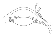
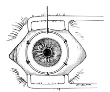
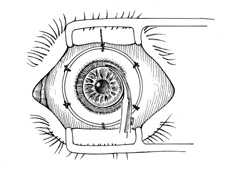
A Flieringa ring or McNeill-Goldmann blepharostat should be sutured to the globe to provide scleral support. One or two of the suture ends can be left long for fixation (Fig. 1). The center of the cornea and eight radial marks can be marked with a surgical pen and a radial keratotomy marker to guide suture placement. Alternatively, some trephines (e.g., Barron Recipient Trephine, Katena) are designed to create evenly spaced marks on the recipient cornea.

Fig. 1. Flieringa ring sutured to the globe. Twelve o'clock suture end is left long for fixation.

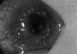
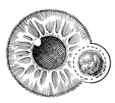
The diameter of the tissue to be removed is determined by the size and position of the pathology and the diameter of the recipient cornea. If possible, all thinned, necrotic, or infected tissue should be removed. The donor button should be at least 5.5 mm in diameter, and it is preferable that it be placed at least 2 mm from the limbus. The donor should be 0.5 to 1.0 mm larger than the recipient bed, to facilitate wound closure. Oversized grafts (>0.5 mm) tend to result in steeper postoperative corneas and therefore can be used to reduce hyperopia in aphakia. Similarly, they could lead to myopia in phakic eyes and should be avoided. Therefore, in infants, with a corneal diameter of 10 to 11 mm, typically a 7-mm donor is placed in a 6.5-mm bed. In a microphthalmic eye, particularly those with axial lengths of 13 mm or less, graft–host disparity of 1.5 mm appears to improve results.16

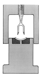
The donor corneoscleral rim is removed from the storage medium and placed in a trephine block, endothelial side up. The cornea is centered in the well, and excess medium is removed. A hollow shaft trephine is carefully centered and aligned vertically over the tissue and then firmly punched through the donor button (Fig. 2). A variety of trephines are available, some of which attempt to ensure centering and verticality of the cut, and some of which place four cardinal marks on the donor epithelium (e.g., Hanna trephine; Barron donor trephine). A drop of storage medium is placed on the endothelial surface, and the cornea is covered. The donor rim should be cultured.

Fig. 2. Trephine for donor cornea that facilitates centration and verticality of cut.

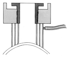
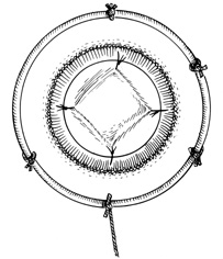
A hollow shaft trephine is also used to cut the recipient cornea. A suction trephine (e.g., Hanna trephine, Barron recipient trephine) facilitates centering and verticality of the cut, and may leave marks for suture placement (Figs. 3 and 4). The trephine is centered on the corneal mark, and a cut of approximately two-thirds depth is made. The anterior chamber is then entered with a knife at the 9 to 10 o'clock (for a right-handed surgeon) position. Right- and left-handed corneal scissors are used to extend the incision (Fig. 5). The scissors are tilted slightly to create a posterior ledge of host cornea, which helps seal the posterior wound. Care is taken to cut the full thickness of the cornea, so Descemet's membrane is not left in the eye, and to avoid damage to the iris and lens. Introduction of viscoelastic material into the anterior chamber may facilitate excision of the host cornea. It may be necessary to separate anterior synechiae, using blunt or sharp dissection. Vitreocorneal and corneolenticular adhesions are cut with scissors.

Fig. 3. Suction trephine on recipient cornea, cross section. Suction is formed between two outer rings. Central blade is lowered into cornea along threads.

Fig. 4. Suction trephine on recipient cornea, surgeon's view. Cross hairs facilitate centering of trephine.

Fig. 5. Completion of vertical incision with scissors. Scissor blades are kept vertical, following the curve of the trephine incision.

Usually the lens and iris move forward as soon as the eye is opened, and visualization and division of peripheral iris synechiae is difficult. Often it is best to close the wound first and lyse synechiae afterward. If iris-lens prolapse occurs, external pressure on the globe from the lids and speculum should be reduced as much as possible. Instead of a speculum, sutures can be placed in the lid margin before trephination, to obtain exposure. Hyperventilation of the patient also can help. Of course, the graft should be sutured in place as expediently as possible. If necessary, shallow sutures can be placed initially and replaced once more control is obtained. Pars plana vitrectomy can be performed to reduce vitreous pressure. Pars plana incisions in an infant should be 2 to 3 mm from the limbus and lens damage can occur during the procedure.

Bleeding from the cornea or iris usually ceases with time and irrigation. Dilute solutions of epinephrine (e.g., 3 mL 1:10,000 in 500-mL saline) may be helpful. If iris bleeding continues, wet field cautery or a retinal diathermy unit can be applied to the vessel.

If cataract extraction is necessary, an anterior capsulorrhexis is optimum. This is started with a cystotome and completed with Utrata forceps. However, the capsulorrhexis often extends peripheral and capsulotomy must be performed with a needle, knife, or scissors. The soft lens nucleus is expressed or removed by a mechanical cutting and suction instrument, and cortical material is removed by irrigation and aspiration. A central posterior capsular opening is created.

Anterior vitrectomy is performed with a mechanical suction and cutting device. Constant irrigation is not necessary. Vitrectomy is continued until the remaining vitreous is well posterior to the iris plane. The anterior iris surface is dabbed with cellulose sponges, to ensure removal of all vitreous strands.

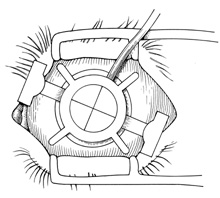
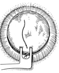
The anterior chamber is re-formed with balanced salt solution, and viscoelastic material is placed centrally over the lens, if present, and on the peripheral recipient rim. The donor cornea is transferred to the bed and sutured into place with four interrupted 10-0 nylon or 9-0 silk sutures (Fig. 6). A double-pronged forceps (e.g., Pollock) facilitates placement of the initial 12 o'clock suture in the donor (Fig. 7). Care must be taken to minimize endothelial damage. The graft is stabilized against the recipient opening superiorly by pulling the tissue with the forceps. The needle is passed radially between the tips of the two-pronged forceps, minimizing movement of the donor tissue.

Fig. 6. Donor cornea held in position with four cardinal sutures. The distribution of donor tissue among quadrants should be symmetric.

Fig. 7. Double-pronged forceps facilitates placement of the first suture in the donor. The needle is placed between the tips of the forceps.

Alignment of the second suture at the 6 o'clock position is crucial to ensure proper distribution of the tissue in the bed. Donor and recipient marks created with the trephines can facilitate suture placement. Anterior iris adhesions can be severed once four to eight cardinal sutures are in place. This is often best done with the chamber filled with a viscoelastic material, particularly in phakic eyes. The peripheral angle is swept with an iris spatula, avoiding contact with the posterior surface of the donor cornea. In some cases sharp dissection is required. If viscoelastic material is not sufficient to maintain the chamber, then additional interrupted sutures or part of the continuous suture is placed before the dissection is performed.

A continuous suture is preferable in most cases because it facilitates rapid wound closure, induces less postoperative inflammation, and is easier to remove when the wound is healed. Interrupted sutures are used when there is localized corneal vascularization (present in some quadrants and not in others) or there is some other reason to expect uneven wound healing. In these cases interrupted sutures can be removed from healed areas while leaving sutures in unhealed areas. Continuous sutures are indicated when symmetric healing is expected, the cornea is totally avascular, or all quadrants are vascularized to a similar extent. Interrupted sutures can also be used when the child is old enough to cooperate with selective suture removal at the slit-lamp to reduce astigmatism.

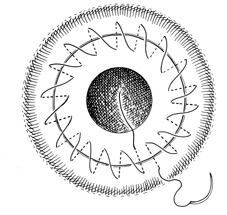
The continuous suture consists of 16 to 24 bites placed in the posterior quarter of the cornea (Fig. 8). The two suture ends are tied on the graft surface with one triple throw, and the interrupted sutures are removed. The chamber is filled with saline solution. The continuous suture is tightened with non-toothed forceps, and the wound is checked for leakage, using topical application of fluorescein dye if necessary. If leakage is present, the running suture is tightened further. The knot then is completed with two additional single throws. If leakage is detected at this point, superficial interrupted sutures are placed as necessary.

Fig. 8. Continuous suture in place.

The scleral fixation ring is removed and subconjunctival injections of antibiotic (e.g., gentamicin, 20 mg) and long-acting corticosteroid (e.g., methylprednisolone acetate, 40 mg) are given. Antibiotic ointment is placed in the eye, which is then covered with a pressure patch and shield. If necessary, elbow restraints are placed on young children to avoid eye manipulation.

The following technique can be helpful in eyes with severe posterior pressure: When excising the host cornea a small hinge is left uncut (on the left for right-handed surgeons). The cornea is lifted temporarily to lyse synechiae and inject viscoelastic, and then reattached with one 7-0 silk suture on the side opposite the hinge. A thick layer of viscoelastic is placed on top of the recipient's cornea, and the donor cornea is placed on top and sutured into place with two 9-0 nylon cardinals. The recipient cornea is then cut free and slid out from underneath.

Back to Top
POSTOPERATIVE CARE
The postoperative care of the young, uncooperative patient is challenging. Administration of medications is difficult for the parents, and they must be taught how to do this safely and expediently. Children are prone to eye trauma caused by rubbing the eye or injuring it during play, and enforcement of wearing glasses or shields is often difficult.

EXAMINATIONS

Young patients are unable to report the symptoms of postoperative complications; therefore, parents should be taught to recognize warning signs such as squinting, photophobia, eye rubbing, and increased irritability. Parents should regularly examine the child's eye for redness, cloudiness of the graft, focal opacities, watering, mucus accumulation on the sutures, and purulent discharge. In addition, the child needs to be examined by the ophthalmologist more frequently than would an adult. A suggested schedule includes examination on the first postoperative day and every 2 to 3 days over the first 2 weeks, then weekly until all sutures are removed, and then every 2 to 4 weeks until the end of the first postoperative year.

Postoperative examinations are conducted in the same manner as described for preoperative examinations. They should be as nonthreatening as possible for the child. A hand-held slit-lamp is used to examine children who cannot cooperate for standard slit-lamp examination. If an adequate examination cannot be obtained with the child awake, the examination must be performed with the child under anesthesia. Typically, children between 2 and 4 years of age are most difficult to handle because they are too young to cooperate and too old to restrain. Multiple examinations under anesthesia are frequently required to remove interrupted sutures.

MEDICATIONS

Initial topical medications typically include a corticosteroid (e.g., prednisolone acetate) every 1 to 2 hours while awake, an antibiotic four times daily, and a cycloplegic agent (e.g., atropine) 1% once daily. Antibiotic treatment is discontinued after 1 week or once the epithelium is healed. Topical corticosteroids are gradually tapered, based on the amount of inflammation observed, over 8 to 12 months. More intensive and prolonged treatment is necessary for vascularized corneas. One study suggested that the addition of topical cyclosporine 2% could reduce the rate of graft rejection.17 For patients who underwent keratoplasty for herpetic scarring, antiviral prophylaxis appears to reduce the risk of recurrence and should be administered for at least 1 year, and possibly for the rest of the patient's life.18,19 Antiviral prophylaxis is also indicated during intensive corticosteroid treatment for graft rejection. If a patient becomes so uncooperative that the parents are unable to administer drops, depot injections of corticosteroids are given during examinations with the patient under anesthesia. Vaccinations should be avoided until 1 year after surgery, because they appear to increase the risk of rejection.

SUTURE REMOVAL

Wound healing is more rapid in children than it is in adults. As the wound heals it contracts, causing suture loosening. Loose sutures cause irritation and induce vascularization, mucus accumulation, rejection, and microbial infection (Fig. 9). Therefore, loose sutures should be removed immediately. Timing of routine suture removal depends on the patient's age and the degree of vascularization. Typically, suture removal is performed 4 to 6 weeks postoperatively in children younger than 1 year of age, 3 to 4 months postoperatively in young children, and 4 to 6 months postoperatively in older children.

Fig. 9. Loosened continuous suture with injection and mucus accumulation.

VISUAL REHABILITATION

Refractive errors should be corrected as soon as possible. This is especially important for younger children and infants, to enhance visual development and avoid amblyopia. In infants, refractive errors should be corrected as soon as sutures are removed (Fig. 10). In older children, when sutures are left in place for 6 months or longer, optical correction can be prescribed even with sutures in place.

Fig. 10. Clear corneal graft after suture removal.

Spectacle correction is preferred for children without significant anisometropia. Contact lenses may provide better vision, but they pose an increased risk to the eye, and there is substantial effort associated with their wear. However, if there is significant anisometropia or astigmatism, contact lenses are necessary. When significant astigmatism is present, rigid gas-permeable lenses may be necessary. These can be worn up to 1 week at a time, if necessary. Contact lens fitting can be performed with the child under anesthesia with the aid of a quantitative surgical keratometer and a trial lens set.20 Alternatively, a conventional keratometer can be mounted vertically from the ceiling. In monocular aphakia, epikeratophakia can be performed if contact lens wear is not possible. Theoretically, LASIK can be performed to reduce anisometropia or astigmatism.21,22 It has been fairly successful in adults after keratoplasty.23–25 Amblyopia therapy should begin as soon as the graft is clear and optical correction is achieved.

Regardless of the means of optical correction, refractions must be repeated frequently, because changes can occur rapidly as the wound heals. In addition, infants can undergo large myopic shifts as the eye continues to grow. The authors prefer to follow these patients in conjunction with a pediatric ophthalmologist, who directs optical correction and amblyopia therapy.

Back to Top
COMPLICATIONS
Injury to or extrusion of a clear lens is a serious complication, because aphakic eyes have a significantly worse visual prognosis. The performance of lensectomy and vitrectomy are also associated with a significantly worse prognosis for graft survival.6,8,10,14 Nevertheless, a lens that appears to have been injured should be removed, because any opacity worsens after surgery. Deposition of fibrin on the lens surface should not be confused with injury to the lens.

Postoperative elevations of intraocular pressure are common, both de novo and in eyes with previously controlled glaucoma. Transient elevations often occur in the first few days after surgery, presumably owing to inflammation, retained viscoelastic materials, and changes in angle configuration. Prolonged elevation of intraocular pressure can result from iris synechiae and congenital anomalies. Medical therapy includes topical β-blockers, prostaglandin agents, miotics, and carbonic anhydrase inhibitors.

If these therapeutic measures are not sufficient, filtering procedures or cyclodestructive procedures can be performed. Both of these procedures are associated with increased intraocular inflammation and therefore increased risk of graft failure or rejection. In addition, cyclodestruction exposes the patient to the risk of phthisis and apparently to a higher risk of sympathetic ophthalmia as well. Trabeculectomy may have a lower risk of graft reaction and phthisis.

Epithelial defects are common immediately after surgery, but most will heal without specific measures. Tear deficiency, corneal anesthesia, endophthalmos, lid malposition or impaired closure, limbal injury, medication toxicity, and trauma can delay healing. Recognition and correction of any abnormalities present are essential. Otherwise, one proceeds sequentially with patching, bandage lens placement, and temporary tarsorrhaphy, as necessary.

Anterior displacement of the graft or wound dehiscence must be surgically repaired. Anterior displacement of the graft can be caused by improper suture placement (host bites not deep enough), sutures being too loose, impaired healing, cheese-wiring of sutures, premature removal of sutures, or thinning of the recipient cornea. Poor wound healing and high astigmatism can result if the situation is not corrected. Wound gape can be caused by tying the sutures too loosely or cheese-wiring of the sutures. Replacement of sutures in the operating room usually is required in both conditions.

Infiltrates around sutures occur commonly in children, particularly in eyes with corneal vascularization. The epithelium may be intact over the infiltrates. They usually are controlled with topical corticosteroids. Infiltrates that are not adjacent to a suture are increasing in size, or are associated with purulent discharge should be presumed to be infectious. Cultures should be obtained and antibiotic treatment initiated.

Immunologic rejection is the most common reason for graft failure. It can occur at any time beyond 10 days after primary graft surgery, and sooner in previously immunized patients. In children older than 8 years of age, graft reactions are similar to those seen in adults. The child often exhibits squinting, photophobia, and tearing. Vision usually is decreased, and the eye is injected. Biomicroscopic signs of graft rejection may include graft edema, keratic precipitates, an endothelial rejection line, subepithelial infiltrates, and anterior chamber reaction. The reaction may be diffuse or may start in one quadrant, usually near an area of vascularization or iris synechiae, and progress across the cornea. A line of precipitates may be present at the advancing edge (Khodadoust line). In younger children, rejections tend to present differently. Most commonly, they are characterized by sudden or gradual onset of corneal edema with minimal signs of inflammation. Keratic precipitates and endothelial rejection lines are uncommon.

Prompt and aggressive treatment can stop the reaction and preserve the graft. Treatment should be given if there is any suspicion that a reaction may be present. Unless the inflammation is very mild, topical corticosteroids should be given every hour. Once the reaction is controlled the dose is gradually tapered. Oral corticosteroids (1 mg/kg prednisone), intravenous corticosteroids, and periocular injections of depot corticosteroids can be considered in cases that are not responsive to topical treatment.

Very often a clear graft is achieved but astigmatism prevents visual correction. The average astigmatism, after all sutures have been removed, is between 3 and 4 diopters. Usually this can be corrected with spectacles. Higher degrees of astigmatism may necessitate contact lens correction. As mentioned, rigid gas-permeable contact lens fitting is preferred. If contact lens wear is not possible, then surgical correction (e.g., relaxing incisions or wedge resection) can be attempted. It is worth stressing that early optical correction and aggressive amblyopia therapy are essential in young children.

Cataracts can result from inadvertent injury during surgery, postoperative intraocular inflammation, or topical corticosteroid treatment. Cataract extraction should be delayed until the graft is well healed. Even then there is a significant risk of graft failure or rejection after the surgery. (The risk in adults is approximately 10%.)26

The treatment of postkeratoplasty infectious keratitis is the same in children as in adults. Infections of the graft require prompt recognition and aggressive antibiotic treatment. Cultures and gram stains are obtained, and broad-spectrum fluoroquinolones or fortified antibiotic drops are administered. If anesthesia is required for examination and obtaining samples for cultures, antibiotics should be injected subconjunctivally. Repeated injections or antibiotic-soaked collagen shields can be helpful in cases in which drops cannot be administered.

Other complications include trauma, endophthalmitis, graft failure from unknown cause, and herpes simplex infection. The treatment of these conditions is similar in children and adults; the reader is referred elsewhere for further information.27,28

LAMELLAR KERATOPLASTY

Lamellar keratoplasty is the transplantation of the anterior layers of the cornea, over a thin posterior stromal layer, Descemet's membrane, and endothelium. Its advantages over penetrating keratoplasty are that healing is more rapid, intraocular surgery is avoided, and endothelial rejection does not occur. Nevertheless, lamellar keratoplasty has seldom been performed. This is because the posterior stroma and endothelium must be healthy, the visual acuity achieved is rarely better than 20/40, because of opacification of the interface, and the results of penetrating keratoplasty are generally good.29,30

However, in recent years the use of lamellar keratoplasty has been increasing, primarily because of the use of the microkeratome for donor and recipient preparation.31–34 The stromal surface created with a microkeratome is much smoother than hand-dissected surfaces, and can result in clear interfaces and 20/20 (6/6) visual acuity. A whole donor globe or artificial anterior chamber and corneoscleral rim are necessary for donor preparation. Current microkeratome suction rings may not fit on very small (e.g., infant) eyes, and hand dissection of the recipient may be necessary.

Lamellar grafting can be performed when opacities are limited to the anterior stroma, such as in superficial postinfectious or traumatic scarring or Reis-Bücklers dystrophy. Lamellar keratoplasty is also used for tectonic purposes: to seal perforations and support thinned or weakened tissue. In children lamellar keratoplasty probably has been most commonly used after excision of corneal dermoids.

Back to Top
SPECIAL SITUATIONS

CORNEAL DERMOIDS

Dermoids are congenital tumors that arise from ectodermal tissue that was displaced during embryologic development.35 They are covered by keratinized epithelium and can contain hair follicles, glands, fat, smooth and skeletal muscle, nerves, blood vessels, bone, cartilage, and teeth. In the eye, they most often present as yellowish-white solid vascularized, elevated nodules straddling the corneal limbus. They also can lie centrally in the cornea, involve the entire cornea, or form a ring around the limbus.36,37 They typically extend into the deeper stroma without affecting Descemet's membrane and the endothelium, but in some cases they replace all tissue anterior to the iris pigment epithelium.38 Gonioscopic examination of the angle beneath the tumor or ultrasonic biomicroscopy can indicate the depth of extension.39 Dermoids may increase in size with age.

Corneal dermoids may be associated with other developmental abnormalities. Approximately 30% of persons with Goldenhar's syndrome (oculoauriculovertebral dysplasia) have epibulbar dermoids.40 Other eye findings include lid colobomas, Duane's syndrome, iris coloboma, and microphthalmos. Abnormalities of structures derived from the first and second branchial arches are also common. Preauricular skin appendages and fistulas, vertebral anomalies, and mandibular and malar hypoplasia can be seen.

Limbal dermoids usually are a cosmetic rather than a visual problem; however, they may induce astigmatism, cause irritation (because of a hair or mass effect), or produce drying of the surrounding cornea by lifting of the lid during blinking. Reduction of the mass of the tumor by shaving it at the level of the surrounding corneal surface reduces astigmatism and improves cosmesis, but some opacity remains. A better cosmetic and astigmatic result can be achieved with a lamellar graft. The dermoid tissue often is not solid enough to retain sutures; therefore, grafts must encompass the entire tumor. Improved cosmesis usually can be achieved, but astigmatism is not consistently decreased.41–43

Technique

A peritomy is performed close to the dermoid, and hemostasis is obtained. The smallest trephine that encompasses the tumor is chosen. It is placed over the corneal and conjunctival sides of the tumor, and a partial-thickness cut is made (Fig. 11). Dissection is continued with a knife until clear stroma is reached. A lamellar dissection blade (e.g., Desmarres, Troutman, or Martinez) is used to perform a lamellar dissection beneath the dermoid (Fig. 12). The cornea is kept dry, and steady traction is used to pull upward on the lamellar disc. The dissector is kept parallel to the stromal lamellae, pushing more than cutting. If opacities remain in the bed, a deeper plane can be created and the dissection repeated. The dissection should extend beyond the diameter of the bed to facilitate placement of a thicker donor, construction of a perpendicular wound, and visualization of suture placement.

Fig. 12. Lamellar dissection is performed beneath the tumor. Gentle upward traction is exerted on the tumor as the bed is sharply dissected.

Fig. 11. Removal of corneal dermoid. After a peritomy a partial-thickness trephination (dotted line) is created around tumor.

A slightly larger (e.g., 0.25-mm) corneal lamellar graft is sewn into the bed. This can be dissected from a whole globe as follows. The donor eye is wrapped tightly in gauze and the intraocular pressure is elevated. A small incision is made at the limbus with a razor or diamond knife to the necessary depth. A duckbill lamellar dissection spatula is pushed into the incision, rotating it back and forth in the lamellar plane. The surgeon should not push downward. In this manner the entire corneal area is dissected. A trephine of the desired diameter then is used to cut through the entire corneal thickness. The lamellar button usually remains attached to the trephine.

If only a corneoscleral rim is available the cornea can be trephined to the desired size and then trimmed to the desired thickness by removal of posterior layers using Vannas scissors. A microkeratome can be used to create the donor lamella. The thickness of the donor tissue is controlled by selecting the appropriate blade head. A suction ring can be used if a whole globe is available. An artificial anterior chamber (e.g., ALTK System, Moria, Paris, France) must be used to create the donor lamella from a corneoscleral rim. The donor button then can be trephined to the correct diameter.

The button then is sewn into the bed using interrupted or running sutures. A paracentesis may be necessary to allow good wound apposition. Depending on the age of the recipient, sutures can be removed in 1 to 3 months.

Penetrating Graft

Penetrating keratoplasty can be performed for central dermoids if they are 7 mm or less in diameter. Larger central dermoids require a two-stage procedure. First the tumor is excised and a large lamellar graft is placed in the bed. Once that is healed, a smaller central penetrating keratoplasty is performed.44

CORNEAL LACERATIONS

Children are particularly prone to penetrating ocular injuries. In the Israeli Ocular Injuries study, 47% of patients hospitalized for ocular injuries were younger than 18 years of age, with children 6 to 10 years of age the most commonly affected group.45 In a study at the Wilmer Eye Institute, 43% of penetrating ocular injuries occurred in patients 18 years of age or younger.46 Sports equipment, wooden sticks, stones, toys, and air-powered BB guns commonly cause the injuries.

Preoperative evaluation of the injured child is often hampered by the child's inability or reluctance to provide a detailed description of the injury. Children guilty of misbehavior may misrepresent the facts, even if they bear no responsibility. Gentle examination usually is sufficient to determine whether a penetrating injury is present. A papoose board, portable slit-lamp, and Desmarres retractors may be required for examination in young children. If a penetrating injury is suspected, the examination should be performed in the operating room with the use of general anesthesia.

The anatomic repair of corneoscleral lacerations in children is no different than in adults. Postoperative care in children younger than 9 years of age may be complicated by amblyopia. Occlusion therapy should be initiated as soon as optical correction can be obtained. Corneal sutures can be removed in 1 to 3 months, depending on the child's age. If there is excessive corneal irregularity or anisometropia, rigid contact lenses may be necessary.

When a corneal scar crosses the pupil, the degree to which vision is impaired is quite variable. In young children the physician must make a subjective assessment of the child's view, and this can be difficult. Eccentric scars usually do not require penetrating keratoplasty, whereas central scars often do, even if they are narrow. In many cases vision is reduced more by surface irregularity than opacification. This can be assessed by keratoscopy and by determining acuity before and after hard contact lens correction.

Rotational autografts should be considered for correction of small central scars. An eccentric circular trephination is made in the donor cornea, the button is rotated to bring the scar out of the pupil, and the wound is sutured closed. The advantage is that the risk of rejection is eliminated. The disadvantages are that astigmatism is more likely and the wound is closer to the visual axis than in a central penetrating graft.

Back to Top
REFERENCES

1. Beauchamp GR: Pediatric keratoplasty: problems in management. J Pediatr Ophthalmol Strabismus 16:388, 1978

2. Brown SI: Corneal transplantation of the infant cornea. Trans Am Acad Ophthalmol Otolaryngol 78:461, 1974

3. Gordon YJ, Mokete M: Penetrating keratoplasty in children. J Pediatr Ophthalmol Strabismus 16:297, 1979

4. Picetti B, Fine M: Keratoplasty in children. Am J Ophthalmol 61:782, 1966

5. Schanzlin DJ, Goldberg DB, Brown SI: Transplantation of congenitally opaque corneas. Ophthalmology 87:1253, 1980

6. Stulting RD, Sumers KD, Cavanagh HD, et al: Penetrating keratoplasty in children. Ophthalmology 91:1222, 1984

7. Waring GO, Laibson PR: Keratoplasty in children. In Kwitko ML (ed):Surgery of the Infant Eye. New York: Appleton-Century-Crofts, 1979

8. Dana MR, Schaumberg , Moyes AL, Gomes JA: Corneal transplantation in children with Peters' anomaly and mesenchymal dysgenesis. Multicenter Pediatric Keratoplasty Study. Ophthalmology 105:1353, 1998

9. Yang LL, Lambert SR, Lynn MJ, Stulting RD: Long-term results of corneal graft survival in infants and children with Peters' anomaly. Ophthalmology 106:833, 1999

10. Comer RM, Daya SM, O'Keefe M: Penetrating keratoplasty in infants. JAAPOS 5:285, 2001

11. Aasuri MK, Garg P, Gokhle N, Gupta S: Penetrating keratoplasty in children. Cornea 19:140, 2000

12. Dana MR, Schaumberg DA, Moyes AL, et al: Outcome of penetrating keratoplasty after ocular trauma in children. Arch Ophthalmol 113:1503, 1995

13. Schaumberg DA, Moyes AL, Gomes JA, Dana MR: Corneal transplantation in young children with congenital hereditary endothelial dystrophy. Multicenter Pediatric Keratoplasty Study. Am J Ophthalmol 127:373, 1999

14. Zaidman G, Ramirez T, Kaufman A, et al: Successful surgical rehabilitation of children with traumatic corneal laceration and cataract. Ophthalmology 108:338, 2001

15. Gloor P, Keech RV, Krachmer JH: Factors associated with high postoperative myopia after penetrating keratoplasty. Ophthalmology 99:775, 1992

16. Feldman ST, Frucht-Pery J, Brown SI: Corneal transplantation in microphthalmic eyes. Am J Ophthalmol 104:164, 1987

17. Cosar CB, Laibson PR, Cohen EJ, Rapuano CJ: Topical cyclosporine in pediatric keratoplasty. Eye Contact Lens 29:103, 2003

18. Barney NP, Foster CS: A prospective randomized trial of oral acyclovir after penetrating keratoplasty for herpes simplex keratitis. Cornea 13:232, 1994

19. van Rooij J, Rijneveld WJ, Remeijer L, et al: Effect of oral acyclovir after penetrating keratoplasty for herpetic keratitis: a placebo-controlled multicenter trial. Ophthalmology 110:1916, 2003

20. Saunders RA, Ellis FD: Empirical fitting of hard contact lenses in infants and young children. Ophthalmology 88:127, 1981

21. Haw WW, Alcorn DM, Manche EE: Excimer laser refractive surgery in the pediatric population. J Pediatr Ophthalmol Strabismus 36:173, 1999

22. Rashad KM: Laser in situ keratomileusis for myopic anisometropia in children. J Refract Surg 15:429, 1999

23. Busin M, Zambianchi L, Garzione F, et al: Two-stage laser in situ keratomileusis to correct refractive errors after penetrating keratoplasty. J Refract Surg 19:301, 2003

24. Barraquer CC, Rodriguez-Barraquer T: Five-year results of laser in-situ keratomileusis (LASIK) after penetrating keratoplasty. Cornea 23:243, 2004

25. Hardten DR, Chittcharus A, Lindstrom RL: Long term analysis of LASIK for the correction of refractive errors after penetrating keratoplasty. Cornea 23:479, 2004

26. Binder PS: Intraocular lens implantation after penetrating keratoplasty. J Refract Corneal Surg 5:224, 1989

27. Brightbill FS: Corneal Surgery: Theory, Technique, and Tissue. St. Louis: Mosby-Year Book, 1999

28. Krachmer JH, Mannis MJ, Holland EJ (eds): Cornea, 2nd Ed. St. Louis: Mosby-Year Book, 2004

29. Paton D: Lamellar keratoplasty. In: Symposium on Medical and Surgical Diseases of the Cornea. Transactions of the New Orleans Academy of Ophthalmology. St. Louis: CV Mosby, 1980

30. Richard JM, Paton D, Gasset AR: A comparison of penetrating keratoplasty and lamellar keratoplasty in the surgical management of keratoconus. Am J Ophthalmol 86:807, 1978

31. Wiley LA, Joseph MA, Springs CL: Tectonic lamellar keratoplasty utilizing a microkeratome and an artificial anterior chamber system. Cornea 21:661, 2002

32. Jimenez-Alfaro I, Perez-Santonja JJ, Gomez Telleria G, et al: Therapeutic lamellar keratoplasty with an automated microkeratome. J Cataract Refract Surg 27(8):1161, 2001

33. Hanna KD, David T, Besson J, Pouliquen Y: Lamellar keratoplasty with the Barraquer microkeratome. Refract Corneal Surg 7:177, 1991

34. Haimovici R, Culbertson WW: Optical lamellar keratoplasty using the Barraquer microkeratome. Refract Corneal Surg 7:42, 1991

35. Mansour AM, Barber JC, Reinecke RD, et al: Ocular choristomas. Surv Ophthalmol 33:339, 1989

36. Mattos J, Contreras F, O'Donnell FE: Ring dermoid syndrome: a new syndrome of autosomal dominantly inherited bilateral, annular limbal dermoids with corneal and conjunctival extension. Arch Ophthalmol 98:1059, 1980

37. Henkind P, Marinoff G, Manas A, et al: Bilateral corneal dermoids. Am J Ophthalmol 76:971, 1973

38. Mann I: Developmental Abnormalities of the Eye. London: W Clowes & Son, 1957

39. Lanzi IM, Augsberger JJ, Hertle RW, et al: The role of ultrasound biomicroscopy in surgical planning for limbal dermoids. Cornea 17:604, 1998

40. Baum JL, Feingold M: Ocular aspects of Goldenhar's syndrome. Am J Ophthalmol 75:250, 1973

41. Scott JA, Tan DT: Therapeutic lamellar keratoplasty for limbal dermoids. Ophthalmology 108:1858, 2001

42. Watts P, Michaeli-Cohen A, Abdolell M, Rootman D: Outcome of lamellar keratoplasty for limbal dermoids in children. JAAPOS 6:209, 2002

43. Panda A, Ghose S, Khokhar S, Das H: Surgical outcomes of epibulbar dermoids. J Pediatr Ophthalmol Strabismus 39:20, 2002

44. Zaidman GW, Johnson B, Brown SI: Corneal transplantation in an infant with corneal dermoid. Am J Ophthalmol 93:78, 1982

45. Koval R, Teller J, Belkin M, et al: The Israeli Ocular Injuries Study: a nationwide collaborative study. Arch Ophthalmol 106:776, 1988

46. Sternberg P, DeJuan E, Michels RG: Penetrating ocular injuries in young patients: initial injuries and visual results. Retina 4:5, 1984

Back to Top