Chapter 105
Surgical Management of Congenital Obstruction of the Lacrimal Collecting System
RICHARD M. ROBB
Main Menu   Table Of Contents

Search

PROBING AND IRRIGATION
SILICONE TUBE INTUBATION
DACRYOCYSTORHINOSTOMY
REFERENCES

PROBING AND IRRIGATION

INDICATIONS AND TIMING

The indication for probing and irrigation of the lacrimal collecting system is persistent obstruction with tearing and discharge dating from the early months of the first year. Probing primarily is the initial procedure to consider for such obstruction. Except for the lacrimal puncta, the drainage system cannot be visualized directly, and probing helps to locate the level and nature of the obstruction. Fortunately, in children the probing also is highly effective in relieving the blockage,1–3 and in most instances, further procedures are not necessary.

Congenital nasolacrimal duct obstruction has a high rate of spontaneous resolution during the first year,4–7 There is some difference of opinion about how early to resort to probing to clear persistent obstructions. Early probing may clear the symptoms faster, but a routine of early probing leads to performing many procedures that would be unnecessary. I usually advise delay of probing until near the end of the first year. Mucopurulent discharge from infection in the collecting system usually is present to some degree. It can be managed with lid hygiene and the application of antibiotic ointments. Pressure over the lacrimal sac can be used to prevent accumulation of discharge. This pressure usually results in retrograde flow of the sac contents onto the eye, where they can be wiped away. Occasionally, the pressure is transmitted hydrostatically down the nasolacrimal duct, and with a sudden popping sensation the obstruction may be relieved.8 Failing this, persistent purulent discharge and lid irritation are reasons to proceed with probing. An episode of frank dacryocystitis with swelling and redness in the area of the lacrimal sac is a more pressing indication for probing. The dacryocystitis is first treated with systemic antibiotics, but subsequent probing to relieve the obstruction is necessary to prevent recurrence. A further indication to probe on an accelerated timetable is the need to clear the lacrimal system infection before anticipated ocular, facial, or intracranial surgery. Occasionally, the timing of a probe may be influenced by the need for general anesthesia for some unrelated surgery and the convenience of doing both procedures under one anesthesia.

A special indication for early probing is distention of the lacrimal sac at birth, caused by a combination of nasolacrimal duct obstruction and a physiologic canalicular blockage of retrograde flow, the so-called congenital dacryocystocele. Probing should be undertaken in the newborn period rather than later because secondary infection is likely to develop in the closed system.4,9 Beyond this, dacryocystoceles also may be associated with distention of the nasolacrimal duct and intranasal cysts, which cause respiratory distress in young infants.10,11 The lacrimal puncta and canaliculi are small at this early age, but even without anesthesia, the inferior canaliculus usually can be dilated and probed to decompress the lacrimal sac, and the distal duct obstruction can be broken by passing the probe on into the nose. Sometimes, an intranasal cyst requires a more extensive procedure, including marsupialization of the cyst with fiberoptic nasal endoscopy.11

Finally, the timing of probing and irrigation may be affected by the decision whether to perform the procedure under anesthesia. In the newborn period, the procedure usually is done without anesthesia, and many ophthalmologists prefer to perform the probe in the office with topical anesthesia until the infant is 6 months of age.1,12 Beyond 6 months, I routinely use general anesthesia in an ambulatory operating room setting.13 Probings done under general anesthesia are more comfortable for the patient, and they are technically easier and better controlled for the surgeon. An endotracheal tube usually is not required for standard probing, although without a tube, irrigation must be limited to prevent aspiration. With mask anesthesia, access to the nose is limited. A skilled pediatric anesthesia staff helps to make probing under general anesthesia a safe and reliable procedure.

CONTRAINDICATIONS

Whereas there are no absolute contraindications to probing and irrigation for persistent obstruction of the lacrimal drainage system, several considerations may affect the timing of the probe. There is no reason to perform a probe in the early months of the first year if the lacrimal sac is not distended and symptoms are manageable. Spontaneous clearing occurs in most obstructions up to at least 12 months of age.4–7 If parents are anxious about a surgical procedure, especially one under general anesthesia, additional time can be allowed for clearing. Probing should be delayed if there is an upper respiratory tract infection or if there is acute dacryocystitis. Any contraindications to general anesthesia, such as severe congenital heart disease, may delay treatment. If probing is done in the presence of congenital heart disease, prophylactic antibiotics are advisable because of the possibility of transient bacteremia.14

PROCEDURE

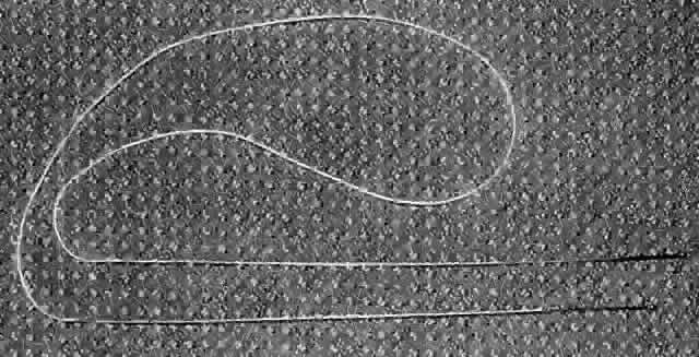
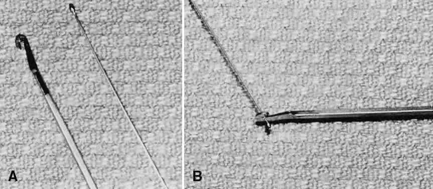
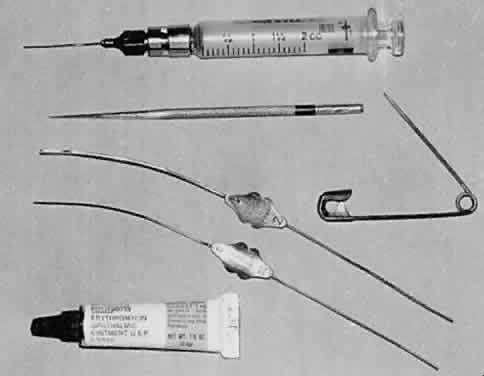
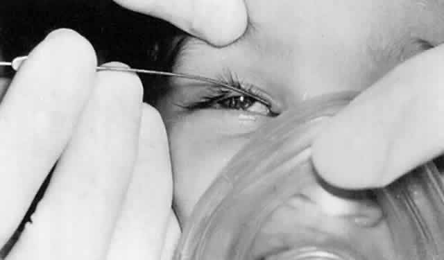
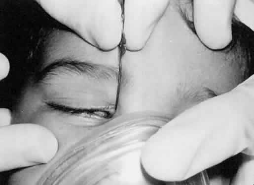
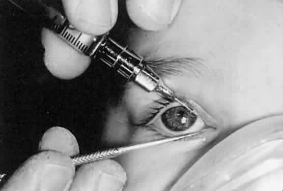
When performing a probe without anesthesia, the lower canaliculus usually is easier to enter, but under general anesthesia the upper canaliculus is the preferred access to the system. The upper punctum is dilated, and a Bowman no. 0 (0.7-mm diameter) or no. 1 (0.8-mm diameter) probe (Fig. 1) is passed through the canaliculus into the lacrimal sac (Fig. 2). Occasionally, the obstruction is at the level of the punctum itself, a blockage signaled by symptoms of tearing without discharge, and when the punctum is opened, tear flow is normalized. More often, the obstruction is at the lower end of the lacrimal duct, and the probe can be passed to that point relatively freely. It is helpful to bend the probe to a gentle curve so that after it reaches a firm stop at the medial wall of the sac in the lacrimal fossa, it can be directed down and posteriorly into the duct (Fig. 3). Lubricating the probe with an ophthalmic ointment also eases its passage. At the lower end of the lacrimal duct, slightly firmer pressure is necessary to break through the membranous obstruction into the nose. It may help to direct the curve of the probe posteriorly and medially to break into the nose under the inferior turbinate. Then the probe either stops at the floor of the nasal cavity or runs posteriorly along the surface of the hard palate. If uncertainty exists as to whether the probe has entered the nasal cavity, a second Bowman probe can be placed in the nose under the inferior turbinate to feel the contact of metal on metal. The probe then is withdrawn, and saline is irrigated through the system using a glass syringe and a 23-gauge cannula threaded through one canaliculus into the sac (Fig. 4). The other canaliculus is plugged with a lacrimal dilator to prevent reflux. Only a small amount of fluid (approximately 1 ml) is required to establish that the lacrimal duct is open; larger amounts increase the risk of aspiration. Fluorescein can be used to stain the irrigant so that it can be visualized using a clear suction catheter in the nose, but patency of the system usually can be recognized by the feel of the freely moving plunger of the glass syringe.

Fig. 1. Instruments required for lacrimal probe: a 23-gauge cannula attached to a glass syringe to irrigate the lacrimal drainage system; a punctum dilator or safety pin to dilate the canaliculus; the Bowman probes, which are bent to a gentle curve; and ophthalmic ointment to lubricate the probes.

Fig. 2. A Bowman probe is passed through the upper canaliculus into the lacrimal sac in an anesthetized patient. The probe can be presumed to be in the sac when its tip is stopped by the firm medial wall of the lacrimal fossa.

Fig. 3. The Bowman probe has been directed down and posteriorly along the lacrimal duct. Slightly more pressure may be necessary to break through the membranous obstruction at the lower end of the duct.

Fig. 4. A 23-gauge cannula attached to a glass syringe is used to irrigate saline from the lacrimal sac into the nose while the free canaliculus is plugged with a dilator to prevent reflux.

Recently, a nasolacrimal duct probe with a balloon catheter has become available as an adjunct to standard probing.15 Several reports of its use have been published,16,17 but it is not clear whether or in which cases this new instrumentation will be advantageous.

EXPECTED RESULTS

After a successful probing, tearing and discharge usually clear within 1 to 2 days. A topical antibiotic ointment or solution can be used if there is much mucopurulent discharge at the time of probing, but these are discontinued as soon as the eye appears to be clear. The normal passage of tears through the lacrimal drainage system helps to clear residual discharge. Initial probings are successful in over 90% of patients.13,12 Some surgeons have found a decrease in the success rate if the probing is delayed beyond the second or third year,18 but many patients can be cured by probing up to 5 years of age or older.2,3,19 If tearing and discharge persist after apparently successful probing, a second probing can be done in a month with the option of placing silicone tubing in the lacrimal system at that time. If it has not been possible to pass the probe into the nose because of bony obstruction at the lower end of the lacrimal duct, dacryocystorhinostomy is required to establish satisfactory tear drainage. Occasionally, the probe seems to pass into the nose, but irrigation is not free. In this case, it is unlikely that the probing will be successful. Repeat probing at the time, or reprobing with the option of silicone intubation after a 1-month interval, may resolve the issue.

COMPLICATIONS

Most probings are free of complications. A false passage of the probe may be signaled by extravasation of saline into surrounding tissues at the time of irrigation. This usually subsides in a short time, but prophylactic systemic antibiotics are advisable. Postoperative fever occasionally occurs after an otherwise unremarkable probe and probably is secondary to bacteremia induced by the probing.20 The fever subsides with antipyretics, and antibiotics are not routinely required.

Back to Top
SILICONE TUBE INTUBATION

INDICATIONS

Intubation of the lacrimal drainage system with a silicone tube may be useful when a probing has been performed through to the nose but the drainage system has failed to remain open.21,22 Since standard probing has such a high rate of success in resolving congenital nasolacrimal duct obstruction, intubation is necessary only occasionally. Yet, for some persistent obstructions, silicone tubes can be helpful.

CONTRAINDICATIONS

If it has not been possible to pass a Bowman no. 0 probe into the nose, silicone tube intubation will not be possible, and a dacryocystorhinostomy should be performed. Some uncooperative patients may not tolerate the tubing at the inner canthus and may pull it out themselves. Such patients are not good candidates for intubation.

PROCEDURE

It is advisable to use general anesthesia with an endotracheal tube or laryngeal mask for lacrimal intubation, since unhurried access to the nasal cavity through the anterior nares is essential. Two flexible probes with a length of silicone tubing swaged over their trailing ends (Fig. 5) are passed in turn through the lacrimal system, one through the upper and one through the lower canaliculus. The first probe passed is retrieved from the nose by grasping its end with a hook or a hemostat slid under the inferior turbinate. The probe designed by Crawford, with a small olive tip at its end, facilitates retrieval with a hook and minimizes trauma to the nasal mucosa21 (Fig. 6). Care should be taken to ensure that the silicone tube is not stripped from the end of the probe as it is drawn through the lacrimal system. The second probe then is passed and retrieved from under the inferior turbinate so that both ends of the silicone tubing, now emerging from the anterior naris, can be tied with a simple square knot in such a position that the knot retracts back into the nose when released. One version of the Crawford silicone tubing has a suture in its lumen, and the sutures (rather than the tubing) can be tied together when the tubing is trimmed to a suitable length. An alternate means of retrieving silicone tubing from the nose is offered by the Ritleng system, which uses a monofilament Prolene guide thread rather than the Crawford wire probes at the ends of the tubing.23 The Prolene guide can be hooked and brought forward as it emerges under the inferior turbinate. (A monocanalicular version of the Ritleng tube also is available.24) The intranasal portion of the silicone tubing can be fixed in place by an absorbable suture to the lateral wall of the nose,25 or it can be tied over a stent that will not pass up into the nasolacrimal duct. The silicone tubing is left in place for 3 to 4 months, during which time antibiotic solution or ointment can be used to reduce discharge and irritation. It is a good sign if tearing and discharge disappear while the tube is in place. Eventually, the tube is removed by cutting the loop between the two lacrimal puncta and pulling on one end until the knot is passed retrograde through the duct, sac, and canaliculus. Gentle tension on the tubing stretches it and thins the knot so that it passes through the canaliculus with relatively little resistance. If a stent has been used, it must be identified under the inferior turbinate and pulled out through the nose, a maneuver requiring general anesthesia in most cases.

Fig. 5. Crawford lacrimal intubation set with silicone tube swaged over the trailing ends of two olive-tipped probes.

Fig. 6. A. Olive tip of Crawford probe and hook for retrieval of probe from under the inferior turbinate. B. Hook engaging olive tip.

EXPECTED RESULTS

If the lacrimal drainage system remains open after the silicone tube has been withdrawn, tearing and discharge will disappear. Persistence of symptoms of obstruction suggests that a dacryocystorhinostomy is necessary.

COMPLICATIONS

Erosion of the canaliculi may occur if the tubing is tied too tightly or is left in place too long. Evidence of erosion should prompt consideration of tube removal. Rarely, the tube or a portion of it may be lost in the lacrimal sac, and it may be necessary to explore the sac to remove the lost portion.

Back to Top
DACRYOCYSTORHINOSTOMY

INDICATIONS

The indication for dacryocystorhinostomy is persistent obstruction of the nasolacrimal duct that cannot be relieved by probing or silicone tube intubation. In the pediatric population, patients with congenital lacrimal duct obstruction requiring dacryocystorhinostomy often have congenital craniofacial deformities.2 Persistent acquired obstructions in children frequently are the result of accidental trauma or surgery in the area of the lacrimal drainage system.26 Mucopurulent discharge from the lacrimal system usually is present with chronic obstruction and may be considered an indication for surgery, but acute dacryocystitis generally is treated medically before surgery is performed. Since other procedures usually are used before dacryocystorhinostomy, it is unusual to be in a position to undertake a dacryocystorhinostomy before 1 year of age. Beyond that time, the procedure can be performed if due respect is paid to the smaller scale of infant anatomy and if it is kept in mind that blood loss in an infant becomes significant at lower volumes than in an adult. (The total blood volume of a 2-year-old is approximately 900 ml.) If bilateral dacryocystorhinostomies are required, it may be wise to perform them on separate occasions, depending on the experience of the surgeon.

CONTRAINDICATIONS

Acute dacryocystitis and intolerance of general anesthesia require postponement of dacryocystorhinostomy. If orbital reconstruction for a more general facial abnormality is planned, a dacryocystorhinostomy may be postponed so that it can be done at the same time as the orbital surgery, since the surgical exposure for the latter procedure often includes isolation of the lacrimal sac.

PROCEDURE

The dacryocystorhinostomy procedure is not described here in detail, since it has been dealt with extensively elsewhere in these volumes. Pediatric dacryocystorhinostomy can be performed with a success rate comparable with that in adults.26 Careful apposition of the lacrimal sac and nasal mucosa with sutures27,28 is my preference, although some surgeons consider this less important.29,30 Silicone tube intubation at the time of the dacryocystorhinostomy is helpful in patients with unusual anatomy and in reoperations.26 Endonasal laser dacryocystorhinostomy is a newer technique that avoids an external incision, but the success rate with this technique has fallen short of that reported for the traditional external approach.31 Remember that the best chance for a successful operation is the first attempt, so the initial surgery should be carried out with careful preparation and forethought.

EXPECTED RESULTS

Tearing and discharge can be expected to clear after the initial dressing is removed if adequate drainage has been established. A success rate of approximately 85% has been reported in pediatric cases.26 In treatment failures, recurrence of symptoms may not develop until several months have elapsed, the most common cause of failure being granulation tissue at the osteotomy site.26,27 Reoperation with the use of silicone tubes also has a success rate of approximately 80%.26

COMPLICATIONS

Persistent nasal bleeding may occur intraoperatively or in the postoperative period and may require nasal packing. Wound infections may occur but are not common. The main complication is failure to achieve adequate tear drainage, and this usually requires reoperation.

Back to Top
REFERENCES

1. Kushner BJ: Congenital nasolacrimal system obstruction. Arch Ophthalmol 100:597, 1982

2. Robb RM: Probing and irrigation for congenital nasolacrimal duct obstruction. Arch Ophthalmol 104:378, 1986

3. ElMansoury J, Calhoun JH, Nelson LB et al: Results of late probing for congenital nasolacrimal duct obstruction. Ophthalmology 93:1052, 1986

4. Petersen RA, Robb RM: The natural course of congenital obstruction of the nasolacrimal duct. J Pediatr Ophthalmol Strabismus 15:246, 1978

5. Paul TO: Medical management of congenital nasolacrimal duct obstruction. J Pediatr Ophthalmol Strabismus 22:68, 1985

6. Nelson LB, Calhoun JH, Menduke H: Medical management of congenital nasolacrimal duct obstruction. Ophthalmology 92:1187, 1985

7. MacEwen CJ, Young JDH: Epiphora during the first year of life. Eye 5:596, 1991

8. Crigler LW: The treatment of congenital dacryocystitis. JAMA 81:23, 1923

9. Weinstein GS, Biglan AW, Patterson J: Congenital lacrimal sac mucoceles. Am J Ophthalmol 94:106, 1982

10. Edmond JC, Keech RV: Congenital nasolacrimal sac mucocele associated with respiratory distress. J Pediatr Ophthalmol Strabismus 28:287, 1991

11. Grin TR, Mertz JS, Stass-Isern M: Congenital nasolacrimal duct cysts in dacryocystocele. Ophthalmology 98:1238, 1991

12. Baker JD: Treatment of congenital nasolacrimal system obstruction. J Pediatr Ophthalmol Strabismus 22:34, 1985

13. Robb RM: Treatment of congenital nasolacrimal system obstruction. J Pediatr Ophthalmol Strabismus 22:36, 1985

14. Eippert GA, Burnstine RA, Bates JH: Lacrimal-ductprobing-induced bacteremia: Should children with congenital heart defects receive antibiotic prophylaxis? J Pediatr Ophthalmol Strabismus 35:38, 1998

15. Becker BB, Berry FD: Balloon catheter dilatation in pediatric patients. Ophthalmic Surg 22:750, 1991

16. Becker BB, Berry FD, Koller H: Balloon catheter dilatation for treatment of congenital nasolacrimal duct obstruction. Am J Ophthalmol 121:304, 1196

17. Hutcheson KA, Drack AV, Lambert SR: Balloon dilatation for treatment of resistant nasolacrimal duct obstruction. J AAPOS 1:241, 1997

18. Katowitz JA, Welch MG: Timing of initial probing and irrigation in congenital nasolacrimal duct obstruction. Ophthalmology 94:698, 1987

19. Robb RM: Success rates of nasolacrimal duct probing at time intervals after 1 year of age. Ophthalmology 105:1307, 1998

20. Schaeffer AR, Gordon RA, Sood SK: Bacteremia following nasolacrimal duct probing. Invest Ophthalmol Vis Sci 31(suppl):610, 1990

21. Kraft SP, Crawford JS: Silicone tube intubation in disorders of the lacrimal system system in children. Am J Ophthalmol 94:290, l982

22. Dortzbach RK, France TD, Kushner BJ et al: Silicone intubation for obstruction of the nasolacrimal duct in children. Am J Ophthalmol 94:585, 1982

23. Pe MRL, Langford JD, Linberg JV et al: Ritleng intubation system for treatment of congenital nasolacrimal duct obstruction. Arch Ophthalmol 116:387, 1998

24. Kaufman LM, Guay-Bhatia LA: Monocanalicular intubation with Monoka tubes for the treatment of congenital nasolacrimal duct obstruction. Ophthalmology 105:336, 1998

25. Hunter LR: Crawford tubes (with suture) secured with absorbable suture. J Pediatr Ophthalmol Strabismus 32:197, 1995

26. Nowinski TS, Flanagan JC, Mauriello J: Pediatric dacryocystorhinostomy. Arch Ophthalmol 103:1226, 1985

27. Chandler PA: Dacryocystorhinostomy. Trans Am Ophthalmol Soc 34:240, 1936

28. Harrison MS, Mukherjee AK: Dacryocystorhinostomy in children and infants. J Laryngol Otol 81:45, 1967

29. Kasper KA: Dacryocystorhinostomy. Surv Ophthalmol 6:95, 1961

30. Iliff CE: A simplified dacryocystorhinostomy. Arch Ophthalmol 85:586, 1971

31. Hartikainen J, Reidar G, Puukka P et al: Prospective randomized comparison of external dacryocystorhinostomy and endonasal laser dacryocystorhinostomy. Ophthalmology 105:1106, 1998

Back to Top