Chapter 101
Intraocular Lens Implantation in Pediatric Patients
KENNETH P. CHENG
Main Menu   Table Of Contents

Search

INDICATIONS
CONTRAINDICATIONS
OCULAR GROWTH AND CALCULATION OF IOL POWER
PROCEDURE
IOL DESIGN
POSTOPERATIVE CARE
COMPLICATIONS
VISUAL RESULTS
FOOD AND DRUG ADMINISTRATION STATUS
SUMMARY
REFERENCES

Cataract surgery is accompanied by aphakia. The capability of the intraocular lens (IOL) to provide a high quality, instant, constant, no-maintenance optical correction of magnification similar to that of the natural lens is an important advantage in children, in whom visual rehabilitation and de-velopment are influenced by the pediatric issuesof amblyopia, development of binocular function,compliance, and the need for parental care and su-pervision. Although the first implantation of an IOLin a child was performed over 40 years ago,1 questions remain that have not yet been fully answered concerning growth of the eye, power considerations, and long-term safety.

Available forms of aphakic correction for the pediatric patient include glasses, contact lenses, and IOLs. Each method of correction has its advantages as well as its limitations, and the choice for any particular patient must be made individually. Aphakic glasses are useful only in the binocular situation and have optical, practical (high cost, breakage, and fitting difficulties), and cosmetic disadvantages. Difficulties with the use of contact lenses are encountered in many patients. Extensive emotional, physical, and financial commitment is required for the insertion, removal, and care of contact lenses, and many children are or become resistant to the use of lenses to the point at which their use becomes impossible. Patients with dry eyes, lid abnormalities, irregular or scarred corneas, or large-amplitude nystagmus are also poor candidates for contact lenses.

Modern, open-loop posterior chamber (PC) lenses and flexible, one-piece, open haptic or footplate-supported anterior chamber (AC) lenses that are designed and manufactured well have established safety records that have made them ubiquitous in the correction of adult aphakia. This practice pattern is gradually, but not completely, being adopted in the care of children with cataracts. The most significant obstacle to more widespread use of IOLs in pediatric patients involves ocular size and growth. The small size of the newborn infant's eye or of a moderately microphthalmic eye makes use of an adult-size IOL problematic. Ocular growth complicates the choice of IOL power and, as discussed later in this chapter, at least partially negates one of the major advantages of IOL use.

The life expectancy of a child is much longer than that of an adult, and long-term safety remains a consideration. Information on modern IOLs, especially those employing new materials in their construction, gained from decades of use is not completely available, and although the risk of a complication may be acceptably low for a patient with a life expectancy of 10 to 30 years, extra consideration must be made for the patient with a life expectancy of 60 to 90 years. Additionally, improvements in IOL design are continually occurring, and some consideration must be given to waiting for improved designs and secondary implantation.

Because of the limitations encountered with aphakic glasses and contact lenses, IOLs are an essential option for the surgeon faced with the correction of aphakia in a pediatric patient, even with unanswered questions remaining regarding their use.

Back to Top
INDICATIONS
Indications for IOL implantation in children have changed dramatically during the past decade and I now believe that IOLs should be considered to be the primary means of both monocular and binocular aphakic correction in all children over 1 to 2 years of age. Some contraindications to IOL implantation in children do exist and these are discussed here subsequently. The advantages of IOLs over contact lenses and aphakic glasses are significant and as the experience with IOL implantation in children continues to demonstrate its safety, the benefits of the procedure now appear to outweigh whatever concerns still exist over unanswered long-term safety questions. The advantages of IOLs include the following:
  • Provision of constant optical correction, especially important in preventing and treating amblyopia
  • Provision of optical correction immediately after implantation
  • Minimization of ongoing maintenance and cost
  • Superior optical quality
  • Minimization of aniseikonia

These advantages are not, however, absolute and are diminished by the need to provide for accommodation with bifocal or reading glasses and to correct residual refractive errors, either at the time of implantation or later, secondary to ocular growth, with glasses alone or combined with the use of contact lenses. Other disadvantages include the nature of IOL implantation as a surgical procedure with the potential for complications and questions regarding long-term safety.

For the optical correction of monocular or binocular aphakia in infants younger than 12 months of age, the contact lens remains the current modality of choice for the pediatric patient because of its safety, optical quality, and, perhaps most important, its ability to be changed in power with growth of the eye. In a study of patients undergoing unilateral IOL implantation before 6 months of age, 8 of 11 eyes had postoperative complications requiring reoperation and significant myopic shifts occurred by 1 year after surgery, which would make overcorrections with glasses or contact lenses necessary.2 It is to be hoped that the complication rate seen in this early study of patients undergoing IOL implantation during infancy will decrease with increased surgical experience with the procedure in young infants. In patients unsuited for contact lens use, an IOL must be used to avoid amblyopia. Few infants are, however, truly intolerant of contact lenses if the resources are available to provide the lenses and if the parents are adequately trained and motivated for contact lens use. Use of monocular aphakic spectacles is not acceptable because of the aniseikonia produced and the physical problem of keeping eyeglasses on an infant's face. At present, IOL implantation in patients younger than 1 year of age should be approached with great caution.

Children with traumatic cataracts are frequently found to be poor candidates for contact lens wear because of sensitivity of their eyes after injury, high or irregular astigmatism (although rigid contact lenses are often required in such cases to achieve best optical correction), or psychological resistance after their experiences with the trauma, followed by one or more surgical procedures. IOL rehabilitation should be strongly considered either primarily or secondarily in children with traumatic cataracts in whom the nature of the injury does not produce a contraindication to lens implantation. In cases in which the risk of marked postoperative inflammation, hemorrhage, retinal detachment, or endophthalmitis is high (e.g., lens trauma with release of cortical material and nuclear fragments throughout the AC and vitreous), or in cases with extensive iris trauma or loss of tissue, a prudent surgical decision might be to perform a primary surgical repair and leave IOL implantation to be performed as a secondary procedure if necessary.

An IOL is also the only alternative for the patient with a monocular radiation-induced cataract.3 Keratitis sicca associated with a history of radiation therapy is an obstacle to successful contact lens wear. Radiation-induced keratitis sicca may be present at the time of cataract surgery or may develop unpredictably months to years later.

Patients with bilateral cataracts and large-amplitude nystagmus are especially good candidates for IOL implantation. Aphakic glasses are less than optimal in this situation because of the ring scotoma produced by the high hyperopic lenses and the movement of the eyes may make contact lens wear difficult in some cases. IOLs are also particularly valuable in patients whose behavior makes them poor candidates for glasses or contact lens use. An example is the resistant, combative, or self-abusive, mentally retarded child.

Back to Top
CONTRAINDICATIONS
There is a relative contraindication to the use of IOLs in children with chronic inflammatory disease. Although a small series of children with chronic uveitis and IOLs has been reported without any IOL complications,4 the investigational nature of pediatric lens implantation suggests the need for a cautious approach. The same logic encourages a cautious approach in aphakic children with glaucoma. Use of an IOL may, however, be advantageous, compared with use of a contact lens, in a patient with a filtration bleb. Use of AC lenses in children with glaucoma is contraindicated, just as it is in adult patients.

Lens implantation is also contraindicated in cases of microcornea, nanophthalmos, or microphthalmia, with corneal diameters of less than 9 mm, because of difficulties with lens size. Eyes with corneal endothelial disease must be approached cautiously when considering secondary lens implantation, and AC lenses are contraindicated in these cases.

AC lenses are contraindicated in cases of aniridia, either congenital or traumatic, and in cases in which trauma has left the angle and iris unable to provide support to the lens. AC lenses are also contraindicated in eyes with shallow ACs (e.g., in retinopathy of prematurity). Proximity of an AC IOL to the cornea also makes use of this type of lens unadvisable for patients who are prone to, or cannot be made to understand not to, rub their eyes. This exception applies to many of the very young, uncooperative, or mentally retarded children in whom the use of a contact lens is impossible. PC IOLs are advisable in these situations because of their greater margin of safety for the cornea and AC angle.

Back to Top
OCULAR GROWTH AND CALCULATION OF IOL POWER
The normal newborn eye has a mean axial length of 16.8 mm and a mean keratometric power of 51.2 D; in adults, the mean axial length is 23.6 mm and the mean keratometric power is 43.5 D. More than half of this growth in axial length occurs before 1 year of age, with slower rates of increase in axial length until 15 years of age.5,6 The change in mean keratometric power occurs almost completely within the first 6 months of life, with only minor changes after that.6 Growth of the eye raises two issues in regard to pediatric IOL implantation: the problem of implanting a lens of fixed haptic size in a growing eye and the problem of implanting a lens of fixed optic power in a growing eye.

The haptics of modern PC IOLs are flexible, and this allows for some latitude in ocular diameter without problems of tissue erosion and distortion being caused by placing an IOL designed for an adult's eye in a child's. This is not true for Kelman-style, 4-foot multiflex AC lenses, which must be sized appropriately to avoid chronic pain or tissue erosion from a lens that is too large, or to avoid the later development of lens movement and damage to the iris or angle (or both) caused by a lens that is too small. These lenses are best reserved for patients older than 2 years of age in whom the anterior segment has essentially reached its adult size.

The second problem of implanting a lens of fixed power in a growing eye is more complex. Some authors recommend implantation of IOLs with powers calculated to produce emmetropia at the time of implantation.7–10 This strategy results in a myopic shift with ocular growth.11 Every 1 mm of increase in ocular length results in approximately 2.50 D of myopic shift. An implant of 28 D producing emmetropia in an 8-month-old infant may very well induce 6 to 7 D of myopia and anisometropia when the child is 3 years of age, necessitating the use of a contact lens to correct the residual refractive error and minimize the resultant aniseikonia. For older children with ocular growth essentially completed at the time of surgery, this plan produces the desired result.

Other authors have advocated implantation of adult-power IOLs (20-D PC IOL or 19-D AC IOL) in young children, presumably allowing the child to grow toward emmetropia.12–14 In a child younger than 2 years of age, and certainly in an infant younger than 1 year of age, this results in significant residual hyperopia in the years following implantation, a condition that must be corrected. In the very young child, this often requires use of a contact lens if a minimization of aniseikonia is to be achieved, defeating the purpose of implantation rather than aphakic contact lens use. Presuming that the eye eventually attains an axial length and corneal keratometric power close to 23 mm and 44 D, the eye ultimately becomes close to emmetropic; in addition, the fellow eye must also be close to emmetropic for anisometropia not to occur. These two presumptions are not true in all cases and may not be true in even most cases.

Ocular growth has many influencing factors, and no study to date provides the information necessary to predict with any certainty how an aphakic or pseudophakic eye will grow. The effect that cataract surgery and aphakia have on the growth of the eye, as well as the effect of the patient's age at the time of surgery, remains unclear. Wilson and colleagues, in a study examining lensectomized monkeys, reported a shortening of aphakic eyes compared with fellow eyes.15 In contrast, Rasooly and BenEzra have reported axial elongation with unilateral aphakia in children with congenital cataracts, concluding that this was related to amblyopia rather than aphakia.16 Axial elongation can be induced in animal models by visual deprivation; however, this association may be less significant in humans than in animals.17,18 Sinskey and associates have presented a case report of a 7-year-old bilaterally aphakic patient who had intermittent contact lens correction in one eye and a PC IOL in the fellow eye.19 Both eyes were correctable to 20/20. The axial length of the eye with the contact lens increased compared with that of the pseudophakic eye; the authors proposed that the difference resulted from different visual input quality in the two eyes.

A study reporting the change in aphakic refraction of children with unilateral congenital cataracts showed a decrease in mean spheric equivalent during the first year of life from + 30.75 D to + 26.36 D, with a less rapid decrease after that; the mean at 2 years was + 23.06 D, at 3 years + 21.29 D, and at 4 years + 20.86 D.20 McClatchey and groups of coworkers have developed a logarithmic model of refractive growth of the aphakic eye showing that the mean refraction follows a logarithmic decline from birth through 20 years of age.21–23 These studies do not address the effect of amblyopia or the role that initial diagnosis (e.g., persistent hyperplastic primary vitreous, microcornea, microphthalmia, among others) may have on the rate of growth.

The refractive status of the fellow eye and familial patterns of myopia or hyperopia are also likely to be factors in growth of the pseudophakic eye. Additionally, virtually no information is available to determine the effect of how cataract surgery is performed (posterior capsule intact, partially removed, completely removed; vitrectomy performed, extensive or limited), or whether the presence of an IOL (in the capsular bag, in the ciliary sulcus, or in the AC) has a role in growth of the eye. Kora and colleagues have studied the effect of IOL implantation on ocular growth in 16 children.24 They found a tendency for the operated eyes to become myopic, without a significant difference in the postoperative increase in axial length in the operated and unoperated eyes. ACs of the operated eyes tended to be deeper. In a multicenter study, McClatchey and associates demonstrated that pseudophakic eyes showed a lesser rate of refractive growth than aphakic eyes (-4.6 D versus -5.7 D). The mean quantity of myopic shift was greater in pseudophakic eyes than in aphakic eyes (-5.26 D versus -4.54 D) as a result of the optic effects of the change in distance of the IOL from the nodal point of the eye with growth of the eye.23

At present, questions remain unanswered concerning how to best determine what power IOL should be implanted in a young child. This is one of the major obstacles in pediatric lens implantation and will remain so until methods are available for easy and safe modification of the refractive status of the pseudophakic eye, or until implants are available that “grow” as the natural crystalline lens does. For now, the goal in determining IOL power must be to achieve a compromise between providing emmetropia at the time of implantation and providing emmetropia after ocular growth has completed. Fortunately, most of the ocular growth and resulting “compromise” in IOL power necessary occurs during the first year of life. Beyond the age of 2 years, the clinical significance of potential IOL power problems is reduced.

Although it may be argued that few patients with monocular congenital cataracts develop any great degree of binocular function, promoting the development of at least gross fusion and stereopsis by minimizing aniseikonia is recommended. Minimizing aniseikonia is even more important in patients with developmental or traumatic cataracts so that binocularity can be preserved.25 With these compromise goals in mind, my recommendation for determination of IOL power takes into consideration the age of the patient at the time of surgery, presumed patterns of ocular growth, and the refractive status of the fellow eye.

As already discussed, lens implantation in patients younger than 1 year of age, who are in a rapid phase of ocular growth, is seldom required and is best avoided if at all possible. For patients less than 1 year of age, the IOL power is calculated using axial length and keratometric measurements to produce a postoperative refraction of + 6.00 D. Many of these patients have hyperopia in their pseudophakic eyes more than 5 D greater than their fellow eyes and these patients will be required to wear a contact lens in addition to the IOL for some months until some ocular growth has occurred. For patients between 1 and 2 years of age, postoperative refraction of + 4.00 is chosen as the target. Patients between 2 and 4 years of age have lens calculations performed to obtain a spherical equivalent refraction equal to that of the fellow eye, and this resulting lens power is reduced by 1.25 D to allow for ocular growth. Patients older than 4 years of age receive IOLs with powers calculated to match the spherical equivalent refraction of the fellow eye. In patients older than 10 years of age, lens power is calculated for emmetropia, and adjustments are made to avoid greater than 3.00 D of postoperative anisometropia. Additional adjustments from the calculated lens power are made in all cases if it appears as though the patient has a strong likelihood, based on current refraction and heredity, of developing high myopia or hyperopia. Correction of residual postoperative refractive errors is made with single vision glasses to produce a net refraction of -2.50 D (for clear vision at near) until 2 years of age and after that with the true refraction for distance and a + 2.50 bifocal for both eyes. Hyperopia in a normal phakic eye is undercorrected by 1.00 D.

An additional source of error in IOL power selection that is more likely to occur in pediatric patients than in adult patients is inaccuracy in measurement of axial length or keratometric power. Special attention must be made to obtain visual axis measurements of axial length and keratometry in poorly cooperative children or in children who are being examined under anesthesia. A 1-mm error in measurement of axial length results in a 2.50-D refractive error, and a 1.00-D error in keratometry changes the calculated implant power by 0.9 D. Errors of this magnitude may easily be made if measurements are off-axis. A study comparing the predictive accuracy of four common IOL power formulas (SRK-II, SRK-T, Holladay, and Hoffer Q) in children did not reveal any significant predictive differences between the formulas.26

Back to Top
PROCEDURE
With the IOL now being the preferred means of aphakic correction for most patients with cataracts, most such patients will now have cataract surgery with primary PC lens implantation within the capsular sac. The exceptions to this are infants less than 1 year of age and some patients with acute trauma and cataracts. The major advantages of capsular fixation compared with sulcus fixation of the haptics include placement of the IOL the maximum distance from the cornea, posterior iris pigment epithelium, iris root, and ciliary processes; reduction of iris chafing and pigment dispersion; no contact with the ciliary body, or erosion of lens haptics into the ciliary body; avoidance of chronic uveal tissue chafing with breakdown of the blood-aqueous barrier; and easier explantation if necessary.27–29 Because of these advantages, every effort possible should be made to place the IOL haptics within the capsular sac rather than in the ciliary sulcus.

Procedure decisions in secondary IOL implantation for aphakic patients previously rehabilitated with contact lenses depend on whether capsular support for a PC lens is present. If the cataract surgery has been performed leaving a rim of capsule, secondary implantation of a PC IOL into the ciliary sulcus can be performed without difficulty. In cases in which such support is absent, for example following trauma, a choice must be made between scleral fixation of a PC IOL or use of an AC IOL. Although many complications have been described with the use of older AC IOL designs (many of which are the result of poor quality of manufacture), the safety record of the modern flexible, one-piece, all-polymethyl methacrylate (PMMA), open-haptic AC IOL suggests that these lenses are safe and that they may be preferable to sulcus fixation of PC IOLs with iris or ciliary body sutures.27,30 Both types of lenses have definite advantages and disadvantages, and the answer as to which is best will not be the same for all patients.

AC IOL implantation has the advantage of being technically much easier and faster than suture fixation of a PC IOL. In many cases, secondary implantation of an AC IOL can be performed without any vitrectomy. Suture fixation of a PC IOL requires additional vitrectomy in all patients except those who have undergone extensive vitrectomy already. The presence of iridocapsular synechiae complicates placement of a PC IOL and in many cases greatly adds to the intraoperative manipulation necessary for lens implantation. Implantation of an AC IOL may be performed much more easily in such cases. Removal of an AC IOL is also much simpler than removal of a PC IOL. This advantage may be of particular importance when considering the potential need for lens exchange due to power inaccuracy.

AC IOLs are contraindicated in cases of corneal endothelial disease, microcornea, distortion of the iris or angle architecture (e.g., trauma, glaucoma), and in patients in whom eye rubbing may be a chronic problem. Many of these patients may also be poor candidates for contact lens wear and suture fixation of a PC IOL may be their only option for aphakic correction.

Suture fixation of a PC IOL to the iris is accompanied by all of the potential complications seen with the outdated styles of iris fixation lenses, such as iris chafing, pigment dispersion glaucoma, and chronic uveitis and is not advised. Suture fixation to the sclera is a technically more difficult procedure entailing much more intraoperative manipulation and potential trauma than implantation of an AC IOL. Passage of fixation sutures carries with it the potential for hemorrhage from the major arterial circle of the iris and the ciliary body. Additionally, the haptics not uncommonly may be located not in the ciliary sulcus but on the face of the ciliary body, with the potential for chronic irritation, erosion, and possible hemorrhage. Knowledge of the anatomy of the ciliary sulcus can increase the likelihood of accurate haptic placement and decrease the potential for problems.24,31 However, variations in the anatomy of the area occur, with placement of surgical landmarks less than absolute, especially in pediatric patients. Sulcus fixation of a PC IOL does not have any advantage over the use of a modern open-haptic AC IOL in terms of disruption of the blood-aqueous barrier.29 It is doubtful whether a lens with scleral fixation placed into the sulcus would affect the blood-aqueous barrier any less, and whether it remains equivalent to the AC IOL remains to be determined.

A disadvantage to scleral fixation of a PC IOL is the possibility of lens decentration or dislocation. In addition to problems with placement of the sutures causing lens decentration, tilting, or iris chafing complications, potential late loosening or breakage of the suture itself may lead to cases of lens dislocation, because stability of the lens results primarily from the presence of the suture and not necessarily from fibrous encapsulation or ciliary sulcus placement of the haptics.31 Evidence also exists for some long-term biodegradation and alteration of polypropylene.32 These are particular concerns in pediatric patients.

Another potential problem with scleral fixation is the late development of endophthalmitis.33 The suture track produces this risk by providing a pathway for the intraocular entry of organisms. Partial thickness scleral flaps to cover the suture knots and ends have been described to eliminate the potential for suture erosion;34 however, erosion of suture knots through scleral flaps has been reported, and a technique for rotation of the suture knot into the eye has been advocated that appears to have advantages over covering the knots with scleral flaps.35 In one report on the use of scleral fixation of PC lenses in children, six of seven eyes had improved visual acuity but three eyes had complications related to the scleral fixation.36 Long-term data remain unavailable, and over the lifetime of a child the absence of this information must remain a concern.

Back to Top
IOL DESIGN
The material used in the manufacture of IOLs and the design of IOLs continues to evolve. IOLs with optics made of PMMA have been used for over 30 years without any reports of complications caused by biodegradation or structural failure. The PC IOL design of choice is an evolution of the lens design of Shearing, an optic with two flexible J-shaped loops, introduced in 1977. Since the introduction of this lens, there have been myriad modifications. Desirable features at this time include all PMMA construction, 10-degree forward angulation of the haptics and ultraviolet-radiation-blocking properties. Heparin surface-modified lenses with hydrophilic surfaces theoretically reduce cell adherence to the IOL and this theoretically decreases adhesions between the iris and the IOL or possibly posterior capsular opacification. This has not been proven in susceptible adults, however.37 PMMA haptic construction is a valuable feature because the memory retentive nature of PMMA, compared with polypropylene, avoids distortion of the haptic shape with possible effects on lens centration.27 Virtually all lenses are designed now with a 10-degree forward angulation of the lens haptics, which reduces the possibility of iris contact and pupil capture. Lens decentration is more common in children than in adults because of their propensity for proliferation of residual lens epithelial cells, secondary membrane formation, and synechiae formation. Because of this, lenses with 6-mm or larger diameters are preferred in children because the optic effects of lens decentration are reduced compared with lenses of smaller diameters.

Lenses made of acrylic and silicone material, which can be folded to allow implantation through a smaller incision, have become popular with adult cataract surgeons and acrylic IOLs are currently the most commonly implanted IOLs in adult patients. Although long-term safety of these lenses is not yet as conclusively proven as that of PMMA lenses, the ACRYSOF acrylic IOL (Alcon Surgical) is being used commonly by pediatric cataract surgeons and no complications related to its material have been reported to date. In a pilot study on IOL implantation in infants, Lambert and colleagues used ACRYSOF and PMMA lenses in infants less than 6 months of age and found similar complications in both groups. None of the complications could be attributed to the IOL material used.2 Special care should be exercised in folding the high (greater than + 24.00 D) power lenses frequently needed in young children to avoid lens damage. Heating the lenses to slightly above room temperature with a warming lamp or by having the surgical assistants hold the packaged lens in their hands before use aids in folding the lens. Taking time to fold the lens particularly slowly is advantageous.

For primary lens implantation with placement within the capsular bag there may be some advantage to using a lens with more gently curved C-shaped loops with a shorter total diameter (12.5 mm) to theoretically produce more gentle and evenly distributed force on the capsular bag.27 A convex posterior surface or a so-called laser ridge theoretically produces a peripheral barrier to the migration of residual epithelial cells; however, opacification of the posterior capsule is unfortunately not prevented.38 A space between the lens and capsule is produced, which decreases the possibility of lens damage with nedodymium:yttrium-aluminum-garnet (Nd:YAG) laser capsulotomy. For lens implantation within the ciliary sulcus, it is slightly easier to direct the haptics of a modified J-shaped loop lens; however, the difference is not significant. Ideally, if the IOL is to be placed in the ciliary sulcus, the total diameter should be 13 to 14 mm.

For secondary PC lens implantation in the absence of capsular support, lens haptics should be modified with positioning holes at the vertex of each haptic to facilitate suture fixation of the lens.

AC lenses should be of the one piece, all-PMMA, flexible, open-haptic design originated by Choyce and Kelman, because the closed loop and small-diameter open-loop lens designs are clearly associated with many more complications.27,28,39 Both three-point and four-point fixation styles of open-haptic lens designs are available and appear to be equivalent in safety. The four-point fixation multiflex style of lens is more widely used and may be less subject to rocking or torsional movement. This style of lens is typically available in three diagonal-length sizes (S, M, and L) with fitting according to the so-called white-to-white + 1 mm corneal diameter. Smaller size lenses are avoided except in patients in whom corneal diameter is less than 10.5 mm to allow for ocular growth.

PRIMARY PC IOL IMPLANTATION

The procedure for primary PC IOL implantation in a pediatric patient is similar to that in an adult patient, although with several special considerations for the pediatric patient. A traditional fornix-based conjunctival flap and corneal-scleral incision or a clear-corneal incision are recommended. Because of the possibility of trauma or eye rubbing, if a scleral tunnel incision is used interrupted sutures should still be used for closure of the incision rather than leaving the wound unsutured. Children do not appear to be as prone to development of permanent incision-induced astigmatism as adult patients and the position, that is, temporal versus 12 o'clock, or construction of the incision do not appear to be as critical in children as in adult patients.

Capsulorrhexis is much more difficult in children younger than 5 years of age compared with adultsbecause of elasticity of the anterior capsule. If the tear begins to extend too far to the periphery of the lens, the technique should be abandoned rather than risk an area of zonulolysis. The anterior capsulectomy is then fashioned with a cutting irrigation/aspiration instrument (Ocutome), leaving a peripheral rim of anterior capsule and zonular attachments 1 to 2 mm wide. The integrity or resistance to tearing of anterior capsular openings created with this technique has been established.40,41 Phacoemulsification alone or in combination with irrigation/aspiration is similar to that in an adult patient, but the lens nucleus is seldom hard, and only a minimum of phacoemulsification power (if any) is used. Hydrodissection may be performed if desired, but in my experience it does not seem to be of any significant benefit. Removal of all, or as much as possible, of the lens cortex is required, even more so than in the adult, because of the vigorous inflammatory response of children to retained cortex and the rapid development of synechiae. If any appreciable cortex is retained, there is a significant risk of lens decentration or malposition because of the proliferation of lens epithelial cells and secondary membrane formation.

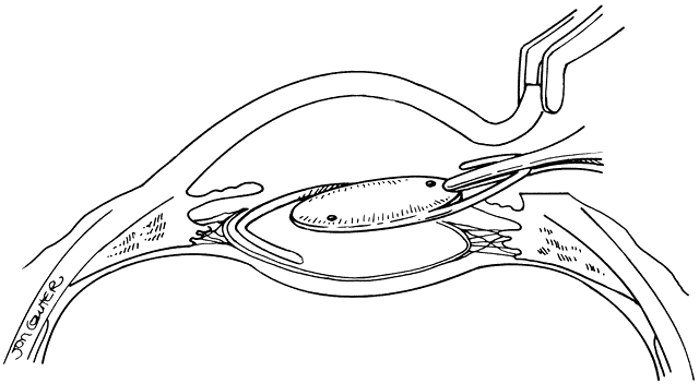
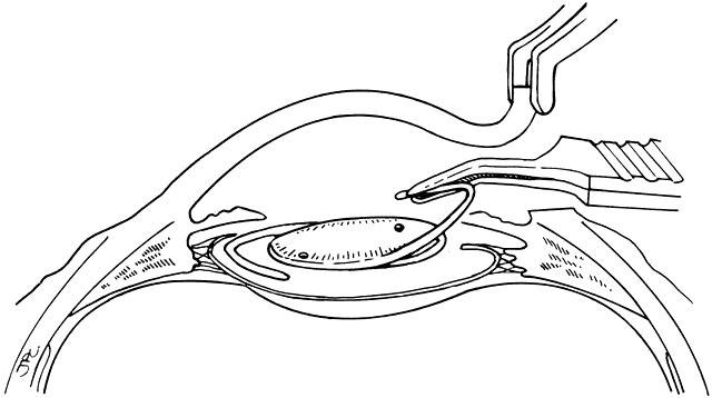
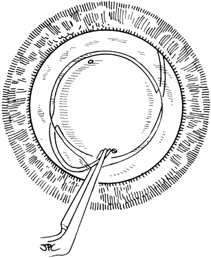
Lens implantation is then performed with capsular or so-called in-the-bag haptic placement (Figs. 1 through 5). Special care must be taken to ensure that both haptics are within the capsular sac. If haptic placement is asymmetric, with one loop in the capsular sac and one loop in the sulcus, lens decentration may occur. In the pediatric patient, the vigorous proliferation of residual lens epithelial cells leads to a greater tendency for uneven force on the lens haptics and a greater chance for lens decentration. Having both haptics in the ciliary sulcus is probably preferable to asymmetric placement. If a foldable Acrysof IOL is used, the so-called moustache style of lens fold, with both haptics placed down on the posterior capsule to unfold into the capsular bag, should not be used in cases of posterior lenticonus where the posterior capsule abnormality may catch one or both of the haptics, which would lead to posterior capsule rupture.

Fig. 1. Placement of a posterior chamber lens within the capsular bag. An anterior capsulorrhexis or capsulotomy has been performed, and all of the lens cortex has been removed. The capsular bag is filled with viscoelastic, with special attention directed toward separating the anterior capsule remnants from the posterior capsule. The lens is grasped with a straight McPherson forceps, holding the edge of the optic and the superior haptic for stability. The inferior haptic is directed into the capsular bag.

Fig. 2. Placement of a posterior chamber lens within the capsular bag. With the inferior haptic within the capsular bag, the tip of the superior haptic is grasped with an angled McPherson forceps.

Fig. 3. Placement of a posterior chamber lens within the capsular bag. The optic is directed inferiorly into the capsular bag, and the haptics are compressed so that the superior haptic clears the edge of the anterior capsule.

Fig. 4. Placement of a posterior chambers lens within the capsular bag. After the curve of the superior haptic has cleared the anterior capsule and is within the capsular bag, the tip of the haptic is rotated slightly upward to keep the superior haptic within the capsular bag, and clockwise rotational force is applied to the lens as the superior haptic is further directed into the capsular bag and the tip is released into the bag.

Fig. 5. Placement of a posterior chamber lens within the capsular bag. The lens is further “dialed” into the haptic's horizontal position with a Sinsky hook placed either at the inferior lens haptic junction or within a positioning hole on the lens.

A peripheral iridectomy is performed in all patients as an added safety precaution and viscoelastic is removed before the wound is closed. In older children able to cooperate at the slit lamp microscope for suture removal, the wound is closed with interrupted 10-0 nylon sutures; suture removal is planned for 5 to 6 weeks after surgery. In young or uncooperative children, the sutures may be removed with an examination under anesthesia, also affording the opportunity for detailed examination of the eye, or interrupted 9-0 Polyglactin absorbable sutures may be used. The activity level of the young patient and possible eye rubbing are kept in mind during wound closure, and extra sutures are placed for added strength if there is any question about the integrity of the wound.

A decision must then be made regarding the posterior capsule, PC opacification after extracapsular cataract surgery is caused by proliferation and migration of residual lens epithelial cells.42 This occurs as an age-related tendency, and virtually all pediatric patients develop capsular opacification over time.43 In an infant less than 1 year of age this may occur within the first several postoperative weeks. In an older child, opacification may take years to develop. One study of IOL use in children reported an average time to opacification of 2 years, regardless of patient age at the time of surgery and suggested primary capsulectomy-anterior vitrectomy at the time of implant surgery for children who are not expected to be candidates for YAG capsulotomy within 18 months of surgery.44 In patients less than 2 years of age, the risk of developing a thick membrane on which it would be difficult to perform YAG capsulotomy is significant and all these patients should have primary posterior capsulotomy-anterior vitrectomy procedures performed. If the patient can be expected to be cooperative enough for subsequent Nd:YAG laser capsulotomy, or if the facilities are available for performing YAG laser procedures on supine anesthetized patients, the capsule is left intact to avoid the potential complications of intraocular capsulotomy. It should be emphasized here that if this is the strategy taken, YAG capsulotomy must be performed in a timely fashion to avoid problems with amblyopia induced by the visual deprivation of capsular opacification and to prevent development of a thick membrane that may be resistant to YAG laser disruption using a reasonable amount of energy.

If posterior capsulectomy-anterior vitrectomy is to be performed at the time of implant surgery, after the implant wound is closed, a vitreous cutting instrument is used from the pars plana, posterior to the posterior capsule and IOL.45 An infusion cannula is placed into the AC and a stab incision is made for the cutting instrument 1.5 to 2 mm posterior to the limbus. A central posterior capsulectomy is then made with removal of the anterior vitreous. Generous vitrectomy should be performed in infants and young children.

SECONDARY PC IOL IMPLANTATION

Capsular Support Present

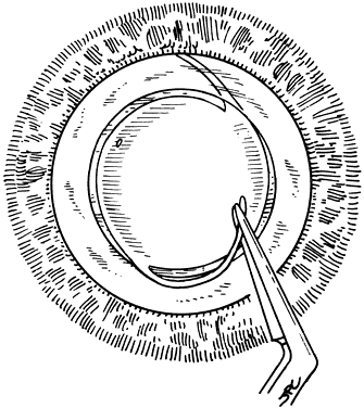
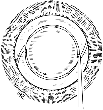
If the cataract surgery has been performed, leaving a rim of capsule present, secondary implantation of a PC IOL into the ciliary sulcus is then performed. The anterior and posterior capsular flaps having sealed together will thus form a so-called shelf to help guide and support the haptics in the ciliary sulcus.46,47 Preoperative evaluation of pupillary dilation, the integrity of this capsular shelf, and examination for iridocapsular synechiae are important. If pupillary dilation is seen to be poor or if synechiae are present, a paracentesis tract should be planned. An iris or Sinskey hook is used for retraction of the iris to aid in examination of the iridocapsular relationships. Synechiae may be lysed with viscoelastic, an iris spatula, or, frequently, they may need to be cut with a discission knife (Fig. 6).

Fig. 6. Lysis of iridocapsular synechiae in preparation for secondary posterior chamber intraocular lens implantation into ciliary sulcus. A paracentesis tract has been made before making the superior surgical incision. A Sinsky hook is used to retract the iris and provide traction while iridocapsular synechiae are cut with a discission knife.

A technique for in-the-bag secondary IOL implantation is selected patients has been described by Wilson and coworkers.48 In this technique, if Sommering's ring is present, with a ring of reproliferated cortex present between the lens equator and the fused anterior and posterior capsular leaflets, the capsular bag may be reopened to allow placement of the haptics within the bag. Those authors suggest limiting the anterior and posterior capsulectomy performed during the initial cataract surgery to a smaller than usual 4 to 5 mm and the performance of generous anterior vitrectomy to aid in preventing closure of the capsulectomy. The success of this technique in reliably producing the required capsular conditions without producing problems with synechiae or secondary membranes has not yet been demonstrated.

Capsular Support Absent

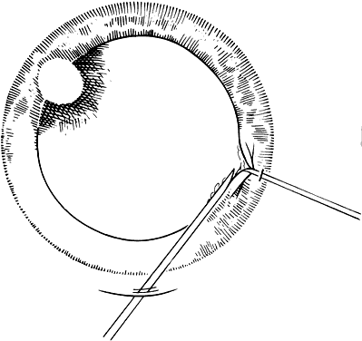
Several techniques have been advocated for suture fixation of PC IOLs into the ciliary sulcus since the concept was first described by Girard.34,35,46–59 Important improvements are the use of an implant with holes at the apex of each haptic to make fixation of the sutures to the haptics easier and less likely to malposition, and incorporation of conjunctival-scleral flaps to sequester the suture knot to prevent erosion and the increased possibility of endophthalmitis. Fixation of the haptics at slightly oblique meridians helps avoid the major arterial vessels of the iris and ciliary body and also decreases the chances for hemorrhage with the passage of the fixation sutures.24

SECONDARY AC IOL IMPLANTATION

Secondary AC IOL implantation in pediatric patients is performed in the same manner as that used in adults. There may be some advantage to a horizontal orientation of the haptics over a vertical orientation. If a peripheral iridectomy is present superiorly, horizontal orientation avoids it. There is also some suggestion that chronic discomfort after AC IOL implantation, particularly if too large a lensis implanted, may be avoided with horizontal placement.

Back to Top
POSTOPERATIVE CARE
All cases are given subconjunctival injections of dexamethasone (4 mg/ml) 0.5 ml and cefazolin (50 mg/ml) 0.15 ml at the conclusion of surgery. Topical antibiotic steroid combination drops are given, and the eye is pressure patched. The eye is covered with a clear plastic shield.

The pressure patch is removed on the first postoperative day. The plastic shield or glasses are worn continuously for 1 to 2 weeks, and physical activity is restricted for 1 month. Postoperatively, all patients are initially given an antibiotic steroid combination drop four times daily. Patients with PC IOLs are given tropicamide 1% three times daily. The medications are tapered as the postoperative inflammation subsides. Systemic steroids are seldom required.

Patients are examined on the first and third postoperative days, at 1 week and then as indicated by the level of postoperative inflammation. Depending on the regression of postoperative astigmatism and whether sutures are to be removed, overcorrections and bifocals are given about 1 month after surgery. As soon as the inflammatory response and refractive status will allow reasonable visual acuity, most often within 1 week of surgery, amblyopia occlusion therapy, if required, is initiated.

Back to Top
COMPLICATIONS
Any complications that may occur after adult cataract surgery or lens implantation may also occur in a child undergoing lens implantation. It is equally important to note that no complications have been reported that are unique to children when compared with adults. However, the vigorous inflammatory response of the child's eye to retained cortical material and the propensity for secondary membrane formation, even where posterior capsulectomy and anterior vitrectomy have been performed, both with and without IOL implantation,60,61 produce a greater tendency for the development of synechiae and IOL dislocations.54–58 It is imperative that surgery be performed as meticulously as possible58 and that postoperative follow-up, shortly after surgery as well as over the ensuing years, be conservative (in terms of frequency of visits) and guarded in nature. Inflammatory pupillary block glaucoma has been reported after PC IOL implantation in children,62 and the tendency for eyes with pediatric cataracts to develop glaucoma has been well documented with a reported prevalence as high as 32%.63–65 Some evidence suggests, however, that primary IOL implantation may decrease the incidence of glaucoma in children following surgery for congenital or developmental cataracts (1 of 377 pseudophakic eyes compared with 14 of 124 aphakic eyes).66 Whether this result is due to patient selection remains undetermined. The special pediatric concerns of ocular growth and secondary membrane formation have already been discussed in this chapter.

A discussion of complications in the pediatric patient also cannot ignore the fact that few patients with lenses implanted in childhood have had follow-up for longer than 10 to 15 years and that there are essentially no published long-term data available. Many published reports on IOL use in children are of little value when discussing complications today because most of this information pertains to iris support and AC lens designs, which have been shown in the adult population to be associated with currently unacceptable complications. Only a limited amount of published information is available concerning the use of PC or modern AC lenses in children, and only a small amount of long-term information is available.7,9,13,14,67–74

Back to Top
VISUAL RESULTS
The cautions already discussed, which limit our ability to examine the current potential complications of pediatric IOL use, also limit our ability to use currently available data to examine the visual results of lens implantation in children. Again, most available data pertain to the use of lenses that are no longer used. One can hopefully assume that the visual results obtained with current technology and surgical techniques are better than those in the no longer current literature.

Hiles presents data comparing the visual results achieved with glasses, contact lenses, IOLs, and epikeratoplasty over a decade of use.14 For traumatic cataracts and for unilateral cataracts of varying diagnoses, the results are similar for all these modalities. These data do not, however, help in providing a more definitive answer to the question of whether lens implantation should be used more widely or for any particular patient or type of cataract.

Any examination of visual results after cataract surgery in the pediatric age group must take into consideration the multiple variables involved in visual outcome. Beginning with diagnosis, especially in the traumatic and congenital groups, the coexistence of other anatomic impairments to the development of vision may profoundly influence visual result. The age of the patient at development of visual deprivation and the delay until surgery and optic correction are performed may have a much greater impact on visual result than which optical rehabilitation modality has been selected. The definitions of congenital and developmental types of cataracts are not used in a uniform fashion, thus affecting the interpretation of visual results. For example, posterior lenticonus cataracts are considered congenital cataracts by some authors, leading to much better results than those achieved in series of congenital cataracts limited to complete opacities present at birth. Finally, and perhaps most significantly, the level of compliance with optical correction and amblyopia occlusion therapy certainly influence visual results greatly.

Examination of only those reported series of patients, in all diagnostic categories, who have received modern PC or AC IOLs reveals that 52% achieved visual acuity of 20/40 or better, 34% achieved visual acuity between 20/40 and 20/200, and 13% achieved visual acuity worse than 20/200 (Table 1). In the compilation of this data, patients who were too young to have formal visual acuity measurements recorded and who were reported as having central fixation responses were, conservatively, considered as having less than 20/200 visual acuity. Trauma caused the cataract in 55% of these patients, decreasing the effect of amblyopia in many of these patients.


Because most patients reported in the literature who have received IOLs have been intolerant to use of contact lenses or have been presumed to be poor candidates for such use, the results achieved by these patients may be skewed toward a poor result. One may assume that a patient intolerant to contact lens wear or without family capabilities for contact lens wear would also be less compliant with amblyopia occlusion, again skewing the visual results. With these thoughts in mind, the comparable visual results that have been reported may be taken as testimony in support of more primary IOL implantation in children. The situation may not be so clear, however, and it may certainly be true that, even after overcoming optical correction compliance problems, the final visual results achieved do not improve because of continued problems with amblyopia occlusion therapy. Patching in the situation of profound amblyopia is almost always met with greater resistance than wearing a contact lens. For developmental cataracts and in traumatic cataracts for which amblyopia is less of a problem, IOLs may play a significant role in preventing the development of deprivation amblyopia caused by the uncorrected aphakia of contact lens noncompliance.

Back to Top
FOOD AND DRUG ADMINISTRATION STATUS
There are currently no IOLs that have received premarket approval (PMA) from the FDA as safe and effective devices for use in patients under the age of 18 years. All IOL implantation in pediatric patients is considered investigational, and patients and their families must be made aware of this as part of their informed consent. There are three manners in which a physician may implant an IOL in a child:

  [*]1. An IOL manufacturer that has received FDA approval for an Investigational Device Exemption (IDE) for a clinical trial of IOL use in children may sponsor a clinical study (necessary to obtain a PMA for the pediatric indication) in which a physician may act as a clinical investigator and perform IOL implantations, within the study protocol, in children. IOLAB Corporation (Claremont, California) was the only company publicly using an IDE for a clinical trial, and as of July 1994 they had stopped enrollment in their study. Between 1981 and 1994, over 1260 eyes were enrolled in the IOLAB study by 361 US investigators. Some of the results of this study were published in 1999.75 PMA has, unfortunately, not been obtained.
  [*]2. A physician may implant in a child an IOL that has PMA approval only for use in adults, in the belief that it is within the scope of his or her licensed practice of medicine or use of approved products for unapproved indications. This is currently the simplest manner of performing IOL implantation in a child. Use of an IOL in this manner may require approval by the surgeon's local Institutional Review Board (IRB) and may be the most risky for the surgeon if a malpractice suit should be filed relating to complications caused by the use of an IOL in a child.
  [*]3. A physician may ask an IOL manufacturer without an ongoing pediatric IDE to request from the FDA, under their IDE for adults, a waiver of the age indication based on the case of a specific patient. Surgery performed in this manner will also require IRB approval.

Back to Top
SUMMARY
Use of IOLs in children has changed dramatically over the past decade and the IOL has now become the primary means of optical correction of aphakia for most pediatric patients affected by cataracts. IOLs are not recommended for all patients, however, and the treatment of a child, especially an infant, with a cataract remains quite different from the adult cataract patient. This chapter attempts to define the differences in treatment strategies needed to allow for the safest and most effective use of IOLs in children.
Back to Top
REFERENCES

1. Choyce P: Correction of uniocular aphakia by means of anterior chamber acrylic implants. Trans Ophthalmol Soc UK 78:459, 1958

2. Lambert SR, Buckley EG, Plager DA et al: Unilateral intraocular lens implantation during the first six months of life. J AAPOS 3:344, 1999

3. Morgan KS, Braverman DE, Baker ID: The correction of unilateral aphakia in children treated for orbital rhabdomyosarcoma. J Pediatr Ophthalmol Strabismus 27:70, 1990

4. BenEzra D, Cohen E: Cataract surgery in children with chronic uveitis: Ophthalmology 107:1255, 2000

5. Swan KC, Wilkins JH: Extraocular muscle surgery in early infancy—Anatomical factors. J Pediatr Ophthalmol Strabismus 21:44, 1984

6. Gordon RA, Donzis PB: Refractive development of the human eye. Arch Ophthalmol 103:785, 1985

7. Dahan E, Salmenson B: Pseudophakia in children: Precautions, technique and feasibility. J Cataract Refract Surg 16:75, 1990

8. BenEzra D, Paez JH: Congenital cataract and intraocular lenses. Am J Ophthalmol 96:311, 1983

9. Sinskey RM, Karel F, Dal Ri E: Management of cataracts in children. J Cataract Refract Surg 15:196, 1989

10. Aron JJ, Aron-Rosa D: Intraocular lens implantation in unilateral congenital cataract: A preliminary report. Am Intra-Ocular Implant Soc J 9:306, 1983

11. Dahan E, Drusedau MU: Choice of lens and dioptric power in pediatric pseudophakia. J Cataract Refract Surgery 23(Suppl 1):618, 1997

12. Hiles DA: Intraocular lens implantation in children with monocular cataracts 1974-1983. Ophthalmology 91:1231, 1984

13. Burke JP, Willshaw HE, Young JDH: Intraocular lens implants for uniocular cataracts in childhood. Br J Ophthalmol 73:860, 1989

14. Hiles DA: Visual rehabilitation of aphakic children III. Intraocular lenses. Surv Ophthalmol 34:371, 1990

15. Wilson JR, Fernandes A, Chandler CV et al: Abnormal development of the axial length of aphakic monkey eyes. Invest Ophthalmol Vis Sci 28:2096, 1987

16. Rasooly R. BenEzra D: Congenital and traumatic cataract. The effect on ocular axial length. Arch Ophthalmol 106:1066, 1988

17. Raviola E, Wiesel TN: An animal model of myopia. N Engl J Med 312:1609, 1985

18. Von Noorden GK, Lewis RA: Ocular axial length in unilateral congenital cataract and blepharoptosis. Invest Ophthalmol Vis Sci 28:750, 1987

19. Sinskey RM, Stoppel MD, Amin PA: Ocular axial length changes in a pediatric patient with aphakia and pseudophakia. J Cataract Refract Surg 19:787, 1993

20. Moore BD: Changes in the aphakic refraction of children with unilateral congenital cataracts. J Pediatr Ophthalmol Strabismus 26:290, 1989

21. McClatchey SK, Parks MM. Myopic shift after cataract removal in childhood. J Pediatr Ophthalmol Strabismus 34:88, 1997

22. McClatchey SK, Parks MM. Theoretic refractive changes after lens implantation in childhood. Ophthalmology 104:1744, 1997

23. McClatchey SK, Dahan E, Maselli E et al: A comparison of the rate of refractive growth in pediatric aphakic and pseudophakic eyes. Ophthalmology 107:118, 2000

24. Kora Y. Shimizu K, Inatomi M et al: Eye growth after cataract extraction and intraocular lens implantation in children. Ophthalmic Surg 24:467, 1993

25. Katsumi O, Miyajima H, Ogawa T et al: Aniseikonia and stereoacuity in pseudophakic patients—Unilateral and bilateral cases. Ophthalmology 99:1270, 1992

26. Andreo LK, Wilson ME, Saunders RA: Predictive value of regression and theoretical IOL formulas in pediatric intraocular lens implantation. J Pediatr Ophthalmol Strabismus 34:240, 1997

27. Apple DA, Mamalis N, Olsen RJ et al: Intraocular Lenses. Evolution, Designs, Complications, and Pathology. Baltimore: Williams & Wilkins, 1989

28. Apple DJ, Mamalis N, Loftfield K, et al: Complications of intraocular lenses. A historical and histopathological review. Surv Ophthalmol 29:1, 1984

29. Miyake K, Asakura M, Kobayashi H: Effect of intraocular lens fixation on the blood-aqueous barrier. Am J Ophthalmol 98:451, 1984

30. Lyle WA, Jin JC: Secondary intraocular lens implantation: Anterior chamber vs posterior chamber lenses. Ophthalmic Surg 24:375, 1993

31. Lubniewski AJ, Holland EJ, Van Meter WS et al: Histologic study of eyes with transsclerally sutured posterior chamber intraocular lenses. Am J Ophthalmol 110:237, 1990

32. Apple DJ, Loftfield K, Mamalis N, et al: Biocompatibility of implant materials: A review and scanning electron microscopic study. Am Intraocular Implant Soc J 10:53, 1984

33. Heilskov T, Joondeph BC, Olsen KR, et al: Late endophthalmitis after transscleral fixation of a posterior chamber intra-ocular lens. Arch Ophthalmol 107:1427, 1989

34. Hu BV, Shin SH Gibbs KA et al: Implantation of posterior chamber lens in the absence of capsular and zonular support. Arch Ophthalmol 106:416, 1988

35. Lewis JS: Sulcus fixation without flaps. Ophthalmology 100:1346, 1993

36. Sharpe MR, Biglan AW, Gerontis CC: Scleral fixation of posterior chamber intraocular lenses in children. Ophthalmic Surg Lasers 27:337, 1996

37. Tabbara KF, Al-Kaff AS, Al-Rajhi AA et al: Heparin surface-moddified intraocular lenses in patients with inactive uveitis or diabetes. Ophthalmology 105:843, 1998

38. Maltzman BA, Haupt E, Cucci P: Effect of the laser ridge on posterior capsule opacification. J Cataract Refract Surg 15:644, 1989

39. Stark WJ, Worthen DM, Holladay JT et al: The FDA report on intraocular lenses. Ophthalmology 90:311, 1983

40. Assia EI, Apple DJ, Tsai JC et al: Mechanism of radial tear formation and extension after anterior capsulectomy. Ophthalmology 98:432, 1991

41. Wilson ME, Bluestein EC, Wang X et al: Comparison of mechanized anterior capsulectomy and manual continuous capsulorrhexis in pediatric eyes. J Cataract Refract Surg 20:602, 1994

42. McDonnell PJ, Zarbin MA, Green WR: Posterior capsule opacification in pseudophakic eyes. Ophthalmology 90:1548, 1983

43. BenEzra D, Cohen E: Posterior capsulectomy in pediatric cataract surgery: The necessity of a choice. Ophthalmology 104:2168, 1997

44. Plager DA, Lipsky SN, Snyder SK et al: Capsular management and refractive error in pediatric intraocular lenses. Ophthalmology 104:600, 1997

45. Buckley EG, Klombers LA, Seaber JH et al: Management of the posterior capsule during pediatric intraocular lens implantation. Am J Ophthalmol 115:722, 1993

46. Eifrig DE: Two principles for repositioning intraocular lenses. Ophthalmic Surg 17:486, 1986

47. Dahan E, Salmenson BD, Levin J: Ciliary sulcus reconstruction for posterior implantation in the absence of an intact posterior capsule. Ophthalmic Surg 20:776, 1989

48. Wilson ME, Englert JA, Greenwald MJ: In-the-bag secondary intraocular lens implantation in children. J AAPOS 3:350, 1999

49. Girard LJ: Pars plana phacoprosthesis (aphakic intraocular implant): A preliminary report. Ophthalmic Surg 12:19, 1981

50. Stark WJ, Goodman G, Goodman D et al: Posterior chamber intraocular lens implantation in the absence of posterior capsular support. Ophthalmic Surg 19:240, 1988

51. Dahan E: Implantation in the posterior chamber without capsular support. J Cataract Refract Surg 15:339,1989

52. Stark WJ, Gottsch JD, Goodman DF et al: Posterior chamber intraocular lens implantation in the absence of capsular support. Arch Ophthalmol 107:1078, 1989

53. Lindquist TD, Agapitos PJ, Lindstrom RL et al: Transscleral fixation of posterior chamber intraocular lenses in the absence of capsular support. Ophthalmic Surg 20:766, 1989

54. Knight-Nanan D, O'Keefe M, Bowell R: Outcome and complications of intraocular lenses in children with cataract. J Cataract Refract Surg 22:730, 1996

55. Crouch ER Jr, Pressman SH, Crouch ER: Posterior chamber intraocular lenses: Long-term results in pediatric cataract patients. J Pediatr Ophthalmol Strabismus 32:210, 1995

56. Brady KM, Atkinson CS, Kilty LA et al: Cataract surgery and intraocular lens implantation in children. Am J Ophthalmol 120:120, 1995

57. Awner S, Buckley EG, DeVaro JM et al: Unilateral pseudophakia in children under 4 years. J Pediatr Ophthalmol Strabismus 33:230, 1996

58. Ainsworth JR, Cohen S, Levin AV et al: Pediatric cataract management with variations in surgical technique and aphakic optical correction. Ophthalmology 104:1096, 1997

59. Shapiro A, Leen MM: External transscleral posterior chamber lens fixation. Arch Ophthalmol 109:1759, 1991

60. Morgan KS, Karcioglu ZA: Secondary cataracts in infants after lensectomies. J Pediatr Ophthalmol Strabismus 24:45, 1987

61. Spierer A, Desatnik E, Blumenthal M: Secondary cataract in infants after extracapsular cataract extraction and anterior vitrectomy. Ophthalmic Surg 23:625, 1992

62. Vajpayee RB, Angra SK, Tityal JS et al: Pseudophakic pupillary-block glaucoma in children. Am J Ophthalmol 111:715, 1991

63. Simon JW, Mehta N, Simmons ST et al: Glaucoma after pediatric lensectomy/vitrectomy. Ophthalmology 98:670, 1991

64. Chrousos AG, Parks MM, O'Neill JF: Incidence of chronic glaucoma, retinal detachment, and secondary membrane surgery in pediatric aphakic patients. Ophthalmology 91:1238, 1984

65. Johnson CP, Keech RV: Prevalence of glaucoma after surgery for PHPV and infantile cataracts. J Pediatr Ophthalmol Strabismus 33:14, 1996

66. Asrani S, Freedman S, Hasselblad V et al: Does primary intraocular lens implantation prevent “aphakic” glaucoma in children? J AAPOS 3:33, 1999

67. Hiles DA, Hered RW: Modern intraocular lens implants in children with new age limitations. J Cataract Refract Surg 13:493, 1987

68. Gupta AK, Grover AK, Gurha N: Traumatic cataract surgery with intraocular lens implantation in children. J Pediatr Ophthalmol Strabismus 29:73,1992

69. Koenig SB, Ruttum MS, Lewandowski ME et al: Pseudophakia for traumatic cataracts in children. Ophthalmology 100:1218, 1993

70. Kora Y, Inatomi M, Fukado Y et al: Long-term study of children with implanted intraocular lenses. J Cataract Refract Surg 18:485, 1992

71. Gimbel HV, Ferensowicz M, Raanan M et al: Posterior chamber IOL implantation in children. J Pediatr Ophthalmol Strabismus 30:69, 1993

72. Sinskey RM, Stoppel JO, Amin P: Long-term results of intraocular lens implantation in pediatric patients. J Cataract Refract Surg 19:405, 1993

73. Gimbel HV, Basti S, Ferensowicz M et al: Results of bilateral cataract extraction with posterior chamber intraocular lens implantation in children. Ophthalmology 104:1737, 1997

74. Zwaan J, Mullaney PB, Awad A et al: Pediatric intraocular lens implantation: Surgical results and complications in more than 300 patients. Ophthalmology 105:112, 1998

75. Young TL, Bloom JN, Ruttum M et al: The IOLAB, Inc. intraocular lens study. J AAPOS 3:295, 1999

Back to Top