Chapter 100
Pediatric Cataract Surgery
KENNETH P. CHENG and ALBERT W. BIGLAN
Main Menu   Table Of Contents

Search

INDICATIONS FOR SURGERY
CONTRAINDICATIONS TO SURGERY
TIMING OF SURGERY
OPTICAL REHABILITATION
PREOPERATIVE MEASURES AND PREVENTION OF COMPLICATIONS
OPTICAL PROPERTIES OF THE EYE
SURGICAL PROCEDURE
POSTOPERATIVE CARE
VISUAL RESULTS
COMPLICATIONS
ENDOPHTHALMITIS
CAPSULE OPACIFICATION
CYSTOID MACULAR EDEMA
RETINAL DETACHMENT
STRABISMUS
AMBLYOPIA
SUMMARY
REFERENCES

The treatment of an infant or child with a cataract requires different decision processes and modifications in surgical procedures compared with the treatment of an adult with a cataract. The developing visual system, growth, and anatomic differences in the eye and related structures, and psychosocial differences between children and adults contribute to the differences in treatment.

The adult eye is fully developed, and the visual pathways are mature. Management after cataract surgery is principally a process of optical rehabilitation to achieve restoration of vision. In children, however, the eye and the visual pathways are developing, and the operation to remove a cataract is only the first step in a long process aimed at promoting normal development of the visual system and achieving the best possible visual acuity in the affected eye or eyes. This chapter reviews the indications, surgical techniques, complications, and expected results of cataract surgery in infants and children.

Back to Top
INDICATIONS FOR SURGERY
In children affected by either unilateral or bilateral cataracts, the indications for surgery and the risk-benefit ratio are more complex than for an adult patient. In an adult, the presence of a cataract that significantly degrades vision or alters the patient's lifestyle is a sufficient indication for surgery. The risks of the procedure are low, and the benefits are high.

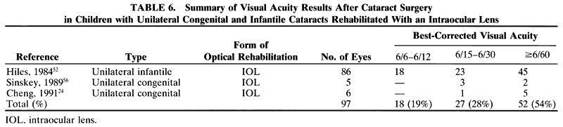
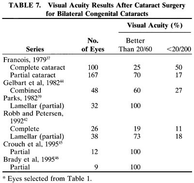
In a child, additional factors must be considered, such as the health of the child, the availability of resources for perioperative care, and the availability of anesthesiologists who are trained in the care of infants and young children. In adults, the surgery can be scheduled to accommodate the convenience of the patient. In children, the timing of surgery may be dictated by the degree of maturation of the visual system. Surgery may need to be performed in a timely fashion to facilitate the effective treatment of amblyopia. In infants with unilateral congenital cataracts, a critical period in visual development exists after birth, during which treatment should be initiated to optimize the possibility for promoting visual development in the affected eye. Evidence suggests that this critical period is before the first 6 to 17 weeks of age.1,2

Another difference is the frequent presence of structural abnormalities of the child's globe and visual pathways. These abnormalities can influence the visual result that is achieved. Inherent defects in the available methods of optical correction and compliance with the correction of aphakia and amblyopia therapy affect the visual result. Additionally, persistence with treatment of strabismus, glaucoma, and other related defects is needed to maintain a successful surgical outcome.

Because of the difficulties with correction of aphakia in children, surgery may not be advisable in some cases. For example, observation of a monocular partial lens opacity that decreases visual acuity to 20/40 in an amblyopia-susceptible 2-year-old child may be preferable to making that child aphakic and intolerant of contact lenses or even pseudophakic without the ability to accommodate, and intolerant of glasses with bifocal lenses. The reverse also can be true. Irreversible deprivation amblyopia can be caused by a partial lens opacity, for example, posterior lenticonus, in an infant or a young child. In these cases, the opportunity to achieve high levels of central vision may be permanently lost if surgery is delayed.3 Because of these factors, the accepted 95% success rate for excellent vision after cataract surgery in adults may not be achieved in children.

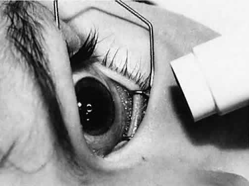
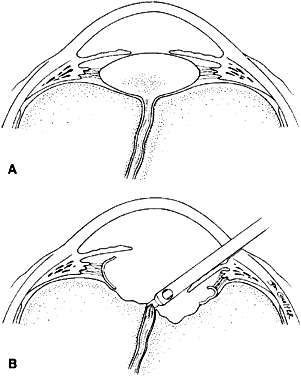
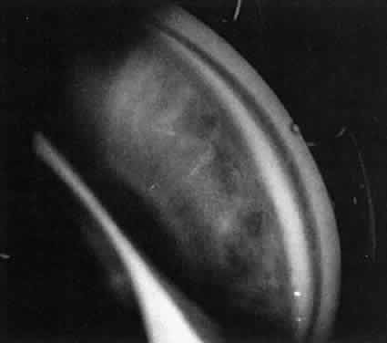
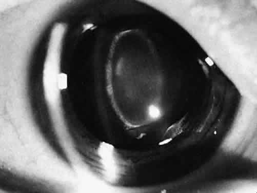
Not all cataracts require surgical treatment. Eyes with punctate or small anterior polar cataracts and others with partial opacification of the lens, such as posterior lenticonus, which only slightly interferes with the refraction of light, are best followed and not surgically treated (Fig. 1). In some cases, it is difficult to determine whether the presence of a partial cataract is responsible for a decrease in visual acuity or whether the refractive error or optical distortion produced by the cataract has produced a mild, reversible form of deprivation amblyopia. In these situations, correction of the refractive error and a trial of occlusion therapy should be attempted. If the visual acuity improves, it may be deduced that amblyopia was responsible for the loss of visual acuity and that the optical distortion produced by the partial cataract is not yet surgically significant.

Fig. 1. Cataracts that do not require surgical treatment. A. Anterior polar cataract. Visual acuity is 20/25. B. Partial punctate or cerulean cataract. Visual acuity is 20/30.

TREATMENT GUIDELINES

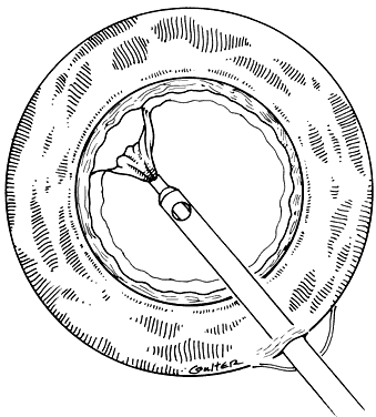
When a cataract is extensive or if a partial cataract is present and the vision does not improve with a trial of occlusion therapy, cataract surgery should be considered. The guidelines for cataract surgery given in this section are based on the extent of the lens opacity and whether the cataract is isolated to one eye or both. Specific indications for cataract surgery, however, should be considered individually for each child.

Unilateral Partial Cataract

Children who have monocular partial cataracts should undergo cataract surgery when visual acuity in the affected eye is judged to be less than 20/70, when there is a loss of central fixation, or when visual deprivation is sufficient to cause strabismus. In questionable cases or when the cataract is small or only partially obstructs the visual axis, a trial of amblyopia treatment along with optical correction of anisometropia and dilation of the pupil, if necessary, to provide a clear visual axis3 should be performed before the decision is made to remove a partial cataract.

Unilateral Complete Congenital Cataract

Infants and children younger than 8 years of age who have monocular complete cataracts that are present from birth should have cataract surgery performed as soon as they are medically stable (preferably before 17 weeks of age). A child who is older than 8 years of age may have a complete monocular cataract removed to visualize the fundi or to improve peripheral vision. Treatment of amblyopia with improvement of central vision in children who have had complete congenital cataracts after 2 years of age usually is unsuccessful. If there was a possibility that the cataract was partial during infancy, with some opportunity for stimulation of the macula with clearly formed images, the cataract should be removed at any age to regain central visual acuity.

Bilateral Partial Cataracts

Children younger than 8 years of age who have bilateral partial cataracts should undergo surgery when vision in either eye is less than 20/70, if visual deprivation is producing strabismus, or if there appears to be a risk for the development of nystagmus as a result of visual deprivation.

Bilateral Complete Cataracts

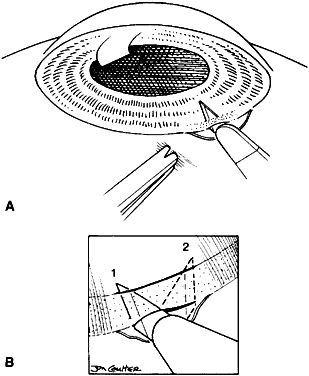
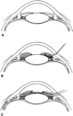
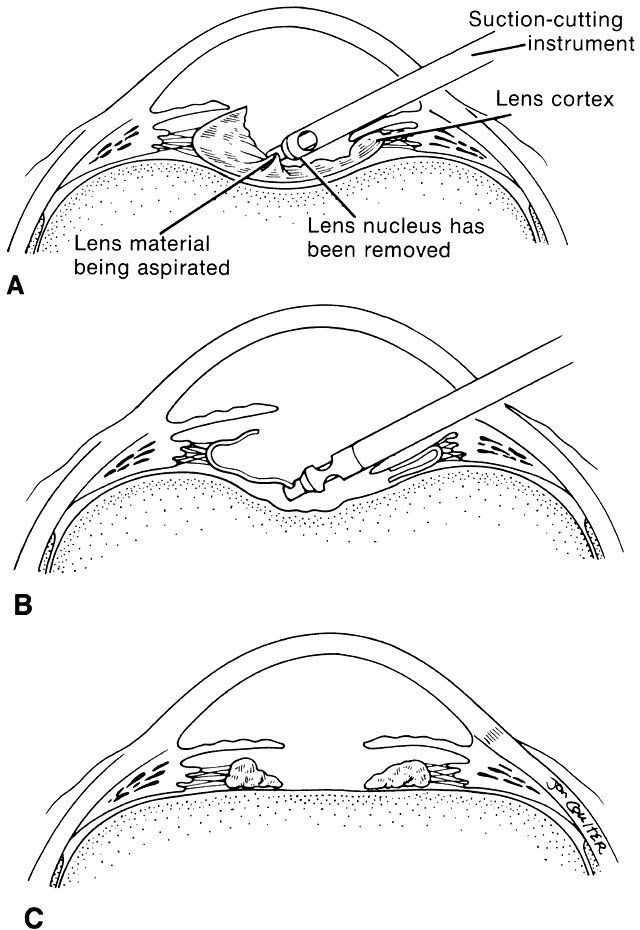
If the lens opacity is complete and involves both eyes, the cataract should be removed in the eye with the most complete lens opacity. The fellow eye should be treated as soon as the first eye recovers from surgery. This interval should be brief (5 to 10 days) in very young children. If a health problem precludes repeated anesthetic sessions, consideration may be given to performing the surgery on both eyes during the same anesthetic session, with the use of different instrumentation and separate operative fields.4,5

OTHER CONTRIBUTING FACTORS

Decisions to perform cataract surgery can be difficult when young or developmentally delayed children cannot provide subjective visual acuity responses. In these situations, the decision to remove a cataract depends on the size, density, type, location, and progression of the opacity and the estimated effect of these factors on vision. Estimates of the effect of a partial cataract on vision can at times be difficult, even for experienced examiners; in difficult cases, second opinions can be helpful.6

Size and Density

Cataracts that are central in location and larger than 1.5 to 2 mm obstruct light entering the eye sufficiently to reduce visual acuity and impede development of the visual system. In this situation, a trial of dilating the pupil with topical applications of 2.5% phenylephrine drops three times a day to allow light to enter around the opacity may result in an improvement in visual acuity. Pupils that dilate poorly with phenylephrine may respond to 0.5% atropine eyedrops; however, the accompanying cycloplegia necessitates the use of bifocals to correct the cycloplegic refractive error and the lack of accommodation. Frequent monitoring of visual acuity and pupil size at different times of the day and under different lighting conditions is recommended. Improvements in acuity may be temporary if the lens opacity progresses in size.

Type and Location

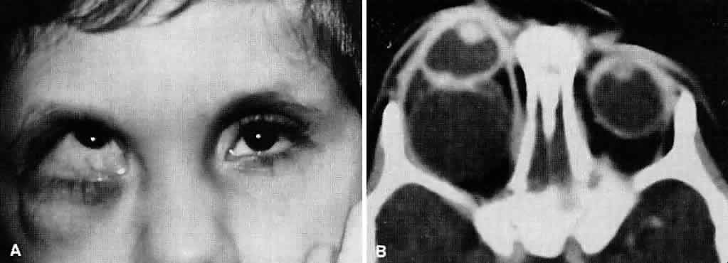
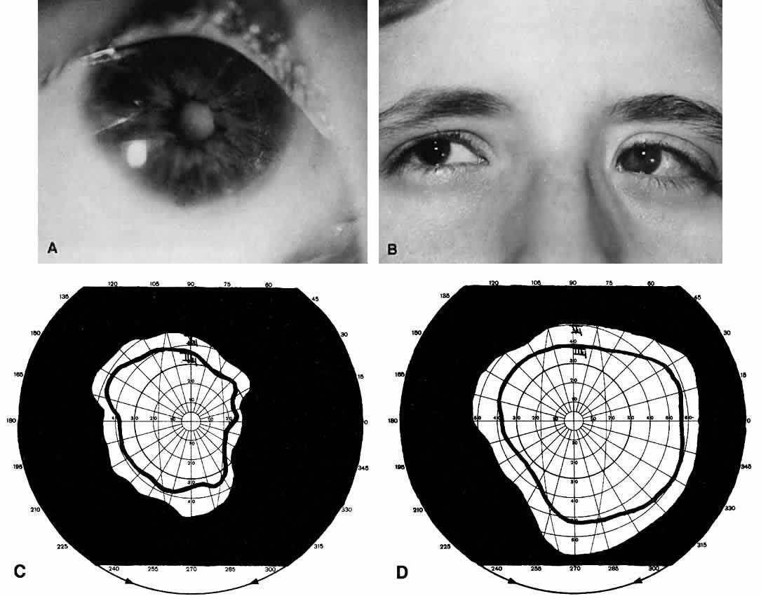
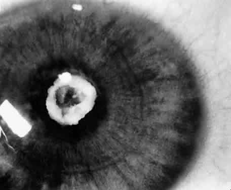
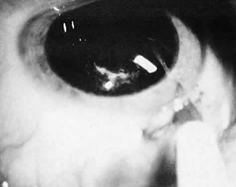
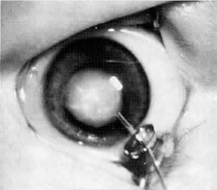
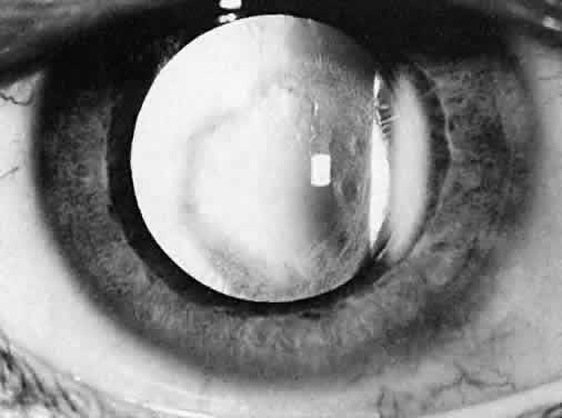
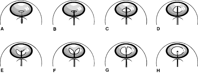
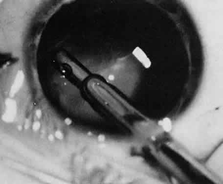
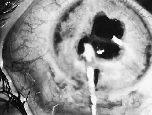
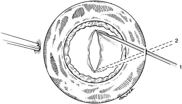
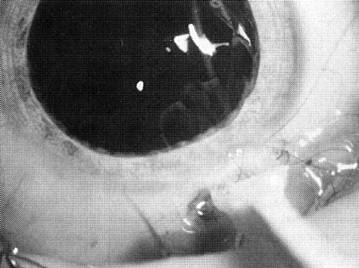
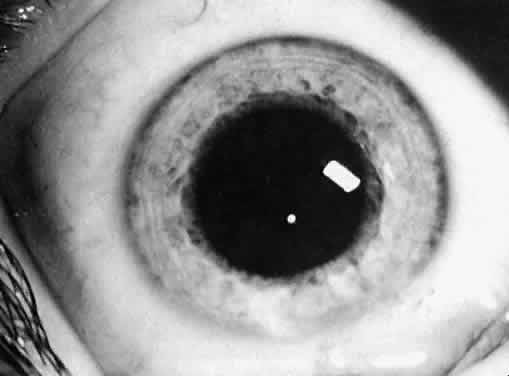
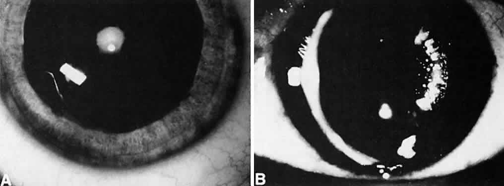
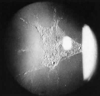
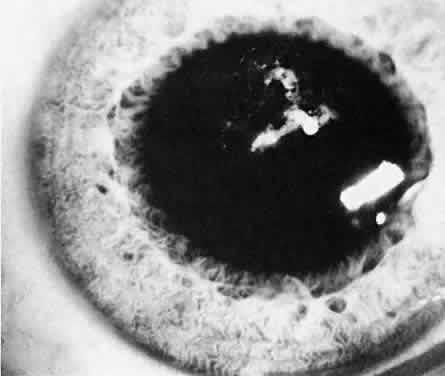
Usually, anterior polar cataracts are visually insignificant and allow normal visual development (see Fig. 1). However, some affect vision, and all require careful monitoring.7 Centrally located cataracts that are on or near the posterior lens capsule have a greater effect on the refraction of light and visual acuity (Fig. 2). Nuclear cataracts associated with metabolic disorders or prenatal infections produce double refracting systems that cause optical distortion and significantly decrease visual acuity (Fig. 3).

Fig. 2. A posterior subcapsular cataract that has reduced visual acuity in healthy illumination to 20/60.

Fig. 3. A nuclear cataract present in both eyes of a child with esotropia. The retinoscopy reflex was irregular, and cataract surgery was recommended. Metabolic evaluation and TORCH titer findings were negative.

Progression

Not all cataracts in children increase in size or density. Premature neonates may have partial opacification (transient neonatal opacities) that may disappear over the first 1 or 2 weeks of life.8 Other cataracts that may regress are those due to galactosemia. These cataracts have the morphologic features of oil droplets. If early dietary restriction of galactose is observed, the opacity may regress.9 Anterior polar cataracts typically remain stable in size and density.

Other types of cataracts characteristically progress in size and density. Cataracts caused by rubella, uveitis, diabetes, exposure to radiation, and use of corticosteroids usually show progression with time.

Visual Acuity and Signs of Visual Function

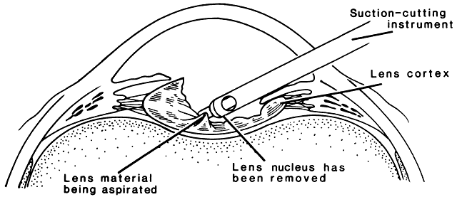
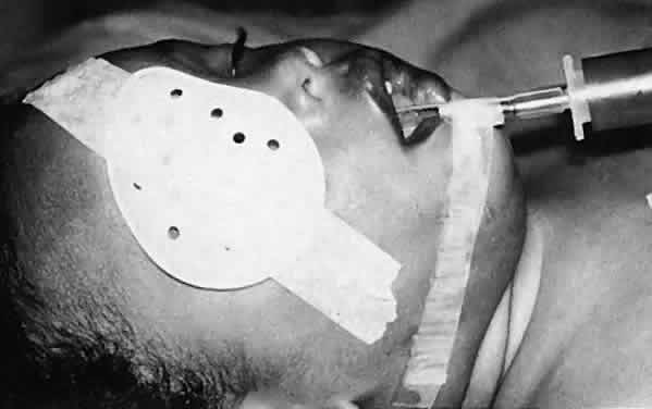
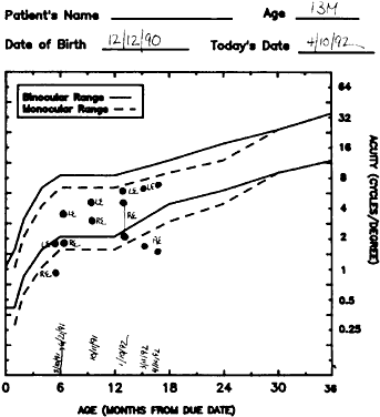
Ultimately, it is the measured or estimated level of visual acuity or acuity potential that determines the need for cataract surgery. If either the measured or the estimated decrease in visual acuity produced by the cataract is sufficient to prevent adequate visual development or if signs of significantly decreased visual acuity, such as strabismus or poor central fixation, are present, cataract surgery is indicated. Use of the Teller visual acuity cards at periodic intervals can help to measure visual acuity.10 In some children, visual acuity that is below normal or decreasing can be documented accurately to support the decision to perform cataract surgery (Figs. 4 and 5). Care must be taken in interpreting Teller visual acuity data because the normal levels in young infants and children are low and span a wide range. The Teller acuity card measurement of grating visual acuity may severely underestimate the level of visual loss in patients with cataracts and amblyopia.11

Fig. 4. Posterior lenticonus type of cataract. When the child was 1 year old, the cataract increased in size, and the posterior portion of the lens became increasingly opaque. Surgery was recommended.

Fig. 5. Acuity card results. Visual acuity was measured repeatedly in the right eye with the Teller visual acuity cards. The visual acuity decreased despite occlusion of the sound eye, and cataract surgery was recommended.

Coexisting Ocular Defects

Infants with cataracts may have coexisting defects of the globe or visual system that influence the decision to proceed with cataract surgery. If a cataract exists in an eye with other major structural anomalies and the fellow eye is normal, the benefits of proceeding with surgery are negligible, and the child should not be subjected to the risk of anesthesia and perioperative morbidity. If both eyes have major structural defects and complete cataracts, cataract surgery may be indicated to allow the child some opportunity for the development of vision, even if it is limited.

Health of the Child

An assessment of the general health of a child is important when considering cataract surgery because general anesthesia is required for the operation. Premature infants with severe bronchopul-monary dysplasia may have unacceptable risksassociated with the use of general anesthesia. Other children who have systemic diseases such as galactosemia, homocystinuria, congenital heart disease, and Marfan's syndrome or who have experienced trauma have additional risks associated with general anesthesia. For these children, appropriate consultation is needed to assess the risks of the procedure before proceeding with surgery.

Social Situation

Because the success or failure of visual rehabilitation of an infant or young child with a cataract depends on postoperative care, acceptance of optical correction, compliance with occlusion therapy, and integrity and capabilities of the family unit and its support structures must be considered. For example, if one parent has bilateral familial cataracts and poor vision and the other parent also has a visual impairment, delivery of postoperative care may be problematic. If the delivery of adequate postoperative care is likely to be a problem, cataract surgery must be considered carefully because the risk for amblyopia and visual loss produced by uncorrected aphakic refractive errors or pseudophakia may outweigh the amblyopia and visual loss produced by a partial cataract.

Back to Top
CONTRAINDICATIONS TO SURGERY
There are few contraindications for performing cataract surgery in children. If a severe life-limiting disease, such as Lowe's syndrome or Edward's syndrome, is present, cataract surgery should be deferred. Surgery is contraindicated in cases of severe microphthalmia (corneal diameter less than 5 mm) and in eyes in which the retina is irreparably detached or the posterior segment is disorganized (Fig. 6). Because of the risk of extraocular spread of tumor, another contraindication is the presence of untreated retinoblastoma. Surgery is not contraindicated in eyes with successfully treated tumors.12

Fig. 6. A. A 2-year-old boy who has microphthalmia with a cyst on the right side and a dense lens opacity. B. CT scan shows a large cyst that communicates with the vitreous. This finding was confirmed with B-scan ultrasonography. Cataract surgery was not performed.

Until three decades ago, it was widely believed that the prognosis for visual rehabilitation of any child with a unilateral congenital cataract was poor.13,14 Frey and associates15 reevaluated the treatment of children with monocular cataracts. They were encouraged when three children who had congenital monocular cataracts had 20/40 or better visual acuity. Some surgeons believe that surgery is not indicated when a monocular cataract is first diagnosed in an adolescent. However, a cataract that is presumed to be congenital or of undetermined onset may not have been visually significant at birth or early in infancy, and some visual development may have occurred. In such cases, visual results after cataract surgery on a visually mature child or adult may be much better than assumed.16 Additionally, cataract surgery should be considered for adults, even when irreversible deprivation amblyopia precludes recovery of central visual acuity. In these situations, the increase in the peripheral visual field on the side of the affected eye may be useful and desirable (Fig. 7). An attempt at visual rehabilitation of an eye with a monocular cataract is warranted unless medical contraindications are present, the globe is structurally disorganized, or the risk of complications is high.

Fig. 7. A. A 31-year-old patient with bilateral congenital cataracts. The left cataract was removed early in life, but glaucoma and retinal detachment occurred after surgery. B. A complete cataract was removed in the fellow right eye when the patient was 31 years old. The resulting visual acuity was counting fingers at 3 feet, but the patient was pleased with her increased field of binocular vision. C. The visual field before cataract surgery. D. The binocular field of vision after cataract surgery.

Back to Top
TIMING OF SURGERY
The immature visual system of a young child is sensitive to defects that interrupt the focusing of light on the retina. Studies conducted in animals as well as in humans have identified a critical period in visual development during which the immature visual system must be stimulated with focused images on the retina to permit normal visual development.17,18 If this stimulation does not occur, profound amblyopia develops. When early treatment of a cataract is followed by accurate optical correction and occlusion therapy, stimulation of the retina occurs during the critical period, and a complete reversal of deprivation amblyopia is possible. Conversely, if treatment of a complete cataract is delayed beyond the critical period, complete reversal of the amblyopia may not be possible.

In infants who have complete unilateral congenital cataracts, the critical period for visual development ends at around 17 weeks.2 Therefore, the length of the critical period removes infantile cataract surgery from the category of emergency surgery. However, amblyopia is more amenable to treatment when treatment begins as early as possible.1 An extension of this rationale applies when monocular partial cataracts occur after birth. In these patients, the amblyopia is reversed more easily and completely when treatment begins as soon as possible after the onset of visual deprivation. Therefore, we can assume the existence of similar critical periods in visual development related to visual deprivation, either complete or partial, with an onset not at birth, but later during the course of visual development. These periods may be much longer or less rigid because at least some stimulation and development of the visual system occurred. In children who are susceptible to amblyopia, cataract surgery should be performed soon after a visually significant lens opacity is identified.

The critical period for complete reversal of deprivation amblyopia caused by complete bilateral congenital cataracts has not been defined as clearly as in the monocular case. Surgery must be performed before the patient is 2 to 3 months old. After this time, the onset of nystagmus due to sensory deprivation occurs.19 Once this form of nystagmus develops, obtaining visual acuity better than 20/50 is unlikely. Young children with bilateral cataracts should undergo surgery on the eye with the most opaque lens as early in life as safety permits. After surgery has been performed and no untoward effects have been identified, the fellow eye can be treated.

In adolescent patients who have bilateral partial cataracts, surgery may be indicated for the eye with the worst vision to enhance distance visual acuity. If the near visual acuity in the second eye is acceptable, this eye can be left untreated. This treatment plan allows the patient to have the benefit of accommodation and also may allow the patient to take advantage of future advances in surgical techniques or developments in intraocular lens (IOL) technology if the lens opacity increases.

Back to Top
OPTICAL REHABILITATION
After a decision to perform surgery has been made, it is necessary to decide on the type of aphakic optical correction that will be used. It is also important to provide for future advances in replacement lens technology. For a child, the ideal optical correction for aphakia should be safe and provide a constant, undistorted focused image on the fovea. This correction should remain effective with growth of the eye, should have optical power and magnification effects similar to those of the natural lens of the fellow eye, and should provide accommodation. Finally, it should provide protection from ultraviolet light and be long-lasting and cost-effective.

Available measures for optical correction of aphakia include glasses, contact lenses, and IOL implants. Epikeratoplasty, once used for the optical correction of pediatric aphakia, is essentially no longer performed. All of these forms of aphakic correction have advantages as well as limitations. The decision as to which method of correction is best for any particular patient must be made on a case-by-case basis. Consideration should be given to whether the patient is affected by monocular or binocular cataracts, both now and in the foreseeable future; the patient's age; and the family or social situation. The optical problems of anisometropia and anisocoria and concerns for safety and complications with the use of bilateral intraocular implants also dictate the choice of optical correction (Tables 1 and 2).


Aphakic spectacles with bifocals are used principally to correct binocular aphakia in patients who are unable to tolerate contact lenses. In infants and young children, there are difficulties with fitting thick aphakic glasses, and these glasses have optical disadvantages when compared with contact lenses. Patients with monocular cataracts are better rehabilitated with a contact lens or an IOL, avoiding the anisokonia, which a monocular aphakic eyeglass lens would produce.

Until only recently, contact lenses have been the mainstay of treatment of either unilateral or bilateral aphakia in children. Developing confidence in the feasibility and safety of IOL implantation has led to a more widespread use of IOLs in children in recent years; however, contact lenses remain the optical rehabilitation method of choice in infants younger than 1 year of age because of their excellent optical properties, safety, and ability to be changed in power with the changing needs of the growing infant eye. Although the use of contact lenses in older children is being supplanted by IOL implantation, contact lenses remain a viable alternative to IOLs in all patients and are indicated when contraindications to IOL use are present, such as chronic inflammatory disease and some cases of glaucoma. Silicone soft contact lenses and rigid gas-permeable contact lenses are available in powers high enough for the correction in aphakia in the small infant eye, and each type of lens has its advantages and disadvantages.20 Silicone lenses can be worn on an extended-wear schedule with removal of lenses only every several weeks for cleaning. These lenses are, unfortunately, available only in a limited number of fit and power parameters, and compromises must be made in many patients. Eyes of young infants and especially microphthalmic eyes may have very steep corneal curvatures requiring contact lens base curves of greater than 50.00 D and powers of greater than 30.00 D. Rigid gas-permeable lenses are readily available in custom designs for fit and power, are relatively lower in cost, and with their smaller diameter may be easier for parents to insert and remove. Rigid gas-permeable lenses neutralize astigmatism better than soft contact lenses. Although naps and an occasional night with rigid gas-permeable lenses in place are acceptable, a daily wear schedule is recommended with this type of lens.

Patients with dry eyes, lid abnormalities, or irregular or scarred corneas are poor candidates for contact lenses, as are children with large-amplitude nystagmus. Extensive family commitment is required for the insertion, removal, and care of contact lenses, and lost or damaged lenses are an ongoing financial responsibility. Contact lens intolerance seldom occurs in infants younger than 1 year of age if the parents are motivated and well instructed in the care of the lenses. Some children are, however, psychologically and physically resistant to the insertion, wear, and removal of lenses; either on their initial use or sometimes after an extended period of successful wear. Delayed intolerance of contact lenses occurs most frequently when a child reaches the age of 2 to 3 years and begins to assert more independence. If the family can be motivated to continue over a period of several difficult months, the child frequently gives up fighting over the lenses. Children between the ages of 2 and 5 years are perhaps the most difficult to start using contact lenses, especially traumatic cataract patients, and primary IOL implantation should be considered for these patients. Predicting which children and families will be poorly suited to contact lens wear is difficult, and even the most experienced practitioners and contact lens technicians frequently are surprised by contact lens successes in the seemingly most difficult and resistant of children, provided that the physician, technical help, and parents are motivated for the effort required.

The first contact lens is placed on the eye between 1 and 2 weeks after surgery. The fit of the first lens is determined either from keratometry measurements or from a trial lens fit obtained before surgery. The power of the first lens is determined from the spherical equivalent refraction between 3 and 7 days after surgery. An adjustment in the lens is almost inevitable in the immediate postoperative period, and frequent lens power and fit changes may occur, especially during the first 6 months of life.21 In infants, the power of the contact lens is chosen to produce an overcorrection of -2.50 to -3.00 D spherical equivalent to allow clear vision in the near range. When distance acuity becomes of greater importance and growth of the face allows the wear of glasses incorporating a bifocal, around 18 months to 2 years, the contact lens power is then chosen to produce a spherical equivalent overcorrection equal to whatever eyeglass lens may be required for the fellow eye. A bifocal of + 2.50 to + 3.00 D is then prescribed.

IOL implants provide immediate, constant, high-quality, no-maintenance optical correction of similar magnification to the natural lens. These are all important advantages in children in whom visual rehabilitation and development are influenced by the pediatric issues of amblyopia, development of binocular function, compliance, convenience, and the need for familial care. Although the first implantation of an IOL in a child was performed more than 40 years ago, only over the past 5 to 10 years has IOL use in children approached common practice. IOLs are becoming the preferred means of optically correcting older children.22 Questions, however, remain that are not yet fully answered concerning growth of the eye, power considerations, potential complications, and long-term safety. For economic and legal reasons, the use of IOL implants in patients younger than 18 years of age is not approved by the United States Food and Drug Administration. Nevertheless, with continued investigation and experience, the minimum age at which IOL implantation is recommended and recommendations for bilateral implantation continue to evolve, making IOL use in children more common. IOL implants are an essential option for many, and probably most, pediatric patients older than 1 to 2 years of age.

Back to Top
PREOPERATIVE MEASURES AND PREVENTION OF COMPLICATIONS

EVALUATION

Because children with congenital or developmental cataracts frequently have associated ocular defects as well as physical abnormalities, a comprehensive physical evaluation should be performed along with a complete assessment of the eye and function of the visual system (pupillary responses and visual acuity or behavior). In some infants and children who are not cooperative during a thorough office examination, added information can be obtained by examination of the eyes while the patient is under anesthesia.

Microphthalmia contributes to increased technical difficulty with surgery and limits the forms of optical correction that can be used. Microcornea has been reported as a risk factor for the development of aphakic glaucoma, 45 (94%) of 48 eyes with aphakic glaucoma in one series, and all children undergoing cataract surgery should have their corneal diameters recorded.23 The presence of iris abnormalities, posterior synechiae, and pupillary membrane remnants may alter the placement of the surgical incision or necessitate the placement of additional corneal incisions to lyse adhesions.

When possible, the intraocular pressure (IOP) should be measured in the office. If measurement in the office is not feasible, it should be done in the operating room with the patient under anesthesia with a Perkins handheld tonometer, Tonopen, or a Schiotz tonometer. IOP should be measured as soon as the child has been anesthetized so that manipulation of the globe does not cause an inaccurately low reading. If glaucoma is suspected, gonioscopy is performed with a three-mirror pediatric contact lens and the surgical microscope or a Koeppe lens and a Barkan hand-held illuminator.

If the IOP is greater than 35 mmHg before surgery, topically applied beta-blocking eyedrops, oral or intravenous carbonic anhydrase inhibitors (acetazolamide, 5 to 10 mg/kg), or intravenous mannitol (1 g/kg intravenously over 30 to 60 minutes or 250 mg/kg intravenous push) should be used before the surgical incision is made. This treatment reduces the risk of expulsive choroidal hemorrhage. In eyes with opaque media, ultrasonography should be performed to determine the status of the posterior structures of the globe and to exclude the presence of an intraocular mass.

Back to Top
OPTICAL PROPERTIES OF THE EYE
Before surgery, keratometry and axial length measurements are recorded to fit a contact lens or calculate the power of an IOL. In uncooperative children, these measurements are made in the operating room with the patient under general anesthesia. The central corneal curvature is measured with a handheld or microscope-mounted keratometer. If this equipment is not available, a standard keratometer can be used with the child's head stabilized in the lateral decubitus position. Contact lenses also can be fit in patients with a trial contact lens set, fluorescein dye, and a cobalt blue light24 (Table 3 and Fig. 8).


Fig. 8. The fit of a trial contact lens is being checked with fluorescein and a cobalt blue light. The presence of an air bubble between the lens apex and the cornea indicates that the contact lens is too steep.

PUPIL DILATION

Pupil dilation of 5 mm or more facilitates cataract surgery. When attempting to achieve maximum pupil dilation, care must be taken not to overdose infants younger than 1 year with strong concentrations of anticholinergic eyedrops, such as 2% cyclopentolate. Concentrations of 10% phenylephrine should not be used because it can elevate blood pressure, increase myocardial irritability, and potentiate the effects of atropine used during the anesthetic induction.

For infants younger than 6 months who have little iris pigment, the recommended mydriatic dosage is 1 drop of Cyclomydril (cyclopentolate 0.2% and phenylephrine 1%) repeated two or three times 30 minutes before surgery. In older children and in infants with particularly dark irides, 2.5% phenylephrine and 1% tropicamide or 2% cyclopentolate is used. If the dilation is not adequate, these drops are reinforced by instilling one or two additional dilating drops after intubation of the airway is accomplished. To maintain mydriasis during the procedure, 1 ampule (1 ml) of preservative-free epinephrine 1:1000 can be added to a 500-ml bottle of irrigation solution (BSS-Plus). The preoperative use of nonsteroidal anti-inflammatory agents such as flurbiprofen (Ocufen) is avoided because these agents can interfere with the miotic effect of intracameral acetylcholine that is used to keep the anterior chamber and wound free of vitreous or to pull the iris away from the incision.

Failure of a pupil to dilate fully is common because of the presence of synechiae (Fig. 9) or relatively atonic pupillary dilator muscles (rubella and Marfan's). If the pupil is small or synechiae are noted, the location of the incision can be adjusted or a paracentesis tract or tracts can be placed to allow iris manipulation with instruments or the insertion of iris retracting hooks. If the pupil is 2 mm or smaller, a sphincterotomy may be necessary. This procedure can be accomplished by cutting the iris sphincter with a suction cutting instrument or a Vannas scissors.

Fig. 9. A cataract that resulted from repeated episodes of uveitis. The pupil will not dilate because of lens-iris synechiae.

ANESTHESIA

A dialogue should be maintained between the anesthesiologist and the surgeon so that the surgeon is aware of any manipulation, movement, or difficulties with the patient. The plane or depth of anesthesia has an effect on IOP and position of the eye. Deep levels of anesthesia produce a lower IOP and a more central position of the eye. If the depth of anesthesia is reduced, elevation or sursumduction of the eye occurs. The increased extraocular muscle tone may cause increased vitreous pressure and shallowing of the anterior chamber if the corneal-scleral incision has been made. Monitoring of the plane or the level of anesthesia is especially important in young infants and children because they have small residual lung capacities and emerge from general anesthesia rapidly and occasionally precipitously. Informing the anesthesiologist that a deep plane of anesthesia is required until the surgery is completed avoids problems associated with early emergence.

ANTIBACTERIAL PREPARATION

The eye and the surrounding tissues are prepared with two applications of a povidone-iodide 5% solution (10% Betadine solution diluted 50/50 with warm normal saline solution), which is rinsed from the skin, and then one drop of the povidone-iodide 5% solution is applied to the conjunctiva.25,26

Infants younger than 3 months are at risk for hypothermia due to heat loss, especially from the head. To avoid this occurrence, warmed saline should be used during the preparation, and the irrigating solutions and intravenous fluids should be warmed to body temperature. Heat loss from the head should be minimized by wrapping the head with a sterile towel. The child should be placed on a temperature-adjustable warming blanket for the procedure (Fig. 10).

Fig. 10. A child who has been prepared for cataract surgery on the right eye. The head is wrapped in a towel and slightly extended. The operative field is isolated with a disposable adhesive drape. The child's temperature is controlled by a thermostatically regulated warming pad that is placed on the surgical table.

Back to Top
SURGICAL PROCEDURE
Pediatric cataract surgery can be performed either through an incision placed over the pars plicata (3.5 mm posterior to the surgical limbus) or through a corneal-scleral incision at the surgical limbus.

An advantage of a pars plicata approach is that it avoids difficulties associated with removing lens cortex adjacent to a limbal surgical incision. The pars plicata approach also is advantageous in patients with microcornea, in whom insertion of instruments into the anterior chamber through a limbal incision may be difficult because of the size of the instruments. Disadvantages of a pars plicata incision include the “blind” passage of instruments into the eye and an increased potential for intraocular hemorrhage because entry is made in the area of the ciliary processes. This risk is particularly significant in microphthalmic and buphthalmic eyes because of their anatomic variations. Additionally, there is some theoretic increased risk for retinal detachment after a pars plicata approach should the incision be too posteriorly placed. These considerations have led us to prefer a limbal incision for most cases.

Advantages of a corneal-scleral or limbal approach include visualization of the instruments, the ability to preserve the posterior capsule when desired, and the lack of disruption of the vitreous unless planned.

The following section describes the technique that we use for cataract extraction without primary IOL implantation. This technique includes provisions for possible secondary posterior chamber IOL implantation at a future date.

EXPOSURE

A pediatric or adult-size Barraquer open-wire lid speculum is used to separate the lids. If there is lateral shortening of the lid fissure or ankyloblepharon and exposure of the globe is inadequate, a lateral canthotomy is performed.

The resting position of the eye and the exposure provided by the lid speculum are assessed to determine whether the relationship among the globe, lids, and superior orbital rim allows easy manipulation of instruments in the plane of the lens. A superior rectus bridal, or stay, suture usually is not required unless the globe is relatively enophthalmic or microphthalmic, or the brow is particularly prominent. Should there be any question of its necessity, however, it is safer to pass a 6-0 silk suture on an atraumatic needle beneath the superior rectus muscle and leave it unsecured than it is to attempt to place it after the corneoscleral incision has been made.

CONJUNCTIVAL INCISION

If there is adequate exposure and no other compelling reason, such as posterior synechiae, to place the incision elsewhere, the incision should be placed at the 12-o'clock position. With this placement, any induced astigmatism will not be oblique, and a peripheral iridectomy will be covered by the upper lid. If the globe is recessed in the orbit and the orbital rim is prominent, the incision can be placed one to two clock-hours temporally to the 12-o'clock position to facilitate the surgery. The incision also can be placed one to two clock-hours nasal or temporal to the 12-o'clock position to accommodate the dominant hand of the surgeon.

A 5- to 6-mm conjunctival peritomy is made at the limbus with scissors (Fig. 11). Care is taken not to leave residual conjunctival tissue on the corneal side of the wound and also to dissect Tenon's tissue from the sclera so that sutures used to close the sclera and conjunctiva are easily placed at the conclusion of the procedure. The dissection is performed bluntly 3 to 4 mm posterior to the surgical limbus to keep the wound free of Tenon's tissue during surgery. Otherwise, irrigating solutions may infiltrate into the sub-Tenon's space and cause “ballooning” of Tenon's tissue and conjunctiva. The peritomy is made as small as possible, and the conjunctiva and the subconjunctival tissue are disturbed as little as possible. This approach preserves the tissue in case glaucoma-filtering surgery is laterrequired. Hemostasis is achieved with wet-field cautery.

Fig. 11. A conjunctival peritomy is made with Vannas scissors.

SCLERAL INCISION

A 3-ram chord length single-plane incision is made at the anterior limit of the surgical limbus with a 22.5-degree disposable knife (Figs. 12 and 13) or an appropriately sized keratome. The incision is just wide enough to allow a snug fit for a coaxial irrigating side-cutting (Ocutome) handpiece, a silicone-sleeved phacoemulsification handpiece, or an irrigation aspiration handpiece. Because the rigidity of the sclera of a child's eye is less than that of an adult's eye, an incision that is too large and allows irrigation fluid to leak around the instruments produces difficulties in maintaining adequate anterior chamber depth during the procedure. In this situation, increasing the flow rate of the irrigation fluid by elevating the irrigation fluid bottle helps to maintain the depth of the anterior chamber. Excess fluid flow and turbulence can traumatize the corneal endothelium.

Fig. 12. The anterior chamber is entered at the corneoscleral junction at the anterior portion of the surgical limbus.

Fig. 13. A. The incision is made at the anterior limit of the surgical limbus to minimize trauma to the anterior chamber angle and to prevent iris adhesions to the wound. A single pass of the disposable knife is made to keep the incision uniplanar. A chord length of 3 mm allows a snug fit of the irrigation aspiration handpieces without excess fluid leakage. B. The plane of the incision is parallel to the iris so that it is partially self-sealing. An incision that is too perpendicular to the cornea will not be self-sealing and is more prone to detachment of Descemet's membrane with instrument insertion.

The plane of the incision is made parallel to the iris so that it is partially self-sealing and may be closed easily. In infants, the surgical limbus frequently is wide and poorly defined. Care must be taken not to place the incision too far posteriorly. Immediately after the incision has been made, a viscoelastic agent is injected into the anterior chamber (Fig. 14).

Fig. 14. Viscoelastic is introduced into the anterior chamber to maintain depth and protect the iris and corneal endothelium.

Scleral-tunneled, or sutureless, incisions that are becoming popular in adult cataract surgery are not suited for use in young children because of a higher incidence of wound leakage caused by decreased scleral rigidity.27 Additionally, it should be kept in mind that children are more prone to rubbing of the eyes, fighting with the application of postoperative topical medications, and causing potential trauma. For these reasons, the use of sutures as a safety precaution is suggested, even if a tunneled incision is made and is watertight at the conclusion of surgery.

IRRIGATING SOLUTIONS

A 500-ml bottle of BSS-Plus with 1 ml of preservative-free 1:1000 epinephrine added is used as the irrigating solution. This bottle is kept less than 65 cm above the child's eye. If the incision is tight, a pressure of 65 cmH2O or 50 mmHg is produced in the eye. Persistent elevation of the IOP could reduce flow in the retinal circulation.

ANTERIOR CAPSULECTOMY

Management of the anterior and posterior lens capsule and its zonular attachments is an important step. When the procedure is performed properly, the residual lens remnants provide support that facilitates secondary posterior chamber IOL implantation (Fig. 15). Leaving a peripheral ring of anterior and posterior capsule and their zonular attachments intact facilitates the insertion and support of the haptics of a sulcus-fixated lens.

Fig. 15. An eye 6 months after lens aspiration. The posterior capsule was left intact and is becoming opacified. The peripheral lens remnants and lack of synechiae provide support for a future sulcus-fixated intraocular lens if needed.

The anterior capsule in infants and young children is more elastic and prone to radial tearing to the lens equator when continuous curvilinear capsulorhexis (CCC) is performed than in the typical adult. Because of the elasticity of the capsule, a greater amount of force is required to initiate tearing of the capsule, but then little force seems necessary to continue the tearing. Because of this difficulty and unpredictability, many surgeons forgo attempts at CCC in preadolescent patients. Special techniques are then required for treatment of the anterior capsule in pediatric patients.

When performing CCC using the typical “adult” technique of starting from a relatively central puncture in the anterior capsule, the young child's capsule frequently tears radially at some point in the 360-degree application of tension on the flap edge. When attempting CCC in this way, it is necessary to proceed very slowly and to regrasp frequently the capsule edge near the forefront of the advancing tear if control of the tear's direction is to be maintained. Another technique for CCC that is more successful in infants in this author's experience was first described using a rabbit model for the high-surface tension and elasticity present in pediatric cases.28 In this technique, a puncture is made at the 12-o'clock position and the capsule is then grasped and directed inferiorly toward the 6-o'clock position, producing clockwise and counterclockwise tearing of the capsule until the superior half-circle is completed (Fig. 16A-D). The capsule flap is then pulled back toward the 12-o'clock position and the edges of the tear come together, completing the circular opening (see Fig. 16A, E-H). Especially during this second phase of the capsulorhexis, it may be necessary to regrasp the capsule closer to the advancing right or left side of the tear to direct the tear to complete the circle. This capsulorhexis technique remains difficult, possibly more so in older children than in infants.

Fig. 16. A. The anterior capsule is punctured with the cystotome to create a horizontal oval opening. B-D. The anterior capsule is grasped centrally with the capsulorhexis forceps, and the capsular flap torn toward the 6-o'clock position until a half-circle is completed. After a half-circle is completed, the capsular flap is torn back toward the 12-o'clock position. E-H. The capsule automatically tears to complete a full circle according to its elastic properties. (From Auffarth GU, Wesendahl TA, Newland TJ et al: Capsulorhexis in the rabbit eye as a model for pediatric capsulectomy. J Cataract Refract Surg 20:188–191, 1994.)

An alternative technique for obtaining a smooth-edged opening in the anterior capsule in a predictably controlled manner involves the use of a vitreous irrigation-aspiration cutting instrument (Ocutome) (Fig. 17). A capsulotomy needle is used to make a small opening in the center of the anterior capsule, and the tip of the cutting instrument is inserted through the opening. A small amount of lens material is aspirated within the lens capsule. The port of the cutting instrument is directed upward, and a circular anterior capsulectomy is performed, leaving a rim of peripheral anterior capsule 2 to 3 mm wide. Openings fashioned in this way are easy to fashion with good control over the opening size and are resistant to radial tearing like those produced by CCC, adding a margin of safety during IOL implantation.29 When experiencing a loss of control and a beginning radial tear with a capsulorhexis technique, this author's choice is to convert to the use of vitrectomy instruments as described here to complete the capsulectomy.

Fig. 17. An anterior capsulotomy may be performed with a suction-cutting instrument.

If the iris is bound down to the lens capsule by posterior synechiae, for example, in a uveitis-associated cataract, a peripheral iridectomy is made. Viscoelastic material is injected between the iris and the lens capsule to hydraulically dissect and separate the tissues (Figs. 18 and 19), which then are broken by blunt dissection with a Barraquer spatula.

Fig. 18. A cataract developed in this eye after repeated episodes of uveitis associated with juvenile rheumatoid arthritis. The lens and iris are adherent. A Barraquer spatula is being used to separate the iris from the anterior lens capsule after viscoelastic has been used to hydraulically separate the nonadherent tissues.

Fig. 19. A. The iris is adherent to the anterior lens capsule. B. Through an iridectomy, viscoelastic is introduced to separate the nonadherent iris from the anterior lens capsule. C. A Barraquer spatula is used to break the remaining adhesions between the lens and the iris.

ASPIRATION OF LENS MATERIAL

After the anterior capsulectomy is performed, the surgeon can continue with lens aspiration, cutting as necessary with the irrigation-aspiration cutting handpiece, or this instrument can be exchanged for a 15-degree phacoemulsification handpiece with a 0.5-mm tip. Ultrasound is rarely required because of the soft consistency of the lens nucleus; however, the larger diameter end opening of the phacoemulsification handpiece facilitates the removal of nucleus and cortex. Any hard lens fragments can be phacoemulsified with a minimum amount of power for easier, quicker, and safer aspiration. Meticulous removal of as much cortical material as possible from the fornix of the lens capsule reduces postoperative inflammation and prevents the formation of iridocapsular synechiae (Figs. 20 and 21). Every effort should be made to maintain the integrity of the posterior capsule.

Fig. 20. After the anterior capsule is removed, the suction-cutting instrument is used to remove the lens nucleus.

Fig. 21. After the nucleus has been removed, the cortex is aspirated by engaging it with the aspiration port and drawing it centrally. The lens cortex is reeled in from the peripheral or equatorial lens recess.

Sophisticated instrumentation for irrigation-aspiration that provides the surgeon with control of volume, flow, and pressure increases the ease and safety of the procedure. Most instruments offer linear control of the amount of aspiration pressure with the use of a foot pedal. Accidental incarceration of capsule or iris into the aspiration port is avoided by using the lowest aspiration pressure needed to remove the lens material. These instruments also minimize the amount of irrigation fluid that passes through the eye, thus reducing endothelial trauma.

POSTERIOR CAPSULE MANAGEMENT

If the posterior capsule is left intact in children younger than 12 years of age, it opacifies because of the proliferation of residual lens epithelial cells from the equatorial region of the lens.30 Opacification and secondary membrane formation occur almost immediately in infants, but it can take weeks or months in older children. The secondary membranes that develop in infants and young children usually are thick and may be difficult to laser, cut, or remove. Because of this, infants and children younger than 3 years of age should have posterior capsulotomy and limited anterior vitrectomy performed at the time of cataract surgery. In children 3 years of age or older who are cooperative enough to sit for Nd:YAG capsulotomy, or if a laser is available that can be used on a supine patient who is under anesthesia, the posterior capsule can be left intact and opened secondarily after opacification so that the vitreous is not disturbed during surgery. Laser procedures in children, especially the young, must be performed in a timely fashion to prevent the development of thick membranes, which can require high levels of energy for YAG disruption.

In patients not having IOL implantation, if a posterior capsulotomy is performed during cataract surgery, the capsular openings should be fashioned to leave a peripheral rim of anterior and posterior capsule and their zonular attachments intact to allow for support of a secondary, sulcus-fixated posterior chamber IOL (Fig. 22). To remove the posterior lens capsule, the cutting port of a cutting irrigation-aspiration handpiece is placed face-down over the center of the posterior capsule after all of the nuclear and cortical materials have been removed. Then, a small amount of suction is applied to engage the capsule (see Figs. 22 and 23). The cutting piston is actuated and a small opening made in the capsule. The tip of the instrument is inserted through the opening, and the cutting port is rotated so that it faces upward. A round opening in the posterior capsule is cut, leaving a peripheral rim of posterior capsule that is slightly larger (smaller aperture) than the remaining rim of anterior capsule that was left previously. For this maneuver, low suction pressure and a low cutting speed are used. The cut edge of the anterior capsular flap and the slightly larger posterior capsular flap seal together rapidly. This 360-degree rim of capsule and zonules forms an ideal shelf to provide guidance and support for sulcus fixation of a secondary posterior chamber lens.

Fig. 22. Another option is to remove the anterior and posterior lens capsule with a suction-cutting instrument. A. The lens nucleus is aspirated after the anterior capsule is removed. B. A posterior capsulotomy. The cutting port is rotated so that it faces upward and the opening is enlarged, leaving a posterior rim that is slightly wider than the anterior rim of the capsule. Both the suction pressure and the cutting speed are reduced during this maneuver. C. The anterior and posterior capsular flaps seal together, forming a shelf to provide guidance and support for sulcus fixation of a secondary posterior chamber lens if needed.

Fig. 23. A posterior capsulectomy. The cutting port is being rotated so that it is face down over the center of the posterior capsule.

In the ensuing months after surgery, no matter how carefully initial cortical removal has been performed, relatively clear cortical material may regenerate between the capsular leaflets over some or all of the circumference of the capsular ring. Opening of this space, removal of the regenerated cortical material, and secondary in-the-bag posterior chamber IOL—implantation (with capsular support of the haptics) have been described.31 This is an ideal situation, allowing secondary IOL—implantation, with its advantages of lens power predictability and in-the-bag haptic placement, which otherwise only come with primary IOL implantation. Unfortunately, whether the capsular leaflets seal together or are separated by the regeneration of cortical material is not predictable and no technique currently exists to ensure one outcome or the other.

Before suction cutting devices and the Nd:YAG laser were available, the posterior capsule was opened with a discission procedure using a discission knife. This approach still may be necessary to cut thick membranes and synechiae (Figs. 24 and 25). If the membrane involves the vitreous face or if there is vitreous in the same plane or immediately behind the membrane, the membrane may recur, especially in young children, after a simple discission procedure; these cases probably occur when inadequate vitrectomy has been performed. Further vitrectomy to clear the lens plane of vitreous is indicated.

Fig. 24. To perform a discission with a knife, the entrance to the anterior chamber should be made in the plane of the iris (right), not perpendicular to the cornea (left). Once the discission knife has entered the anterior chamber, it can be directed posterior to cut the secondary membrane, or opaque posterior capsule.

Fig. 25. The posterior capsule is opened by moving the knife blade after it has penetrated the posterior lens capsule. The globe is stabilized with a 0.5-mm toothed Castroviejo forceps (left).

The availability of a YAG laser designed for use with a supine patient (Microruptor III) may change how the posterior capsule is managed (Fig. 26). The ability to perform Nd:YAG laser capsulotomy on a patient who is under general anesthesia has advantages over discission or cutting/removal-of-vitrectomy procedures. These advantages include the avoidance of an incision and a reduced risk for endophthalmitis, intraocular hemorrhage, endothelial cell loss, and postoperative inflammation and glaucoma induced by an intraocular procedure. The reduction of vitreous manipulation also may decrease the potential for developing retinal complications. If an Nd:YAG laser is available for use on young children who are under general anesthesia or intravenous sedation, the posterior capsule can be left intact at the time of cataract extraction, with Nd:YAG laser capsulotomy being performed as a secondary procedure. This should not be done in infants and children younger than 2 to 3 years, since these patients will certainly have secondary membranes develop that may be thick and resistant to Nd:YAG laser capsulotomy or that will require high energy levels for disruption.32 When these thick membranes are treated with the laser, they frequently recur with opacification of the vitreous. These patients require vitrectomy at the time of their cataract surgery.

Fig. 26. YAG laser capsulotomy being performed on a patient who is under general anesthesia.

ANTERIOR VITRECTOMY

The vitreous of a child's eye usually is formed or semisolid, and frequently after opening the posterior capsule, the anterior hyaloid face may still be intact and vitreous may not move forward to the pupillary space. Even when some vitrectomy has been performed, the relatively solid vitreous may not present to the pupillary plane. As alluded to above, the intact hyaloid face or semisolid vitreous, especially in a young child, may opacify and children younger than 2 to 3 years of age must have adequate vitrectomy performed. The vitreous moves forward after surgery. Vitrectomy should be performed to remove the anterior third of the vitreous (i.e., more than simply clearing the lens plane, pupillary space, and wound). Low flow and suction settings and high cutting speeds should be used with the vitrectomy instruments. In cases with a more liquid vitreous, older children, or eyes that have had trauma, the use of intracameral acetylcholine (Miochol) for pupillary miosis, viscoelastics, or air may be valuable to keep vitreous from the anterior chamber and wound. In persistent hyperplastic primary vitreous cases, the stalk, if present, is cut at its attachment to the lens with the vitrectomy handpiece (Fig. 27).

Fig. 27. When the lens has posterior extension into the vitreous (A), as seen in eyes with persistent hyperplastic primary vitreous, the suction-cutting instrument can be used to cut the stalk (B). If bleeding occurs, the irrigation bottle is raised. If it persists, an intraocular cautery tip may be used.

IRIDECTOMY

Pupillary block glaucoma in aphakic children is uncommon, especially if significant anterior vitrectomy has been performed as part of the procedure. Some surgeons chose not to perform an iridectomy routinely on the eyes of infants who have undergone uncomplicated cataract extraction. Infants frequently have prominent iris vasculature, and the risk of intraocular hemorrhage in these patients frequently is judged to be a greater hazard than the low risk of pupillary block glaucoma. If no obvious iris vessels are seen, an iridectomy is simple to perform, of low risk, and provides some added insurance against pupillary block glaucoma should the vitrectomy have been inadequate. An iridectomy is recommended in cases that are secondary to uveitis or trauma, or when the posterior capsule has been opened but the vitreous has not been disturbed.33 If an iridectomy is performed, it is placed at the base of the iris root and kept small and superior in location.

WOUND CLOSURE

In children who are too young to have sutures removed safely and comfortably at the slit-lamp, the wound is closed with a 10-0 synthetic, absorbable suture, such as polyglactin, with either an interrupted or a figure-8 closure technique (Fig. 28). The absorbable suture is adequate because of the rapid wound-healing characteristics of children. The suture knots and incision are covered with conjunctiva. The conjunctiva can be held in place by coaptation of the edges of the peritomy with cautery or closed with interrupted 10-0 polyglactin sutures (Fig. 29).

Fig. 28. A cataract incision closed with a figure-eight closure technique using 10-0 polyglactin suture. The wound is tested for leaks with a cellulose sponge.

Fig. 29. The conjunctiva has been closed with a coaptation forceps.

At the completion of the procedure, subconjunctival injections at separate sites of 0.5 ml dexamethasone (4 mg/ml) and 0.15 ml cefazolin (50 mg/ml) are given. Topical antibiotic steroid drops and atropine drops are applied, and the eye is covered with a protective plastic shield, preferably clear (Fig. 30). In older children, a pressure patch can be applied for comfort. Pressure patches are not needed in infants.

Fig. 30. The eye is protected with a shield. A patch is not routinely used.

Back to Top
POSTOPERATIVE CARE

MEDICATION

A topical antibiotic-steroid combination drop, such as prednisolone acetate 1% and gentamicin or dexamethasone and tobramycin, is administered four times daily. Atropine eyedrops are given twice daily:0.5% concentration for infants younger than 1 year and 1% for children older than 1 year. Atropine drops are discontinued after the first postoperative week, and the antibiotic-steroid preparation is continued as needed to control postoperative inflammation. In most cases, all medications are discontinued by the fourth postoperative week.

RESTRICTIONS

The eye shield is worn 24 hours a day for protection. Because wound healing occurs rapidly in infants and because they are unlikely to traumatize their own eyes with great force, the eye shield is discontinued after 1 week. In older children, protective glasses can be substituted for the shield while the child is supervised. These glasses are used for 3 weeks. Physical activity (e.g., participation in gym class and contact sports) is restricted for 3 weeks. Children are allowed to return to school on the third postoperative day.

FOLLOW-UP

Patients are routinely examined on the first postoperative day and then at 3 days to 1 week. If a contact lens is prescribed, it is dispensed 5 to 7 days after cataract surgery. Examinations are performed to monitor the healing response of the eye and the degree of postoperative inflammation. The fit and power of the contact lens are checked at each visit. Occlusion therapy is begun as soon as the contact lens is worn comfortably and consistently.

In infants younger than 1 year of age, refractions are performed every 2 to 3 months, and appropriate changes in lens power are made.34,35 Bifocals are not used effectively by children who are younger than 2 to 3 years of age. For younger children, lenses are prescribed that provide clear focus for images between 0.3 and 0.6 meters. In older children who can use a bifocal lens, the top segment is corrected for distance and a + 2.50 bifocal is prescribed. When monocular correction is needed, only the aphakic eye receives a bifocal unless an esotropia is present when both eyes are given a bifocal, as well as any hyperopic correction possible. This is to eliminate any accommodative component contributing to the esotropia. A flat-top bifocal placed just beneath the lower pupil border is prescribed.

Back to Top
VISUAL RESULTS
Multiple factors influence the visual results after cataract surgery. Some of these factors can be identified immediately, and others become evident as the child's visual development proceeds. Table 4 lists the factors whose effect on visual acuity can be estimated at initial assessment of the patient and those factors whose effect must be determined over time. One large study has shown that the most important predictor of long-term visual outcome and complications is cataract type, and the cataract type underlies other critical determinants of outcome such as age at onset, preoperative vision, amblyopia, and risk of complications. The best results were obtained in lamellar and posterior lentiglobus types of cataracts.36


The initial evaluation may show associated ocular defects. In traumatic cases, it often is the accompanying ocular defects that limit the visual result rather than the presence of the cataract, the aphakia, or the pseudophakia after surgery. For example, corneal opacification or scarring, retinal detachment, traumatic maculopathy, or optic nerve injury may influence the visual result more than the cataract itself. The effect of other associated congenital or developmental anomalies of the globe must be assessed before cataract surgery.

The cortical visual pathways also may have coexisting defects that influence the visual outcome. Decreased function of the visual pathways associated with global developmental delay, seizure, intraventricular hemorrhage, or damage to the visual cortex resulting from anoxia or head trauma may adversely affect the visual result.

The eyes of patients who have bilateral cataracts have a better prognosis after surgery than the eyes of those affected by unilateral cataracts. In patients with a monocular cataract, the presence of a sound, healthy eye frequently causes a delay in presentation, diagnosis, and treatment. Additionally, parental commitment to provide consistent optical correction and amblyopia therapy when the fellow eye is healthy is a frequent problem. Patients who have unilateral cataracts also have a higher incidence of coexisting defects of the globe. The most frequent defects that occur monocularly are microphthalmia, persistent hyperplastic primary vitreous, and those acquired through trauma.

The age of the patient at the onset of deprivation; the age at the time of presentation, diagnosis, and surgery; and the relation of these events to the development of the visual system affect visual results. Critical periods occur in visual development and visual deprivation. Subsequent efforts toward promoting visual development (e.g., amblyopia occlusion therapy or contact lens use) influence the level of amblyopia that is present at visual maturity and the level of visual acuity that is achieved.

Complications related to surgery also influence visual results. Some conditions that can lead to late complications can be identified before surgery. An example is glaucoma in microphthalmic eyes. However, the development of other complications may be unpredictable. Some complications (e.g., infection, cystoid macular edema, synechiae or secondary membranes) that make accurate refraction difficult may develop in the early postoperative period or years later. Corneal decompensation, retinal detachment, or glaucoma also can occur soon after surgery or several years later. When these complications occur, a good initial result may be diminished.37–40

Compliance with optical correction of aphakia and management of amblyopia are two additional factors that vary with patient age, disposition, and family situation. The form of optical correction used appears to play a small role in determining the final visual outcome22,41 (Tables 5 and 6). Large series of patients with bilateral congenital cataracts have shown similar visual results with the use of spectacles or contact lenses. For eyes with monocular cataracts, similar results have been reported with the use of contact lenses, epikeratophakia, and IOLs. Controlled trials to compare these methods of aphakic correction have not been conducted. Such trials are difficult to design because of the multiple factors that must be considered.


Finally, compliance with treatment of amblyopia is a variable, especially in patients with monocular cataracts. If efforts to treat amblyopia are not carried out, central visual acuity will not develop. The earlier that the treatment of amblyopia is initiated, the more satisfying are the results. Treatment must be continued until the child is visually mature (around 8 years of age). If there is a lapse in the treatment of amblyopia, especially if it is combined with a lapse in optical correction, amblyopia may recur and visual acuity may regress.

Because of these factors, it is difficult to estimate the visual outcome for any individual patient. An estimation of the range of visual acuity that can be achieved can be obtained by examining the visual results gathered in a large series of patients who have been treated with contemporary management techniques and followed for many years. Because cataract surgery in children initiates a developmental process and because late complications may occur, it may take years to determine the “final” visual result in any given patient.

BILATERAL CATARACTS

Progress has been made during the past two decades in improving the visual outcome of children who have bilateral cataracts. In recent years, cataract surgery techniques for children have improved. The ophthalmologist can now remove the cataract with one operation and provide a longlasting, clear, unobstructed optical aperture with few complications. Once this goal is met, precise optical correction can be provided at an earlier age.42,43

The data summarizing the visual acuity in children with bilateral cataracts are separated by the type of cataract. The child with a partial or developmental cataract has a better visual outcome because it allows some level of form vision early in life. A complete congenital cataract provides no opportunity for retinal stimulation other than diffuse illumination.

Table 7 lists the data from six large series of children who were treated for bilateral congenital cataracts. The first series, reported by Francois,37 shows the acuity achieved in patients treated before the introduction of the newer instrumentation techniques. The other five series report the results of the management of complete and partial cataracts with current surgical techniques followed by contemporary methods of treatment of the aphakic refractive error and amblyopia.39,42,44–46 These data show an improvement in the visual outcome. Most important, the number of eyes that have visual acuity less than 20/200 or are considered legally blind has been reduced significantly. Not shown in the table, but reported by the authors, are an increasing percentage of children who have 20/30 visual acuity or better in at least one eye. In 1997, Gimbel and colleagues47 reported 48 eyes of 24 patients with bilateral cataracts treated with posterior chamber IOL implantation. Twenty eyes (42%) had 20/20 visual acuity and 34 eyes (71%) had 20/40 or better visual acuity. The results in this series are skewed by patient selection factors; however, this series does show the excellent results that are possible.


UNILATERAL CATARACTS

A review of visual acuity results obtained in children with mortocular cataracts also shows improvement from the pessimistic results reported in the 1950s and 1960s.

In 1973, Frey and coworkers15 reported visual acuity in 50 children with monocular cataracts whose cases were followed for at least 6 months. If patients who had rubella and eyes that had undergone trauma were excluded, three of 21 eyes with idiopathic cataracts had visual acuity of 20/40 or better.

In 1986, Kushner16 reviewed a series of 217 cataract procedures and found 17 eyes that had a monocular cataract removed when the child was between 1 and 5.5 years of age. Eyes with microphthalmia were excluded. Fourteen of 17 eyes had 20/50 or better visual acuity. Most of these eyes did not have congenital cataracts. Kushner concluded that unless there was a strong reason to believe that a mortocular juvenile cataract was congenital, visual rehabilitation should be attempted.

In 1987, Robb and associates48 reported a 3.5-year follow-up after removal of unilateral congenital cataracts in 12 infants. Five patients had visual acuity of 20/70 or better, three had visual acuity of 20/100 to 20/400, and four had visual acuity of less than 20/400.

In 1988, Birch and Stager49 reviewed 38 patients with congenital unilateral cataracts. Fifteen eyes did not have an associated anomaly. Of the 38 patients in the study, 53% achieved a visual acuity of 20/80, and 74% had a visual acuity of 20/200 or better.

In 1991, we reported visual acuity in 25 consecutive patients who had unilateral congenital cataracts and whose cases were followed for at least 5 years after their operation.2 Eyes with retinal and optic nerve abnormalities were excluded. Twenty percent had best-corrected visual acuity of 20/40 or better, 20% had visual acuity of 20/50 to 20/100, 24% had visual acuity of 20/200, and 30% had visual acuity of less than 20/200. In this series and in the literature, almost all patients who had unilateral congenital cataracts and achieved visual acuity of 20/40 or better underwent surgery before they were 17 weeks old. The need for “early” surgery to obtain a “good” outcome in patients with unilateral congenital cataracts is confirmed by studies by Birch and colleagues in 1993 and 1996.1 The mean acuity for children treated during the first 6 weeks of life was 20/40 compared with 20/100 for children treated at 2 to 8 months of age. The literature review showed that since 1980, when modern surgical techniques were used, 40% of infants with unilateral congenital cataracts who had surgery performed before roughly 4 months of age had visual acuities of 20/80 or better.

The review also showed that many factors determine the visual result, and some of these factors cannot be altered. Conversely, meticulous execution of the cataract operation followed by early and accurate correction of aphakia, continuous monitoring of vision, and treatment of amblyopia ensure that eyes with congenital cataracts achieve their best possible visual acuity.

Back to Top
COMPLICATIONS
Any complication associated with adult cataract surgery can also occur in children. However, detection and diagnosis of the complications can be challenging because of the difficulties encountered in performing detailed postoperative examinations in children, who may be uncooperative.

WOUND LEAK

The luxury of detailed slit biomicroscopy usually is not available to detect subtle wound leaks or shallowing of the anterior chamber. Fortunately, rapid wound healing makes problems of persistent wound leaks and postoperative filtering blebs uncommon. Transient decrease in aqueous production as a response to surgery does not seem to occur in children. If the anterior chamber is shallow, other causes must be searched for and corrected promptly.

SYNECHIAE

Synechiae with inflammation develop more readily in children than in adults. They can occur as posterior iridocapsular synechiae or as anterior iridocorneal adhesions. Microphthalmic eyes and eyes with wound leaks are prone to have synechiae develop between the iris and peripheral cornea, and they can produce angle closure glaucoma. Eyes with cataracts that are caused by rubella or uveitis or that are associated with juvenile rheumatoid arthritis and radiation exposure have an increased inflammatory response to surgery. These eyes may require more topical steroid eyedrops, oral prednisone, or peribulbar injections of corticosteroid preparations (2.0 to 4.0 mg Decadron phosphate, subconjunctival) to suppress postoperative inflammation and to reduce synechiae formation.

Back to Top
ENDOPHTHALMITIS
The difficulty encountered in performing a detailed slit-lamp examination may make differentiating the early signs of endophthalmitis from an exuberant postoperative inflammatory response caused by retained cortical material difficult. Sterile inflammatory endophthalmitis is observed more often after cataract surgery in children than in adults, especially in eyes with uveitis or rubella, or in those that have had radiation treatment. A recent report provided some evidence that patients younger than 18 months of age may be less likely than older children to have sterile inflammatory endophthalmitis. Pain, lid swelling, purulent discharge, and hypopyon suggest bacterial endophthalmitis. Inflammation with a fulminant onset of more than 24 hours is likely to be infectious in nature. A survey of more than 500 pediatric ophthalmologists and glaucoma surgeons yielded 17 cases of endophthalmitis (11 after cataract surgery) of 24,000 surgical cases, yielding an incidence of 7 cases per 10,000, which is similar to that after adult cataract surgery.50 When cultures were positive, gram-positive organisms were identified in all cases, and the infection was diagnosed by the third postoperative day in 82% of the patients.

GLAUCOMA

Glaucoma can occur either in the early postoperative period or years after surgery. In a study of 65 patients with aphakic glaucoma, 87% were found to have glaucomas 2 years or more after lensectomy.51 Immediately after surgery, acute pressure elevation may be caused by viscoelastic material remaining in the anterior chamber, by pupillary block, or by synechial angle closure. The corneal scleral incision described usually can be closed without the aid of viscoelastic. If it is used, viscoelastic material should be removed from the anterior chamber before wound closure. Pupillary block glaucoma is uncommon, and when it occurs, it usually is related to a complicating factor.52 This cause of glaucoma is identified by a shallow anterior chamber, by an immobile pupil, or by visualization of posterior synechiae. Pupillary block is prevented by performing a peripheral iridectomy and by movement of the pupil after the operation with mydriatic drops.

Accurate measurement of IOP in children frequently requires sedation or examination with the patient under general anesthesia. Because of these difficulties, office measurements of the IOP are not performed routinely at each visit. Symptoms of pain, photophobia, irritability, and vomiting or signs of disc cupping, corneal clouding, or decreased tolerance of a contact lens should prompt measurement of the IOP. More subtle suggestions of glaucoma are increases in corneal diameter beyond normal growth and large myopic shifts in the refractive error. Because of the occurrence of late-onset glaucoma, it is important to measure the IOP every 6 to 12 months.

Eyes with pediatric cataracts appear to have an abnormal outflow facility and a propensity for open-angle glaucoma. Simon and colleagues52 reported glaucoma in 24% of eyes treated for cataracts and followed for at least 5 years. Johnson and Keech53 reported glaucoma in 32% of patients with PHPV-type cataracts (eight of 25) and the same 32% in patients with infantile cataract (15 of 47). The mean time of onset of glaucoma was 65 months and 47 months after surgery, respectively. Chrousos and associates38 found chronic glaucoma in 6.1% of treated eyes. Eyes with rubella, uveitis, or microphthalmia are particularly affected. A slightly more anteriorly placed incision and careful wound closure prevent iris-to-cornea adhesions. Figure 31 shows a gonioscopic view of an angle of a 5-year-old child in whom late-onset glaucoma developed after cataract surgery. Walton51 has described a near-constant (96%) but variable angle defect characterized by blockage of the trabecular meshwork by an acquired repositioning of the iris against the posterior trabecular meshwork.

Fig. 31. An open filtration angle with a three-mirror pediatric Goldmann lens in a 5-year-old child. Glaucoma developed in this patient after cataract surgery.

Glaucoma once detected can be difficult to treat. In a study of 64 eyes with aphakic glaucoma, medications alone were successful in controlling the IOP in 21 patients (64%) and surgery was successful in 11 of 14 eyes. Multiple procedures often were required.54 It is hoped that a better understanding of its cause and improved treatment will help to decrease the adverse effects of glaucoma on visual acuity.

Back to Top
CAPSULE OPACIFICATION
Opacification of the posterior lens capsule occurs more rapidly in children than in adults. When the posterior capsule opacifies, it should be treated promptly to permit accurate optical correction and treatment of amblyopia. Early treatment also reduces the need for excessive energy levels if the capsulotomy is performed with an Nd:YAG laser. In patients for whom Nd:YAG laser capsulotomy is not feasible or whose membranes are too thick for Nd:YAG laser capsulotomy, the membrane must be cut with a discission knife or removed with a suction cutting instrument.
Back to Top
CYSTOID MACULAR EDEMA
Cystoid macular edema can occur in children; however, its apparent incidence is less than that in adults.55 This lower incidence can be caused by difficulties in detection because poor visual acuity may be ascribed erroneously to amblyopia. It is also possible that children have some form of protective mechanism that prevents the development of macular edema.
Back to Top
RETINAL DETACHMENT
Retinal detachment occurs in 1% to 2% of eyes after cataract surgery. The prevalence of retinal detachment is related to the length of follow-up study.37–40 Francois37 observed that detachments related to pediatric cataracts can occur decades after surgery. With improvements in surgical instrumentation and technique, however, the incidence of retinal detachment appears to be decreasing.
Back to Top
STRABISMUS
Because of the interruption in fusion produced by a cataract and the anisometropia or aniseikonia caused by aphakic correction,56 strabismus in children with monocular or binocular cataracts is common. Robb and Peterson42 reported strabismus in 68% of children with bilateral congenital cataracts. The prevalence of strabismus in children with unilateral cataracts is even higher. Earlier detection, more effective treatment, and application of accurate aphakic correction should improve the visual results and decrease the incidence of strabismus. Some binocular fusion and stereopsis are even possible after early surgery for monocular congenital cataracts. Wright and coworkers57 reported three of 13 patients who were operated on by 9 weeks of age who had fusion and stereoacuity of up to 200 sec/arc. Birch and coworkers58 reported that three of eight patients who had surgery and optical correction by 6 weeks of age showed stereoacuity between 200 and 310 sec/arc, while none of the patients who were treated between 2 and 8 months of age showed any stereopsis. Brown and coworkers59 reported three of 13 patients who were treated before 4 months of age had 150 sec/arc or better stereo acuity.

Strabismus surgery should be performed when visual acuity has improved to 20/200 or better in the affected eye and the angle of deviation is stable so that some binocular function may develop. If possible, attempts should be made to equalize the visual acuity before strabismus surgery is performed. However, in some situations, the size of the strabismic angle may preclude the satisfactory use of contact lenses. Children who have congenital cataracts and strabismus can undergo strabismus surgery at the same time as cataract surgery. Performing both operations at the same time aids in the treatment of amblyopia and eliminates the need for a second general anesthesia session. In this situation, strabismus surgery, which softens the eye, is performed before cataract surgery.

Back to Top
AMBLYOPIA
Deprivation and strabismic amblyopia can occur. Patients with monocular cataracts also have amblyopia of binocular interaction secondary to their anisometropia and aniseikonia. This secondary condition makes treatment of the amblyopia difficult. The age of the patient, the length of time the cataract is present, and the interval until successful optical rehabilitation of the eye is achieved determine the depth of amblyopia and influence its response to occlusion therapy.
Back to Top
SUMMARY
The treatment of a child with a cataract requires a thorough understanding of the growth and development of the visual system. After cataract surgery, the surgeon and the family must share a commitment to provide prompt and constant optical rehabilitation and to initiate and monitor treatment for amblyopia until visual maturity is reached. Indications for surgery frequently are more complex than simple deterioration of visual acuity. Decisions to perform cataract surgery in children must be made on an individual basis, taking into consideration the clinical findings, the general health of the child, the availability of resources, and the social situation. The ultimate visual result obtained is dependent on many factors, including whether the cataract is monocular or binocular, patient age at the time of onset of the cataract and visual deprivation, patient age at the time of surgery, the presence of associated ocular and systemic conditions, and the degree of compliance with aphakic optical rehabilitation efforts and amblyopia occlusion therapy. Further improvement in surgical technique and instrumentation will lead to decreased postoperative complications and improved visual results.
Back to Top
REFERENCES

1. Birch EE, Stager DR: The critical period for surgical treatment of dense congenital unilateral cataract. Invest Ophthalmol Vis Sci 37:1532, 1996

2. Cheng KP, Hiles DA, Biglan AW et al: Visual results after early surgical treatment of unilateral congenital cataracts. Ophthalmology 98:903, 1991

3. Cheng KP, Hiles DA, Biglan AW et al: Management of posterior lenticonus. J Pediatr Ophthalmol Strabismus 28:134, 1991

4. Guo S, Nelson LB, Calhoun J, Levin A: Simultaneous surgery for bilateral congenital cataracts. J Pediatr Ophthalmol Strabismus 27:23, 1990

5. Zwaan J: Simultaneous surgery for bilateral pediatric cataracts. Ophthalmic Surg Lasers 27:15, 1996

6. Freeman RS, Rovick LP: Cloudy lenses and issues: A pedigree of unoperated congenital cataracts. J Pediatr Ophthalmol Strabismus 31:318, 1994

7. Jaafar MS, Robb RM: Congenital anterior polar cataract: A review of 63 cases. Ophthalmology 91:249, 1984

8. McCormick AQ: Transient phenomena of the newborn eye. In Isenberg SG (ed): The Eye in Infancy. Chicago: Year Book Medical Publishers, 1989:69

9. Stambolian D: Galactose and cataract. Surv Ophthalmol 32:333, 1988

10. Sebris SL, Dobson V, McDonald MA et al: Acuity cards for visual acuity assessment of infants and children in clinical settings. Clin Vision Sci 2:45, 1987

11. Kushner BJ, Luchese NJ, Morton GV: Grating visual acuity with teller cards compared with Snellen visual acuity in literate patients. Arch Ophthalmol 113:485, 1995

12. Portellos M, Buckley EG: Cataract surgery with intraocular lens implantation in patients with retinoblastoma. Arch Ophthalmol 116:449, 1998

13. Falls HF: Developmental cataracts: Results of surgical treatment in 131 cases. Arch Ophthalmol 29:201, 1943

14. Owens WC, Hughes WF: Results of surgical treatment of congenital cataracts. Arch Ophthalmol 39:339, 1948

15. Frey T, Friendly D, Wyatt D: Re-evaluation of monocular cataracts in children. Am J Ophthalmol 76:381, 1973

16. Kushner BJ: Visual results after surgery for monocular juvenile cataracts of undetermined onset. Am J Ophthalmol 102:468, 1986

17. Hubel DN, Wiesel TN: The period of susceptibility to the effects of unilateral eye closure in kittens. J Physiol 206:419, 1970

18. von Noorden GK, Crawford MLJ: The sensitive period. Trans Ophthalmol Soc UK 99:442, 1979

19. Rogers GL, Tishler CL, Tsou BH et al: Visual acuities in infants with congenital cataracts operated on prior to 6 months of age. Arch Ophthalmol 99:999, 1981

20. Amos CF, Lambert SR, Ward MA: Rigid gas permeable contact lens correction of aphakia following congenital cataract removal during infancy. J Pediatr Ophthalmol Strabismus 29:243, 1992

21. McQuaid K, Young TL: Rigid gas permeable contact lens changes in the aphakic infant. CLAO Journal 24:36, 1998

22. Lambert SR, Drack AV: Infantile cataracts. Surv Ophthalmol 40:427, 1996

23. Wallace DK, Plager DA: Corneal diameter in childhood aphakic glaucoma. J Pediatr Ophthalmol Strabismus 33:230, 1996

24. Saunders RA, Ellis FD: Empirical fitting of hard contact lenses in infants and young children. Ophthalmology 88:127, 1981

25. Apt L, Isenberg S, Yoshimori R et al: Chemical preparation of the eye in ophthalmic surgery. III. Effect of povidone-iodide on the conjunctiva. Arch Ophthalmol 102:728,1984

26. Speaker MG, Minikoff JA: Prophylaxis of endophthalmitis with topical povidone-iodide. Ophthalmology 98: 1769, 1991

27. Basti S, Krishnamachary M, Gupta S: Results of sutureless wound construction in children undergoing cataract extraction. J Pediatr Ophthalmol Strabismus 33:52, 1996

28. Auffarth GU, Wesendahl TA, Newland TJ et al: Capsulorhexis in the rabbit eye as a model for pediatric capsulectomy. J Cataract Refract Surg 20:188, 1994

29. Wilson ME, Bluestein EC, Wang X et al: Comparison of mechanized anterior capsulectomy and manual continuous capsulorhexis in pediatric eyes. J Cataract Refract Surg 20:602, 1994

30. Apple DJ, Kincaid MC, Mamalis N et al: Intraocular Lenses: Evolution, Designs, Complications, and Pathology. Baltimore: Williams & Wilkins, 1989:329

31. Wilson ME, Englert JA, Greenwald MJ: In-the bag secondary intraocular lens implantation in children. J AAPOS 3:350, 1999

32. Atkinson CS, Hiles DA: Treatment of secondary posterior capsular membranes with the Nd:YAG laser in a pediatric population. Am J Ophthalmol 118:496, 1994

33. Eustis HS, Walton RC, Ball SF: Pupillary block glaucoma following pediatric cataract surgery. Ophthalmic Surg 6:413, 1990

34. Gordon RA, Donzis PB: Refractive development of the human eye. Arch Ophthalmol 103:785, 1985

35. Moore BD: Changes in the aphakic refraction of children with unilateral congenital cataracts. J Pediatr Ophthalmol Strabismus 26:290, 1989

36. Parks MM, Johnson DA, Reed GW: Long-term visual results and complications in children with aphakia: A function of cataract type. Ophthalmology 100:826, 1993

37. Francois J: Late results of congenital cataract surgery. Ophthalmology 86:1586, 1979

38. Chrousos AG, Parks MM, O'Neill JF: Incidence of chronic glaucoma, retinal detachment and secondary membrane surgery in pediatric aphakic patients. Ophthalmology 91:1238, 1984

39. Parks MM: Visual results in aphakic children. Am J Ophthalmol 94:441, 1982

40. Keech RV, Tongue AC, Scott WE: Complications after surgery for congenital and infantile cataracts. Am J Ophthalmol 108:136, 1989

41. Biglan AW: Pediatric cataract surgery. In Albert DM (ed): Ophthalmic Surgery: Principles and Techniques. Malden, MA: Blackwell Science, 1999:970–1014

42. Robb RM, Peterson RA: Outcome of treatment for bilateral congenital cataracts. Ophthalmology 23:650, 1992

43. Bradford GM, Keech RV, Scott WE: Factors affecting visual outcome after surgery for bilateral congenital cataracts. Am J Ophthalmol 117:58, 1994

44. Gelbart SS, Hoyt CS, Jastroeski G et al: Long-term visual results in bilateral congenital cataracts. Am J Ophthalmol 93:615, 1982

45. Crouch ER Jr, Pressman SH, Crouch ER: Posterior chamber intraocular lenses: Long-term results in pediatric cataract patients. J Pediatr Ophthalmol Strabismus 32:210, 1995

46. Brady KM, Atkinson CS, Kilty LA et al: Cataract surgery and intraocular lens implantation in children. Am J Ophthalmol 120:1, 1995

47. Gimbel HV, Basti S, Ferensowicz M et al: Results of bilat-eral cataract extraction with posterior chamber intraocularlens implantation in children. Ophthalmology 104:1737,1997

48. Robb RM, Mayer DL, Moore BD: Results of early treatment of unilateral congenital cataracts. J Pediatr Ophthalmol Strabismus 24:178, 1987

49. Birch E, Stager DR: Prevalence of good visual acuity following surgery for congenital unilateral cataract. Arch Ophthalmol 106:40, 1988

50. Wheeler DT, Stager DR, Weakley DR: Endophthalmitis following pediatric intraocular surgery for congenital cataracts and congenital glaucoma. J Pediatr Ophthalmol Strabismus 29:139, 1992

51. Walton DS: Pediatric aphakic glaucoma: A study of 65 patients. Trans Am Opth Society 93:403, 1995

52. Simon JW, Mehta N, Simmons ST et al: Glaucoma after pediatric lensectomy/vitrectomy. Ophthalmology 98:670, 1991

53. Johnson CP, Keech RV: Prevalence of glaucoma after surgery for PHPV and infantile cataracts. J Pediatr Ophthalmol Strabismus 33:14, 1996

54. Asrani SG, Wilensky JT: Glaucoma after congenital cataract surgery. Ophthalmology 102:863, 1995

55. Poer DV, Helveston EM, Ellis FD: Aphakic cystoid macular edema in children. Arch Ophthalmol 99:249, 1981

56. Katsumi O, Miyanaga Y, Hirsose T et al: Binocular function in unilateral aphakia. Ophthalmology 95:1088, 1988

57. Wright KW, Matsumoto E, Edelman PM: Binocular fusion and stereopsis associated with early surgery for mon-ocular congenital cataracts. Arch Ophthalmol 110:1607,1992

58. Birch EE, Swanson WH, Stager DR et al: Outcome after very early treatment of dense congenital unilateral cataract. Invest Ophthalmol Vis Sci 34:3687, 1993

59. Brown SM, Archer S, Del Monte MA: Stereopsis and binocular vision after surgery for unilateral infantile cataract. J AAPOS 47:39, 1999

Back to Top