Chapter 99
Neodymium: YAG Capsulotomy in Children
PAOLO NUCCI, ALBERT W. BIGLAN and PAUL REHKOPF
Main Menu   Table Of Contents

Search

HISTORIC PERSPECTIVE
INDICATIONS
CONTRAINDICATIONS
PROCEDURE
COMPLICATIONS
SUMMARY
REFERENCES

HISTORIC PERSPECTIVE
Over the past four decades, the techniques for removing cataracts in children have undergone many changes. This evolution has altered the management of the posterior lens capsule. In 1960, Scheie1,2 reviewed the procedures used to treat children's cataracts and advocated a two-step technique. The first step required a general anesthetic. The surgeon disrupted the anterior lens capsule. This was followed by a second procedure, also requiring a general anesthesia, during which the “fluffy” lens material was aspirated. Occlusion of the pupil by dense membranes was listed as one of the complications. In 1967, Parks and Hiles3 recommended a single-step procedure in which the anterior capsule was opened and lens material was aspirated with a blunt-tip needle attached to a glass syringe. In both of these procedures, attempts were made to leave the posterior lens capsule intact. This was to prevent prolapse of vitreous into the anterior chamber. Both of these procedures required a second anesthetic to open the opacified lens capsule.

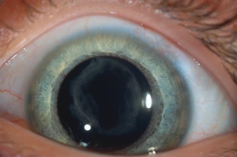
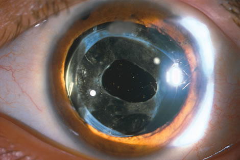
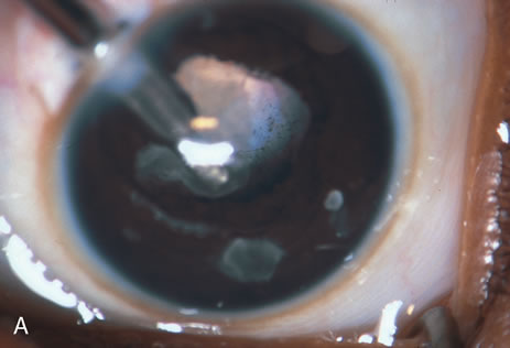
When phacoemulsification became available, it was quickly applied to the removal of children's cataracts because it provided the surgeon with better control of the flow of irrigating solutions and provided improved control of the aspiration flow and pressure.4 The instrument also added a new capability, that of being able to mechanically disrupt the lens nucleus and cortex to facilitate aspiration of the lens. Although the phacoemulsification instrument was helpful for removing lens cortex, it was ineffective in cutting or removing the posterior lens capsule. At the conclusion of the phacoemulsification procedure, the posterior capsule was left intact. When capsular opacification occurred, it was treated with a discission procedure, an operation that consisted of making a cut in the posterior capsule with a bent needle, a Ziegler knife, or a modification of the latter (Fig. 1). If the membrane was thick and resisted opening with a knife, an intraocular scissors was necessary to open the lens capsule.

Fig. 1. A blade of a discission knife is introduced into the anterior chamber through a clear cornea incision. The tip of the knife engages the secondary membrane (posterior lens capsule), and the membrane is incised. Inset shows this procedure with an IOL in place.

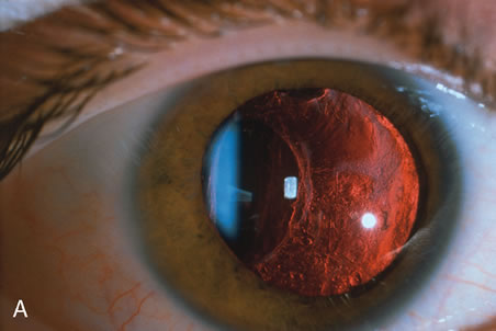
In children, opacification of the posterior capsule of the lens and a subsequent decrease in vision will always occur if the posterior lens capsule is left intact. The terms posterior capsule fibrosis, secondary membrane, and Elschnig's pearls are used interchangeably to describe these changes. Opacification of the posterior capsule results from proliferation and migration of residual lens epithelial cells.5 In adults, the extent and rapidity of capsular opacification can be minimized by reducing surgical trauma, by removing as many of the posterior lens capsule epithelial cells as possible by polishing the posterior capsule, and by selecting implants that reduce posterior capsule opacification.6–9 In children, even when these measures are taken, the lens epithelial cells rapidly proliferate. If a posterior capsulotomy is not performed, or if only a small opening in the capsule is made, the posterior capsule will opacify within weeks to months. The vitreous face is considered to serve as a scaffold for migration of the lens epithelial cells. However, even when this is disrupted with an anterior vitrectomy, the lens material can opacify the visual axis.10–11 Opacification of the posterior lens capsule causes difficulty in the visual habilitative process because it reduces the accuracy of retinoscopy and will cause light entering the eye to be diffracted. These factors will impede visual development and will make management of amblyopia difficult.

The advantage of leaving the posterior capsule of the lens intact after cataract surgery is that it retains a barrier between the anterior chamber and vitreous. This prevents the vitreous from entering the anterior chamber, and it theoretically preserves the ocular anatomic relationships after cataract surgery. The disadvantage of leaving the posterior lens capsule intact is that when the capsule opacifies, a second procedure is needed to re-establish a clear visual axis. To achieve this, a second anesthetic is administered and an incision is made into the clear cornea. The chamber is deepened with a viscoelastic material and a knife or other instruments are introduced into the anterior chamber to cut or tear the posterior capsule so that the visual axis can be cleared (Fig. 1).

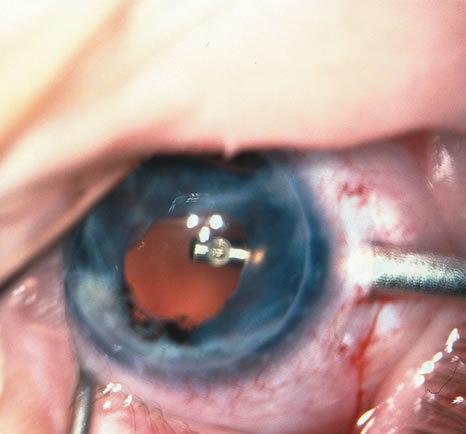
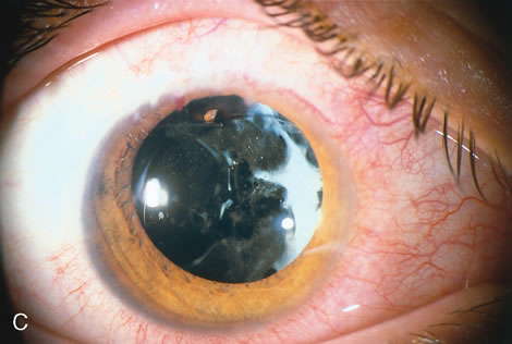
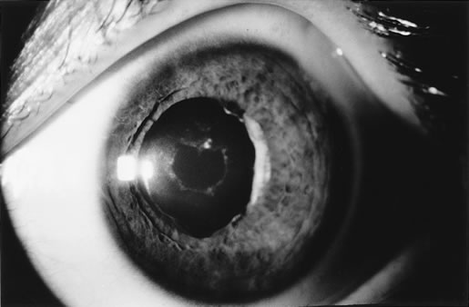
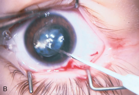
When the vitreous suction-cutting devices became available, they were quickly employed to remove cataracts in children. In addition to providing control of aspiration pressure and control of the flow of irrigation solutions, they also added the ability to remove some or all of the posterior lens capsule, even when the capsule had a thick fibrovascular stalk associated with persistent hyperplastic primary vitreous (PHPV) or a thick capsular plaque (Fig. 2). These fine-tip suction-cutting instruments provided sufficient control of the anterior chamber depth, thus permitting the surgeon to precisely open the posterior capsule and, if necessary, safely remove vitreous from the anterior chamber. Keech and co-workers,12 in a contemporaneous surgical series, showed that having the ability to remove the posterior lens capsule and perform an anterior vitrectomy reduced the need for secondary procedures from 75%, if the capsule was left intact, to 11% after capsulectomy and anterior vitrectomy. They found that when a large section of the posterior lens capsule was removed, it provided a lasting optical opening and reduced the requirement for additional surgery.

Fig. 2. A. A vitreous suction cutting instrument is used to cut and remove a thickened posterior lens capsule. Thickened posterior lens capsules are difficult to treat with the Nd:YAG laser. B. This diagram demonstrates the extent of tissue removal when a vitreous suction cutting device is used to clear the visual axis.

As techniques evolved, concerns were raised about the wisdom of removing a portion of the anterior vitreous with the capsulectomy. Occasional reports postulated that cystoid macular edema may occur with these techniques, but the actual risk of developing cystoid macular edema and the long-term results of an anterior vitrectomy on the eye of a child (and in particular on the retina) remain unknown.13 This relatively new capability of safely removing a portion of the lens capsule, or even the entire posterior lens capsule, gave rise to controversy. If only a small portion of the posterior capsule was removed, opacification could recur. Experience showed that the lens would develop pearls at the edge of the posterior capsulotomy, and these lens epithelial cells could migrate across a scaffold created by the anterior vitreous face. If the entire posterior lens capsule was removed, recurrence of posterior capsule opacification would usually not occur, but the barrier effect of the posterior lens capsule between the anterior chamber and vitreous was lost.

In 1981, Taylor, and later, Parks14–15 reviewed these issues and concluded that it was best to remove the posterior lens capsule and combine this with a small anterior vitrectomy. It was considered that the risk of developing cystoid macular edema was relatively remote and unproved. There was a greater risk of decreased vision if the capsule became opaque and amblyopia became refractory to treatment. Following this, some surgeons began to remove as much of the lens capsule as possible to ensure that regrowth of the lens capsule and subsequent occlusion of the visual axis did not occur. When this technique is taken to its extreme, the entire posterior lens capsule is removed. This can also create a problem. If a child with a monocular cataract becomes intolerant to wearing a contact lens, and a complete lensectomy has been performed, there is insufficient support from the remaining lens remnants for placement of a secondary sulcus-fixated posterior chamber intraocular lens. These eyes pose difficult situations for the surgeon and the child. Optical rehabilitation can be achieved only by placing an anterior chamber IOL, or by suturing the IOL to the sclera in the posterior chamber.16,17

With the incremental acceptance of use of the IOL as the principal method of rehabilitating children's eyes, the issue of the formation of secondary membranes has become more complicated. Authors have advocated opening the posterior lens capsule at the time of the cataract surgery either by using vitrectors, or by performing a posterior lens capsule capsulorhexis, with or without prolapsing the lens optic through the posterior capsular opening.18–24 These measures however, do not ensure that the visual axis will not become opacified.25–27.

The pioneering work of Krasnov, Aron-Rosa, and Fankhauser in developing the neodymium (Nd>YAG laser and the refinement of its technology have resulted in an additional method for management of the posterior capsule.28

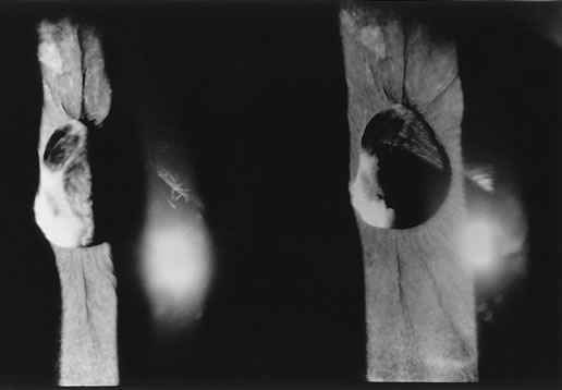
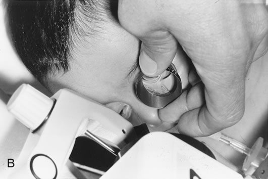
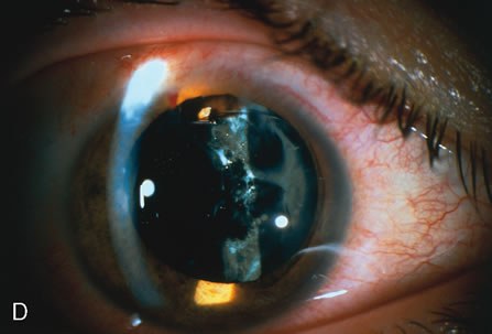
The Nd:YAG laser provides the ophthalmologist with the ability to open the posterior lens capsule without making a surgical incision. Because of this, the risk of infection and of complications related to making the incision and introducing instruments into the vitreous are reduced. In older cooperative children, the procedure can be performed in an office setting (Fig. 3).

Fig. 3. This cooperative child is being treated in a laser center, without sedation or anesthesia.

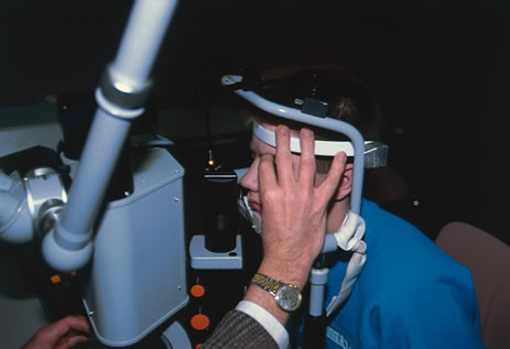
Until recently, application of laser technology for young or uncooperative children has been difficult because the instrumentation is bulky and difficult to transport. To some extent, these problems have been reduced with a reduction in the size of the laser. Use of the Nd:YAG laser to open the posterior lens capsule in young children has required that the patient be treated in a seated position. If the child is young or uncooperative, some surgeons have resorted to anesthetizing the child near the location of the Nd:YAG laser and have performed the laser treatment on the child while he or she is held in position for use of the laser delivery device. With this technique, there can be problems with positioning the eye, and if the child is treated at a location that is remote from the operating room, there may be difficulty in monitoring the child's anesthetic and providing emergency treatment if this would be required.

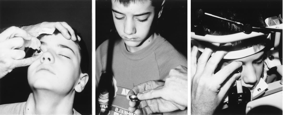
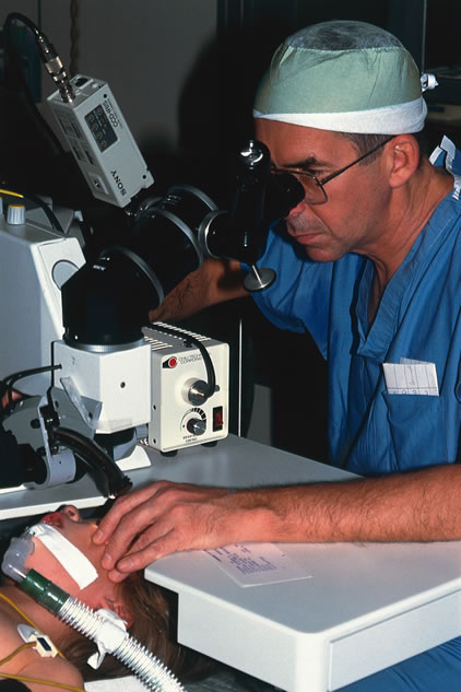
To overcome these difficulties, the Nd:YAG laser has been modified so that a posterior capsulotomy can be performed on a child in the supine position. The laser delivery systems now provide the surgeon with the ability to safely perform an Nd:YAG laser capsulotomy in children in the operating room with the assistance of sedation or, if necessary, general anesthesia (Fig. 4). In 1994, Atkinson and Hiles demonstrated the effectiveness of the Nd:YAG laser in children 3 months of age and older.29 The surgeon may elect to perform the Nd:YAG capsulotomy immediately after cataract surgery or at a later date, when the capsule has become tense or has begun to opacify.

Fig. 4. A secondary membrane is opened with an Nd:YAG laser. The procedure is performed in the operating room. The child is placed in a supine position after anesthesia has been administered.

The term laser is an acronym that stands for “light amplification by a stimulated emission of radiation.” In 1917, Einstein postulated that a photon released from an excited atom could, upon interacting with a second similarly excited atom, trigger a second atom to be excited and release a photon. The photon released by the second atom would be identical in frequency, energy, direction, and phase to the triggering photon. These two photons could then proceed to trigger more atoms, causing an excited state or stimulated emission. Stimulated emission is achieved by “pumping” energy into a lasing medium. This is accomplished in the Nd:YAG by use of a gas discharge lamp. When an appropriate medium contains a great many excited atoms, the emission of light output will be randomized and equal in all directions. Once stimulated radiation is achieved, the energy can be given a preferential direction by placing a fully mirrored surface at one end of the optics cavity and a partial mirror at the opposite end (Fig. 5). The light energy reflected between these two mirrors collides with other excited atoms, causing more photons to be released and emit more light energy. The energy that escapes through the partial mirror is said to be coherent, which means that it has only one orientation or polarization. Other random light produced is trapped within the laser medium. The stimulated emission can be collimated and focused by a laser delivery system.

Fig. 5. Schematic representation of a laser. The gas discharge lamp is used to “pump” the neodymium, which is embedded in a yttrium-aluminum-garnet medium.

Light energy emitted by a laser differs from ordinary light sources. What makes lasers different is their ability to emit light-specific wavelengths. This light energy, the frequency of which is dependent on the laser medium, can be focused on a very small area, and the light can be discharged over very short periods of time. This makes it possible to concentrate light energy in an extremely small space without causing damage to surrounding tissues.

The energy produced is measured as the capacity to do work and is classified as a form of potential or kinetic energy. In the metric system, this is measured in joules (J). Power is a measure of the rate at which work is being done and is measured in watts (W). One watt equals one joule per second. The laser effect on tissue is dependent on the amount of energy, the number of pulses, and the time it takes to deliver the pulse.

The active medium of a Nd:YAG laser consists of neodymium embedded in a yttrium-aluminum garnet (YAG) medium. The advantage of the Nd:YAG laser is that the optical energy can be delivered in the Q-switched mode in a very brief period of time (nanoseconds), and it is readily absorbed by tissue. The Q-switched mode can be thought of as a very fast shutter that lets a pulse of coherent light exit from the laser into the delivery tube. The beam creates a very high temperature when it strikes tissue, and it will cause an acoustic pressure wave as well as thermal and optical breakdown of the tissue (photodisruption). If light energy is focused or concentrated on the posterior lens capsule, there will be destruction of the membrane. Secondarily, but related to these effects, energy is also released in the form of a pressure wave. This causes disruption of the secondary membrane. In addition to its use in disrupting the posterior lens capsule, the Nd:YAG laser may be used to perform peripheral iridotomy and cut fibrous adhesions, iris synechiae, and vitreous bands. In its thermal, continuous wave, or running mode, it may also be used to destroy the ciliary processes and lower intraocular pressure.

In this chapter we describe the use of the Nd:YAG laser to perform capsulotomy of the posterior lens capsule in children.

Back to Top
INDICATIONS
The most common application of the Nd:YAG laser in ophthalmology has been to open an opacified posterior lens capsule. The opacified capsule is commonly referred to as a secondary membrane (Fig. 6). A capsulotomy is recommended when there is a decrease in visual acuity that is due to opacification of the posterior lens capsule. This can cause either partial or complete obstruction of the visual axis. Judging the extent of a capsule's opacity and its effect on the patient's visual development may be difficult, especially in young or nonverbal children or in the presence of amblyopia. Signs that vision is decreased in a child are decreased light reflex on retinoscopy, presence of an ocular deviation, usually esotropia; decreased visual function; failure of the visual acuity to improve with occlusion; and a reduction in vision in spite of occlusion and accurate correction of the refractive error. During the evaluation, it is important to exclude other causes of these signs. Once it has been determined that the posterior capsule of the lens is causing the vision to be decreased (even slight opacity that decreases acuity to 20/30 or 20/40), photodisruption or opening of the posterior capsule is indicated.

Fig. 6. This child developed a secondary membrane after cataract surgery. The posterior capsule was not opened at the time of surgery.

Streak retinoscopy, performed with the pupil well dilated, is helpful in determining the degree of opacification. In severe degrees of opacification, the red reflex may be diminished or absent. The posterior capsule can also be examined by using a slit lamp or by using the indirect ophthalmoscope as a light source and holding a 20D indirect lens close to the eye so that it serves as a magnifying lens. The hand-held portable slit lamp and the direct ophthalmoscope are other methods available to confirm the location and extent of opacification. These techniques are also used to judge the thickness of the secondary membrane. In children, the secondary membranes tend to become thicker with time. Estimation of the degree of thickness is important, because the thicker the membrane, the more energy it will take to photodisrupt it. Also, thick membranes will not retract as much as thin membranes. If the posterior capsule is thickened or the opacity is very dense, capsulotomy will need to be performed with either a discission knife or a vitreous suction-cutting device. Atkinson and Hiles suggested that the procedure should be performed within 3 weeks of the cataract surgery. Longer delays result in thicker membranes that require higher energy applications to perform the treatment. Recurrent membranes must be anticipated. In Atkinson's series, eight of 28 membranes required retreatment.29 When membranes recur, they are treated by repeating the procedure.

Back to Top
CONTRAINDICATIONS
Before recommending a posterior capsulotomy with an Nd:YAG laser, the cooperation of the child must be carefully evaluated. In children who are uncooperative and in children with large-amplitude nystagmus, the procedure can be performed, but it requires the assistance of sedation or general anesthesia. Consideration must be given to the availability of suitable laser delivery sources and modifications in technique that enable satisfactory positioning and stabilization of a young or uncooperative patient.

Other relative contraindications include eyes with corneal opacities or other defects that preclude clear visualization of the posterior capsule and surrounding area. Dense opacities can limit the surgeon's ability to adequately aim and focus the laser, and they can also shadow the cone-shaped beam of the Nd:YAG laser, which will reduce its power. Patients with eyes that have very small pupils or pupils that do not dilate well because of dense synechiae of the iris to the lens capsule are also poor candidates for this procedure. Eyes with opacification of the vitreous due to hemorrhage, those with a thick posterior PHPV stalk, and eyes that have very thick membranes are more suited to removal of the secondary membrane and other defects with a vitreous suction-cutting instrument (Fig. 7). When a secondary membrane is thick, it can be opened with an Nd:YAG laser, but it requires excessive levels of power, and this will produce thermal, light, and acoustic trauma to the eye. If an IOL is in place, pitting of the optic can occur. Use of high energy can precipitate an elevation in intraocular pressure, and it may also cause an increased risk for retinal detachment.

Fig. 7. A very thick membrane formed behind an IOL. This was best removed through a pars plana approach using a suction cutting instrument.

If an eye has persistent inflammation, Nd:YAG laser capsulotomy should be deferred until the inflammation has subsided. Other contraindications include the presence of a significant defect such as a large retinal or optic nerve coloboma, and the presence of other profound sight-limiting defects. The presence of an unrepairable retinal detachment or any condition that would preclude improvement of the visual acuity is also a contraindication to the procedure. Eyes that have an elevated intraocular pressure and those with peripheral retinal defects that predispose to retinal detachment can be treated, but only after careful explanation of the risks related to the procedure. In patients with elongated eyes (low hypermetropic aphakic refractive errors), it may be safer to open the secondary membrane with an Nd:YAG laser than with a knife or suction-cutting instrument.

Back to Top
PROCEDURE
A careful assessment of the composure of the child is made in the office. At this time, a relationship of trust should be nurtured between the ophthalmologist and the child. Applanation tonometry gives valuable information about the child's response to placing instruments near the eye and also establishes a pretreatment level of intraocular pressure. Children who are cooperative enough to sit in front of a slit lamp and hold their eye still (steady fixation) may be treated with one of several Nd:YAG lasers that provide delivery for a seated patient. If the child is old enough, a retrobulbar block may be considered to stabilize the eye of a patient with nystagmus. In children who are anxious, uncooperative, or restless, the use of sedation or general anesthesia is recommended.

The pupil can be dilated according to the surgeon's preference. Some surgeons prefer not to dilate the pupil to ensure that the capsulotomy is placed in the aperture of the pupil (Fig. 8). Dilation, however, provides a margin of safety in a young child who may move unexpectedly. The dilated pupil will permit the surgeon to create a large capsulotomy without injury to the iris. To dilate the pupil, two applications of cyclopentolate HCL 1% or 2% combined with phenylephrine 2.5% are instilled (5 minutes apart) 20 to 30 minutes before the procedure.

Fig. 8. This patient was treated without dilating the pupil to ensure placement of the opening within the aperture of the pupil. (Left) Before treatment. (Right) Two months after treatment.

The child should be introduced to the equipment in a reassuring way (Fig. 9). The function of the safety switch on the head bar is explained, and the head is gently placed in front of the laser. The HeNe aiming beam is demonstrated, and steady fixation is practiced. Topical anesthetic is applied to both eyes. This anesthetic decreases the urge to blink. Some children may permit the application of the Peyman (Fig. 10) or Fankhauser lens on the cornea. The Lasag CGP lens is another lens that may be helpful for performing a capsulotomy. The advantage of using a contact lens is that the opacified capsule is easier to visualize and the contact lens will also help stabilize the eye. The optics of the lens permit capsulotomy to be performed at reduced energy levels, compared with performing the capsulotomy without a lens.30 Other children will not permit the application of a contact lens. In these situations, the Nd:YAG laser capsulotomy can be performed without the use of a contact lens, but applications that require higher laser power may be needed.

Fig. 9. A. Topical anesthetic drops are applied to both eyes. B. The child is introduced to the equipment, in this case the CGP lens. C. Treatment is started after reassurance and trust have been established.

Fig. 10. A Peyman lens (inset) or a CGP lens is applied to the eye with a viscous contact solution. Use of either of these lenses stabilizes the eye and permits photodistribution at lower energy settings.

Before treatment, the lens capsule should be closely inspected, and a strategy for where to apply the laser to perform capsulotomy should be planned (Fig. 11). Areas are noted where the lens capsule is thinnest and where the capsule appears to be under tension. A thin tense lens capsule is desirable because flaps of the capsule will separate when the capsule is photodisrupted. The preferred laser is set, and test pulses of I to 2 millijoules are set to be delivered in two- to three-pulse increments (Fig. 12). Alternatively, the laser can be set at I to 3 mj with single-pulse delivery. The Microruptor III (Lasag, Inc) provides the ability to treat patients in a supine position. For this instrument, settings of 0.7 to 0.9 millijoules of power, delivered in bursts of two to three, is a good starting point. The HeNe aiming beam is focused on the posterior capsule, and test pulses are applied to a peripheral part of the capsule until disruption is observed. There, power is increased until this occurs (Fig 13), If the eye does not have an intraocular lens, the beam is not defocused. When treating eyes with an intraocular lens, with the secondary membrane in close proximity to the posterior surface of the optic, the laser can be defocused. By defocusing the beam, the focal point of the laser in air is 0.4 mm posterior to the focus of the HeNe aiming beam. Using the retrofocusing mode provides an extra margin of safety and helps prevent pitting of the lens.

Fig. 11. A. The secondary membrane shows a vertical strand surrounded by areas of thin capsule. Treatment on either side of the strand (1, 2), followed by treatment of the strand (3) will cause the capsule to open. B. Additional applications (4, 5) will increase the aperture. C. Further opening can be achieved with treatment of the edges of the opening (6, 7). (Drawings courtesy of Kenneth P. Cheng, MD.)

Fig. 12. A. An 11-year-old boy developed a secondary membrane after removal of a traumatic cataract and placement of a posterior chamber 10L. A Retro illumination of membrane. B. The appearance of the membrane just prior to treatment. C. The posterior capsule immediately after disruption with an Nd:YAG laser. D. The vision improved from 20/50 to 20/25 1 week after the Nd:YAG capsulotomy.

Fig. 13. Example of a secondary membrane in a 7 year old child, 1 year after NdYAG laser capsulotomy.

If the capsule is thin, the laser is used to create a vertical opening using 10 to 25 applications (Fig. 14). This is usually enough to create a sufficient opening. As shown by Capone and co-workers,31 it is not necessary to create a round capsulotomy. The posterior capsule will retract and increase the area of the opening, with a tendency toward sphericity. Attempts should be made to create an opening at least 4.5 to 5 mm in size.

Fig. 14. A thin posterior lens capsule can be opened with vertical applications of the Nd:YAG laser. A vertical opening 5 mm in length will usually provide a sufficient aperture. With time, the opening will become round.

When the capsular membrane is thick, or if it does not look as though it will retract, the laser should be used to create either a triangular or a cruciate opening beginning at the 12 o'clock position and progressing down to the 5 o'clock and 7 o'clock positions (Fig. 15). This technique creates a flap that will usually roll posterior and provide a sufficient opening. Attempts should be made to avoid creating a free-floating fragment of lens capsule. Children who have undergone a capsulotomy and anterior vitrectomy may be left with an irregular pupil or one that encroaches upon the visual axis in one meridian. The Nd:YAG laser can be useful in cutting discrete adhesions between the iris and the posterior capsule. Care should be taken to identify any blood vessels when cutting membranes. Cutting a blood vessel with the Nd:YAG laser can cause bleeding in the anterior chamber. When this occurs, the bleeding usually stops spontaneously. If it does not, application of gentle pressure on the contact lens will raise the intraocular pressure and decrease the rate of hemorrhage. Bleeding may occur to such an extent that the anterior chamber becomes turbid and visualization becomes difficult.

Fig. 15. Thick membranes will retract poorly. To open a thick membrane, a triangular opening is created. This will permit retraction of the flap and will provide adequate opening in the capsule.

Turbidity can also occur when pockets of retained cortex are opened with the laser. At higher power settings, bubbles can form in the eye, and in the supine child, they can float up to the posterior surface of the cornea. This process obscures the view and reduces the effective power delivered by the laser. When this occurs, the procedure should be suspended, and often small bubbles are absorbed. Another alternate would be to stop the procedure and complete it at another date.

Postoperative care includes measurement of the intraocular pressure 1 hour after the procedure. If the intraocular pressure is elevated, apraclonidine or a topical beta-blocking drop can be applied twice a day for 2 days, and the pressure should be remeasured.32 Some physicians use one or two applications of apraclonidine prophylactically after the procedure to prevent elevation of intraocular pressure.

After the procedure, the surgeon may elect to dilate the pupil with a short-acting cycloplegic agent such as 1% or 2% cyclopentolate. If the treatment is lengthy or there is liberation of residual lens cortex, adhesions to the cortex and capsule can be prevented by using a short-acting mydriatic that will produce movement of the pupil. An antibiotic-steroid eye drop is applied to the eye four times a day for 2 days to prevent infection. Endophthalmitis has been reported after an Nd:YAG capsulotomy. This is believed to be caused by liberation of bacteria that have been encapsulated within the lens material.33

The patient is re-examined 2 to 10 days after the procedure to assess the effectiveness of the capsulotomy and to update or change the optical correction if needed (Fig. 16).

Fig. 16. This is the appearance of a posterior lens capsule 2 weeks after treatment with an Nd:YAG laser.

Back to Top
COMPLICATIONS
Steinert and associates34 have shown that opening the posterior capsule with an Nd:YAG laser does carry a low but definite risk of complications. An Nd:YAG capsulotomy should be considered intraocular surgery, and the ophthalmologist and the parent or parents should be aware of possible complications, some of which may cause a decrease in vision. Most complications that have been observed have occurred in adults. These include retinal detachment, hemorrhage, and elevated intraocular pressure.35–37 Experience with complications in using the Nd:YAG laser to perform capsulotomy in children is limited. This is in part due to the relatively infrequent occurrence of cataracts in this age group, but it is also due to the previously mentioned difficulties in delivery of this form of treatment. In children, there can be a potential risk of complications related to focusing and directing the laser beam. Eye movement and movement of the patient may cause the procedure to be terminated before a sufficient opening of the capsule has been achieved. In a restless child, confusion can be caused by reflections of the HeNe aiming beam off of structures in the anterior and posterior chambers. If this occurs and the laser is discharged, pitting of an intraocular lens or inadvertent application of the laser to the cornea or iris may occur.

Leff and colleagues35 investigated risk factors for developing a retinal detachment after an Nd:YAG capsulotomy in adults. They studied 24 adults (25 eyes) who developed a rhegmatogenous retinal detachment after their Nd:YAG capsulotomy. The average time from cataract surgery to capsulotomy was 15 months. The mean interval from capsulotomy to retinal detachments was 6 months. In eight of 25 eyes, a risk factor for retinal detachment was found (high myopia, lattice degeneration, or a retinal development in the fellow eye).

If the energy level is too high, excessive heat will be generated and a gas bubble will form. Using the lowest amount of energy to accomplish the capsulotomy helps to avoid this complication.

In adults, an Nd:YAG laser posterior capsulotomy can cause acute elevation in intraocular pressure. This is usually transient and will normally last for less than 24 hours.36,37 Experience with adults has shown that there will be a persistent increase in intraocular pressure in about 1% of patients treated. This may be energy dependent.36 Silverstone observed higher pressures associated with performing large capsulotomies that required high energy levels.32 The mechanism for the increased intraocular pressure is believed to be a decrease in outflow secondary to the shock wave or entrapment of capsular fragments and debris in the filtration angle. In our experience, elevations of intraocular pressure are not common after the procedure. Some pressure elevations may go undetected in very young children because of the difficulties in accurately measuring the intraocular pressure.

If an intraocular lens is in the posterior chamber and close to the membrane, the heat and energy produced by the laser may cause pitting of the lens. When this occurs, it is believed not to interfere with the optical properties of the lens, but it may induce glare. Lenses with glass optics are at risk for cracking; however, these lenses have not been used in children. If one or both haptics of the lens are in the capsular bag, and the bag is under tension, posterior capsulotomy may cause disruption of the posterior capsule and cause the lens to dislocate.

The capsulotomy created by the Nd:YAG laser may be insufficient in size. Attempts should be made to provide a capsulotomy larger in size than the final desired aperture. The edge of the capsulotomy frequently thickens after treatment with a YAG laser, and this may encroach upon the visual axis, necessitating repeat capsulotomy

Recurrence of the secondary membrane is probably the most disappointing complication for the patient's family and the surgeon. In the study by Atkinson and Hiles, regrowth of the membrane occurred in 28% of the children, with a follow up of 9 to 24 months. The Nd:YAG frequently is unable to create a sufficiently large opening so that the membrane reformation is common. This has also been the experience of one of the authors (AWB). We examined the formation of secondary membranes following cataract removal in children. Twenty-six eyes that had the posterior lens capsule left intact and treated with a single Nd:YAG capsulotomy had resolution of the membrane in 65% of eyes, however, 34% required more than one treatment.27 Hutcheson and co-authors observed a 57% recurrence rate after Nd:YAG capsulotomy, and 17% of eyes required more than one treatment.38

Other potential complications include loss of corneal endothelium. This has not been documented in children.

Back to Top
SUMMARY
Careful patient selection and availability of an Nd:YAG laser provide an additional modality for safe management of opacification of the posterior lens capsule after cataract surgery in children.
Back to Top
REFERENCES

1. Scheie HG: Aspiration of congenital soft cataracts: A new technique. Am J Ophthalmol 50:1048, 1960

2. Letocha CE: The knife needle operation for secondary capsular cataract. Arch Ophthalmol 114:1414, 1996

3. Parks MM, Hiles DA: Management of infantile cataracts. Am J Ophthalmol 63:10, 1967

4. Hiles DA, Wallar PH: Phacoemulsification versus aspiration in infantile cataract surgery. Ophthalmic Surg 5:13, 1974

5. Liu CS, Wormstone IM, Duncan G et al: A study of human lens cell growth in vitro, a model for posterior capsule opacification. Invest Ophthalmol 37: 906, 1996

6. Apple DJ, Solomon KD, Tetz MR et al: Posterior capsule opacification. Surv Ophthalmol 37:73, 1992

7. Apple DJ, Peng Q, Visessook N et al: Surgical prevention of posterior capsule opacification . Part 1 : J Cataract Refract Surgery 26:180, 2000

8. Peng Q, Apple DJ, Visessook N et al: Surgical prevention of posterior capsule opacification. Part 2 : Enhancement of cortical cleanup by focusing on hydrodissection. J Cataract Refract Surg 26:188, 2000

9. Peng QM Visessook N, Apple DJ et al.: Surgical prevention of posterior capsule opacification. Part 3: intraocular lens optic barrier effect as a second line of defense. J Cataract Refract Surg 26:98, 2000

10. Spierer A, Desatnik H, Blumenthal M.: Secondary cataract in infants after extracapsular cataract extraction and anterior vitrectomy. Ophthalmic Surg 23:625, 1992

11. Morgan KS, Karcioglu ZA: Secondary cataracts in infants after lensectomies. J Pediatr Ophthalmol Strabismus 24: 45, 1987

12. Keech RV, Tongue AC, Scott WE: Complications after surgery for congenital and infantile cataracts. Am J Ophthalmol 108:136, 1989

13. Poer DV, Helveston EM, Ellis FD: Aphakic cystoid macular edema in children. Arch Ophthalmol 99:245, 1981

14. Taylor D: Choice of surgical technique in management of congenital cataract. Trans Ophthal Soc U K 101:114, 1981

15. Parks MM: Posterior lens capsulectomy during primary cataract surgery in children. Ophthalmology 90:344, 1983

16. Sharpe MR, Biglan AW, Gerontis CC: Scleral fixation of posterior chamber intraocular lenses in children. Ophthalmic Surg Lasers 27:337, 1996

17. Buckley EG: Scleral fixated (sutured) posterior chamber intraocular lens implantation in children. J Pediatr Ophthalmol Strabismus : 3:289, 1999

18. Mackool RJ, Chhatiawala H.: Prediatric cataract surgery and intraocular lens implantation: A new technique for preventing or excising postoperative secondary membranes. J Cataract Refract Surg. 17:62, 1991

19. Buckley EG, Klombers LA, Seaber JH et al: Management of the posterior capsule during pediatric intraocular lens implantation. Am J Ophthalmol 115:722, 1993

20. Gimbel HV. Posterior capsulorhexis with optic capture in pediatric cataract and intraocular lens surgery. Ophthalmology 103:1871, 1996

21. Vasavada A, Desai J: Primary posterior capsulorhexis with and without anterior vitrectomy in congenital cataracts. J Cataract Refract Surg 23:645, 1997

22. Plager DA, Lipsky SN, Snyder SK et al: Capsular management and refractive error in pediatric intraocular lenses. Ophthalmology 194:600, 1997

23. Fenton S, O'Keefe M: Primary poster capsulorhexis without anterior vitrectomy in pediatric cataract surgery: longer term outcome. J Cataract Refrect Surg 25:793, 1999

24. Kugelberg M, Zetterstrom C: Pediatric cataract surgery with or without anterior vitrectomy. J Cataract Refract Surg 28 1776, 2002

25. Tassignou MJ, De Groot V, Smets RM et al: Secondary closure of posterior continuous curvilinear capsulorhexis J Cataract Refract Surg 22:200, 1996

26. Jensen AA, Basti S, Greenwald MJ, Mets MB: When may the posterior capsule be preserved in pediatric intraocular surgery? Ophthalmology 109:324, 2002

27. Hosal BM, Biglan AW: Risk factors for secondary membrane formation after pediatric cataract removal. J Cataract Refract Surg 28:302, 2002

28. American Academy of Ophthalmology: Nd:YAG photodisruptors. Ophthalmology 100:1763, 1993

29. Atkinson SA, Hiles DA: Treatment of secondary posterior capsular membranes with the Nd:YAG laser in a pediatric population. Am J Ophthalmol 118:496, 1994

30. Brancato R, Menchini U: Microchirugia Lasers in Ophthalmologia, p 102. Milano, Ed Ghedini, 1989

31. Capone A, Rehkopf PG, Warnicki JW et al: Temporal changes in posterior capsulotomy dimensions following Neodymium YAG Laser discission. J Cataract Refract Surg 16:451, 1990

32. Silverstone DE, Brint SF, Olander KW et al: Prophylactic use of apraclonidine for intraocular pressure increase after neodymium:YAG capsulotomies. Am J Ophthalmol 113: 401, 1992

33. Carlson A, Koch D: Endophthalmitis following Nd:YAG posterior capsulotomy. Ophthalmic Surg19:168, 1988

34. Steinert RF, Puliafito CA, Kumar SR et al: Cystoid macular edema, retinal detachment, and glaucoma after neodymium:YAG laser posterior capsulotomy. Am J Ophthalmol 112:373, 1991

35. Leff SR, Welch JC, Tasman W: Rhegmatogenous retinal detachment after YAG laser posterior capsulotomy. Ophthalmology 94:1222, 1987

36. Fourman S, Apisson J: Late-onset elevation in intraocular pressure after Neodymium:YAG Laser posterior capsulotomy. Arch Ophthalmol 109:511, 1991.

37. Migliori ME, Beckman H, Channel MM: Intraocular pressure changes after neodymium:YAG laser capsulotomy in eyes pretreated with timolol. Arch Ophthalmol 105:473, 1987.

38. Hutcheson KA, Drack AV, Ellish NJ et al:Anterior hyaloid face opacification after pediatric Nd:YAG laser capsulotomy. J Pediatr Ophthalmol Strabismus 3: 303, 1999.

Back to Top