Chapter 91
The Fadenoperation
JAMES B. SPRAGUE
Main Menu   Table Of Contents

Search

THEORY
TECHNIQUE
INDICATIONS AND RESULTS
INCOMITANT STRABISMUS
DISSOCIATED VERTICAL DEVIATION
ESOTROPIA WITH HIGH ACCOMODATIVE CONVERGENCE:ACCOMODATION RATIO
EXOTROPIA WITH HIGH AC:A RATIO
NYSTAGMUS BLOCKAGE SYNDROME
CONGENITAL ESOTROPIA
HIGH MYOPIA
COMPLICATIONS
ACKNOWLEDGMENT
REFERENCES

In 1974, Cüppers described the “Fadenoperation” at the second meeting of the International Strabismological Association.1 Two years later, Mühlendyck presented this new procedure to a US audience.2 Numerous reports subsequently appeared in the US and European literature on appropriate applications for this new technique, and on their success.3–9

Cüppers called his technique the “Fadenoperation,” or “string operation” because “faden” means suture or string in German. The common English usage, “faden suture,” is thus redundant. The procedure has also been called “arc of contact surgery,” “the posterior fixation procedure,” and “retro-equatorial myopexy.” In this chapter, I have used de Decker's abbreviation “Fd” for the procedure as both noun and adjective.

This chapter describes the theory of Fd, its indications, surgical techniques and complications. Although I have relied primarily on material readily available in English for the US reader, I have expanded the European references in this revision because Fd remains more popular in Europe than in the United States.

Back to Top
THEORY
In 1941, Peter suggested weakening a rectus muscle by suturing it posteriorly to the sclera.10 In 1958, Tour and Asbury11 noted that the rectus muscle has rotational effect that extends as long as its direction of tangency, at which point the muscle becomes progressively more of a retractor and less of a rotator. Cüppers1 reasoned that suturing the muscle to the posterior sclera would create artificial paralysis, or a controlled incomitance, by weakening the rotational ability of a muscle only in its field of action. This was his so-called dynamic angle. He felt that the balance of forces between agonist and antagonist in primary position would not change. If there was strabismus in primary gaze, the so-called static angle, he added a standard recession to the muscle treated with posterior fixation.

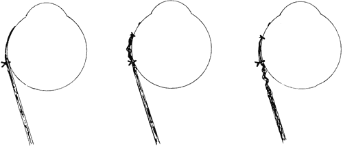
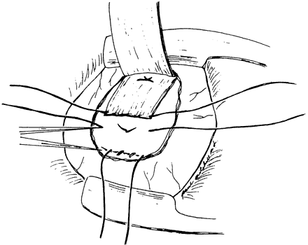
Alan Scott diagrammed the mechanics of Fd.12 He suggested that changing the arc of contact of the muscle with the globe reduces the lever arm formed by the muscle insertion, the center of rotation of the globe, and the origin of the muscle (Fig. 1). Scott's mechanical model suggested that the posterior suture needed to be 10 to 12 mm from the insertion for the medial rectus, 12 to 14 for the superior and lateral recti, and 14 to 16 for the lateral rectus.

Fig. 1. The lever arm between the center of rotation and the muscle's insertion and origin is shortened as the effective insertion of the muscle is moved posteriorly.

Kushner measured saccadic velocity after the Fd.13 He expected to find decreased velocity in the field of the operated muscle but did not. He questioned whether the Fd changed the torque active on the muscle as would be suggested by the lever arm theory and also noted that the lever arm theory would not explain the effect of Fd without a recession. He stressed the effect of the amount of muscle contained in the suture and how effectively it was immobilized to explain the clinical effect of the operation.

Clark and colleagues14 examined the theory of the Fd in the context of the extraocular muscle pulleys previously described by Demer and coworkers,15 who described connective tissue sleeves that envelop and position each rectus muscle. Given that the anterior portion of the sleeve overlies the area at the equator where the posterior fixation suture is placed, they suggested that displacement of the pulley sleeve by the posterior suture itself or by extensive sharp dissection posteriorly results in mechanical restriction rather than a reduction in torque during muscle contraction. Their studies that used magnetic resonance imaging (MRI) did not show a change in tangency and thus did not predict a reduction in torque to explain the effect of the Fd. Mechanical restriction created by the Fd would explain the effect of the procedure without simultaneous recession of the muscle. It could also explain the variable results that many researchers have noted.

Regardless of the theory of how it works, there are five points to consider in applying the theory of the Fd in clinical practice and also in evaluating the literature:

  • First, it is important to determine how far posteriorly the fixation suture ought to be placed from the limbus. From the point of view of the mechanical model, this determines how much the lever arm is shortened, and thus the farther back such placement is made, the better will the result be. With the pulley model, extensive posterior orbital dissection could damage the anterior pulley slings and reduce the desired mechanical restriction to ocular movement. However, many surgeons try to put the suture(s) as far back as possible. For example, Biglan (Biglan AW, personal communication, 2000) has suggested going back 14 mm on the superior rectus for mild dissociated vertical deviation (DVD), 14.5 mm for moderate, and 15 mm for severe deviations. H. Eggers (personal communication, 2000) attempts to go back 15 mm on the medial rectus for high accomodative:convergence accommodation (AC:A) esotropia. Peterseim and Buckley16 placed medial rectus sutures an average of 18.3 mm (range, 15 to 20.5 mm). De Decker4 found that unsuccessful cases on reoperation did not have the suture placed as far posteriorly as the initial surgeon had claimed.
  • Second, the scleral suture (or sutures) needs to be heavy and deep enough to hold firmly4 and the knots into the muscle have to be tight enough to stay posterior.17 The amount of muscle incorporated in the sutures is important and could explain variable results.13
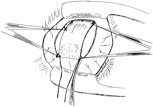
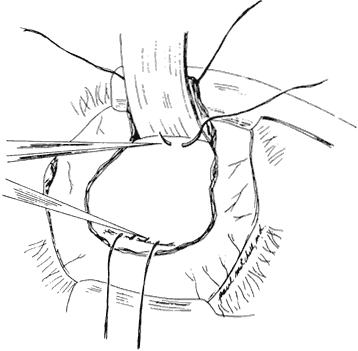
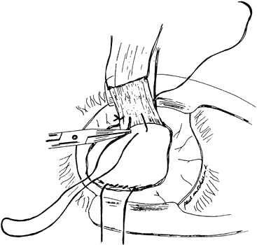
  • Third, simultaneous recession augments the procedure. Sprague and colleagues5 advocatedrecession of the superior rectus for DVD after disappointing results without it. Although undercorrection is a problem with DVD, large simultaneous medial rectus recessions may lead to overcorrections. In 
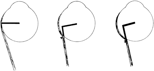
addition, if the posterior suture is placed before the muscle is recessed, the slack in the muscle will be anterior to the posterior attachment. Alan Scott pointed out that it is more effective to allow the slack created by the recession to fall behind the posterior suture, in effect doing the recession first and then placing the posterior suture (Fig. 2).12
  • Fourth, previous recession can be augmented with the Fd alone without the need to recess the muscle further or consider a marginal myotomy.16,18,19
  • Fifth, the Fd may weaken the operated muscle by tissue destruction and scarring.4,7 In rabbit studies, Alio and associates20 found muscle degeneration at the point of posterior fixation as well as anterior to the insertion. The Fd may lead to muscle stiffening, as shown by Castenera.21 De Decker found on reoperation that there was adherence of the muscle to the globe anterior to the posterior suture if a recession had been done.4

Fig. 2. A. Posterior fixation alone. Recession added with the slack created left in front of the suture (B) and with the suture brought forward in the muscle, so that the slack is posterior to the suture (C).

Back to Top
TECHNIQUE
Cüppers1 described disinserting the muscle, placing two sutures though the sclera under the muscle, and then bringing them through the muscle. This is thought to spare the center of the muscle and the circulation of the ciliary vessels. De Decker4 used two sutures at the muscle edge, but preferred to leave the muscle in place if a simultaneous recession was not needed. He also preferred to secure the muscle with a triple loop to prevent it's sliding through the suture. Mühlendyck2,17 used a similar technique including 2 mm of the muscle on each side. He stressed the importance of the knot's being tight and described a patient who had initially done well but whose ocular deviation had returned. The suture had slipped and the knot had migrated forward.17 He also stressed the importance of avoiding the long ciliary arteries and parasympathetic nerves. For the superior rectus, Sprague and colleagues5 preferred a single double armed suture brought up through the middle of the muscle and tied on its orbital surface.8

When simultaneous recession was not indicated, Castilla and colleagues22 passed a suture over the muscle with attachments to the sclera at each edge. Eggers (personal comunication, 2000) has used Supramid sutures for this. Schroeder and Schroeder23 suggested tying a 17-mm section of 2-mm wide silicon band across the muscle. The muscle can then slide under the suture or band, with an effective posterior point of tangency. This technique is claimed to restrict the muscle without increasing muscle stiffness and with minimal scar, thus facilitating reoperation.

Exposure is a major problem in performing the Fd, so as to place the suture posteriorly while at the same time avoiding unnecessary dissection. A fiberoptic headlight and loupe magnification are always used. The technique followed by Sprague and colleagues5 uses a limbal incision. The muscle is isolated on a suture and cleaned of fascial attachments. An extra suture marks the muscle where the posterior fixation suture will engage it (Fig. 3). This indicates the distance back from which that the posterior fixation suture will be placed, less the amount of recession. The slack created by the recession is therefore posterior to the posterior fixation suture.

Fig. 3. The muscle (here the superior rectus) is isolated. The muscle is marked where the suture will come through it (caliper on left), anterior to the point on the sclera where the suture will be placed (caliper on right).

The muscle is released from the globe and the globe is held over with a traction suture woven through the insertion. Small, malleable brain retractors or thin orbital retractors under the muscle hold the muscle and orbital contents back (Fig. 4). The scleral sutures will determine how the muscle is to be displaced: vertically in the case of the horizontal recti, horizontally in the case of the vertical recti. Two sutures are used on the medial rectus to avoid the long ciliary vessels in the sclera that can be cut with a single central suture. On the vertical muscles, a single suture in the middle can be used (Fig. 5). In both the one- and two-suture techniques, the needle pass through the sclera must be deep enough to prevent the suture from pulling out. The suture is brought through the muscle (see Fig. 5) and tied(Fig. 6). The recession is completed (see Fig. 6). There should be no tension on the muscle between the posterior suture and the new insertion of the recessed muscle. A videotape has been produced by Gerhard Cibis, which illustrates this surgical procedure.24

Fig. 4. The muscle is disinserted and the globe is held down with a traction suture woven through the insertion. Scleral exposure is provided by a malleable brain spatula. A nonabsorbable suture is placed in the sclera.

Fig. 5. The suture is brought through the muscle at the mark, so that the laxity created by the recession falls behind the posterior fixation suture.

Fig. 6. The posterior suture is tied, and the recession is completed.

Heavy, nonabsorbable sutures should be used. Supramyd, a nonreactive suture type, does not drag tissue and is easy to tie. It is available in 5-0 diameter with short spatula needles. Mersilene and Dacron are more readily available, nonreactive, and can be procured in a 5-0 size with a short, flat spatula needle, such as the Alcon T-1 or D-5 models. It is helpful to have a short needle because the posterior surgical space is confined and the exposure is usually limited.

The posterior fixation suture is placed at least 14 mm posterior to the original insertion when operating on the medial, superior and inferior rectus muscles. Placement of a suture 14 mm posterior to the insertion of the superior rectus requires identification of the posterior border of the superior oblique tendon and the adjacent vortex veins. Suturing into the sclera through the tendon may not provide adequate posterior fixation of the superior rectus. The tendon should be brought forward and the posterior suture placed behind it. Alternatively, an incision can be made through Tenon's capsule behind the superior oblique to give access to the sclera.8

Because of the long arc of contact of the lateral rectus muscle, the posterior suture would have to be placed at least 17 mm posterior to the original insertion; this is technically challenging because it is close to the macula. Scott25 has described an adjustable hang back technique which can be used on the lateral rectus using simultaneous resection and recession. This allows attachment of the muscle stump where the posterior fixation sutures would be. Because the muscle in front of this point is removed, an adjustable suture technique can be used or a hang back suture can be used to avoid suturing posteriorly. The muscle is allowed to hang back as much or more than the resection. He used scleral tunnels to prevent vertical displacement. His study described three patients with horizontal incomitance in his initial publication and he has since operated on another 20 patients without major complications (A.B. Scott, personal communication, 2000).

Bock and associates26 described five patients in whom the muscle was adjusted and 7 others without adjustment. They found undercorrection but were cautious in the amount of resection done. There is not yet enough experience with this procedure to create a surgical nomogram. Potential serious problems include late overcorrection and difficulty in reoperation.

Hoover27 described an adjustable technique with slip knots on both the posterior sutures and on the hang-back suture for the insertion. Kushner13 found that adjusting the hang back suture was effective without loosening the posterior fixation sutures.

Back to Top
INDICATIONS AND RESULTS
In the United States, the Fd has been used primarily to treat incomitant secondary strabismus, dissociated vertical deviation, high AC:A esotropia, and the nystagmus blockage syndrome. It has also been used as an alternative to the Knapp procedure for double elevator palsy, for the ptosis in same condition, and as an alternative to marginal myotomy.9 In Europe, the Fd has been used as a primary procedure to correct for esotropia.4,17,28,29
Back to Top
INCOMITANT STRABISMUS
The Fd can be used to weaken normal function of the contralateral yoke muscle, which balances the reduced force of a paretic or restricted muscle. It is indicated when surgery on the ipsilateral rectus muscle is likely to cause strabismus in primary position or when surgery would be difficult after retinal detachment surgery or with thyroid myopathy.

Saunders30 treated three patients with incomitant vertical strabismus and Buckley and Meekins reported on 17 additional patients.31 All were successfully managed with posterior fixation of the contralateral nonparetic inferior rectus. Buckley and Meekins also found that a Fd with the posterior suture 13 mm from the insertion was effective for patients with vertical deviations in downgaze that varied from 8 to 20 prism diopters (PDs). Kushner32 successfully used the Fd on the inferior rectus on 10 patients with -1 to -2 limited depression in the other eye. If the involved eye did not depress at all, he found the Fd to be ineffective

Parks and Eisenbaum34 and von Noorden and Murray35 used the Fd to reduce upshoot in lateral gaze as found in Duane's syndrome. Parks and Eisenbaum also detected some improvement in type I Duane's with posterior fixation alone or in combination with a recession of the lateral rectus. They found no benefit with posterior fixation of the vertical rectus muscles. Most of their patients with posterior fixation of horizontal muscles had other horizontal surgery in addition to the Fd.

Von Noorden and Murray34 successfully treated upshoot in four patients with type III Duane's and one with type I using the Fd on both medial and lateral rectus muscles. They also performed appropriate recessions at the same sitting.

Patients with Duane's syndrome who fixate with the involved eye often have unacceptable secondary strabismus of the normal eye. The Fd can be successfully used on the appropriate horizontal rectus muscle of the uninvolved eye. In addition, Saunders et al were able to expand the field of single binocular vision in three patients with type II Duane's and in one patient with type I. Surgery was limited to the normal eye in one patient.

Hypotropia with pseudoptosis with hypotropia can be treated with Fd to the contra lateral superior rectus. Cüppers36 described this initially; more recently Spahn and Klainguti37 reported on two patients.

Grimmett and Lambert38 described two patients with hypertropia in upgaze after cataract surgery without inferior rectus weakness who were treated successfully with a Fd and a recession of the ipsilateral superior rectus. Two additional patients with less vertical incomitance did well with only superior rectus recession.

Back to Top
DISSOCIATED VERTICAL DEVIATION
Shortly after the Fd was first described, several authors reported success treating unilateral and bilateral DVD with the Fd in comparison to historical controls.5,8,39 Sprague and associates5 emphasized that simultaneous recession of the superior rectus was also necessary and that the recessed portion of the muscle had to be behind the posterior suture. Jampolsky40 considered that a large recession was better for DVD than the Fd and that the Fd was only effective because it produced an augmented recession.

Lorenz and associates41 compared Fd with and without recession and 10 mm superior rectus recession for DVD in 42 patients (52 eyes). Their patients were divided into small groups, and the indications they chose for the procedure are not clear in the study as published. They found the Fd more effective over the first 2 months of follow-up; they stressed the need of recession with deviations over 14 PDs. Esswein and coworkers42 compared superior rectus Fd to large superior rectus recessions for DVD and found better results with recession alone. However, their Fd technique did not put the suture 14 mm posterior to the insertion and did not always include a large recession of the superior rectus. They did not specify the position of the suture in relation to the recession.

Unilateral Fd has an advantage over a large unilateral superior rectus recession when concern exists about postoperative hypotropia.5,39 It is also useful in the rare situation where the involved eye is hypotropic in the non dissociated position.

Back to Top
ESOTROPIA WITH HIGH ACCOMODATIVE CONVERGENCE:ACCOMODATION RATIO
Given that the Fd should have its greatest effect in the field of action of the operated muscle, it should be effective for esotropia present only at near or greater than at near, when the medial recti are said to be overacting. Von Noorden18 reported successful results in 11 of 12 patients who had had previous medial rectus recessions. Leitch and partners43 reported on 31 patients with convergence excess esotropia operated on with the Fd or with simultaneous large medial rectus recessions. They asserted that the Fd was an effective primary treatment. Klombers and Buckley44 reported retrospectively on 36 consecutive patients with high AC:A esotropia, which was defined by the gradient method. They successfully used the Fd with and without simultaneous medial rectus muscle recession. They described the procedure as reliable and effective. Schroeder and Schroeder23 used their modification of the “muscle belt” on 206 patients and reported success in reducing the near angle. Klainguti and coleagues45 reported on 50 patients operated with the Fd or with conventional horizontal surgery. They placed the suture 13 to 13.5 mm posterior to the insertion without simultaneous recession and found the Fd effective in reducing both the distant and near deviation. Stärk and associates46 reported a series of 83 patients and reduced the distance-near disparity in 90% of them using the Fd and simultaneous recession. There were only two overcorrections.

However, in a prospective study, Kushner and colleagues47 treated high-AC:A esotropic patients with augmented medial rectus recessions or with recessions with the Fd. They found the Fd to be less successful and less predictable than large bilateral medial recessions.

Unilateral Fd has been used to augment medial rectus rerecession and to treat high AC:A esotropia with marked amblyopia using unilateral surgery.48,49 For example, Elsas and Mays49 operated on 10 patients with 20/200 or worse vision in the esotropic eye and 20 PD or more of esotropia at near than at far. They added a Fd to unilateral medial rectus recession in three patients and to conventional recession-resection surgery in the remaining seven patients. Four of the latter were overcorrected at distance, suggesting that the amount of resection should be decreased. All were improved at near.

Pollard50 reported on 39 patients with small (i.e., 12 to 16 PDs) deviation at distance with 10 to 19 PD more esotropia at near. He recessed one medial rectus 5 mm with a Fd 15 mm posterior to the insertion. Twenty-one patients had severe amblyopia, four required reoperation for consecutive exotropia. Eighteen patients had good vision in the operated eye, and 15 (83%) had a reduction of the distance-near disparity to less than 10 PDs. Exotropia subsequently developed in one patient.

Back to Top
EXOTROPIA WITH HIGH AC:A RATIO
Brodsky and Fray51 described six patients with exotropia and high AC:A. All were treated with lateral rectus recessions for the distant deviation and medial rectus Fd 13 mm posterior to the insertion without recession. Five of the six did not require bifocals postoperatively.
Back to Top
NYSTAGMUS BLOCKAGE SYNDROME
In 1966, Adelstein and Cüppers52 described a syndrome of variable esotropia with nystagmus, theso-called nystagmus blockage syndrome (NBS). They considered that the nystagmus component of the syndrome was reduced when both eyes were in adduction and that the angle of esodeviation typically increased with a decreased amplitude of nystagmus. A head turn was present, even when one eye was occluded. Mühlendyck2 presented this to a US audience in 1975 and advocated standard recession for the basic, or so-called static, angle with the Fd to control the variable, or so-called dynamic angle. Since these initial reports, there has been debate over the true prevalence of this syndrome. Von Noorden and Wong53 summarized their surgical experience treating NBS and found their success to be less predictable than that expected with routine cases of esotropia. They did not find a difference in effect between bilateral medial rectus recessions with or without Fd.
Back to Top
CONGENITAL ESOTROPIA
Medial rectus Fd without recession has been successfully used in Europe for congenital esotropia and is claimed to produce fewer overcorrections than conventional recessions compared with historical controls. Happe and Suleiman29 reported on 1569 children whose initial procedure was a Fd without recession. Of their patients, 49 had consecutive exotropia, of which 34 cases were obvious in the immediate postoperative period. Iuvara-Bommeli and Klainguti followed up on 6 esotropic children after Fd alone and 39 after Fd and bilateral medial recessions of 1 to 3 mm. In their group, 70% had satisfactory results, with an average follow-up of 34 months.
Back to Top
HIGH MYOPIA
Krzizok and colleagues55 have looked for muscle displacement in high myopes with MRI scanning. They found inferior dislocation of the lateral rectus, which they suggest could explain esotropia and hypotropia. In addition to medial rectus recession, they repositioned the lateral rectus in the physiologic meridian with a posterior suture in addition to resection.
Back to Top
COMPLICATIONS
Most complications are related to the problem of poor exposure, leading to inadvertent anterior placement of the suture, a loosely tied suture, or a shallow needle pass though sclera; all lead to a poor result. Hemorrhage can occur from cutting or avulsing a vortex vein. The sutures can presumably pull out, because good initial results can change dramatically. Mühlendyck17 noted scleral perforations in 4 of 2721 cases. Spielmann28 reported 3 eyes with scars in 4000 cases.

Alio and Faci57 found 15% of 187 eyes had fundus scars after uneventful Fd using 4-0 Mersilene. Lyons and partners57 reviewed 100 patients and found only 7 with appropriately located chorioretinal scars. They used 5-0 Ethibond with a semicirclular spatulate needle. De Decker4 reported 3 perforations among 1000 cases without describing the follow-up. Roggenkämper58 found 10 choroidal scars without perforation after 2000 procedures. Endophthalmitis has also been described after the Fd.59

The procedure is described by some as easily reversible. Others have had difficulty releasing scarring under the muscle as well as at the site of the posterior suture. Experimental data in rabbits suggest that significant scarring occurs in the first month.20 Therefore, the procedure may be most reversible early.

The Fd is an important part of the strabismus surgeon's armamentarium, although it is not widely used for simple cases in the United States. It is technically difficult and any claim for good results or bad must be evaluated with the knowledge of precisely how far back the posterior fixation suture was placed, whether a recession was done, and how.

Back to Top
ACKNOWLEDGMENT
The drawings in this chapter were prepared by Paul Mitchell.
Back to Top
REFERENCES

1. Cüppers C. The so-called “Fadenoperation.” In: Fells P, ed. Second Congress of the International Strabismological Association. Marseilles: Diffusion Générale de Librairie, 1976:395–400

2. Mühlendyck H. Diagnosis of convergent strabismus with nystagmus and its treatment with Cüppers' Fadenoperation. In: Moore S, Mein J, Stockbridge L, eds. Orthoptics: Past, Present and Future. New York: Stratton, 1976:143

3. Cüppers C. Contribution au probleme de l'arc de contact. Bull Soc Belge Ophtalmol 1981;196:19–29

4. de Decker W. The Faden operation, when and how to do it. Trans Ophthal Soc UK 1981;101:264

5. Sprague JB, Moore S, Eggers H et al. Dissociated vertical deviation, treatment with the faden operation. Arch Ophthalmol 1980;98:465

6. Guyton DL. The posterior fixation procedure: Mechanism and indications. Int Ophthalmol Clin 1985;25:79

7. Harcourt B. Faden operation. Eye 1988;2:36

8. Shuckett EP, Hiles DA, Biglan AW et al. Posterior fixation suture operation (Fadenoperation). Ophthal Surg 1981;12:578

9. von Noorden GK. The posterior fixation suture in strabismus surgery. In: Symposium on Strabismus. Transactions of the New Orleans Academy of Ophthalmology. St Louis: CV Mosby, 1978:578

10. Peter LC. The Extraocular Muscles. 3rd ed. Philadelphia: Lea & Febiger, 1941:324

11. Tour RL, Asbury T. Overcorrection of esotropia. Am J Ophthalmol 1958;46:644

12. Scott A. The Faden operation: Mechanical effects. Am Orthop J 1977;27:44

13. Kushner BJ. Evaluation of the posterior fixation plus recession operation with saccadic velocities. J Pediatr Ophthalmol Strabismus 1983;20:202

14. Clark RA, Isenberg SJ, Rosenbaum AL et al. Posterior fixation sutures: A revised mechanical explanation for the fadenoperation based on rectus extraocular muscle pulleys. Am J Ophthalmol 1999;128:702

15. Demer JL, Miller JM, Poukens V et al. Evidence for fibromuscular pulleys on the recti extraocular muscles. Invest Ophthalmol Vis Sci 1995;36:1125

16. Peterseim MMW, Buckley EG. Medial rectus Fadenoperation for esotropia only at near fixation. J AAPOS 1997;1:129

17. Mühlendyck H, Goerd J, Kellner S. 20 years experience with the Fadenoperation. In: Lennerstrand G, ed. Update of Strabismus and Pediatric Ophthalmology. Proceedings of the Joint Congress of the 7th meeting of the International Strabismological Association and the 20th meeting of the American Association for Pediatric Ophthalmology and Strabismus, 1994. Boca Raton: CRC Press, 1995

18. von Noorden GK: An alternative to marginal myotomy. Am J Ophthalmol 1982;94:285

19. Vivian A, Kousoudides L, Fells P et al: Posterior fixation sutures for the management of convergence excess esotropia. In: Lennerstrand G, ed. Update of Strabismus and Pediatric Ophthalmology. Proceedings of the Joint Congress of the 7th meeting of the International Strabismological Association and the 20th meeting of the American Association for Pediatric Ophthalmology and Strabismus, 1994. Boca Raton: CRC Press, 1995

20. Alio JL, Chacon M, Faci A et al. Muscular structural changes following Fadenoperation. J Pediatr Ophthalmol Strabismus 1984;21:102

21. Castanera AM. Length-tension diagrams of medial rectus muscles after Cüppers' Fadenoperation. Ophthalmologica 1989;198:46

22. Castilla JC, Zato M, Hernani MJ et al. Operacion del hilo en puente: Variacion a la tecnica de Cüppers. Arch Soc Esp Ofthalmol 1979;39:793

23. Schroeder B, Schroeder W. Ergebnisse des bimedialen Muskelgürtels. Klin Monatsbl Augenheilkd 1992;201:224

24. Cibis GW. Pediatric strabismus surgery videotapes. Available from G.W. Cibis, MD, 4620 JC Nichols Pkwy, Kansas City, MO 64112-1681

25. Scott AB. Posterior fixation: Adjustable and without fixation sutures. In: Lennerstrand G, ed: Update of Strabismus and Pediatric Ophthalmology. Proceedings of the Joint Congress of the 7th meeting of the International Strabismological Association and the 20th meeting of the American Association for Pediatric Ophthalmology and Strabismus, 1994. Boca Raton: CRC Press, 1995

26. Bock CJ, Buckley EG, Freedman SF. Combined resection and recession of a single rectus muscle for the treatment of incomitant strabismus. J AAPOS 1999:3:263

27. Hoover DL. Results of a combined adjustable recession and posterior fixation suture of the same vertical muscle for incomitant vertical strabismus. J AAPOS 1998;2:336

28. Spielmann A. Bilan de 12 années de fadenoperation. Bull Mém Soc Fr Ophtalmol 1986;97:333

29. Happe W, Suleiman Y. Early and delayed consecutive exotropia following a medial rectus faden operation. Ophthalmologe 1999;96:509

30. Saunders RA. Incomitant vertical strabismus: Treatment with posterior fixation of the inferior rectus muscle. Arch Ophthalmol 1984;102:1174

31. Buckley EG, Meekins BB. Fadenoperation for the management of complicated incomitant vertical strabismus. Am J Ophthalmol 1988;105:304

32. Kushner BJ. Management of diplopia limited to down gaze. Arch Ophthalmol 1995;113:1426

33. Parks MM, Eisenbaum A. Posterior fixation suture for the leash effect in Duane's syndrome. Presented at the American Academy of Ophthalmology annual meeting, San Francisco, 1979

34. von Noorden GK, Murray E. Up- and downshoot in Duane's retraction syndrome. J Pediatr Ophthalmol Strabismus 1986;23:212

35. Saunders RA, Wilson ME, Bluestein EC et al. Surgery on the normal eye in Duane retraction syndrome. J Pediatr Ophthalmol Strabismus 1994;31:162

36. Cuppers C, Thomas CH. L'opération du fil sur un oeil pour le traitement du ptosis de l'autre oeil par la provocation d'une impulsion d'élévation sur cet autre oeil. Bull Mém Soc Fr Ophtalmol 1976:318

37. Spahn B, Klainguti, G. Cuppers' Faden operation (posterior fixation) in the management of pseudo ptosis: Two cases. Klin Monatsbl Augenheilkd 1998;212:410

38. Grimmett MR, Lambert SR. Superior rectus muscle overaction after cataract extraction. Am J Ophthalmol 1992;114:72

39. Harcourt B. Dissociated vertical divergence and its treatment. Trans Ophthal Soc UK 1981;101:271

40. Jampolsky A. In: Symposium on Strabismus. Transactions of the New Orleans Academy of Ophthalmology. St Louis: CV Mosby, 1978:579

41. Lorenz B, Raab I, Boergen KP. Dissociated vertical deviation: What is the most effective surgical approach? J Pediatr Ophthalmol Strabismus 1992;29:21

42. Esswein MB, von Noorden GK, Coburn A. Comparison of surgical methods in the treatment of dissociated vertical deviation. Am J Ophthalmol 1992;13:287

43. Leitch RJ, Burke JP, Strachan IM. Convergence excess esotropia treated surgically with fadenoperation and medial rectus muscle recessions. Br J Ophthalmol 1990;74:277

44. Klombers LA, Buckley EG. Fadenoperation for high gradient AC/A esotropia. Presented at the American Association of Pediatric Ophthalmology and Strabismus, Maui, 1992

45. Klainguti G, Strickler J, Presset C. Traitement chirurgical de l'excès de convergence accommodative: étude de 50 cas opérés. Klin Monatsbl Augenkeilkd 1998;212:291

46. Stärk N. Vanselow K, Stahl E et al. Retroequatorial myopexy combined with bimedial recession for near-distance esotropia. Ophthalmologe 1999;96:513

47. Kushner BJ, Preslan MW, Morton GV. Treatment of partly accommodative esotropia with a high accommodative convergence-accommodation ratio. Arch Ophthalmol 1987;105:815

48. Mims JL, Wood RC. A method for graduated re-recession of the medial recti for late recurrent esotropia: Results in 25 cases. Binocular Vis 1988;3:77

49. Elsas FJ, Mays A. Augmenting surgery for sensory esotropia with near/distance disparity with a medial rectus posterior fixation suture. J Pediatr Ophthalmol Strabismus 1994;31:162

50. Pollard Z. Unilateral medial rectus recession with retroequatorial myopexy for small angle esotropia with distance near disparity. Binocular Vis 1994;9:301

51. Brodsky MC, Fray KJ. Surgical Management of intermittent exotropia with high AC:A ratio. J AAPOS 1998;2:330

52. Adelstein FE, Cüppers C. Zum Probleme der echten und der schienbaren Abducenslähmung (das sog. Blockierungssyndrom). Büch Augenarzt 1966;46:271

53. von Noorden GK, Wong S. Surgical results in nystagmus blockage syndrome. Ophthalmology 1986;93:1028

54. Iuvara-Bommeli A, Klainguti G. Chirurgie précoce du strabisme convergent. Klin Monatsbl Augenheilkd 1994;204:366

55. Krzizok T, Kaufmann H, Traupe H. New approach in strabismus surgery in high myopia. Br J Ophthalmol 1997;81:625

56. Alio JL, Faci A. Fundus changes following faden operation. Arch Ophthalmol 1984;102:211

57. Lyons CJ, Fells P, Lee JP et al. Chorioretinal scarring following the faden operation: A retrospective study of 100 procedures. Eye 1989;2:401

58. Roggenkämper P. The Faden operation. Bull Soc Belge Ophtalmol 1989;232:25

59. Bialasiewicz AA, Ruprecht KW, Naumann GOH. Staphlokokken-Endophthalmitis nach Schieloperation. Klin Monatsbl Augenheildk 1990;196:86

Back to Top