Chapter 90
Transposition Procedures
DAVID A. PLAGER and DANIEL E. NEELY
Main Menu   Table Of Contents

Search

MUSCLE TRANSPOSITIONS
SIXTH CRANIAL NERVE PALSY
THIRD CRANIAL NERVE PALSY
LOST MUSCLES
REFERENCES

MUSCLE TRANSPOSITIONS

GENERAL INDICATIONS

A muscle transposition procedure may be indicated when the function of one or more of the rectus muscles is absent or severely deficient as a result of third or sixth cranial nerve (CN-3, CN-6) palsy, lost muscle, type I Duane syndrome, supranuclear defects (as occur in double-elevator palsy), or in the rare case of congenital absence of one or more extraocular muscles.

BACKGROUND

Hummelsheim is credited with describing the first rectus muscle transposition procedure for treatment of paralytic strabismus in 1908.1 His procedure for treating the esodeviation that results from CN-6 palsy involved splitting the vertical recti and reattaching the temporal halves of each vertical rectus muscle adjacent to the insertion of the lateral rectus muscle. Several modifications to this procedure have been described, including those of O'Connor (1921),2 Berens and Girard (1950),3 Wiener and Scheie (1952),4 Hildreth (1953),5 Costenbader (1958),6 Schillinger (1959),7 Jensen (1964),8 Uribe (1968),9 and Knapp (1969).10 These modifications were reviewed by Helveston in 1971.11 More recently, alternatives have been suggested by Carlson and Jampolsky (1979),12 Kushner (1979),13 Rosenbaum and associates (1989),14 and Foster (1997).15 Even with these modifications, the original principle remains the same. All extraocular muscle transpositions that involve the rectus muscles shift forces of antagonist muscles to a rectus muscle that lies between them and acts in the opposite plane. For example, vertical recti are transposed nasally or temporally to enhance horizontal function, and horizontal recti are transposed superiorly or inferiorly to enhance vertical function. In this manner, a surgeon is able to alter both active contractile and passive elastic force vectors to augment the action of a deficient rectus muscle, improving alignment, ductions, and field of single binocular vision.

The most widely used procedures for rectus muscle transposition are the full-tendon transfer combined with either chemodenervation or recession of the antagonist of the paretic muscle, the Hummelsheim procedure, and the Jensen procedure. The most common indication for muscle transposition is lateral rectus weakness secondary to acquired CN-6 palsy. Transposition is indicated less often for superior rectus weakness such as occurs in double-elevator palsy and least often for inferior rectus palsy. Transposition is also useful when a rectus muscle has lost attachment to the globe and cannot be found and reattached. This occurrence most commonly is a complication of medial rectus surgery. Superior oblique transposition can be used for medial rectus paralysis when the vertical recti also are compromised, an occurrence most often associated with CN-3 palsy. Techniques for this procedure have been described by Jackson16 and also by Scott.17

CONTRAINDICATIONS

Restriction

Muscle transposition alone is not effective when free movement of the eye is restricted. Some mild restriction, or stiffness, of the antagonist of a paretic muscle invariably occurs after several months. This restriction is due to the effects of the chronic contracture of the antagonist of the paretic muscle. In most cases, it can be relieved by recessing the muscle or treating it with botulinum. When restricted movement is caused by abnormal attachments of the orbital fascia to other orbital fascia or to the globe, the attachments must be freed by surgery before an extraocular muscle transposition is performed if the best result from this surgery is to be obtained.

Vascular Insufficiency

This surgery is relatively contraindicated in older patients who have significant vascular disease because such patients have an increased risk of developing anterior segment ischemia after surgery. This topic is covered more fully in the section of this chapter on complications.

TECHNIQUES

Full-Tendon Transfer

Our preferred technique for muscle transposition is a full-tendon transfer. Any two antagonist rectus muscles can be used for the transfer, but the most common method involves transposing the vertical recti temporally to treat lateral rectus palsy. This procedure can be performed through two fornix incisions, for example, superotemporal and inferotemporal, or through a large, 240-degree limbal peritomy, such as from the 5-o'clock position clockwise around to the 1-o'clock position in the right eye.

The superior rectus muscle is located, engaged on a muscle hook, and dissected from the overlying anterior Tenon's capsule and the intermuscular membrane at least 10 mm posteriorly. Attachments to the underlying superior oblique muscle also are divided. The muscle insertion is secured with a double-armed 6-0 absorbable suture (Vicryl, Biosorb) and disinserted from the globe. The inferior rectus muscle is exposed, secured, and disinserted in a similar fashion. Thorough dissection of the inferior rectus muscle from attachments to Lockwood's ligament is necessary to minimize lid fissure changes postoperatively.

The superior and inferior rectus muscles are then reattached to the globe in juxtaposition to the lateral rectus insertion. We prefer to suture the vertical recti so that the temporal corner of the transposed muscle abuts the adjacent corner of the lateral rectus insertion. The nasal corner of the transposed muscle is attached so that the new insertion of this muscle coincides with the spiral of Tillaux (Fig. 1). The antagonist muscle, in this case the medial rectus, must often be weakened. Our preferred method is by chemodenervation with botulinum A toxin. This method, when successful, obviates the need for recession of a third rectus muscle. A single injection of up to 5 U of botulinum may be given at the time of surgery, with the site of injection in the muscle belly under direct observation. As an alternative, botulinum may be given before or after the transposition procedure with the aid of electromyographic localization.

Fig 1. Full-tendon transposition of the rectus muscle.

Some surgeons believe that there is an advantage to pretreating the restricted antagonist with botulinum several days before the transposition so that the drug has a chance to initiate relaxation of the antagonist muscle before the transposition and thereby enhance the effect of surgery. Usually, we inject the antagonist at the time of the transposition; it also can be effective if given after surgery. An initial overcorrection after muscle transposition with botulinum injection is desirable. This overcorrection resolves in several weeks as the effect of the botulinum diminishes. A potential side effect of botulinum treatment is ptosis from diffusion of the botulinum into the levator, which can persist for several weeks or months, but usually resolves.

If the surgeon chooses not to use botulinum, or if restriction persists despite treatment with botulinum, then recession of the antagonist muscle is indicated. In the case of a lateral rectus palsy, a recession of the ipsilateral medial rectus muscle, either with or without an adjustable suture, would be performed.

Anterior segment ischemia is always a potential complication of full-tendon transfer procedures. Saunders and Sandall18 reported two cases of anterior segment ischemia after a full-tendon transfer alone, but in both cases, the two horizontal recti had been operated on several years earlier. It is well known that anterior ciliary artery circulation is not re-established when a rectus muscle has been detached and reattached. Olver and Lee19 pointed out the greater risk of anterior segment ischemia after removal of the vertical recti because these muscles do not have an associated long posterior ciliary artery.

Normally transposed rectus muscle bellies are known to approach their new insertion at an angle approximating 45 degrees.20 This produces a significant gap between the borders of the paretic muscle and the two transposed rectus muscles (see Fig. 1). Magnetic resonance imaging (MRI) studies confirm that, despite significant displacement of the anterior insertion sites, transposition procedures produce relatively small changes in more posterior portions of the rectus muscle. Following vertical rectus muscle transposition to the lateral rectus, the path of the muscle belly posterior to the equator has been found to shift 3 mm or less.21

A modification of the traditional full-tendon transfer is Foster's procedure,15 which involves vertical muscle transposition augmented with a lateral fixation suture (Fig. 2). The goal of Foster's procedure is to close the gap between the transposed rectus muscles and the nondisplaced paretic muscle, thereby enhancing the desired force vectors of the transposed muscles (Fig. 3). For a lateral rectus palsy, a full-tendon transfer is performed as described above, with the addition of a 5-0 nonabsorbable, polyester fiber suture placed through the sclera adjacent to the border of the lateral rectus. This suture is placed 16 mm posterior to the limbus or approximately 8 mm posterior to the insertion of the lateral rectus; separate sutures are placed along the upper and lower borders of the muscle. After being anchored in the sclera, the sutures are passed through the adjacent transposed vertical rectus, incorporating 25% of its width. When the suture is pulled tight and tied, the gap between the muscles is closed as the borders are drawn into approximation and securely fixed at the equator.

Fig 2. Foster modification of the fulltendon transfer procedure.

Fig 3. Effect of augmented transposition on the force vectors produced. (Foster RS: Vertical muscle transposition augmented with lateral fixation. J AAPOS 1:20, 1997.)

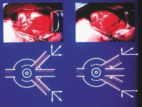
Lateral fixation suture-augmented transposition has been shown to increase the tonic force of the transposed vertical muscles significantly in cases of CN-6 palsy and type I Duane syndrome.15 This frequently results in satisfactory alignment and a larger diplopia-free visual field without requiring ipsilateral medial rectus recession (Fig. 4). Leaving the medial rectus unoperated reduces the risk of anterior segment ischemia or loss of adduction ability.

Fig 4. Diplopia-free visual field of patient with type I Duane syndrome after undergoing left augmented transposition. (Foster RS: Vertical muscle transposition augmented with lateral fixation. J AAPOS 1:20, 1997.)

The Hummelsheim Procedure

The technique for the Hummelsheim procedure is similar to that already described for the full-tendon transfer, except that the muscle is divided along its long axis, and only half the muscle is transposed. The other half remains attached to the globe with some vasculature presumably intact. This procedure is indicated for cases in which preservation of anterior segment circulation is a special concern.

Once the vertical recti have been identified, engaged on a muscle hook, and freed from surrounding attachments to intermuscular membrane and anterior Tenon's capsule, a small muscle hook is used to split the muscle and tendon, beginning at the insertion and extending approximately 15 mm posteriorly. The anterior ciliary arteries should be identified and the splitting done between the vessels, leaving intact the vessel in the half of the muscle that was not detached. That half of the muscle to be transposed is secured on a double-armed, absorbable 6-0 suture and disinserted. The same splitting procedure is performed on the direct antagonist. In a fashion similar to that used in a full-tendon transfer, the halves of the two muscles are then secured to the sclera, concentric with the limbus, at the edges of the insertion of the palsied or lost muscle (Fig. 5). The ipsilateral antagonist to the lost or palsied muscle should be weakened by chemodenervation or surgical recession, as already described above.

Fig 5. The Hummelsheim procedure.

The Jensen Procedure

The Jensen procedure is performed by splitting the palsied muscle and joining the midportion of this muscle to the similarly split midportions of the two adjacent rectus muscles. The dissection and splitting are performed as described for the Hummelsheim procedure, but the muscles are not disinserted. Instead, a 5-0 nonabsorbable polyester fiber suture is used to join the muscle bellies to effect a union of the two muscles just anterior to the equator, as shown in Figure 6.

Fig 6. The Jensen procedure.

This procedure has little advantage over the full-tendon transfer or Hummelsheim procedure, other than theoretically reducing the risk of anterior segment ischemia. However, anterior segment ischemia has been reported after the Jensen procedure,22,23 so choosing this technique does not ensure avoidance of this complication. Significant scar tissue formation occurs with the Jensen procedure, making subsequent surgical exploration more difficult when compared with other tendon transfer techniques.

As with full- or half-tendon transfers, a weakening of the ipsilateral antagonist muscle is performed by chemodenervation or recession.

Superior Oblique Muscle Transposition

Before the superior oblique transposition is performed, the surgeon must verify that superior oblique muscle function is present. In the patient with complete CN-3 palsy, this function can be determined reliably by noting incyclotorsion of the involved exodeviated eye when the patient attempts to look down.

A limbal peritomy is made in the superonasal quadrant sufficient to provide exposure to both medial and superior recti. Traction sutures placed at 11- and 4-o'clock positions in the right eye (1- and 8-o'clock positions in the left eye) can facilitate exposure before the limbal incision is made. Alternatively, exposure to both the superior oblique and medial rectus can be obtained through a superonasal fornix incision. The superior oblique is isolated and dissected free of surrounding fascial attachments along the tendon, following underneath and along the nasal side of the superior rectus. After the superior oblique tendon is engaged on a small muscle hook on the nasal side of the superior rectus muscle, a suture is placed around the tendon, passed beneath the superior rectus, and pulled temporally, thus exposing the superior oblique insertion. The tendon is freed of surrounding attachments from the insertion toward the trochlear cuff. Dissection is limited to the sub-Tenon's space to avoid fat prolapse, which can produce postoperative restrictions.

A double-armed 6-0 absorbable suture is used to secure the superior oblique tendon at its insertion. The tendon is disinserted and brought down toward the superior edge of the medial rectus insertion. The superior oblique tendon will be redundant at this point. It is sutured to the sclera at the superior border of the medial rectus insertion at the point on the tendon that brings the eye to a centered position. It is unwise to shorten the superior oblique tendon excessively because doing so may produce an unwanted hypertropia or esotropia. Conversely, the tendon should not be left so long that the tonic contraction of the superior oblique muscle is not transmitted to the globe. After an appropriate length for the tendon is determined and it is sutured to the sclera, the excess tendon is excised (Fig. 7). A more physiologic, but also more difficult, superior oblique tendon transfer can be done after fracture of the trochlea. The trochlea may be fractured by passing a fine-tipped hemostat, spatula, or other instrument nasally along the tendon into the trochlea and then twisting or prying toward the orbit to force the arch of the trochlea open by fracture. The tendon then can be pulled free from the trochlea so that it passes straight to the new insertion site at the superior border of the medial rectus insertion. In adult eyes, the trochlea resists fracture, and the effort required to fracture it can lead to unintentional severance of the superior oblique tendon, making the transfer procedure impossible. For practical reasons, trochlea fracture should be confined to young patients, if it is done at all. We favor leaving the trochlea intact in most cases.

Fig 7. Superior oblique transposition to the superior border of the medial rectus muscle.

RESULTS

The results obtained from the transposition procedure vary with both the technique and the underlying problem. Specific results are covered in the following discussions of several common indications.

COMPLICATIONS

Anterior Segment Ischemia

Anterior segment ischemia is caused by compromising more than a critical amount of anterior segment circulation during strabismus surgery. This complication has been reported after detachment of only two rectus muscles, conversely, all four rectus muscles have been detached in other patients without producing this clinical picture. The risk of this complication increases whenever a third rectus muscle is detached in the same eye, especially if two of these muscles are vertical recti, because vertical recti do not have an associated posterior ciliary artery. If surgery on a fourth rectus muscle becomes necessary, a procedure that spares the anterior ciliary artery can be used,24 or botulinum injection to the muscle can be considered.

Back to Top
SIXTH CRANIAL NERVE PALSY
Sixth cranial nerve palsy causes weakness of the lateral rectus muscle, which results in esotropia, decreased abduction, and in some cases, head turn. When some lateral rectus function remains, recession of the medial rectus and resection of the lateral rectus are indicated. When paralysis with minimal or no lateral rectus function is present, a transposition procedure is indicated. Measurement of saccadic velocity and force generation or clinical assessment of forced ductions, observed saccadic velocity, and binocular visual fields can be useful in the presurgical assessment of these patients. A simple and useful clinical technique for evaluation of CN-6 palsy involves assessment of maximum abduction. If the eye cannot abduct past the midline, and this response is coupled with a floating saccade, then a transposition procedure will likely be required. In such cases, it is unwise to do a recession-resection procedure on the horizontal recti as an initial procedure because this will compromise the horizontal ciliary vessels that will invariably be needed when vertical transposition becomes necessary.

INDICATIONS

Chronic (i.e., longer than 6 months), stable esotropia that results from CN-6 palsy is an indication for surgical correction. Surgical goals are elimination of diplopia or face turn and enlargement of the field of single binocular vision. A transposition is not indicated for partial palsies in which sufficient lateral rectus function is present for a successful recession-resection procedure on the horizontal recti.

PROCEDURE

Any rectus muscle transfer procedures discussed earlier can be used if it is determined that a recession-resection procedure would not be sufficient. Our preference for transposition is the full-tendon transfer of the vertical recti, with or without injection of botulinum to the ipsilateral medial rectus. In long-standing cases in which significant medial rectus contracture has occurred, or if botulinum injections have proved ineffective, then recession of the medial rectus is indicated. Recession is done in the range of 4 to 7 mm (8.5 to 11.5 mm when measuring from the limbus), depending on the preoperative angle of deviation and degree of restriction shown on forced duction testing.

RESULTS

Muscle transposition will not likely produce normalization of ductions, versions, and a full field of single binocular vision, and patients should be made completely aware of this situation before surgery is performed. An excellent surgical result is achieved if the eyes are straight in the primary position, there is no face turn, and an enlarged area of single binocular vision is present after surgery. A good result is single binocular vision with a minimal head turn (5 to 10 degrees).

After an initial good or excellent surgical result, a recurrent esodeviation may develop, requiring further surgical intervention. Further weakening of the medial rectus usually is the logical next step. Bilateral CN-6 palsy creates a special problem because of diplopia, which if present persists in most cases postoperatively, even when good primary position alignment is attained.

COMPLICATIONS

Overcorrection

Initial overcorrection is a favorable sign after a transposition procedure, especially when botulinum is used. Permanent overcorrection after a transposition is rare. Potential causes include excessive weakening of the medial rectus, restriction to adduction caused by adhesions associated with the transposed muscle, and delayed (or unexpected) return of lateral rectus function. A persistent overcorrection is managed surgically according to standard surgical techniques, including weakening overacting muscles, strengthening underacting muscles, and freeing restrictions.

Undercorrection

Undercorrection (residual esodeviation) is far more common than overcorrection. Undercorrection is treated most effectively by further weakening of the ipsilateral medial rectus. Injection or reinjection of botulinum into the medial rectus can be done under electromyographic control, but if mechanical restriction to abduction is present, then further medial rectus recession is indicated. If an initial full- or half-tendon transfer was performed without lateral fixation sutures (Foster modification), these can be added to the transposed vertical recti at a later date to enhance an undercorrection.

Induced Vertical Deviation

Induced vertical deviation can occur if the forces of the vertical recti are not balanced, or if botulinum has spread to affect a vertical muscle. The cause of the induced vertical deviation can be determined only if the transposition procedure and the botulinum injection are performed separately. Rosenbaum and colleagues14 advocated using adjustable sutures on the vertical recti to help avoid residual vertical deviation and injecting botulinum into the medial rectus after the adjustment of the transposed vertical recti has been done.

Back to Top
THIRD CRANIAL NERVE PALSY
Third cranial nerve palsy involves four of six extraocular muscles in addition to the levator palpebrae and the constricting muscles of the pupil, thus making it the most difficult isolated CN palsy to treat. The difficulty is compounded by the fact that the involvement of the included muscles ranges from total and complete to partial and incomplete, producing a varied clinical picture. In addition, about two thirds of traumatic or congenital CN-3 palsies are complicated by aberrant regeneration. Several surgical procedures are available to treat CN-3 palsy. The choice of procedure depends on the severity of the palsy and the presence or absence of aberrant regeneration. The most realistic result of successful surgery for severe CN-3 palsy is realignment of the affected eye in the primary position and single binocular vision within a very limited range, without much hope for creating normal or near-normal ductions.

INDICATIONS

Congenital palsies in children can be treated at any age after the condition is determined to be stable and amblyopia has been treated. We always obtain preoperative imaging of the head with computed tomography (CT) or MRI in cases of congenital CN-3 palsy. Adults can be treated surgically after the alignment has stabilized and the underlying etiology has been assured. We routinely wait a minimum of 6 months after the onset of the CN-3 palsy before undertaking surgical correction.

PROCEDURE

The choice of surgical procedure for the treatment of CN-3 palsy depends on the specific nature and the severity of the paresis or palsy. In the case of paresis where some medial rectus function remains, a recession of the lateral rectus and resection of the medial rectus can restore primary position alignment, and therefore, is the procedure of choice. If hypotropia is present, vertical upshift of both horizontal recti should be done at the same time as the recession-resection procedure. An estimate of the amount of upshift required can be made by anticipating 2 prism diopters of vertical change in the same direction as the shift for each millimeter that the horizontal recti are shifted upward. For example, upshifting both horizontal recti 4 mm would be expected to elevate the eye approximately 8 prism diopters.25 Larger amounts of hypotropia can be corrected by performing larger vertical shifts.26 Eventually, this becomes a semantic dilemma about whether the procedure is best described as a recession-resection procedure with vertical shift, or a transposition procedure modified by recessing or resecting the muscles. This type of combined vertical and horizontal correction works best when medial rectus function permits horizontal correction to be accomplished by a recession-resection procedure that does not exceed the usual limits of 8 mm for lateral rectus recession and 10 mm for medial rectus resection. In selected cases, a superior oblique weakening procedure may be done to treat the hypotropia without adverse consequences.

The most important requirement in horizontal muscle surgery for severe CN-3 palsy is to weaken (recess) the lateral rectus muscle adequately. In some cases, “super recessions” as large as 12 to 15 mm from the insertion must be done, depending on the amount of antagonist function present in the palsied medial rectus muscle. A lateral rectus recession of this size places the new insertion posterior to the equator and the anterior border of the inferior oblique insertion. For complete medial rectus paralysis, it can be advantageous to excise the sub-Tenon's portion of the lateral rectus and to suture closed the Tenon's penetration site to prevent any attachment of the lateral rectus to the globe, thus creating an iatrogenic lost muscle.

If a large resection (12 to 15 mm) of a paralyzed medial rectus muscle has not maintained alignment, or if a recession-resection procedure is considered likely to be insufficient to align the eye because the medial rectus function is poor, then a superior oblique transposition procedure, with or without fracture of the trochlea, is indicated.

EXPECTED RESULTS

Unlike fourth and CN-6 palsies, in association with which a fairly wide range of single binocular vision is obtainable, adults with severe CN-3 palsy have little chance of obtaining useful single binocular vision. Nearly all these patients will continue to have diplopia. Often, such patients are more disturbed by diplopia after alignment has been improved because the two images are closer and therefore more difficult to ignore. These patients must be warned about the problems that are associated with postoperative diplopia, and when it occurs, the surgeon has the responsibility to provide the patient with adequate support, including use of various occluding contact or eyeglass lenses. No patient in our experience has exercised the option given them to have their eyes returned to the preoperative angle.

The goal of surgery for CN-3 palsy in the adult is to align the eyes in the primary position, with the hope of providing a useful area of single binocular vision. When patients with paralysis undergo a supermaximum recession-resection procedure, with or without superior oblique transposition, this goal is obtained at the expense of creating a nearly immobile eye.

The goal of treatment of CN-3 palsy in children is also to align the eyes in the primary position. Some fusional ability can develop in children and may result in an abnormal head posture. However, most children with CN-3 palsies do not achieve either fusion or measurable stereopsis despite surgical realignment.27 Treatment of amblyopia is critical and should not be overlooked in children. It is best to treat the amblyopia sufficiently to allow good vision in the palsied eye but not to the extent that fixation is switched to the palsied eye. Interestingly, we have seen several children who preferred fixation with the palsied eye. This has usually been due to anatomic abnormality of the other eye, but in several cases no reason for the paradoxic fixation was evident.

COMPLICATIONS

Undercorrection

Undercorrection of surgically treated CN-3 palsy is common if medial rectus function is absent. If undercorrection occurs, further surgery should be performed based on what procedure was done initially. The lateral rectus muscle can be recessed further, or even detached and sutured into the extraconal space. The superior oblique muscle should be transposed if this procedure was not done initially, or the previously transposed superior oblique tendon could be tightened, which is a technically difficult procedure. Further medial rectus resection also can be performed so that the medial rectus can act as a tether. We generally do not advise surgery on the contralateral sound eye.

If all these measures result in an unacceptable exotropia, the eye may be fixed in better alignment using a traction suture. The medial rectus is exposed through a limbal incision, and the muscle insertion and adjacent sclera are secured on a double-armed 5-0 nonabsorbable suture. The two needles are passed through Tenon's capsule under the conjunctiva and brought out on each side of the medial canthal tendon, which has been exposed by means of a small skin incision. Care must be taken to avoid damaging the lacrimal drainage apparatus. The eye is pulled into position nasally, the sutures are tied over the medial canthal tendon, and the skin incision is closed.

Overcorrection

Persistent overcorrection in cases of CN-3 palsy is rare. When they occur, they usually are the result of underestimation of the preoperative medial rectus function. Botulinum toxin can be injected into the medial rectus muscle in an attempt to correct the deviation. If overcorrection persists, the recession-resection procedure must be revised by an appropriate amount.

Vertical Deviation

Vertical deviation is common after procedures to treat CN-3 palsy. A small vertical deviation can be treated with prisms if the patient is made more comfortable by them. If the superior oblique was not already transposed, larger hypodeviations can be reduced by superior oblique tenotomy, by upshifts of the horizontal recti, by recession of the inferior rectus, or by resection of the superior rectus. Anterior segment ischemia is a concern if a third rectus muscle is operated on, particularly in older patients. However, this complication is rare, so we do not avoid operating on a third rectus muscle solely to avoid this complication.

Hypertropia can result if the superior oblique is pulled too tightly after it is transposed. This placement can be readjusted if needed, although the indication is rare. An increasing hypertropia can result from scarring and fibrosis in an adherence syndrome that is caused by a disturbance of the orbital fat. Because the adherence syndrome is difficult to repair, fat should be avoided at the time of surgery by careful application of surgical technique.

Cyclodeviation

Although a good functional result rarely is limited only by cyclodiplopia, this diagnosis should not be overlooked. Incyclotorsion accompanied by a residual hypodeviation can be treated by weakening the superior oblique muscle.28

Back to Top
LOST MUSCLES
An extraocular muscle is considered lost if it has lost its attachment to the globe. This situation can occur after strabismus surgery, as a result of other surgery around the eye and orbit, or from trauma. Because the medial rectus has no fascial attachment to an oblique muscle, it is the muscle that most readily will retract posteriorly through its Tenon's penetration site. Both the superior and inferior rectus muscles have attachments to an adjacent oblique muscle or tendon, and the inferior rectus muscle has a firm attachment to Lockwood's ligament. Because of these anatomic factors, the vertical recti are less likely to retract from the sub-Tenon's space unless vigorous dissection around the muscle has been performed. Lateral rectus attachment to the inferior oblique is less pronounced; however, it can help to hold a detached lateral rectus forward in the sub-Tenon's space, where it can be identified more readily. In the treatment of a lost muscle, the best results are obtained when the muscle can be found and reattached to the sclera. When a lost rectus muscle cannot be found and reattached to the sclera, a transposition procedure is indicated.

INDICATIONS

The diagnosis usually is apparent, with a history of ocular surgery or trauma combined with clinical examination that shows strabismus and limitation of ductions in the field of action of the suspected lost muscle. In the course of evaluation, other causes for limited ductions, such as CN palsy or a restrictive process, must be excluded. It may be necessary to test forced ductions and evaluate generated muscle force. Differentiating a slipped muscle, which is attached to the globe through its empty muscle capsule, from a lost muscle, which has no functional attachment to the globe, is not always possible with a clinical examination alone.29 MRI or CT studies can be helpful. These imaging studies can provide the surgeon with increased confidence that a lost muscle is retrievable; however, the results of these scans can be misleading. Ultimately, only after adequate surgical exploration will the surgeon have sufficient information to make the diagnosis and then carry out the appropriate surgical treatment.

FINDING LOST MUSCLES

A surgeon who is dealing with a lost muscle must realize that the muscle is not really lost; it has just become detached from the globe and disappeared from view, probably temporarily. Adequate lighting and sufficient exposure at the surgical field are imperative. A fiberoptic headlamp, an experienced assistant, and proper instruments, including malleable retractors, provide a good start. The search for the medial rectus should be carried out along the medial orbital wall, not at the medial aspect of the globe. Tenon's capsule and the intermuscular septum should be handled gently with fine-tooth forceps to prevent excessive swelling of these tissues. Swelling can obscure tissue relationships and make a hard job even more difficult. The extraconal fat pad must not be traumatized during a frantic effort to locate the muscle. Exposure of this fat, with subsequent hemorrhage into the fat and dislocation of this tissue, leads to irreversible scarring and restriction. This condition has been labeled adherence syndrome. Instead, gentle hand-over-hand manipulation of Tenon's capsule should be carried out to find the opening in the posterior Tenon's capsule through which the muscle has retracted.

After what is thought to be the muscle tissue has been found, placing gentle traction on it may slow the heart rate (oculocardiac reflex).30 This sign helps to confirm that the tissue that is being pulled is indeed muscle. This sign can be obscured if the anesthesiologist has administered atropine.

If the muscle cannot be found after a careful search, muscle transposition can be performed, or the operation can be terminated, with another surgical exploration planned to take place after the tissues settle. The original surgeon may have more success in locating the muscle during a second procedure, or the patient can be referred to a surgeon who has more experience in treating such cases. Again, the most important guideline for the primary surgeon is to avoid disrupting the normal anatomic relationships so that fat prolapse and its consequences are avoided.

The vertical recti, and to a lesser extent, the lateral rectus muscle, share attachments to an oblique muscle, so the posterior retraction of the detached rectus muscle frequently will be checked at that point. This junction is the best place to find these lost muscles.

If the lost muscle is found, it usually will be contracted to a degree proportionate to how long it has been detached. When a muscle has been detached or lost for months or years, it may be difficult to bring the muscle forward sufficiently to reattach it to the globe at the intended position. In general, the muscle should be advanced as far toward the original insertion as possible, provided that this placement does not cause unwanted restriction.

If the large deviation associated with a lost muscle persists for an extended time, contracture of the antagonist muscle also will occur. In this case, the antagonist must be recessed when the lost muscle is repaired.

PROCEDURES

If the lost muscle cannot be retrieved and reattached to the globe, then a transposition procedure is indicated. Conventional transpositions, such as the full-tendon transfer with injection of botulinum into the antagonist muscle described earlier, may be performed. However, anterior segment ischemia becomes an important consideration because the blood supply from the lost muscle is already missing. When vertical tendon transposition is performed for a lost medial rectus muscle, only one anterior ciliary vessel, that associated with the lateral rectus, would be left undisturbed. If recession of the lateral rectus subsequently becomes necessary, it can only be done at the expense of the last remaining anterior ciliary vessel. One solution to this problem is to use a vessel-sparing technique on the vertical recti, as described by McKeown and associates.24 Another alternative is to use a split muscle transposition, such as the Hummelsheim procedure. This procedure has the theoretical advantage of preserving one anterior ciliary vessel in each of the transposed vertical recti. Plager and Parks31 reported acceptable primary position alignment, although limited duction, in 10 patients who underwent this procedure for a lost medial rectus muscle. No cases of anterior segment ischemia were encountered.

EXPECTED RESULTS

The goal of transposition surgery for treatment of a lost muscle is restoration of alignment in the primary position. Some limitation of ductions with incomitance always will be present.

An excellent result from transposition is alignment in the primary position with more than 25-degree horizontal abduction and adduction. The result is good if alignment is achieved with ductions to either side of the midline limited to 20 degrees or less. A fair result is achieved if single vision is obtained with a cosmetically acceptable head posture (e.g., less than 10-degree head turn). Failure to achieve either alignment in the primary position or comfortable fusion with a minimal head posture is a poor outcome. Unfortunately, this result is not unusual.

COMPLICATIONS

Anterior Segment Ischemia

Anterior segment ischemia is a significant risk because the blood supply from the lost muscle is gone. This topic is covered in the discussion of procedures.

Undercorrection

Undercorrection is common. If the residual deviation is unacceptable, then further recession or chemodenervation of the antagonist can be done.

Overcorrection

Overcorrection after treatment for a lost muscle is rare; however, in this case, a recession of the transposed muscles can be done.

Induced Vertical Deviation

Induced vertical deviation can be the result of an asymmetrical transposition procedure. Although adjustment of the transpositions is possible, it is technically easier and perhaps more predictable to adjust the fellow eye vertically.

Back to Top
REFERENCES

1. Hummelsheim E: Weitere Erfahrungen mit partieller SehnenuberpÂlanzung an den Augenmuskeln. Arch Augenheilkd 62:71, 1908-1909

2. O'Conner R: Transplantation of ocular muscles. Am J Ophthalmol 4:838, 1921

3. Berens C, Girard LJ: Transplantation of the superior and inferior rectus muscles for paralysis of the lateral rectus. Am J Ophthalmol 33:1041, 1950

4. Jackson E: Operations on muscles of the eye. In Wiener M, Scheie HG (eds): Surgery of the Eye, p 402. 3rd ed. New York: Grune & Stratton, 1952

5. Hildreth HR: The tendon-transplanting operation. Am J Ophthalmol 36:1267, 1953

6. Costenbader FD: In Allen JH (ed): Strabismus Ophthalmic Symposium 2, p 502. St Louis: CV Mosby, 1958

7. Schillinger RJ: A new type of tendon transplant operation for abducens paralysis. J Int Coil Surg 31:593, 1959

8. Jensen CDF: Rectus muscle union: A new operation for paralysis of the rectus muscle. Trans Pac Coast Otoophthalmol Soc 45:359, 1964

9. Uribe LE: Muscle transplantation in ocular paralysis. Am J Ophthalmol 65:601, 1968

10. Knapp P: The surgical treatment of double-elevator paralysis. Trans Am Ophthalmol Soc 67:304, 1969

11. Helveston EM: Muscle transposition procedures. Surv Ophthalmol 16:92, 1971

12. Carlson MR, Jampolsky A: An adjustable transposition procedure for abduction deficiencies. Am J Ophthalmol 87:382, 1979

13. Kushner BJ: Jensen procedure modified. Ann Ophthalmol 11:1255, 1979

14. Rosenbaum AL, Kushner BJ, Kirschen D: Vertical rectus muscle transposition and botulinum toxin (Oculinum) to medial rectus for abducens palsy. Arch Ophthalmol 107: 820, 1989

15. Foster RS: Vertical muscle transposition augmented with lateral fixation. J AAPOS 1:20, 1997

16. Jackson E: Operations on muscles of the eye. In Wiener M, Scheie HG (eds): Surgery of the Eye, p 405. 3rd ed. New York: Grune & Stratton, 1952

17. Scott AB: Transposition of the superior oblique. Am Orthoptic J 5:11, 1977

18. Saunders RA, Sandall GS: Anterior segment ischemia syndrome following rectus muscle transposition. Am J Ophthalmol 93:34, 1982

19. Oliver JM, Lee JP: The effects of strabismus surgery on anterior segment circulation. Eye 3:318, 1989

20. Scott AB: Action force tests in lateral rectus palsy. Arch Ophthalmol 85:397, 1971

21. Miller JM, Demer JL, Rosenbaum AL: Effect of transposition surgery on rectus muscle paths by magnetic resonance imaging. Ophthalmology 100:475,1993

22. von Noorden GK: Anterior segment ischemia following Jensen procedure. Arch Ophthalmol 94:845, 1976

23. Frey T: Anterior segment ischemia caused by Jensen's procedure. J Ocul Ther Surg 3:242, 1985

24. McKeown CA, Lambert HM, Shore JW: Preservation of the anterior ciliary vessels during extraocular muscle surgery. Ophthalmology 96:498, 1989

25. Biglan AW, Walden PG: Results following surgical management of oculomotor nerve palsy with a modified Knapp procedure. Ophthalmic Surg 16:759, 1985

26. Buckley EG, Townshend LM: A simple transposition procedure for complicated strabismus. Am J Ophthalmol 111: 302, 1991

27. Schumacher-Feero LA, Yoo KW, Solari FM et al: Third cranial nerve palsy in children. Am J Ophthalmol 128: 216,1999

28. Helveston EM: Atlas of Strabismus, pp 252–253. 3rd ed. St Louis: CV Mosby, 1985

29. Plager DA, Parks MM: Recognition and repair of the slipped rectus muscle. J Pediatr Ophthalmol Strabismus 25:270, 1988

30. Apt L, Isenberg SJ: The oculocardiac reflex as a surgical aid in identifying a slipped or “lost” extraocular muscle. Fr J Ophthalmol 64:362, 1980

31. Plager DA, Parks MM: Recognition and repair of the “lost” rectus muscle. Ophthalmology 97: 131, 1990

Back to Top