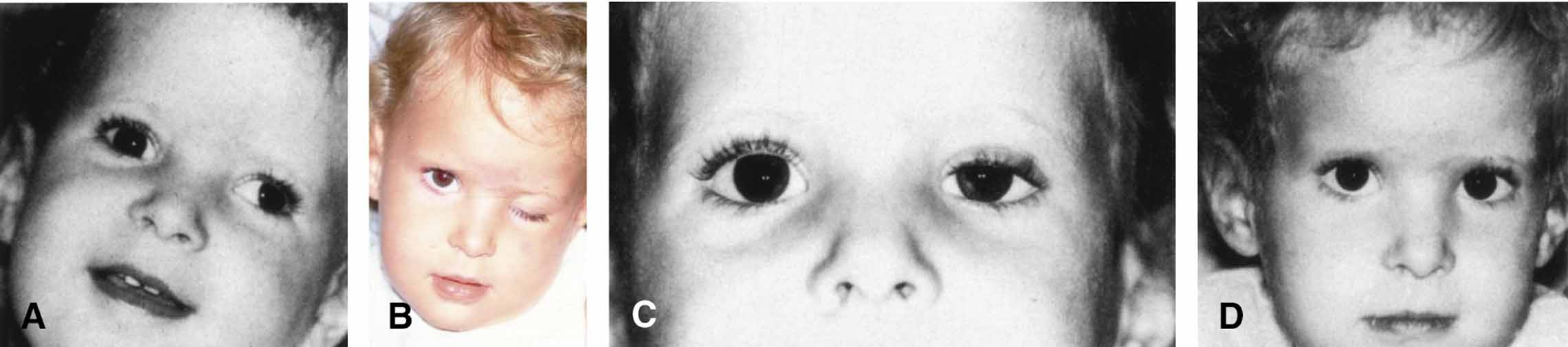
Chapter 89
Chemodenervation of Extraocular Muscles
EDWARD G. BUCKLEY, LUCAS TRIGLER and DEREK B. HESS
Main Menu   Table Of Contents

Search

MECHANISMS OF ACTION
INDICATIONS
CONTRAINDICATIONS
PATIENT SELECTION
ANESTHESIA
PROCEDURE
EXPECTED RESULTS
COMPLICATIONS
REFERENCES

In the early 1970s, Dr. Alan B. Scott discovered that when botulinum A toxin was injected into an extraocular muscle, a prolonged, dose-related reversible paralysis was caused that could be used to treat strabismus.1 Beginning in 1977, hundreds of investigators injected thousands of patients with botulinum A toxin to treat a wide variety of ocular motility disorders. After establishing its efficacy and safety, botulinum A toxin was approved in December 1989 as a treatment for strabismus in adults and children over the age of 12. The addition of botulinum A toxin has greatly expanded our capabilities for treating strabismus.
Back to Top
MECHANISMS OF ACTION
Six antigenically distinct forms of botulinum toxin (A to F) are produced by the bacterium Clostridium botulinum. Botulinum toxin type A is used clinically because it retains its toxicity, yields highly potent culture fluids, and can crystallizes easily into a stable form. Botulinum A toxin exerts its paralytic effect by rapidly and strongly binding to the presynaptic nerve terminal of cholinergic axons.2,3 The toxin is then internalized into the intracellular compartment where it inhibits the release of acetylcholine-containing vesicles.4 Within 6 hours after injection a complete decline of miniature end plate potentials occurs, but the clinical effect does not peak for approximately 5 to 7 days.1,5 This delay may be related in part to spontaneous release of nonvesicular acetylcholine.6 Eventually, the muscle becomes denervated and atrophic changes occur. The paralytic effect usually subsides over 3 to 4 months but some effects may persist for as long as 6 months. Muscle function slowly returns when the axon terminal forms new sprouts that result in new synaptic contacts on adjacent muscle fibers.7

The clinical effectiveness of botulinum A toxin is related to the completeness and duration of the paralysis of the injected muscle and to the condition that is treated (i.e., nonparalytic versus paralytic strabismus). Prior to the use of botulinum A toxin it had been observed that patients who recovered from sixth-nerve palsies often had a residual esotropia even though lateral rectus muscle function had apparently returned to normal. In such cases, the longer the paralysis persisted the more likely it was that a permanent deviation would occur. This change in ocular alignment was thought to be secondary to a mild residual weakness of the involved muscle (i.e., lateral rectus) plus contracture of its antagonist muscle (i.e., medial rectus). A similar mechanism is postulated to be responsible for the effects seen after botulinum A toxin injection, that is, weakness of the injected muscle plus contraction of its antagonist.

Once injected into the extraocular muscle botulinum A toxin binds rapidly to the nerve terminals, thereby limiting exposure to the systemic circulation. Until recently, antibodies could not be detected in humans exposed to therapeutic botulinum A toxin injections. However, an in vivo mouse neutralization assay has been developed that can detect serum antibodies to botulinum A toxin.8 Although not yet documented in patients treated for strabismus, clinical resistance to the effects of subsequent injections of botulinum A toxin has been correlated with the presence of antibodies to botulinum A toxin detected by this bioassay.9

The commercial preparation of botulinum A toxin (Oculinum) is distributed by Allergan Pharmaceuticals, Inc. (Irvine, CA) in a frozen, lyophilized form that is very stable. Once reconstituted into a solution, it must be used within hours because its effectiveness deteriorates rapidly. Care must be used in handling the reconstituted toxin because of its susceptibility to damage from preservatives, fluctuations in pH, heat, vigorous shaking, and rapid injection.10 The standard unit for measuring the potency of botulinum A toxin is derived from a mouse assay.11 One unit of botulinum A toxin is the lethal dose for 50% of a group of 18- to 20-g female Swiss-Webster mice. The median lethal dose (LD50) for humans is estimated at approximately 40 U/kg.1 The toxin available in the United Kingdom (Dysport, Ipsen Limited, Slough, Berkshire, UK) is much more potent; 1 ng of British toxin contains 40 mouse units, whereas 1 ng of American toxin contains 2.5 mouse units.12 This may explain the higher incidence of side effects with the more potent British toxin. The doses used in strabismus applications are roughly proportional to the mass of the muscle being injected and are less than 1/100 of the estimated human LD50.13

Back to Top
INDICATIONS
A wide variety of ocular motility disorders can be successfully treated with botulinum A toxin and the list is still evolving (Table 1). Originally it was thought that botulinum A toxin would only be useful for comitant deviations and in situations in which extraocular muscles were functioning normally. However, with time it has become apparent that its use can be extended to such complicated entities as postretinal detachment strabismus, thyroid myopathy, as an adjunct to transposition procedures for paralytic strabismus, in the treatment of oscillopsia from acquired nystagmus and stable deviations secondary to myasthenia gravis. In addition, botulinum A toxin has been found to be effective in small-angle deviations of less than 10△ to 15△ which might be considered too small for surgical intervention but still bothersome to the patient. Botulinum A toxin may also be a vessel-sparing alternative to strabismus surgery after trauma or other situations in which anterior segment blood flow has been compromised.


TABLE 1. Strabismus Categories Treatable with Botulinum A Toxin


Comitant strabismus
 Esotropia
  Infantile
  Comitant
  Sensory
 Exotropia
  Intermittent
  Sensory
 Hypertropia
Paralytic
 Acute sixth-nerve palsy
 Third nerve palsy
Restrictive strabismus
 Dysthyroid myopathy
 Retinal detachment
Postoperative
 Overcorrections
 Undercorrections
Adjunct to surgery
 Transpositions
Acquired nystagmus


Botulinum A toxin can also be a useful alternative in those patients in whom general or local anesthesia is contraindicated. Those individuals with a history of malignant hyperthermia, chronic obstructive pulmonary disease, or significant cardiac abnormalities can often be treated successfully with botulinum A toxin avoiding anesthesia and its potential hazards in these high-risk patients.

Back to Top
CONTRAINDICATIONS
As a general rule, ocular motility abnormalities caused by tight and inelastic muscles or by chronic, complete nerve palsies do not respond well to botulinum A toxin injections. For a permanent change in ocular alignment to occur after a botulinum A toxin injection, there must be contracture of the antagonist to the injected muscle and this will not occur when the injected muscle is stiff or when its antagonist muscle is weak. In conditions such as chronic thyroid myopathy with fibrotic muscles, entrapped muscles after blowout fractures, or restrictive strabismus secondary to multiple operations to correct strabismus, only minimal relaxation results after injection allowing no opportunity for the antagonist muscle to shorten. In patients with chronic sixth-nerve palsies with no lateral rectus function, injecting the ipsilateral medial rectus will only result in temporary improvement because no permanent contracture of the paretic lateral rectus can be expected. This is also true for patients with Duane's syndrome, with an inactive lateral rectus, or in persistent strabismus secondary to excessive surgical weakening procedures (e.g., recessions, myotomies).

Anatomic considerations must also be considered in selecting patients for botulinum A toxin injection. Those patients with an abnormal orbital anatomy caused by posttraumatic (e.g., blowout fractures) or congenital conditions (e.g., Crouzon's disease), must be approached with caution because the extraocular muscles are often in abnormal positions. The presence of other orbital abnormalities such as tumors, vascular malformations, or bony defects may make botulinum A toxin injection difficult, thereby increasing the risk of significant complications after injection.

Back to Top
PATIENT SELECTION
Care must be taken in selecting patients because cooperation during the injection is essential. Because the procedure is somewhat threatening, it is imperative that the patient be considered capable of remaining in control in this stressful situation. Although age is certainly a factor, it alone cannot determine who will tolerate this procedure. Time spent before the injection educating the patient about the amount and duration of discomfort will help avoid surprises at the time of injection. The ability of the patient to tolerate an office forced duction test can be used to select which patients will be cooperative for the injection. Inability to remain calm and follow directions during forced duction testing will surely be amplified during an injection procedure. This is especially helpful in trying to decide whether or not a child or teenager will be a good candidate.

Until the ophthalmologist is familiar and experienced with the injection procedure, consideration should be given to treating only the horizontal rectus muscles. The vertical rectus muscles, because they have opposite functions from the nearby oblique muscles, require a higher level of expertise to avoid unwanted complications. Likewise, experience should be gained on adults before attempting injections in children.

Back to Top
ANESTHESIA
The conjunctival surface is anesthetized with several repeated installations of topical anesthesia (proparacaine, tetracaine, or cocaine) given liberally over several minutes. This should be placed in both eyes to reduce the urge of the untreated eye to blink during the procedure. Occasionally, 1% lidocaine can be injected over the muscle insertion in patients with significant scar formation secondary to multiple operations, scleral buckling procedures, or trauma. This does not interfere with the electromyographic (EMG) signal if given anteriorly. Because placement of the needle into the muscle is guided by an EMG signal, peribulbar or retrobulbar anesthesia has the risk of blocking the signal and is not usually necessary. Ketamine is recommended for children when sedation is required, since it tends to preserve the EMG signal. For children 1 to 6 years of age, intravenous ketamine at a dose of 0.5 to 1 mg/kg provides adequate sedation and still preserves EMG activity.14,15 Topical anesthetic drops must be used because low-dose ketamine provides sedation but not full anesthesia. Higher doses of ketamine (up to 2 mg/kg) have also been recommended.16

When inhalation anesthesia with nitrous oxide or halothane is used, the EMG recording is diminished or extinguished. Placement of the drug in the muscle belly can be done under direct visualization or during the waking-up process. If this is attempted, careful attention to eye position and anatomy are important. Usually the medication is delivered approximately 2.5 cm posteriorly in the muscle. Without the use of EMG, a more anterior injection may minimize adjacent muscle involvement. If EMG guidance is desired, an alternative approach requires waiting until the child is coming out of light stage (stage 2–3) anesthesia. At the point when extraocular movements are seen, the electrode is inserted rapidly in an attempt to locate the muscle before the patient becomes completely awake. This requires considerable skill and is somewhat cumbersome if mask anesthesia is used. In infants under 12 months of age injections can be performed using topical anesthesia and restraint. A hungry infant can sometimes be fed a bottle and remain quiet and comfortable throughout the entire procedure.17

Back to Top
PROCEDURE
The best results are obtained when botulinum A toxin is placed into the muscle in the region of the motor end plate, which for rectus muscles, is approximately 2.5 cm posterior to the muscle insertion. Recording an EMG response from the tip of a monopolar injection needle is an important aid in accurately localizing this position. Special 27-gauge, 1½-inch long, Teflon-coated, monopolar injection needles can be obtained for this purpose (Fig. 1). When attached to an audio EMG amplifier, a distinct, recognizable noise pattern results that can be used during the injection process to locate the appropriate muscle (Fig. 2). It is desirable to place the needle into the muscle on the first attempt because the drug diffuses readily along needle tracts and there is a risk of hemorrhage.

Fig. 1 Special 27-gauge, 1½-inch monopolar injection needle with Teflon coating. and positive electromyographic lead. The wire position can also be used to mark the position of the needle bevel.

Fig. 2 Audio electromyographic device used for botulinum A toxin injections. A wire attaches to a special 27-gauge, Teflon-coated, monopolar injection needle attached to a tuberculin syringe. The device is grounded through a pediatric electrocardiographic electrode and fastened to the forehead.

LOCATION

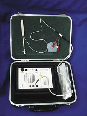
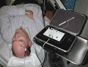
The patient should be made comfortable in the supine position (Fig. 3). Although this can be performed in an office chair, a flat examination table or similar setup is advisable both for patient comfort and stability of the head during the procedure. The room should be as free as possible from electrical signals that can interfere with the audio EMG recording device. Certain fluorescent fixtures and overhead wiring produce signals that can be picked up by the recorder, thereby interfering with assessment during the injection process. Loud background noise often occurs when the EMG device is turned on prior to inserting the needle into the conjunctiva. However, as soon as the needle touches the conjunctiva, this noise is greatly diminished. If this does not occur, then accurate localization of the muscle will be difficult. In this situation, reducing the illumination or turning off nonessential lights can be helpful.

Fig. 3 Patient ready for botulinum A toxin extraocular muscle injection. Note the placement of the electrocardiographic electrode on the patient's forehead medially for a medial rectus muscle injection. The patient is in the supine position and the head is supported comfortably on a pillow. A tray, table, or other support is necessary for the audio electromyographic device since the electrode wires are only 24 inches long.

PREINJECTION PREPARATION

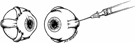
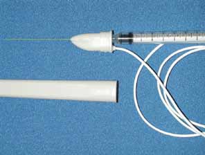
The dosage to be administers should be calculated (Table 2). Nonpreserved saline 0.9% is used to mix the Oculinum so that the desired number of units to be given are in a volume of 0.1 mL or less. Two milliliters of the reconstituted fluid is drawn into a tuberculin syringe; the monopolar electrode needle should not be used for this process. The 27-gauge, 1½-inch, Teflon-coated, monopolar electrode needle is then attached to the end of the tuberculin syringe. The total volume in the tuberculin syringe is reduced to to one-tenth of a milliliter by injecting the excess drug through the needle end. Because of the size of the needle and the small volume used, the needle barrel must be loaded with the drug to avoid undermedicating the patient. The location of the attachment of the wire onto the needle hub can be used as a reference point to determine the position of the needle bevel.


TABLE 2. Botulinum Toxin Dosage for Horizontal and Vertical Rectus Muscles (in units)


Deviation Amount Adults Children
in △ (> 12 yr) < 6 kg 6–9 kg 10–12 kg > 12 kg
< 201.25–2.5 1.251.251.25
20–502.5–511.251.25–1.751.25–2.5

△, prism diopter. (Scott AB, Magoon EH, McNeer KW et al: Botulinum treatment of childhood strabismus. Ophthalmology 97:1434, 1990 Published courtesy of Ophthalmology)


A pediatric electrocardiographic electrode is then attached to the patient's forehead, making sure that a good contact is present (see Fig. 3). It should be placed medially for the medial rectus and laterally for the lateral rectus. If not already done, the patient should be informed that an audio signal will be used during the procedure so that he or she will not become startled when the amplifier is turned on. Next, turn on the amplifier and test the connections by touching the needle tip to the conjunctiva. Any background noise should become extinguished and an audible click should be heard. If this does not happen, the ground connection to the needle hub should be checked and possible interference from overhead lights, etc., investigated. This is also the time to make sure that adequate topical anesthesia has been achieved.

INJECTION TECHNIQUE

Before beginning, it should be explained to the patient that he or she will have to move the eyes into various gaze positions during the procedure. Therefore, it is important that the patient keep both eyes open and fixed on some target. It is helpful to have an assistant during the injection procedure. Standing at the head of the patient, the lids are held open with the fingers. The patient should look away from the field of action of the injected muscle, for example, when injecting the medial rectus have the patient begin by looking laterally with that eye (Fig. 4). The assistant can use a hand or other fixation target for this purpose. The patient should be reminded to keep both eyes open so that fixation is maintained.

Fig. 4 Patient with a left sixth-nerve palsy undergoing left medial rectus muscle injection of botulinum A toxin. A. The eyelids are held open with the index finger and thumb and the patient is directed to look out of the field of action of the injected left medial rectus muscle. B. Once the electrode is inserted through the conjunctiva, the patient is asked to look into the field of action of the injected muscle to increase the electromyographic signal. The needle is advanced until the loudest signal is heard and then the fluid is injected. There is often attenuation of signal with injection.

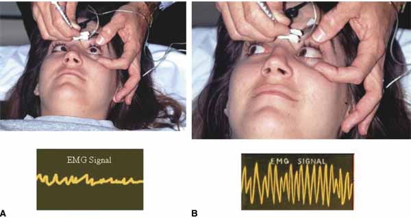
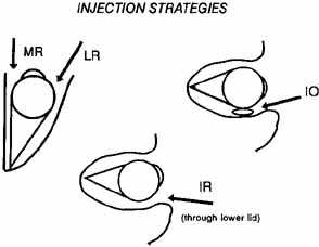
With the bevel facing the muscle, the needle is inserted through the conjunctiva 8 to 10 mm from the limbus. Once through the conjunctiva, the needle is advanced several more millimeters until the tip is beyond the equator. The assistant should move the fixation target slowly to the primary position so that the injection-muscle EMG signal can be activated. If the medial rectus is being injected, continue to advance the needle straight posteriorly aiming for the optic canal (Fig. 5). When injecting the lateral rectus the syringe has to be angled toward the ear at approximately a 45-degree angle to avoid touching the periosteum of the lateral orbital wall with the needle point, which is quite uncomfortable (see Fig. 5). A slight angle is also necessary when injecting the superior rectus. The inferior rectus is often easier to inject through the lower lid rather than the conjunctiva. As the needle is advanced and while listening to the EMG signal, continue to advance until a loud crackling noise occurs. If uncertainty exists as to whether this is from the appropriate muscle, have the patient slowly look out of the field of action of the muscle to be injected. The signal should decrease. Care must be taken to ensure that slow eye movements are made. This may be somewhat difficult because the patient may be anxious or apprehensive and may experience some discomfort. When the electrode is at the appropriate location, the reconstituted toxin is injected slowly. The EMG signal should diminish at this point as the tissue is pushed away from the tip by the entering fluid. Allow the needle to remain in this position for 10 to 15 seconds and then slowly withdraw the needle. The patient should be checked for signs of hemorrhage or other potential complications.

Fig. 5 The direction in which the monopolar electrode needle is advanced depends on the muscle injected. Top left. For medial rectus (MR) muscle injections the needle should be directed straight backward toward the orbital apex. For lateral rectus (LR) injections a 45-degree angle is necessary once the needle tip is past the equator. Top right. Inferior oblique (IO) injections should be directed slightly inferiorly and through the conjunctiva fornix. Bottom. Inferior rectus (IR) injections are somewhat difficult through the conjunctiva and may be easier to inject through the lower lid, especially in patients with restrictive disorders such as thyroid ophthalmopathy.

POSTINJECTION MANAGEMENT

Once finished, the patient should be asked to sit up on the edge of the examination table but should not be allowed to stand until postinjection anxiety and bradycardia have subsided. Often the patient is so relieved that the process is over that an episode of lightheadedness or even syncope can occur. One to two minutes is usually all that is necessary at this time. It is not necessary to patch the eye or use antibiotics unless there is some concern about the injection process. The patient should be informed that topical anesthesia has been applied to the eye and ocular manipulation for the next 30 to 45 minutes should be discouraged. The patient should be observed for 5 to 10 minutes to make sure that no retrobulbar bleeding has occurred. Acetaminophen should suffice for any postinjection discomfort.

Back to Top
EXPECTED RESULTS

HORIZONTAL STRABISMUS

As a group, patients with horizontal strabismus as respond well to treatment with botulinum A toxin injection (Fig. 6).18–24 The results of several large studies are summarized in Table 3. As a general rule botulinum A toxin injections reduce a deviation by approximately 65%, and a little over half of patients have their deviation reduced to less than 10 △. The larger the deviation the greater the prism diopter change, but the less likely that the total deviation will be reduced to 10△ or less. Overcorrections are rare and have occurred in fewer than 1% of patients. A single injection can be expected to reduce a deviation to less than 10△ in approximately 30% to 35% of patients.20 Most patients, however, need more than one injection with the average number of injections varying anywhere from 1.3 to 2, depending on the type of strabismus and the size of the original deviation.19,20,23 After the toxin effect has disappeared, the stability of the postinjection deviation appears to be comparable to that achieved with surgery and is enhanced if there is potential to establish or regain fusion.


TABLE 3. The Effect of Botulinum A Toxin Injection on Horizontal Strabismus


      % Change F/U
Author Deviation No. Pre-inj (△) Post-inj (△) Deviation % <10 △ (mo)
Scott20Esotropia38431 10 68 68 17
 Exotropia29332 12 60 52 17
Biglan19Esotropia3223   14.5   36.5 34 21
 Exotropia1527 20   24.5   13.3 21
Carruthers53Esotropia525  51 40  
 Exotropia1234  50 25  
Buckley*Esotropia5836 14 64 52 15
 Exotropia7839 16 63 54 15
Sener54Esotropia2539   32 3–6
 Exotropia4538   22 3–6

△, prism diopter; pre-inj, preinjection; post-inj, postinjection; F/U, follow-up.
*E.G. Buckley, unpublished data.


Fig. 6 Top. A 68-year-old patient with an esotropia after cataract extraction. Middle left. Three weeks after botulinum A toxin injection into the right medial rectus muscle, the patient developed a complete ptosis of the right upper lid. Middle center. The injection resulted in profound weakness of the right medial rectus. Middle right. Interestingly, superior rectus function was not affected. Bottom. Three months after injection the ptosis and esotropia have resolved.

VERTICAL STRABISMUS

There is little information in the literature about the results from injections into vertical rectus or oblique muscles. The superior rectus is rarely treated because ptosis occurs in almost every patient. Similarly, inferior and superior oblique injections are rarely attempted because of the high incidence of undesirable associated effects on the adjacent muscles. The inferior rectus is the only muscle routinely injected for vertical deviations. Many of these patients are status postretinal detachment surgery or have thyroid myopathy and are discussed later in the chapter. In uncomplicated vertical deviations, injection into the inferior rectus tends to result in the same amount of correction as seen in horizontal deviations. In a recent series of 20 patients with a vertical hypotropia the average preinjection deviation was 30 △. Botulinum A toxin reduced the deviation to an average of 17△ resulting in a 41% reduction. These patients received on the average of 1.7 injections and were followed for at least 6 months. Forty percent of these patients were able to regain fusion postinjection (E.G. Buckley, unpublished data).

SIXTH-NERVE PALSY

The effectiveness of botulinum A toxin to correct an esotropia caused by sixth-nerve palsy is determined by the amount of residual function of the lateral rectus muscle, the amount of medial rectus muscle contracture, and the duration of the palsy prior to injection. During the acute phase, botulinum A toxin can be injected into the medial rectus to prevent permanent contracture and restore binocularity (Fig. 7). Because the time to recovery after botulinum A toxin injections and sixth-nerve palsies are approximately the same, reinjection is usually not necessary and up to 75% of patients have complete recovery of their ocular motility25–29 Botulinum A toxin can also be used as an adjunct to transposition surgery in patients with a complete lateral rectus palsy.30–32 In these patients, botulinum A toxin is injected into the medial rectus either prior to or during the surgical procedure (Fig. 8). In this role it functions as a chemical traction suture. It allows the transposition procedure to achieve its maximum lateral position and enables the tissue to heal before it is subjected to the pulling forces of the medial rectus (Fig. 9). It also reduces the chances of anterior segment ischemia by limiting the incisional surgery to movement of only two extraocular muscles. The size and location of the field of single binocular vision has been shown to be larger and more central with this technique when compared with other procedures (Table 4).

Fig. 7 A 7-year-old child with a right sixth-nerve palsy after head trauma. Top row. Eight weeks after head trauma and prior to botulinum A toxin injection. Note significant restriction to abduction of the right eye. Middle row. Four weeks after botulinum A toxin injection into the right medial rectus muscle. Note mild ptosis of the right upper lid and severe restriction of adduction of the right eye. Lateral rectus function has already improved significantly. Bottom row. Three months after botulinum A toxin injection, the patient's esotropia has resolved and medial and lateral rectus function are now normal.

Fig. 8 Illustration showing the technique for botulinum A toxin injection plus transposition surgery for chronic sixth-nerve palsies. Botulinum A toxin is injected into the medial rectus muscle prior to or at the time of surgery. A full tendon transposition of the superior and inferior rectus to the involved lateral rectus is performed. Note that the transposed muscles are sutured adjacent to the lateral rectus insertion rather than perpendicular to it. (McManaway JW, Buckley EG, Brodsky MC: Vertical rectus muscle transposition with intraoperative botulinum injection for treatment of chronic sixth nerve palsy. Graefes Arch Clin Exp Ophthalmol 228:402, 1990)

Fig. 9 Patient with a chronic left sixth nerve palsy. Top row. Preoperatively, note significant lack of abduction of the left eye. Middle row. One month postoperatively the patient has a significant paralysis of his left medial rectus muscle secondary to the botulinum A toxin injection. Note the amount of abduction. Bottom row. Six months postoperatively, the patient had 70-degree of single binocular vision and 3 degrees of abduction. (McManaway JW, Buckley EG, Brodsky MC: Vertical rectus muscle transposition with intraoperative botulinum injection for treatment of chronic sixth nerve palsy. Graefes Arch Clin Exp Ophthalmol 228:403, 1990)


TABLE 4. Comparison of Results of Methods of Treating Unilateral Acquired Sixth Nerve Paralysis


      Average Change in Position Average Improvement in Abduction Average Improvement in SBV
Author Method No. Patients (△) (%) (°) (°)
Jensen Jensen 6 29.2 108 19.2  
Selezinka Jensen 11 55.2 97.3 27.3  
Freuh Jensen 4 32.5 81.6 28.8  
Cline Jensen 10 55.2 91.8   34.3
Berens Hummelsheim 1 40 100 50 60
Gifford Hummelsheim 4 38  100  21.1  
Rosenbaum Transposition + medial rectus recession 8       43.3
Rosenbaum Transposition + medial rectus botulinum 8 55   90  20  51 
McManaway Transposition + medial rectus botulinum 6 36   96.7 30.8 69.2

SBV, single binocular vision; △, prism diopter. (McManaway JW, Buckley EG, Brodsky MG: Vertical rectus muscle transposition with intraoperative botulinum toxin injection for the treatment of chronic sixth nerve palsy. Graefes Arch Clin Exp Ophthalmol 228:404, 1990)


RETINAL DETACHMENT

Persistent strabismus after retinal reattachment surgery is difficult to correct and has additional surgical risks over other types of strabismus surgery. Recent reports have established the effectiveness of botulinum A toxin in eliminating diplopia and restoring binocular vision in these patients (Table 5).33–35 The effectiveness of botulinum A toxin injections depends on a variety of factors including visual acuity, macular abnormalities, retinal reoperations, and the presence of significant restrictions. Even with all these obstacles, approximately 70% of patients obtain significant benefit. The injection procedure itself is more difficult because the normal anatomic position of the extraocular muscles has been altered by the retinal detachment surgery. Theoretically this increases the possibility of globe perforation with the electrode needle and extra care should be exercised when injecting these patients.


TABLE 5. Results of Botulinum A Toxin Injections in Strabismus After Retinal Detachment Surgery


  Petitto and Buckley34 Scott33 Lee37 Maurino35
Cases 20 2027 26
Visual acuity    
20/400–LP  1 113  4
20/200  5 3 6  5
20/20 to 20/100 14 16 7 15
Retinal reoperations  0 518 18
Strabismus surgery  0 3 5  0
Fusion restored 85% 60%15% 50%

LP = light perception.


DYSTHYROID MYOPATHY

Botulinum A toxin has been shown to be useful in the management of patients with both acute and chronic dysthyroid myopathy. During the acute phase, injections into the involved muscles can relieve contracture, improve ocular rotations, and eliminate diplopia. Repeated injections and larger quantities of drug are required because of the enlargement of the affected muscle.36 With time, fibrotic changes occur in the muscle making botulinum A toxin less effective. In a series of 25 patients with acute thyroid myopathy, only 6 patients had long-term single vision with botulinum A toxin injections alone.37 Scott20 reported similar results with 16 of 27 patients with vertical strabismus initially treated with botulinum A toxin later requiring surgical intervention. Botulinum A toxin injections are less effective if the motility disturbance has been present for more than a year. In patients with small vertical or compound horizontal and vertical deviations, botulinum A toxin injections may reduce the number of operated muscles or decrease the size of the deviation to that which can be corrected by prisms.

NYSTAGMUS

Botulinum A toxin has been used to control severe oscillopsia in patients with acquired nystagmus. Different procedures have been suggested depending on the nature and severity of the nystagmus. Injection into the involved horizontal or vertical muscle has been successful for a uniplanar jerk or pendular nystagmus. In patients with a combined vertical, horizontal, and rotary nystagmus, retrobulbar botulinum A toxin injection has been attempted. Helveston and Pogrebniak38 reported on two patients who developed severe incapacitating oscillopsia after brain stem infarct. Each received 25 units of botulinum A toxin into the retrobulbar space. Visual function improved significantly in both patients. The effect lasted from 5 to 13 weeks and no adverse side effects were observed. As with other entities treated with botulinum A toxin, repeat injections provided similar results. I personally have treated several patients with multiple sclerosis and one patient with oculopalatal myoclonus with similarly good results. Ptosis can occur after retrobulbar botulinum A toxin injection and may be a hindrance to visual function. Concerns about intravascular injection of botulinum A toxin are unwarranted because the dosage is well below the amount necessary to cause systemic toxicity and the molecule is too large to pass through the blood–brain barrier.38

OVERCORRECTIONS AND UNDERCORRECTIONS

Botulinum A toxin can be used when it becomes obvious that a residual deviation after strabismus surgery is not going to improve with time or when other types of supportive therapy such as Fresnel prisms, patching, or exercises yield no benefit. Patients with overcorrections appeared to do exceptionally well with up to 87% obtaining adequate alignment.19,39 Undercorrections responded the same as other types of horizontal strabismus with a 40% to 60% satisfactory alignment. As might be expected, those patients who had multiple re-operations or had a significant restrictive component to their strabismus, did poorly, but some patients had a permanent improvement in their deviation averaging approximately 10△ of correction.39

CHILDHOOD STRABISMUS

While botulinum A toxin is still considered experimental in children under the age of 12, results to date indicate that it may be effective in this age group and may be a suitable alternative to traditional strabismus surgery (Fig. 10).14,17,40 Scott and collaborators15 recently reported that 61% of 362 children who underwent botulinum A toxin injections achieved an alignment of within 10△ of orthophoria. (This is comparable to adults who achieve 65%.) When subdivided into types of strabismus, children with esotropia did better than those with exotropia (65% versus 45%), and smaller deviations (less than 20 △) were more frequently corrected than larger deviations (73% versus 54%) (Table 6). The change in alignment was more stable than that achieved in adults and was felt to be caused by the binocular fusion obtained in children after the injection.15,17 The frequency of complications was similar to that seen in adults except for ptosis, which occurred in over a third of patients. No amblyopia or visual loss was produced by the injection procedure or by the drug effects in Scott's series. Tejedor and Rodriguez41 also cite small angle of esotropia, as well as high hypermetropia and minimal amblyopia as best predictors of favorable outcome. In botulinum A toxin treatment of acquired childhood esotropia motor success rates were 53% after one injection and 88% after two or three injections. In regards to binocularity in this group, 47% had at least 400 seconds of arc and 71% had peripheral fusion.41

Fig. 10 A. Child with a large exotropia. B. Two weeks after botulinum A injection into the left lateral rectus muscle the patient developed complete ptosis. C. Six weeks after injection the patient developed a head position and appeared binocular. Note mild ptosis of the left upper lid and chin-elevated head position. D. Six months after botulinum A toxin injection the ptosis cleared, the exotropia resolved, and the patient remained binocular.


TABLE 6. Botulinum Treatment of Childhood Strabismus


  No. of Age (mo) Prior Surgery Deviation % Final within 10 △ of Ortho No. of Eventual Surgery Follow-up Since Last Injection (mo)
  Patients Average Range No. (%) Initial Final Change No. (%) Injections No. (%) Average Range
Total362 58   2–144128 (35) 30 11 64219(61) 1.778(22)  266–65
Total esotropes               
 No prior operations185 47   3–141(0) 34 10 70121(65) 1.835(19)  286–65
 Prior operations80 65   6–14480  (100) 25 10 6254(68) 1.620(25)  256–65
Infantile esotropes               
 No prior operations61 25   4–96(0) 43 10 7640(66) 2.215(25)  296–65
 Prior operations46 51   6–11446  (100) 28 11 6130(65) 1.612(26)  256–65
Sensory esotropes14 58  13–123(21) 34 19 434(29) 1.74(29)  317–65
Residual               
Accommodative esotropes90 67   8–141(9) 27  8 7164(71) 1.511(12)  286–63
Other esotropes               
 No prior operations31 43   3–113(0) 36 13 6418(58) 1.47(23)  258–56
 Prior operations23 80  11–14423  (100) 20  7 6519(83) 1.76(26)  226–52
Total exotropes               
 No prior operations49 73   2–140(0) 32 15 5119(39) 1.38(16)  257–50
 Prior operations48 72  12–14448  (100) 20 11 4525(52) 1.615(31)  226–54
Intermittent exotrope21 72  16–134(14) 28 15 476(29) 1.35(24)  197–50
Sensory exotrope118 111–126(33) 25  5 813(100)  1.30(0)  156–32
Other exotropes               
 No prior operations29 74   2–140(0) 33 17 4911(38) 1.44(14)  2912–49
Prior operations44 68 12–14444 (100)2011472455)1.614(32) 236–54
Paralytic2147  2–113(10) 40 16 5912(57) 1.38(38)  226–56
Neurologic50  21–96(44) 22  7 686(67) 1.83(33)  246–65

△, prism diopter.
(Scott AB, Magoon EH, McNeer KW et al: Botulinum treatment of childhood strabismus. Ophthalmology 97:1435, 1990. Published courtesy of Ophthalmology)


Successful treatment of essential infantile esotropia has been described by several authors.7,13,21,29 Adequate alignment was achieved in 68% to 100% of patients with varying age groups and length of follow-up. In a study of 76 patients, McNeer and coworkers42 report successful alignment in 89% with a mean follow-up of 37 months (Table 7). With respect to timing of botulinum A toxin injections, Campos et al.43 conclude that treatment prior to the age of 7 months is optimal while Ruiz et al.44 caution against treatment prior to 18 months of age because of a higher rate of decompensated DVD. In addition, it is well known that reoperation rates for infantile esotropia are significant. Tejedor and Rodriguez45 compared botulinum A toxin injections to traditional surgery for retreatment of infantile esotropia. Of 55 patients, 28 had surgery and 27 had injections with 3 years of follow-up. Successful alignment within 8△ was achieved in 68% versus 59% (p = 0.72), respectively.45


TABLE 7. Botulinum Toxin Management of Infantile Esotropia*


Total Population (n = 76)      ≤ 12 mo# (n = 41) >12 mo# (n = 35)
Age at injection 16.0±1.37.8±0.425.6±1.8
No. of injections 1.8±0.12.0±0.21.5±0.1
Preinjection deviation, △ 33.4 ± 1.636.3±2.230.0±2.2
Maximum exotropia, △ -27.9 ± 2.5-26.7±3.7-29.4±3.3
Postinjection (final) deviation, △ 2.0±1.11.7±1.72.3±1.4

*Data are given as mean±SE; △, prism diopters;
#age at first injection; mo, months.
(McNeer KW, Tucker MG, Spencer RF: Botulinum toxin management of essential infantile esotropia in children. Arch Ophthalmol 115:1411–17, 1997)


Botulinum A toxin has not been found to be useful in childhood strabismus characterized by incomitancy such as in A- and V-patterns or Duane's syndrome. Some success has been achieved with dissociated vertical deviation when it is not associated with inferior oblique overaction. McNeer46 injected botulinum A toxin into the superior rectus of the nondominant eye in five patients. A marked reduction in the deviation from 20△ to 5△ occurred. Close follow-up after injection is necessary because all patients developed a significant transient ptosis.

SENSORY STRABISMUS

Achieving ocular alignment in patients with sensory strabismus can be difficult. Botulinum A toxin has been recognized as an alternative to surgical correction in these patients. A recent study of 12 patients with 10△ to 50△ of sensory esotropia or exotropia, who underwent botulinum A toxin injection of either the medial or lateral rectus muscle, reported a 75% success rate with a mean of 7 months of follow-up. The mean number of injections was 1.6 and 3 patients (25%) required surgery.47 Long-term follow-up of patients with sensory and consecutive exotropia demonstrated a much higher frequency of surgery (59%) in those previously treated with botulinum A toxin.48

Correction of large-angle exotropia (> 80 △) often requires surgery on three or four extraocular muscles. In patients with monocular sensory deprivation, this can present a dilemma because of the preference to operate on the eye with poor vision. Owens et al.49 reported three patients with large-angle sensory exotropia (100 to 110 △ who underwent simultaneous botulinum A toxin injection to the lateral rectus muscle followed by a 10-mm recession of the lateral rectus muscle and a 10-mm resection of the medial rectus muscle. Follow-up ranging from 7 months to 4 years demonstrated a satisfactory cosmetic result in all three patients. This technique obviates the need to operate on the normal eye and augmenting the surgical amounts with botulinum A toxin prevented large ductional deficits.49

Back to Top
COMPLICATIONS
The side effects and complications from a botulinum A toxin injection into an extraocular muscle can be grouped into three categories (Table 8). The first are related to the actual injection procedure. Scleral perforation, the most worrisome complication, occurred in 9 of 8300 injections.20 Three of the 9 were treated with cryotherapy or laser. Only 1 patient lost vision from 20/25 to 20/30. Myopia and previous surgery are considered risk factors. There does not appear to be any toxic effects from intravitreal injection of botulinum A toxin and animal studies support this conclusion.24,50


TABLE 8. Complications and Side Effects


Type Percent
Procedure complications 
 Conjunctival hemorrhage0.5
 Retrobulbar hemorrhage0.2
 Scleral perforation0.11
 Headache0.1
Undesirable adjacent muscle effects 
 Ptosis 
  Temporary16
  Permanent0.16
 Hypertropia 
  Temporary17
  Permanent (> 2 △)2
 Pupillary dilation0.15
Sensory effects 
 Diplopia* 
 Spatial disorientation/past pointing* 

*Not available


The second category encompasses unwanted effects of botulinum A toxin on adjacent muscles. The most common is ptosis that appears to be caused by botulinum A toxin's selective effect on fast-twitch fibers, which are quite abundant in the levator muscle.51 The incidence of ptosis increases in direct relationship to the injected muscle's proximity to the levator, with superior and medial rectus muscle injections having the highest likelihood.10,20 While disconcerting to the patient, the ptosis usually clears rapidly and resolves completely by 6 months (see Fig. 6). Only six cases of slight residual ptosis have been reported. The second most common muscle disturbance is a vertical deviation after horizontal rectus injection (Fig. 11). This is seen much more frequently when the muscle anatomy has been altered such as after strabismus or retinal detachment surgery. Most of these vertical deviations will improve, but in a small percentage a residual deviation persists. As with ptosis, it appears to occur more often with medial rectus injections.52 Particular caution must be exercised when injecting the inferior rectus. The inferior oblique, because of its close proximity to the inferior rectus and its large size, may be inadvertently injected instead of the inferior rectus. The likelihood of this happening can be decreased by carefully listening to the EMG signal and remembering that the inferior oblique is an elevator muscle and should have a much louder signal on upgaze than the inferior rectus, which is a depressor muscle. Carefully testing the EMG response in both positions helps to avoid this complication. Pupillary dilation is a rare side effect that probably results from injury to the ciliary ganglion. To date, two patients have gone on to develop an Adies tonic pupil.20

Fig. 11 Top left. A 25-year-old man with a 30△ esotropia. Four weeks after left medial rectus muscle injection, the patient has an exotropia and left hypertropia. Top center. Note significant paralysis of the left medial rectus. Top right. Also note weakness of the left inferior rectus secondary to botulinum A toxin. Bottom left. Patient 6 months after injection with complete resolution of his hypertropia and esotropia. Bottom center. Note recovery of the left medial rectus. Bottom right. The left inferior rectus has completely recovered with no residual weakness or vertical deviation.

Last, sensory symptoms can occur after an extraocular muscle has been paralyzed. In patients with a long-standing strabismus the sudden change from an exotropia to an esotropia may result in extremely bothersome diplopia. Often the potential for this to occur can be detected prior to the injection. In patients with fusion potential the lack of movement in one eye will almost certainly result in diplopia and the patient may adopt a head position in order to correct for it. While most cases of diplopia postinjection are transitory, the possibility should be investigated in the same manner as in patients undergoing strabismus surgery. Spatial disorientation and past pointing can also be seen after botulinum A toxin injection and are particularly common in patients who have a muscle in the preferred eye injected. These effects clear as the botulinum A toxin-induced paresis resolves.

Back to Top
REFERENCES

1. Scott AB: Botulinum toxin injection of eye muscles to correct strabismus. Trans Am Ophthalmol Soc 79:734, 1981

2. Melling J, Hamblelin P, Shone CC: Clostridium botulinum toxin: nature and preparation for clinical use. Eye 2:16, 1988

3. Simpson LL: The origin, structure, and pharmacologic activity of botulinum toxin. Pharmacol Rev 33:155, 1981

4. Sellin LC: The pharmacological mechanism of botulism. Trends Pharmacol Sci 6:80, 1988

5. Kao I, Drachman DB, Price DL: Botulinum toxin: Mechanism of presynaptic blockade. Science 193:1256, 1976

6. Stanley EF, Drachman DB: Botulinum toxin blocks quantal but not nonquantal release of ACh at the neuromuscular junction. Brain Res 261:172, 1983

7. Pamphlett R: Early terminal and nodal sprouting of motor axons after botulinum toxin. J Neurol Sci 92:181, 1989

8. Hatheway CG, Snyder JD, Seals JE, et al: Antitoxin levels in botulism patients treated with trivalent equine botulism antitoxin to toxin types A, B, and E. J Infect Dis 150:407, 1984

9. Schwartz KS, Jankovic J: Predicting the response to botulinum toxin injections for the treatment of cervical dystonia [abstract]. Neurology 40(Suppl 1):382, 1990

10. Osako, M, Ketner JL: Botulinum A toxin (Oculinum) in ophthalmology. Surv Ophthalmol 36:28, 1991

11. Schantz EJ, Johnson EA: Dose standardisation of botulinum toxin. Lancet 335:421, 1990

12. Quinn N, Hallett M: Dose standardisation of botulinum toxin. Lancet 1:964, 1989

13. Jankovic S, Brin MF: Therapeutic uses of botulinum toxin. N Engl J Med 324:1186, 1991

14. Magoon EH: Botulinum toxin chemodenervation for strabismus in infants and children. J Pediatr Ophthalmol Strabismus 21:110, 1984

15. Scott AB, Magoon EH, McNeer KW, et al: Botulinum treatment of childhood strabismus. Ophthalmology 97:1434, 1990

16. Hague S, Lee JP: Botulinum toxin: an alternative to squint surgery in children? Int Pediatr 5:258, 1990

17. Magoon EH: Chemodenervation of strabismic children. Ophthalmology 96:931, 1989

18. Bernstein JM, Lee JP: Botulinum toxin: an alternative to surgery in symptomatic exodeviations. Br Orthoptic J 47:31, 1990

19. Biglan AW, Burnstine RA, Rogers GL, et al: Management of strabismus with botulinum A toxin. Ophthalmology 96:935, 1989

20. Scott AB: Botulinum toxin treatment of strabismus. In Focal Points 1989: Clinical Modules for Ophthalmologists. Vol 7. San Francisco: American Academy of Ophthalmology, 1989:1–11

21. Scott AB: Antitoxin reduces botulinum side effects. Eye 2:29–32, 1988

22. Scott AB: Botulinum toxin injection into extraocular muscles as an alternative to strabismus surgery. J Pediatr Ophthalmol Strabismus 17:21, 1980

23. Gammon JA, Jemmill M, Tigges J, et al: Botulinum chemodenervation treatment of strabismus. J Pediatr Ophthalmol Strabismus 22:221, 1985

24. Hoffman RO, Helveston EM: Botulinum in the treatment of adult motility disorders. Int Ophthalmol Clin 26:241, 1986

25. Metz HS, Mazow M: Botulinum toxin treatment of acute sixth and third nerve palsy. Graefes Arch Clin Exp Ophthalmol 226:141, 1988

26. Metz HS, Dickey CF: Treatment of unilateral acute sixth nerve palsy with botulinum toxin. Am J Ophthalmol 112:381, 1991

27. Wagner RS, Frohman LP: Long-term results: Botulinum for sixth nerve palsy. J Pediatr Ophthalmol Strabismus 26:106, 1989

28. Scott AB, Kraft SP: Botulinum toxin injection in the management of lateral rectus paresis. Ophthalmology 92:676, 1985

29. Holmes JM, Beck RW, Kip KE, et al: Botulinum toxin treatment versus conservative management in acute traumatic sixth nerve palsy or paresis. J AAPOS 4:145–9, 2000

30. Rosenbaum AL, Kushner BJ, Kirschen D: Vertical rectus muscle transposition and botulinum toxin (Oculinum) to medial rectus for abducens palsy. Arch Ophthalmol 107:820, 1989

31. McManaway JW, Buckley EG, Brodsky MC: Vertical muscle transposition with intraoperative botulinum toxin injection for the treatment of chronic sixth nerve palsy. Graefes Arch Clin Exp Ophthalmol 228:401, 1990

32. Fitzsimons R, Lee JP, Elston J: Treatment of sixth nerve palsy in adults with combined botulinum toxin chemodenervation and surgery. Ophthalmology 95:1535, 1988

33. Scott AB: Botulinum treatment of strabismus following retinal detachment surgery. Arch Ophthalmol 108:509, 1990

34. Petitto VB, Buckley EG: Use of botulinum toxin in strabismus after retinal detachment surgery. Ophthalmology 98:509, 1991

35. Maurino V, Kwan A, Khoo BK, et al: Ocular motility disturbances after surgery for retinal detachment. J AAPOS 2:285–92, 1998

36. Dunn WJ, Arnold AC, O'Connor PS: Botulinum toxin for the treatment of dysthyroid ocular myopathy. Ophthalmology 93:470, 1986

37. Lee J, Elston J, Vickers S, et al: Botulinum toxin therapy for squint eye. Eye 2:24, 1988

38. Helveston EM, Pogrebniak AE: Treatment of acquired nystagmus with botulinum A toxin. Am J Ophthalmol 106:584, 1988

39. McNeer KW: Investigation of the clinical use of botulinum toxin A as a postoperative adjustment procedure in the therapy for strabismus. J Pediatr Ophthalmol Strabismus 27:3, 1990

40. Magoon E, Scott AB: Botulinum toxin chemodenervation in infants and children: an alternative to incisional strabismus surgery. J Pediatr 110:719, 1987

41. Tejedor J, Rodriquez JM: Long-term outcome and predictor variables in the treatment of acquired esotropia with botulinum toxin. Invest Ophthalmol Vis Sci 42:2542, 2001

42. McNeer KW, Tucker MG, Spencer RF: Botulinum toxin management of essential infantile esotropia in children. Arch Ophthalmol 115:1411, 1997

43. Campos EC, Schiavi C, Bellusci C: Critical age of botulinum toxin treatment in essential infantile esotropia. J Pediatr Ophthalmol Strabismus 37:328, 2000

44. Ruiz MF, Moreno M, Sanchez-Garrido CM, et al: Botulinum treatment of infantile esotropia with abduction nystagmus. J Pediatr Ophthalmol Strabismus 37:196, 2000

45. Tejedor J, Rodriguez JM: Early retreatment of infantile esotropia: comparison of reoperation and botulinum toxin. Br J Ophthalmol 83:783, 1999

46. McNeer KW: Botulinum toxin injection into the superior rectus muscle of the nondominant eye for dissociated vertical deviation. J Pediatr Ophthalmol Strabismus 26:162, 1989

47. Han SH, Lew H, Jeong CW, et al: Effect of botulinum toxin A chemodenervation in sensory strabismus. J Pediatr Ophthalmol Strabismus 38:68, 2001

48. Lawson JM, Kousoulides L, Lee JP: Long-term results of botulinum toxin in consecutive and secondary exotropia: outcome in patients initially treated with botulinum toxin. J AAPOS 2:195, 1998

49. Owens PL, Strominger MB, Rubin PA, et al: Large-angle exotropia corrected by intraoperative botulinum toxin A and monocular recession resection surgery. J AAPOS 2:144, 1998

50. Wienkers K, Helveston EM, Ellis FD, et al: Botulinum toxin injection into rabbit vitreous. Ophthalmic Surg 15:310, 1984

51. Burns CL, Gammon A, Jemmill MC: Ptosis associated with botulinum toxin treatment of strabismus and blepharospasm. Ophthalmology 93:1621, 1986

52. Shippman S, Wiseley AC, Cahen KR, et al: Secondary vertical deviations after Oculinum injection. Am Orthoptic J 36:120, 1986

53. Carruthers JD, Kennedy RA, Bagaric D: Botulinum vs. adjustable suture surgery in the treatment of horizontal misalignment in adult patients lacking fusion. Arch Ophthalmol 108:1432, 1990

54. Sener EC, Sanac AS: Efficacy and complications of dose increments of botulinum toxin-A in the treatment of horizontal comitant strabismus. Eye 14:873, 2000

Back to Top