Chapter 87
Inferior Oblique Muscle Surgery
PAUL R. MITCHELL
Main Menu   Table Of Contents

Search

SURGICAL PROCEDURES
PREFERRED TECHNIQUES
ACKNOWLEDGMENT
REFERENCES

The history of inferior oblique muscle surgery has been reviewed in detail by Dyer,1 Parks,2 and Weakley and Stager.3 Various inferior oblique strengthening procedures have been described4–8 but they are no longer useful or recommended. Therapy for inferior oblique underaction includes techniques that involve the antagonist superior oblique muscle or the contralateral yoke muscle,3,9 and they are not discussed here.

This chapter discusses surgery on the inferior oblique muscles for primary inferior oblique overaction, secondary inferior oblique overaction associated with palsy of the ipsilateral superior oblique or contralateral superior rectus, A and V patterns, and dissociated vertical deviations. More detailed and thorough descriptions of the etiology, diagnosis, and management of these conditions are provided in other chapters.

Back to Top
SURGICAL PROCEDURES
Surgical procedures that are performed on the inferior oblique muscles include myotomy, myectomy, disinsertion, recession, denervation, denervation and extirpation, and anterior transposition.10

MYOTOMY

An inferior oblique myotomy usually is performed between the nerve to the inferior oblique muscle and the insertion of the muscle. After the muscle is crushed with one or two hemostats, transverse surgical sectioning of the muscle is performed. Suture ligatures or cautery may be used for hemostasis. If the inferior oblique muscle is isolated properly, this procedure is not complicated and is performed rapidly. The severed ends of the inferior oblique muscle tend to reunite, however, and the preoperative state may be resumed within several months.2,10

MYECTOMY

An inferior oblique myectomy includes removal of a section of the muscle to reduce the tendency of the myotomized ends to reunite. Two hemostats are used, as with the myotomy, except that a space of 5 mm or more is maintained between the hemostats, so that the segment of muscle between the hemostats can be removed. Cautery, ligature, or both are used for hemostasis. This procedure offers the same advantages as the myectomy but the cut ends still tend to reunite.10 Parks2,11 performed a controlled prospective study comparing the various methods of weakening the inferior oblique muscle. The study included 638 consecutive patients who had a minimum follow-up of 2 years. Parks performed 19 myectomies at the origin of the inferior oblique, 86 myectomies at the insertion, 89 disinsertions, and 444 recessions of inferior oblique muscles. The overaction of the inferior oblique muscle returned in 79% of the 19 eyes that underwent a myectomy at the origin. Because the recurrence rate is high and the procedure is difficult to perform, this procedure is no longer performed. Myectomy at the insertion produced a 37% return on overaction, also causing that procedure to be abandoned by Parks. Many authors, however, still prefer this procedure and advocate its use. Dyer1 reported a 91% success rate in reducing the hypertropia to 10 diopters or less, and Davis and coworkers12 reported a 93% success rate.

DISINSERTION

Disinsertion occurs at the scleral attachment of the inferior oblique muscle. Assuming proper exposure of the inferior oblique, this procedure is performed rapidly. The reattachment rate of the inferior oblique tendon is not predictable, however. It has a tendency to attach at or near the original insertion site or to the inferior border of the lateral rectus muscle.10 Parks2,11 found a 53% rate of return of overaction, worse than that for the myectomy procedure at the insertion end. Jones and associates,13 however, reported a success rate of 88% with inferior oblique disinsertion.

RECESSION

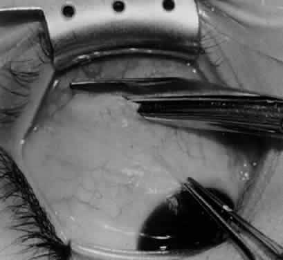
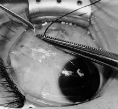
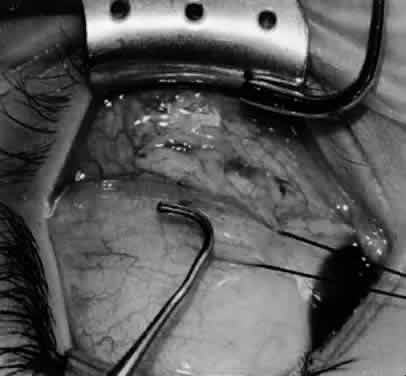
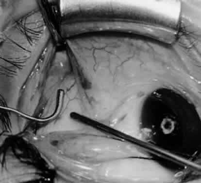
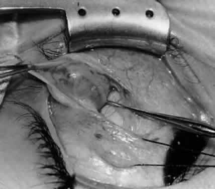
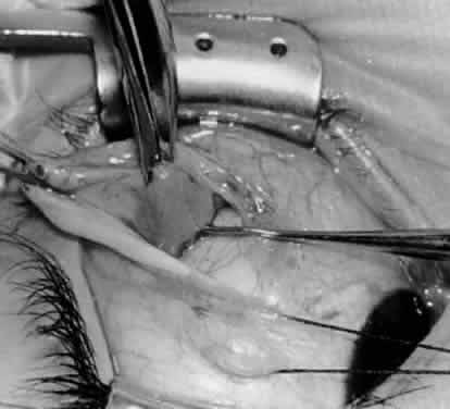
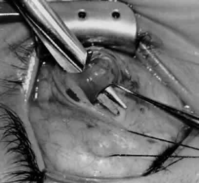
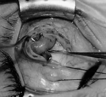
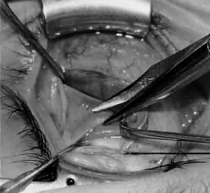
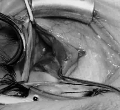
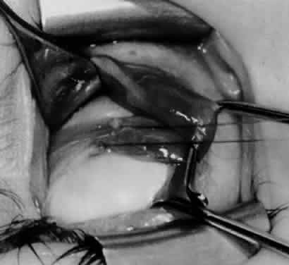
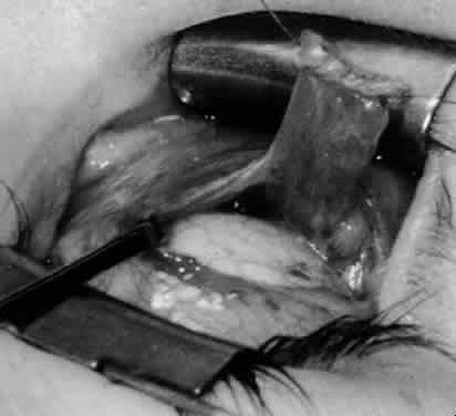
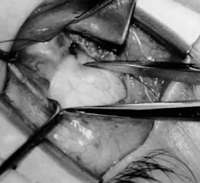
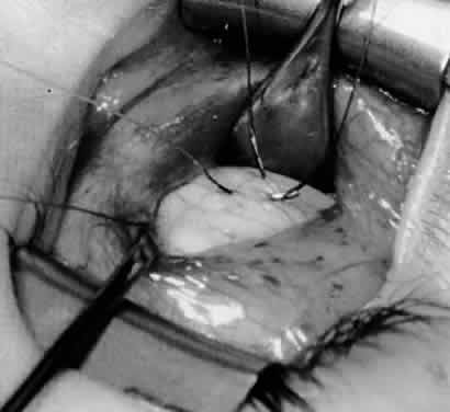
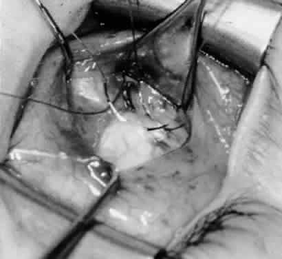
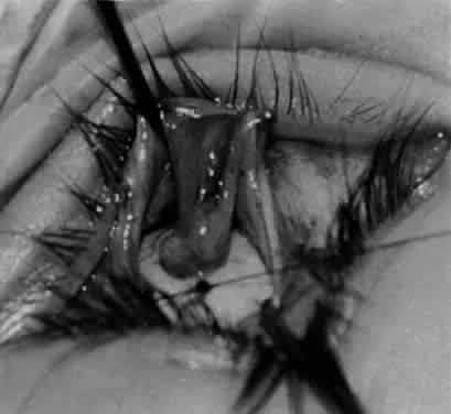
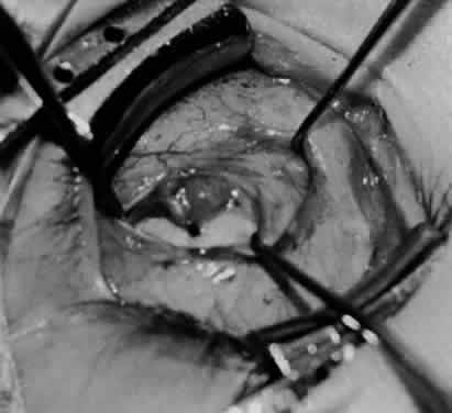
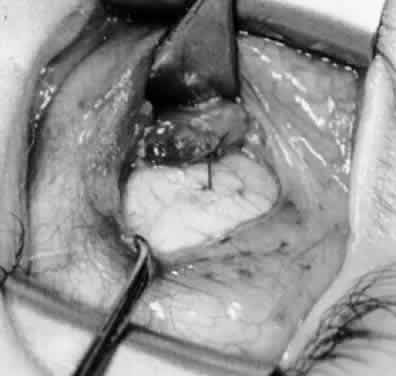
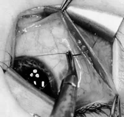
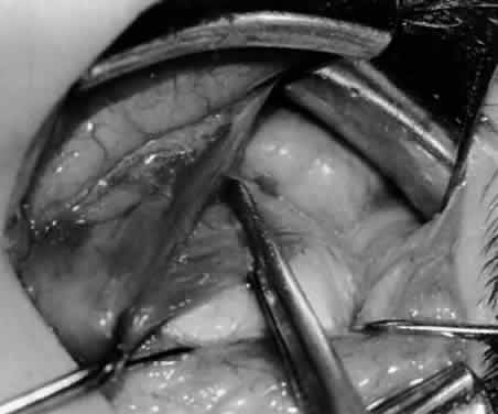
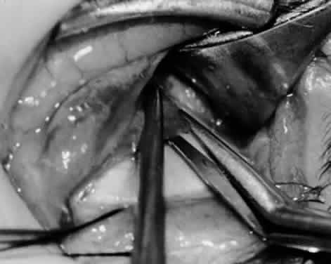
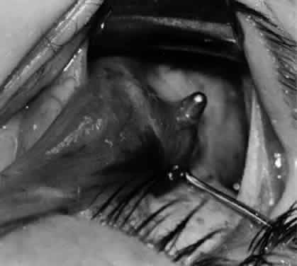
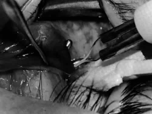
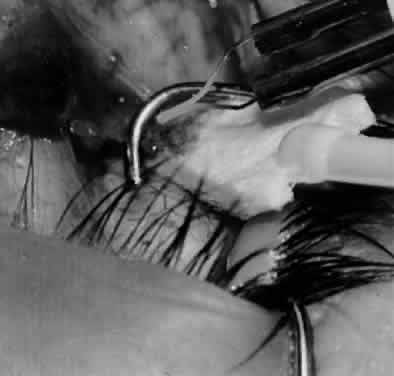
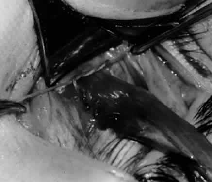
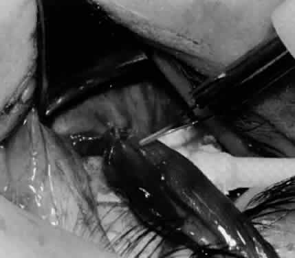
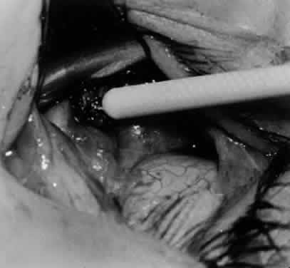
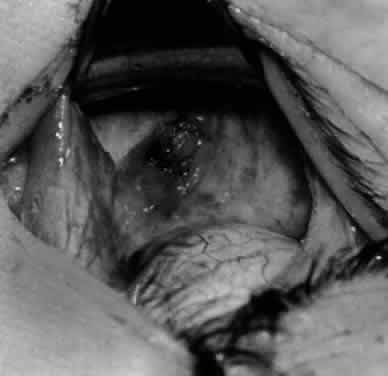
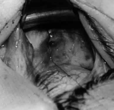
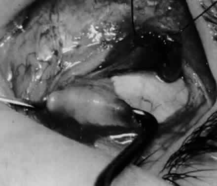
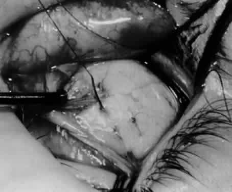
In comparing the various weakening procedures, Parks2,11 concluded that the recession procedure was superior (Figs. 120). The major advantage of the recession is that it allows the weakening procedure to be titrated according to the severity of the overaction. For 1+ overaction, the inferior oblique muscle is recessed 6 mm; for 2+ overaction, 10 mm; and for 3+ overaction, 14 mm, which is the maximum recession. A double-armed 6-0 synthetic suture, such as polyglactin (Vicryl [J-562]*) with a half-circle spatula needle (S-28*), is placed within the insertion of the inferior oblique with a locked bite at the anterior and posterior border (see Fig. 11). With an Aebli scissor, the muscle is sectioned from the globe (see Fig. 12) and recessed 6 mm by placing the anterior suture 4 mm lateral to the lateral insertion of the inferior rectus muscle and the posterior suture 3 mm more laterally. The 10-mm recession (see Figs. 15, 17, 18, 20) requires placement of the anterior suture 2 mm temporal and 3 mm posterior to the temporal insertion of the inferior rectus muscle, with the posterior suture placed 3 mm more posteriorly. The 14-mm recession (see Figs. 14, 16, 19, 20) requires placement of the anterior and posterior sutures parallel to the inferior-temporal vortex vein, which usually is found 8 mm posterior to the temporal insertion of the inferior rectus muscle.

Fig. 1. The standard inferior-temporal fornix incision is made through the conjunctiva and Tenon's capsule with a Westcott scissors. The incision is placed about 8 mm posterior to the limbus, anterior to the fat pad.

Fig. 2. After the lateral rectus muscle is hooked with a Stevens and then a Green muscle hook, the lateral rectus muscle is tented to allow placement of a 4-0 silk suture beneath the insertion of the lateral rectus muscle. The needle tip should glide along the inferior surface of the muscle hook to avoid laceration of the lateral rectus muscle or laceration or penetration of the globe.

Fig. 3. The eye is retracted superonasally with the 4-0 silk traction suture. The suture is attached to the drapes with a hemostat. A Stevens hook is placed along the inferior border of the lateral rectus muscle and drawn temporally. A von Graefe hook is placed within the incision and drawn inferotemporally to expose the inferior oblique muscle.

Fig. 4. A stiff iris repositor can be placed against the sclera to depress the sclera and enhance the view of the inferior oblique muscle. The inferior temporal vortex vein adjacent to the Stevens hook is retracting tissue along the inferior border of the lateral rectus muscle. The second Stevens hook is pointing to the inferior oblique muscle.

Fig. 5. The Stevens hook is placed adjacent to the posterior edge of the inferior oblique muscle and rotated as the inferior oblique muscle is drawn into the operative field. A forceps is used to retract the redundant Tenon's capsule, encapsulated fat, and intermuscular septum as the muscle is drawn further into the operative field with the Stevens muscle hook.

Fig. 6. The Westcott scissor is used to open the intermuscular septum along the posterior border of the inferior oblique muscle. Care is taken to cut adjacent to the tip of the Stevens muscle hook. This incision is important because it helps to avoid damaging the Tenon's capsule and releasing fat, which leads to the adherence syndrome and postoperative hypotropia.

Fig. 7. A Westcott scissor is used gently to open a space beneath the elevated inferior oblique muscle and allow placement of a Green muscle hook.

Fig. 8. The Green muscle hook is placed beneath the belly of the inferior oblique muscle, and the eye is retracted nasally.

Fig. 9. A Westcott scissor is used to dissect the intermuscular septum and expose the insertion of the inferior oblique muscle.

Fig. 10. The inferior oblique muscle is placed on the Green muscle hook. The exposed insertion is ready for myotomy, myectomy, disinsertion, placement of sutures for recession, or removal for the extirpation procedure.

Fig. 11. Sutures are placed into the insertion of the inferior oblique muscle before it is sectioned from the globe for the recession procedure.

Fig. 12. An Aebli scissor is placed beneath the inferior oblique muscle insertion before it is sectioned from the globe.

Fig. 13. The inferior oblique muscle is sectioned from the globe. A preplaced 6-0 Vicryl suture is attached to the insertion.

Fig. 14. The inferior temporal vortex vein, 8 mm posterior to the temporal insertion of the inferior rectus muscle, is held on a Green muscle hook.

Fig. 15. The placement of sutures within the sclera for a recession procedure of 10 mm. The anterior suture is placed 3 mm temporal and 2 mm posterior to the lateral insertion of the inferior rectus muscle, and the posterior suture is placed 3 mm further posteriorly.

Fig. 16. Two sutures straddle the inferior temporal vortex vein insertion for a 14-mm recession procedure.

Fig. 17. Knots are tied to show the placement. The inferior oblique muscle is held on a Stevens muscle hook.

Fig. 18. The final position, with the inferior oblique muscle recessed 10 mm. The inferior rectus muscle is retracted with a Green muscle hook. As described in the text, the 14-mm recession would place the inferior oblique muscle over the inferior temporal vortex vein, and the 6-mm recession would place the inferior oblique muscle anterior and nasal to this position.

Fig. 19. A 14-mm inferior oblique muscle recession is placed over the inferior temporal vortex vein.

Fig. 20. Optional closure of the incision with a 6-0 Vicryl suture. The incision may be allowed to heal without a suture.


Ethicon, Somerville, NJ.

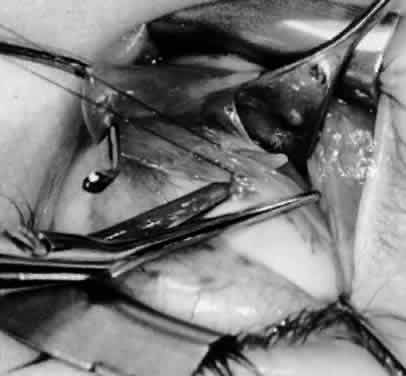
Because the inferior oblique muscle inserts near the macula, there may be concern about penetration of the macula with a preplaced suture, which can cause macula hemorrhage, visual impairment, or visual loss. An alternative is to place a hemostat near the insertion of the inferior oblique muscle and then section the muscle from the globe with the Aebli scissor. The 6-0 Vicryl suture can be placed within the muscle, which is grasped by the hemostat and a locked bite placed at the anterior and posterior borders of the muscle, before the muscle is reattached to the sclera at the intended position. The technique of removing the inferior oblique muscle with the hemostat is part of the denervation and extirpation procedure.

The disadvantage of the recession procedure is the difficulty involved in performing the procedure within a small operative field with limited visibility, compared with the myectomy or disinsertion. With proper illumination, however, such as the surgeon wearing the operating headlight and exposure with the Desmarres or Conway lid retractor, the inferior oblique insertion readily can be seen for placement of sutures and separation of the inferior oblique from the sclera.

Parks2,11 found that 15% of patients had a return of overaction with the recession procedure, compared with 79% with myectomy at the origin, 53% with disinsertion, and 37% with myectomy at the insertion. Short-term follow-up is not helpful with oblique muscle surgery because inferior oblique overaction can return gradually over 2 years.10 Therefore, long-term follow-up is necessary for proper evaluation of success. In a more recent report, Wilson and Parks14 found a 25% recurrence rate of inferior oblique overaction after surgery, with an average follow-up of 3 years. Only 6% of patients required repeat inferior oblique surgery, however.

DENERVATION

Gonzalez15 first described denervation of the inferior oblique muscle as a weakening procedure but also reported the return of overaction within the first postoperative year.16,17 The nerve to the inferior oblique is attached tightly to the posterior belly of the inferior oblique muscle, lateral to the inferior rectus muscle border. At this site is a fusiform enlargement of the belly. After the nerve is hooked posteriorly with a Stevens hook, cautery is used to sever the nerve and the accompanying artery and vein. The procedure of denervation is no longer used alone because of the likelihood of recurrence of the inferior oblique overaction but it is used as the foundation for the denervation and extirpation procedure.

DENERVATION AND EXTIRPATION
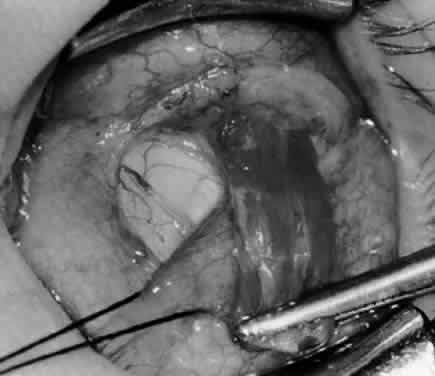
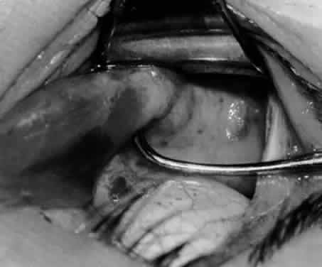
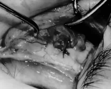
The denervation procedure (Figs. 2132) allows the inferior oblique muscle to be released further into the operative field once the nerve has been sectioned (see Figs. 26 and 27). A 3-0 Vicryl suture ligature is placed around the inferior oblique muscle (see Fig. 28) as close to the Tenon's capsule penetration as possible. The inferior oblique is sectioned by cautery (see Fig. 29), just distal to the suture ligature. The cauterized stump is pushed through the opening in the Tenon's capsule (see Figs. 30 and 31) and a 6-0 or 7-0 Vicryl suture is used to close the opening, with either a running or a purse-string suture (see Fig. 32). The purse-string suture can be preplaced before the inferior oblique muscle is sectioned and closed after the muscle is sectioned. The advantage of the denervation and extirpation procedure is its ability to permanently correct 4+ overaction of the inferior oblique.10,18 In addition, it is the best operative procedure to eliminate recurring inferior oblique overaction after disinsertion, myectomy, or recession.10 Disadvantages include permanent underaction of the inferior oblique muscle in some patients. Pupil dilation can occur but the dilation usually is transient, clearing in 3 to 6 months.10,18

Fig. 21. A hemostat is placed adjacent to the insertion of the right inferior oblique muscle.

Fig. 22. An Aebli scissor is used to section the inferior oblique muscle from the globe.

Fig. 23. The inferior oblique muscle is drawn into the operative field with a hemostat. This step may be used for the recession procedure if the surgeon is concerned about the placement of the 6-0 Vicryl suture while the muscle is attached to the globe in the proximity of the macula. The recession sequence would follow after the placement of the suture while the muscle is grasped in the hemostat.

Fig. 24. The eyelid speculum is removed and a Desmarres lid retractor is placed inferiorly. The inferior oblique muscle is drawn superiorly and nasally. The fusiform expansion of the inferior oblique muscle is evident on the posterior border, where the nerve to the inferior oblique muscle and artery and vein enter the inferior oblique muscle. The Stevens muscle hook is used to hook the neurovascular bundle.

Fig. 25. The hook is advanced further for identification of the neurovascular bundle on the inferior oblique muscle.

Fig. 26. Cautery is applied to sever the neurovascular bundle, and a sponge is used to protect the sclera and lids from cautery.

Fig. 27. As the neurovascular bundle is severed completely, the inferior oblique muscle will be released of the normal tension provided by the neurovascular bundle on the posterior border of the inferior oblique muscle.

Fig. 28. The inferior oblique muscle is drawn further into the operative field, and a 3-0 Vicryl suture ligature is applied about the belly of the muscle.

Fig. 29. Cautery is used to sever the inferior oblique muscle and to provide hemostasis.

Fig. 30. The stump of muscle is placed through the Tenon's capsule opening. The surgeon must apply sufficient cautery to prevent postoperative bleeding.

Fig. 31. The inferior oblique muscle stump is visible at the opening in the Tenon's capsule before closure with 6-0 or 7-0 Vicryl suture. The S-28 half-circle curved needle is preferred to the S-29 needle, which has a 3–8-circle curve.

Fig. 32. The Tenon's capsule opening is closed with 6-0 or 7-0 Vicryl suture to place the inferior oblique muscle entirely outside Tenon's capsule.

ANTERIOR TRANSPOSITION

Elliott and Nankin19 modified the standard recession procedure by transposing the inferior oblique muscle anteriorly toward the insertion of the inferior rectus muscle (Figs. 3335). This procedure reduced the persistent inferior oblique overaction that frequently is noted after recession surgery and reduced marked inferior oblique overaction when performed as the initial surgical procedure. Seventy-three percent of patients, however, had a postoperative deficiency in primary position elevation, compared with only 25% who underwent the usual inferior oblique recession surgery. Bremer and coworkers20 performed a recession and anteriorization of the inferior oblique muscle on 3 patients who had fourth nerve palsy. They suggested that this procedure would benefit patients with dissociated vertical deviations because the inferior oblique is converted to a depressor muscle by the anterior transposition. Ziffer and associates21 also suggested that the anterior transposition converts the inferior oblique muscle from an elevator to a depressor on attempted elevation. Because of its powerful weakening ability, the anterior transposition should be reserved for patients with moderate to severe inferior oblique muscle overaction and should be performed on both eyes to avoid postoperative hypotropia in upgaze. Gonzalez and Cinciripini22 proposed the anterior transposition procedure for treatment of unilateral superior oblique palsy. None of their 3 patients developed primary position hypotropia but all demonstrated some elevation deficiency, with elevation of the lower lid in upgaze.

Fig. 33. A 6-0 Vicryl suture is placed in the inferior oblique muscle in preparation for anterior transposition. A Green muscle hook is placed beneath the inferior rectus muscle.

Fig. 34. The eye is retracted nasally with a Green muscle hook. The anterior suture is placed just anterior and temporal to the insertion of the inferior rectus muscle. The posterior suture is applied about 10 mm temporal to the inferior rectus muscle on a line that would be continuous with the inferior rectus insertion if it were extended temporally.

Fig. 35. The suture is tied, and the inferior oblique muscle is in the position for anterior transposition. The inferior rectus muscle is retracted nasally on the Green muscle hook, and the Desmarres lid retractor is placed inferior temporally.

Stager and coworkers23 described clinical, radiologic, and histologic evidence that the neurovascular bundle supplying the inferior oblique muscle serves as the functional origin of the inferior oblique after anterior transposition of the insertion and converts the distal portion of the inferior oblique muscle from an elevator to a depressor. Stager and Weakley24 described a new surgical approach for management of recurrent inferior oblique overaction after the anterior transposition procedure. A myectomy of about 1 cm or more of a portion of the inferior oblique, nasal to the inferior rectus, is performed using a temporal incision and approach. This preserves the distal inferior oblique fibers attached to the neurovascular bundle, which provide the depressor function of the inferior oblique in its anterior transposed position.

Mims25 performed an anterior transposition on 61 children who had bilateral overaction of the inferior oblique muscle with concurrent or previous infantile esotropia. A substantial reduction in the dissociated vertical deviation occurred in all cases when it was present, and only one child required subsequent surgery for dissociated vertical deviation. In a prospective study, Elliott and Parks26 compared the effectiveness of denervation and extirpation with that of anterior transposition in the management of patients who had maximum inferior oblique overaction. They concluded that the anterior transposition procedure was effective for eliminating bilateral superior oblique overaction but that it should be performed identically on both eyes because of its tendency to restrict elevation. The anterior transposition procedure was recommended for the treatment of patients with overactive inferior oblique muscles in association with dissociated vertical deviation. In a series of 21 patients receiving an anterior transposition of the inferior oblique with overaction of the inferior oblique and dissociated vertical deviation, Seawright and Gole27 reduced the preoperative incidence of inferior oblique overaction from 84% to 16% postoperatively. Inferior oblique overaction was absent in 43% and improved in 86% of eyes. At the latest postoperative visit, 57% did not demonstrate any evidence of dissociated vertical deviation and 68% of eyes had no dissociated vertical deviation or improved dissociated vertical deviation. There was no evidence of primary position hypotropia in any patient who had unilateral anterior transposition, and there was no evidence of inferior oblique underaction. Three patients required repeat inferior oblique surgery.

Burke and colleagues28 also found the anterior transposition of the inferior oblique to be an effective treatment for dissociated vertical deviation with inferior oblique overaction but the long-term results may be less stable if the preoperative dissociated vertical deviation is in excess of 15 prism diopters.

Back to Top
PREFERRED TECHNIQUES
Although myectomy and disinsertion procedures have their advocates, I prefer recession of the inferior oblique muscle for 1+ , 2+ , or 3+ overaction and denervation and extirpation for 4+ overaction of the inferior oblique muscles. Because the anterior transposition limits elevation, the use of this procedure should be limited. It is recommended for patients who have bilateral marked inferior oblique overaction with dissociated vertical deviation.

Whether the inferior oblique overaction is primary, secondary, or associated with V pattern, these guidelines should be observed consistently. A 1+ overaction of an inferior oblique muscle is not a justification for surgical intervention unless a more severe overaction is present in the fellow eye. A 2+ overaction merits a 10-mm recession of the inferior oblique, a 3+ overaction merits a 14-mm recession, and a 4+ overaction merits a denervation and extirpation procedure. A 6-mm recession would be performed for a 1+ overaction. In the presence of a 1+ overaction, this type of bilateral surgery would be indicated for treatment of a V pattern or in anticipation that the 1+ overactive eye may become more overactive in time.

INFERIOR OBLIQUE SURGERY

Indications

Whether inferior oblique overaction is primary, secondary, or associated with the V pattern, surgery is indicated to treat hypertropia, diplopia, binocular vision compromise in the field of adduction, torticollis, and to improve cosmetic appearance. These indications are not repeated for the subsequent sections on primary inferior oblique overaction, secondary inferior oblique overaction, V pattern, and dissociated vertical deviations.

Contraindications

In asymmetric inferior oblique overaction, an inferior oblique without overaction should not be weakened surgically.2,9,29 It is not possible to predict which inferior oblique muscle will become overactive at a later date. Weakening a normal inferior oblique muscle leads to subsequent vertical deviation in the primary position, and further corrective surgery may be needed.

In a V pattern, the normal inferior oblique muscle should not be weakened, and surgery should be confined to vertical displacement of the horizontal rectus muscles.2,9,29

It is not prudent to perform an anterior transposition procedure unless the patient has both markedly overactive inferior oblique muscles and dissociated vertical deviation. In the absence of dissociated vertical deviation, the anterior transposition procedure can lead to limitation in upgaze movements, which is not a desired effect if the patient had only overaction of the inferior oblique muscle preoperatively.

PRIMARY INFERIOR OBLIQUE OVERACTION

There are two clinical types of inferior oblique overaction: primary, without ipsilateral superior oblique palsy or contralateral superior rectus palsy, and secondary, with palsy of a cyclovertical muscle.2,11,29 In primary overaction of the inferior oblique, there is overelevation of the adducting eye, which increases with increasing adduction. Usually, there is no vertical deviation in the primary position and no cyclodeviation. Therefore, there is no torticollis. The Bielschowsky head-tilt test result is negative.30,31 Primary inferior oblique overactions are not congenital and rarely are noted in patients younger than 1 year.14,31 The patient may or may not have an associated horizontal deviation of the eyes. In a series of 50 patients who had congenital esotropia and underwent surgical alignment before the age of 1 year, Parks and Mitchell2,29 found 65% with inferior oblique overaction. Hiles and associates32 reported a 78% incidence of overaction of the inferior oblique muscle in one or both eyes in a series of 54 patients who had congenital esotropia. Wilson and Parks14 found primary overaction of the inferior oblique muscle in 72% of patients with congenital esotropia at an average age of 3.6 years, 34% of patients with accommodative esotropia at an average age of 5.2 years, and 32% of patients with intermittent exotropia—also at an average age of 5.2 years. When detected, inferior oblique overaction was asymmetric in 44% of patients and unilateral in 23%.14 If one inferior oblique muscle is overactive, the second may or may not overact.29 When the second inferior oblique muscle overacts, the onset usually is evident 2 to 6 months after the onset of overaction in the first muscle, regardless of whether inferior oblique surgery has been performed.29 Overaction may occur many years later, however.14

Indications

Surgical weakening is the treatment of choice for the overactive inferior oblique muscle. Excyclodeviation is not associated with primary overaction, and weakening the inferior oblique produces neither significant incyclodeviation nor a vertical deviation in the primary position. No significant change in the horizontal alignment of the eyes in primary position is produced by weakening the primary overacting inferior oblique muscles.33 The previous terminology for overaction—mild, moderate, marked, and supermarked—should be classified as 1+ , 2+ , 3+ , and 4+ overaction, respectively.14

Eustis and Nussdorf34 photographed the posterior pole at the time of surgery in 27 eyes of 14 patients with infantile esotropia. A masked observer graded each photo for the presence or absence of fundus torsion. Of the 27 eyes, 15 developed inferior oblique overaction, and 6 of the 15 demonstrated fundus excyclotorsion before the inferior oblique overaction was clinically recognized. Therefore, the presence of fundus torsion with infantile esotropia may serve as a marker or a predictor of subsequent overt inferior oblique overaction.

When unilateral inferior oblique overaction occurs, only the overactive muscle is weakened. If asymmetric inferior oblique overaction is present, asymmetric surgery is performed. Weakening procedures on the overactive inferior oblique muscles are performed simultaneously with surgery on the horizontal muscles if the misalignment is a combined horizontal tropia with the overactive inferior obliques.

Weakley and coworkers35 studied 368 patients with infantile esotropia, acquired accommodative or partially accommodative esotropia, or acquired nonaccommodative esotropia. Of the 126 patients (34%) with amblyopia, 93 patients (74%) had symmetric inferior oblique overaction. Thirty-one patients (25%) had more inferior oblique overaction in the eye with the amblyopia, however. The authors stress that asymmetric inferior oblique overaction in esotropia correlates highly with amblyopia in the eye with greater inferior oblique overaction, regardless of the subtype of esotropia, the duration of the esotropia, or the angle of deviation.

Contraindications

An inferior oblique that is not overactive should not be weakened because it is not possible to predict if and when an inferior oblique muscle will become overactive. Bilateral weakening in the presence of unilateral overaction produces a vertical deviation in the primary position. This deviation can persist and become symptomatic,31 necessitating further surgery to alleviate the symptoms.

SECONDARY INFERIOR OBLIQUE OVERACTION

A secondary inferior oblique overaction is an early finding in patients with contralateral superior rectus palsy or a late finding in patients with ipsilateral superior oblique palsy. Overelevation of the adducting eye is seen, as in primary overaction of the inferior oblique. In addition, there is a significant vertical and cyclovertical deviation in the primary position, regardless of whether there is hypertrophy or contracture of the secondarily overactive inferior oblique.2,29 The V pattern is associated with the secondary overaction of the inferior oblique muscle. Secondary overaction usually occurs 6 months or more after the onset of superior rectus or superior oblique muscle palsy. Hypertrophy or contracture can occur within several weeks of the onset of the palsy, however.2,29 Torticollis tends to develop to maintain single binocular vision, and the Bielschowsky head-tilt test result is positive.

Treatment

The ideal treatment is weakening of the inferior oblique muscle, preferably by a recession procedure. Usually, the recession procedure precedes a tuck of the ipsilateral palsied superior oblique tendon or resection of the palsied superior rectus, or a weakening of the yoke muscle.29 Improvement will occur in the overelevation of the adducting eye in the vertical position and in the cyclodeviation in the primary position, and the torticollis should be reduced or eliminated. The result of the Bielschowsky head-tilting test should be improved but the positive finding will not be eliminated.

The trochlear nerve palsy may be bilateral or unilateral. If it is bilateral, the involvement may be asymmetric, with the minimally involved side being masked by the maximally involved side. This effect may become apparent only after the maximally involved eye is corrected surgically. Bilateral superior oblique palsy shows 10° or more of excyclotorsion on double Maddox rod testing, with left hypertropia in right gaze, right hypertropia in left gaze, V pattern, chin depression, and a preferred upgaze posture. Bilateral superior oblique palsy occasionally may be present when less than 10° of excyclotorsion is found on the double Maddox rod test. The treatment of choice is not inferior oblique weakening but a Harada-Ito procedure or a modification. This procedure requires sagittalization and advancement of the anterior half of the superior oblique tendons toward or adjacent to the superior border of the lateral rectus muscles.31,36

V PATTERN

Overaction of the inferior oblique muscle frequently is associated with the V pattern, regardless of whether the primary position measurements include orthophoria, esotropia, or exotropia.2 The deviation is measured at distance in the primary position, 30° chin elevation, and chin depression. A difference of 10 prism diopters in the horizontal alignment between the upgaze and downgaze position is considered diagnostic of A or V pattern. Clinical evidence supports the association of abnormality in A and V patterns with abnormalities of the cyclovertical muscles.

Treatment

Procedures to weaken the inferior oblique muscles improve the V pattern, but must be used only when the oblique muscles are overactive. When a V pattern is present without overaction of the inferior obliques, vertical displacement of the horizontal muscles is indicated,2,9,29,37,38 rather than weakening of normal inferior oblique muscles. Weakening of normal muscles leads to underaction and an undesirable surgical result.9

DISSOCIATED VERTICAL DEVIATIONS

The usual treatment for surgical correction of dissociated vertical deviations includes recession of the superior rectus muscle, resection of the inferior rectus muscle, placement of superior rectus posterior fixation sutures, supermaximum recession, or a hang-back technique with a superior rectus weakening.

The child who has overaction of the inferior oblique muscle in addition to dissociated vertical deviation can benefit from an anterior transposition of the inferior oblique muscle. The tethering effect of the anterior transposition is the result of the physiologic conversion of the inferior oblique to a depressor muscle.21–26

INFERIOR OBLIQUE SURGERY

Complications

Inferior oblique surgery has several complications, all of which can be avoided by the application of proper and judicious surgical technique under direct vision. A common approach in the past was the blind sweeping of the eye-muscle hook in an attempt to grasp the inferior oblique without proper exposure of the operative field. The blind sweeping technique can cause the inferior rectus or lateral rectus muscle to be hooked inadvertently, and torn or severed by vigorous “fishing” for the inferior oblique muscle. The inferior temporal vortex vein can be hooked accidentally and severed, causing extensive orbital hemorrhage. Accidental severing of the vortex vein is not vision-threatening, and cautery should be applied to stop the bleeding and reduce the possibility of postoperative hematoma. The inferior oblique can be torn partially from repeated attempts to hook the muscle without direct vision.2 Therefore, if posterior fibers of the inferior oblique muscle remain intact, the recession of only the anterior fibers will not reduce the overaction. Postoperatively, full overaction will be present, and reoperation will be necessary. The entire inferior oblique insertion must be seen before surgery is performed.

The adherence syndrome2 described an iatrogenic syndrome caused by rupturing Tenon's capsule and allowing orbital fat to protrude from the normal position into the operative field. The result was fibrofatty proliferation and adherence to the sclera and the inferior rectus muscle.

Treatment requires extensive, careful dissection of fibrofatty scarring from the sclera and inferior rectus muscle capsule. The goal of this treatment is to halt the relentless progression of the hypotropia in the affected eye. In the best of hands, the surgical results are limited. Residual hypotropia consistently is present, despite the most aggressive approach in removing scar tissue and attempting to elevate the affected eye. Parks2 reported that the average preoperative hypotropia in 23 patients was 19 diopters, ranging from 4 to 40 diopters. The average postoperative hypotropia was 8 diopters, even though an average of 2.6 vertical muscles per patient required surgery to reduce the hypotropia.

These conditions can be avoided with the use of proper illumination, such as that provided by a fiberoptic headlight, and with adequate surgical direct vision assistance that allows direct vision of the vortex vein and posterior border of the inferior oblique muscle.

Grasping only a portion of the inferior oblique muscle leads to residual overaction if the most posterior fibers are left intact. Proper placement of hooks reduces the likelihood that the inferior rectus or lateral rectus would be grasped and severed in a myotomy or myectomy procedure if the inferior oblique is identified correctly before the muscle is sectioned.

In the recession procedure, the inferior oblique insertion is exposed and is located adjacent to the macula. When a suture is placed within the insertion of the inferior oblique muscle, the macula can be damaged if the suture is placed improperly or too deeply. Unless the assistance is excellent, the best approach may be to grasp the insertion of the inferior oblique muscle with a hemostat, section the muscle from the globe with an Aebli scissor, and place the 6-0 Vicryl suture within the insertion ends before performing the recession procedure.

Regardless of which procedure is selected, adequate hemostasis is essential to avoid postoperative bleeding, which can lead to a hematoma in the orbit or within the eyelid tissue.

Internal ophthalmoplegia has been reported. Usually, it is transient10,18,39; however, it may last many years.40

Technique

All inferior oblique surgery requires the same initial steps to the point of isolation of the inferior oblique muscle (see Figs. 110). At that time, any one of several procedures can be performed. These procedures are described individually (see Figs. 1135). The figures are presented from the view of the surgeon seated at the head of the patient.

Back to Top
ACKNOWLEDGMENT
The author thanks Marshall M. Parks, MD, for many of the surgical photographs.
Back to Top
REFERENCES

1. Dyer JA: Tenotomy of the inferior oblique muscle at its scleral insertion: an easy and effective procedure. Arch Ophthalmol 68:56, 1962

2. Parks MM: The overacting inferior oblique muscle: the XXXVI DeSchweinitz lecture. Am J Ophthalmol 77:787, 1974

3. Weakley DR, Stager DR: Inferior oblique weakening procedures. Ophthalmol Clin North Am 5:57. 1992

4. Wheeler JM: Advancement of the superior oblique and inferior oblique ocular muscles. Trans Am Ophthalmol Soc 32: 237, 1934

5. White JW, Brown HW: Occurrence of vertical anomalies associated with convergent and divergent anomalies. Arch Ophthalmol 21:999, 1939

6. Wagman OH: Resection of the inferior oblique muscle in hypotropia. Am J Ophthalmol 28:1226, 1945

7. Pranger AH: Some observations on the surgical treatment of the extraocular muscles. Trans Am Ophthalmol Soc 4: 251, 1946

8. McLean JM: Direct surgery of underacting oblique muscles. Trans Am Ophthalmol Soc 46:622, 1948

9. Parks MM, Mitchell PR: A and V patterns. In Tasman W. Jaeger EA (eds): Clinical Ophthalmology, vol 1. Philadelphia, JB Lippincott, 1991

10. Parks MM: Inferior oblique weakening procedures. In Nelson LB, Wagner RS (eds): International Ophthalmology Clinics: Strabismus Surgery, vol 1, pp 107–117. Boston, Little, Brown & Co, 1985

11. Parks MM: The weakening surgical procedures for eliminating the overaction of the inferior oblique muscle. Am J Ophthalmol 73:107, 1972

12. Davis G, McNeer KW, Spencer RF: Myectomy of the inferior oblique muscles. Arch Ophthalmol 104:855, 1986

13. Jones TW, Lee DA, Dyer JA: Inferior oblique surgery: experience at the Mayo Clinic from 1962 to 1981. Arch Ophthalmol 102:714, 1984

14. Wilson ME, Parks MM: Primary inferior oblique overaction in congenital esotropia, accommodative esotropia, and intermittent exotropia. Ophthalmology 96:950, 1989

15. Gonzalez C: Denervation of the inferior oblique (as a weakening surgical procedure). Trans Am Acad Ophthalmol Otolaryngol 78:816, 1974

16. Gonzalez C: Denervation of the inferior oblique: current status and long term results. Trans Am Acad Ophthalmol Otolaryngol 81:899, 1976

17. Gonzalez C: Reinnervation of the nerve to the inferior oblique after iatrogenic denervation. J Pediatr Ophthalmol Strabismus 18:21, 1981

18. DelMonte MA, Parks MM: Denervation and extirpation of the inferior oblique: an improved weakening procedure for marked overaction. Ophthalmology 90:1178, 1983

19. Elliott RL, Nankin SJ: Anterior transposition of the inferior oblique. J Pediatr Ophthalmol Strabismus 18:35, 1981

20. Bremer DL, Rogers GL, Quick LD: Primary position hypotropia after anterior transposition of the inferior oblique. Arch Ophthalmol 104:229, 1986

21. Ziffer AJ, Isenberg SJ, Elliott RL, Apt L: The effect of anterior transposition of the inferior oblique muscle. Am J Ophthalmol 116:224, 1993

22. Gonzalez C, Cinciripini G: Anterior transposition of the inferior oblique in the treatment of unilateral superior oblique palsy. J Pediatr Ophthalmol Strabismus 32:107, 1995

23. Stager DR, Weakley DR Jr, Stager D: Anterior transposition of the inferior oblique—anatomic assessment of the neurovascular bundle. Arch Ophthalmol 110:360, 1992

24. Stager DR, Weakley DR Jr: A new temporal surgical approach to the nasal portion of the inferior oblique muscle. Binocular Vision & Eye Muscle Surgery Qtrly 7:211, 1992

25. Mims JL: Bilateral anterior transposition of the inferior obliques. Arch Ophthalmol 107:41, 1989

26. Elliott RL, Parks MM: Inferior oblique weakening by denervation-extirpation and anterior transposition. Presented at the annual meeting of the American Association for Pediatric Ophthalmology and Strabismus, Bolton Landing, New York, August 2, 1990

27. Seawright AA, Gole GA: Results of anterior transposition of the inferior oblique. Aust NZJ Ophthalmol 24:339, 1996

28. Burke JP, Scott WE, Kutschke PJ: Anterior transposition of the inferior oblique muscle for dissociated vertical deviation. Ophthalmology 100:245, 1993

29. Parks MM, Mitchell PR: Oblique muscle dysfunctions. In Tasman W, Jaeger EA (eds): Clinical Ophthalmology, vol 1, pp 1–6. Philadelphia, JB Lippincott, 1991

30. Parks MM: Isolated cyclovertical muscle palsy. Arch Ophthalmol 60:1027, 1958

31. Parks MM, Mitchell PR: Cranial nerve palsies. In Tasman W, Jaeger EA (eds): Clinical Ophthalmology, vol 1, pp 1–17. Philadelphia, JB Lippincott, 1991

32. Hiles DA, Watson BA, Biglan AW: Characteristics of infantile esotropia following early bimedial rectus recession. Arch Ophthalmol 98:697, 1980

33. Stager DR, Parks MM: Inferior oblique weakening procedures: effect on primary position horizontal alignment. Arch Ophthalmol 90:15, 1973

34. Eustis HS, Nussdorf JD: Inferior oblique overaction in infantile esotropia: fundus extorsion as a predictive sign. J Pediatr Ophthalmol Strabismus 33:85, 1996

35. Weakley DR, Urso RG, Dias CL: Asymmetric inferior oblique overaction and its association with amblyopia in esotropia. Ophthalmology 99:590, 1992

36. Mitchell PR, Parks MM: Surgery for bilateral superior oblique palsy. Ophthalmology 89:4484, 1982

37. Knapp P: Vertically incomitant horizontal strabismus. The so-called “A” and “V” syndromes. Trans Am Ophthalmol Soc 57:666, 1959

38. Knapp P: A and V patterns symposium on strabismus. Transactions of the New Orleans Academy of Ophthalmology, pp 242–254. St Louis: CV Mosby, 1971

39. Damanakis AG, Theodossiadis GP: Internal ophthalmoplegia caused by inferior oblique myectomy. J Pediatr Ophthalmol Strabismus 22:117, 1985

40. Bajart AM, Robb RM: Internal ophthalmoplegia following inferior oblique myectomy: a report of three cases. Ophthalmology 86:1401, 1979

Back to Top