Chapter 84
Surgery for Esotropia
COLLEEN J. CHRISTIAN and JOSEPH H. CALHOUN
Main Menu   Table Of Contents

Search

PREOPERATIVE CONSIDERATIONS
OPERATIVE CONSIDERATIONS
POSTOPERATIVE CONSIDERATIONS
SURGERY FOR INFANTILE ESOTROPIA
SURGERY FOR ACCOMMODATIVE ESOTROPIA
SURGERY FOR ACQUIRED NONACCOMMODATIVE ESOTROPIA
SURGERY FOR SENSORY ESOTROPIA
SURGERY FOR SIXTH-NERVE PALSY
SURGERY FOR A AND V PATTERNS
REFERENCES

Esodeviations are the most common forms of strabismus. It is useful to classify the many different forms of esotropia according to certain characteristics, including age of onset, comitance or pattern, response to treatment of a hyperopic refractive error, and relationship to prior surgery.

Types of esotropia include infantile, accommodative, partially accommodative, and acquired nonaccommodative. Sensory esotropia occurs in patients with poor vision in one or both eyes. Other types include esotropia include those related to a sixth cranial nerve palsy, and A or V pattern esotropia.

Strabismus is comitant when the measured deviation is similar in all positions of gaze; it is noncomitant when the measured deviation varies in different gaze positions. Common forms of comitant esotropia include infantile esotropia, accommodative and partially accommodative esotropia, and sensory esotropia. In patients with nystagmus blockage syndrome a variably comitant esodeviation is seen as convergence is intermittently used to dampen the nystagmus. A small comitant esodeviation can also occur in patients with monofixation syndrome.1,2 With noncomitant esotropia, the measured deviation varies in right and left, or up and down-gaze. For example, patients with a sixth cranial nerve palsy have an esodeviation greater when looking in the direction of the affected lateral rectus muscle. In A and V patterns, the deviation varies from up to down-gaze. Other causes of noncomitant esotropia include restriction of the globe due to prior orbital or extraocular muscle surgery, an intraorbital mass, or orbital bony abnormalities.

Accommodative esotropia is caused by uncorrected hyperopia or a high accommodative convergence to accommodation (AC/A) ratio. It can frequently be corrected with hyperopic glasses; bifocals may be added to correct an esodeviation that is greater at near.

Esotropia can occur postoperatively. Consecutive esotropia occurs after surgery to correct exotropia, while an esodeviation remaining after surgery to correct esotropia is termed residual esotropia.

Back to Top
PREOPERATIVE CONSIDERATIONS
Before surgical correction can be recommended, an attempt should be made to correct the deviation with nonsurgical methods. Visual acuity should be equalized by treating amblyopia; any accommodative component should be treated with glasses when the hyperopic refractive error is greater than 2.50 diopters. Good and equal visual acuity will help to stabilize the ocular alignment once surgery has been performed.

SIZE OF THE DEVIATION

Surgical correction is recommended when esotropia is greater than 15 prism diopters. Esodeviations of less than 15 prism diopters are not considered large enough to impede the development of fusion and not unsightly enough to require surgical correction.

TIMING OF THE OPERATION

Timing of surgical correction for esotropia varies according to cause. Surgical correction of infantile esotropia generally is not recommended until the size of deviation is stable on repeated examinations. Ing3 showed that children with this problem have a better chance of developing fusion and binocularity when they undergo surgery before the age of 2. Surgery for sensory esotropia is delayed until cosmetic alignment of the eye becomes a concern for the patient or parents. Patients with esotropia related to a sixth-nerve palsy are usually observed for 6 months before surgery is recommended because recovery of lateral rectus function may occur during this period.

Back to Top
OPERATIVE CONSIDERATIONS
In general, the goal of surgical treatment for esotropia is to correct alignment to orthotropia. Tables developed by experienced ophthalmologists provide guidelines for surgery, but each surgeon should perform a periodic review of the correction achieved for specific amounts of corrective muscle surgery. Differing surgical technique and variation in measurement from the original muscle insertion will produce different but consistent variations in effect. The number of millimeters of surgery performed per prism diopter of deviation should be adjusted accordingly so that surgery corrects the deviation to within 10 prism diopters of orthotropia. Both overcorrection and undercorrection of the angle of strabismus will occur; if the surgeon performs the correct amount of surgery, the number of overcorrections should roughly equal the number of undercorrections.

Over the past 25 years, there has been a rethinking of the amount that the medial rectus muscle may be recessed. Formerly, this amount was limited to a maximal recession of the muscle to 5 mm posterior to the original insertion.4,5 However, medial rectus recessions of up to 7.5 mm have been shown to predictably correct large angle esotropia without compromising adduction of the eye and are commonly used now for this.6 Tables 1, 2, and 3 provide a guide to the quantity of surgery we perform.


TABLE 1. Two Symmetric Muscle Surgery for Esotropia


Prism diopters esotropia Medial rectus recession in both eyes (mm) Lateral rectus resection in both eyes (mm)
153.03.5
203.54.5
254.05.5
3056
355.56.5
4067
506.5Not recommended
607.0*Not recommended
707.5*Not recommended

*Adduction may be compromised.


TABLE 2. Monocular Recession–Resection Surgery for Esotropia


Prism diopters esotropia Medial rectus recession (mm) Lateral rectus resection (mm)
1533.5
203.54
2545.5
304.56
3557
405.57.5
5068


TABLE 3. Unilateral Medial Rectus Recession for Esotropia


Prism diopters esotropia Medial rectus recession (mm)
125
185.5
226
256.5


TYPE OF PROCEDURE

Surgical correction of comitant esotropia usually consists of symmetric recession of the medial rectus muscles. With large-angle esodeviation, a large symmetric recession can be done or simultaneous resection of one or both lateral rectus muscles can be combined with medial rectus recessions.

A recession of the medial rectus muscle combined with a resection of the lateral rectus muscle in the same eye is termed a recess–resect procedure. This is preferred for patients with poor vision in one eye, those who have had a similar prior procedure on the other eye, or those who have an acquired noncomitant form of esotropia, such as sixth-nerve palsy. In A or V pattern esotropia, surgery is designed to eliminate the pattern in addition to the esotropia. When overaction of the oblique muscles is observed, surgery to weaken these muscles is included with horizontal muscle recession or resection. When overaction is not present, A or V pattern esotropia is corrected by supraplacing or infraplacing the new horizontal muscle insertion.

ANESTHETIC CONSIDERATIONS

General, local, or topical anesthesia may be used for surgical correction of esotropia. General anesthesia is customarily used for children or adults requiring surgery on both eyes. Local or topical anesthesia supplemented by intravenous sedation is used frequently for adults who require surgery on one eye.

Back to Top
POSTOPERATIVE CONSIDERATIONS
Some surgeons use topical antibiotics or combination antibiotic–steroid combinations postoperatively. However, instillation may be difficult, and effective absorption through inflamed conjunctiva has been questioned. If risk factors such as blepharitis or immune suppression are present, the use of antibiotics administered in drop form or by mouth may be considered. Each patient should receive individual consideration. The decision as to whether to use postoperative medication is best left up to the individual surgeon.

POSTOPERATIVE VISITS

Traditionally, the effect of surgical correction of esotropia is evaluated within 1 to 3 days of surgery and again at 4 to 8 weeks. The rare complication of endophthalmitis begins to be apparent to the patient and to the ophthalmologist 1 to 3 days after surgery, and it is prudent to either see the patient or speak with them during that interval. If the phone conversation suggests any undue inflammation the patient should be examined and treated based on that examination.

If there are no concerns in the immediate postoperative period, the next examination should take place after an interval that allows all swelling and inflammation from surgery to subside, usually 4 to 6 weeks after surgery. Once stability of ocular alignment has been achieved, annual eye examinations to monitor visual acuity, alignment and binocular function are recommended.

SURGICAL COMPLICATIONS

The incidence and treatment of surgical complications after rectus muscle surgery are discussed in another chapter. Complications include scleral perforation, anterior segment ischemia, endophthalmitis, suture granuloma, conjunctival inclusion cyst, conjunctival scarring, slipped muscle, and the lost muscle. With a slipped muscle, the muscle slips back away from its newly attached site but does not retract into the posterior orbit. A lost muscle occurs when the muscle slips out of the suture or clamp during surgery and retracts into the posterior orbital tissues.

Back to Top
SURGERY FOR INFANTILE ESOTROPIA

INDICATIONS

Surgery is indicated for the treatment of infantile esotropia when the size of the deviation is stable, amblyopia has been treated, and an accommodative component of the esodeviation has been eliminated.

Every effort to correct amblyopia should be made prior to surgery; however, Lam and associates7 showed that amblyopia therapy could be successfully continued after surgery is done and that realignment itself can help reverse amblyopia in some cases. Parents should be advised that occlusion might be necessary postoperatively. Occlusion therapy for amblyopia does not seem to result in measureable angle variation in patients with infantile esotropia.8 An accommodative component is said to be present in 15% of patients with infantile esotropia,9 and may be treated with glasses when the hyperopic refractive error is greater than 2.25 diopters. When the refractive error is less than 2.25 diopters, echothiophate iodide (phospholine iodide 0.125%, Wyeth, Philadelphia, PA) instilled in the eye each morning may reduce the size of the deviation by reducing the amount of accommodative convergence. However, its use for this is decreasing in popularity by pediatric ophthalmologists.

The measured deviation should be stable over two office visits. The Pediatric Eye Disease Investigator Group10–12 showed that esotropia with an angle of less than 40 prism diopters and with onset in early infancy frequently resolves; esotropia with a deviation greater than or equal to 40 prism diopters presenting after 10 weeks of age has a low likelihood of resolution. Although surgery technically can be performed safely in patients who are as young as 3 to 4 months of age, many surgeons prefer to wait until the patient is 6 months of age to permit stability of the ocular deviation.

Ing3 reported that patients with infantile esotropia who underwent surgical alignment before the age of 24 months showed improved binocular function compared with those who achieved alignment after the age of 2. Birch and associates13 showed that although surgical correction of infantile esotropia during the first year of life is not associated with a higher prevalence of steroacuity, it is associated with better stereoacuity in children who achieve stereopsis after surgery. Early surgical alignment is thought to be associated with better stereopsis because the duration of misalignment is shortened.14,15

CONTRAINDICATIONS

Surgery should not be performed to correct infantile esotropia when the patient has a life-limiting condition or a medical condition that precludes the safe administration of anesthesia. Surgery should be delayed when the accommodative component has not been treated or when measurements are unstable.

PROCEDURE

Surgical techniques used to correct infantile esotropia include bilateral medial rectus recession,16 unilateral recession of the medial rectus combined with resection of the lateral rectus,17 and bilateral medial rectus recession combined with resection of one or both lateral rectus muscles3,18,19

Recession of both medial rectus muscles is the most commonly performed operation to correct infantile esotropia. However, when one eye is preferred, some surgeons treat esotropia with a recession of the medial rectus muscle combined with a resection of the lateral rectus muscle on the nonpreferred eye.17 Because of concern that large recessions of the medial rectus muscle (greater than 7 mm) will limit adduction and that smaller recessions will produce undercorrection in large angle infantile esotropia, some surgeons combine bilateral medial rectus recessions with resection of one or both lateral rectus muscles when the angle of esotropia is 70 prism diopters or more.18,19 Other surgeons feel that in children younger than 1 year of age, esotropia up to 70 prism diopters can safely be corrected with surgery on both medial rectus muscles.20 Table 1 shows a guide to the quantity of surgery we perform.

The operation should be designed so that it corrects the entire deviation and places the eyes in parallel alignment at distance and near. Measurements are made with the alternate prism and cover test when possible; in infants, the Krimsky test is used.

The amount of correction achieved by recessing the medial rectus muscle can be augmented by recessing the conjunctiva and anterior Tenon's tissue with the use of techniques advocated by Knapp21 and Helveston and colleagues.22 This en bloc surgical technique helps to reduce the restrictive component caused by contracted conjunctiva and Tenon's capsule when a deviation has been present for a long time

The medial rectus muscle can be approached through either a cul-de-sac (fornix) incision23 or an incision directly along the limbus.24 Long-term cosmetic differences between these two surgical approaches are insignificant, but patients seem to be more comfortable in the earlier postoperative period with the fornix-based incision, and the amount of swelling and scarring visible when the eyelids are open is less. The choice of conjunctival incision is best left to the individual surgeon.

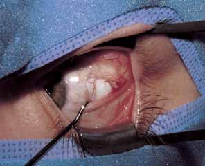
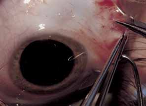
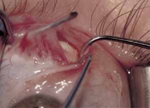
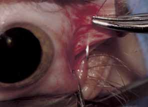
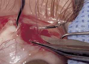
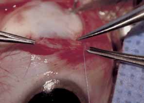
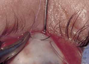
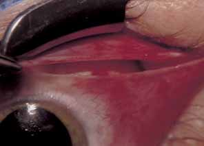
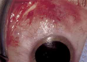
The muscle is then exposed; Tenon's capsule and the intramuscular septum are then dissected free (Figs. 1, 2, and 3). For a rectus muscle recession, a double-armed absorbable suture with a half circle needle is passed transversely through the muscle tendon, approximately 1 mm posterior to its insertion (Fig. 4 and 5). This suture is secured in place at both the superior and inferior poles of the muscle with locking bites. The muscle is disinserted from the globe.

Fig. 1 After creating a direct opening through conjunctiva and Tenon's capsule, the rectus muscle is located using a series of muscle hooks.

Fig. 2 The intermuscular septum is cut open, exposing the superior pole of the rectus muscle insertion.

Fig. 3 The rectus muscle is exposed and checked for further tendinous attachments.

Fig. 4 A suture is passed through the anterior insertion of the muscle, with a locking bite both superiorly and inferiorly.

Fig. 5 The muscle is disinserted from the globe by cutting through the tendon.

A Castroviejo caliper is used to measure a point on the sclera posterior to the insertion. When measurements are made from the original insertion, the posterior ridge of the original insertion is used (Fig. 6). This landmark is convenient and distinct. The effect of recession may vary if measurements are not always made from the same location. The sclera is marked directly posterior to the upper and lower poles of the original insertion to avoid any infraplacement of supraplacement effect. Some authors prefer to use a curved Scott ruler to achieve these measurements; others measure the distance for recession from the limbus.25 Each method is acceptable, and if used consistently will yield reproducible results.

Fig. 6 A caliper is used to measure and mark the sclera. Care must be taken to measure exactly from the posterior margin of the original insertion.

The previously placed intramuscular suture is passed within the sclera where marked, reattaching the muscle to the globe (Fig. 7). The needles are passed in a crossed-swords fashion so that their tips cross. This maneuver creates one site where both needles exit the sclera, and facilitates tying the knot.

Fig. 7 Each needle is passed through the sclera, beginning at the previously marked scleral point. The tips should exit the sclera in a “crossed swords” fashion, taking care not to cut previously placed suture.

The conjunctiva is pulled over the muscle and closed according the surgeon's preference (Fig. 8). Some prefer to leave the small cul-de-sac incision unsutured; others close this incision with interrupted sutures (see Fig. 9) Differences in postoperative discomfort or complications between these two approaches are insignificant.

Fig. 8 Conjunctiva is pulled up over the muscle insertion.

Fig. 9 The surgical wound can be swept closed with a muscle hook, or sutured.

Placement of scleral sutures may be difficult when large recessions are performed to correct congenital esotropia. Difficulty results from the poor exposure caused by the relatively small size of the globe and the confined space of the orbit and its surrounding tissues. In this situation, the hang-back recession is favored by some surgeons.26,27 Sutures are placed through the original insertion, and a series of knots is tied. The muscle is allowed to slide back a measured amount to its new insertion. Studies have shown this approach to be as effective as traditional recession techniques.26

Esotropia up to 28 prism diopters can be corrected with recession of just one medial rectus muscle.28 The advantages of unilateral surgery include the opportunity to use local anesthesia in adults, shortened operating and anesthesia time, and exposure of just one eye to the risks of strabismus surgery. Table 3 provides a guide to the quantity of surgery we perform.

Some surgeons prefer to combine bilateral medial rectus recession with resection of the lateral rectus muscle tendon. The lateral rectus muscle is isolated; Tenon's tissue and the intramuscular septum are dissected away for approximately 3 to 4 mm more than the desired resection. The amount of muscle tendon to be resected is measured from the insertion with a caliper. A double-armed absorbable suture is placed through the center of the muscle; it is passed transversely to the margin, where a locking bite is taken. A second suture is placed across the remaining muscle half and locked at the margin. A hemostat is then placed across the entire muscle, just anterior to the sutures. The muscle is disinserted from the globe, leaving a small stump. Muscle anterior to the hemostat is excised. The previously placed sutures are passed through the original insertion, each in a crossed swords fashion, so that the needle tips cross. The muscle is then pulled up and tied in place. Conjunctiva is pulled over the muscle and closed according to the surgeon's preference.

EXPECTED RESULTS

Ideally, children who are surgically treated for infantile esotropia should have no deviation of the visual axis at distance and near. However, surgery generally is considered successful when the postoperative deviation is within 10 prism diopters of orthotropia. Caputo and co-workers29 reported better long-term results with postoperative measurements of up to 10 prism diopters of esotropia. In their study group, all of the patients who had consecutive exotropia were wither orthophoric or exotropic during the immediate postoperative period.

Ing and associates30 reported at 54% success rate with bimedial recession in 50 patients who had esotropia of 50 prism diopters or more; however, this report was published in 1966, when maximal recession of only 5 mm was done.

More recent series advocate larger amounts of medial rectus recession Hess and Calhoun31 were early proponents of recessing the medial rectus muscles more than the traditional 5 mm. They reported a success rate of 84% in patients who underwent 6-mm recession for esotropia that averaged 57 prism diopters, and a success rate of 60% in patients who underwent 7-mm recession for an average preoperative deviation of 78 prism diopters. Prieto-Diaz32 and Nelson and co-workers33 reported similar success with 6- and 7-mm recessions of the medial rectus muscles in patients followed for 3 years or more.

In an earlier study, von Noorden and colleagues reported a success rate of 42% with a recess–resect procedure performed on the nonfixating eye. The 58% undercorrection rate in this study probably can be attributed to inadequate amounts of medial rectus recession; this study was reported in 1972, when 5-mm recession was considered maximal. Long-term controlled studies are not available to compare the effect of the recess-resect procedure with symmetric medial rectus recession for the correction of infantile esotropia.

Foster and associates34 reported a success rate of 79% with bilateral medial rectus recession and resection of one or both lateral rectus muscles for correction of congenital esotropia. Lee and Dye18 reported a success rate of 61% in their series with this same approach. Scott and associates19 compared recession of the medial recti with medial rectus recession combined with resection of one or both lateral rectus muscles. Good surgical results were found in 37.3% of patients who underwent bimedial recession compared with 64.5% of those who underwent surgery on three or four muscles. However, maximal recession in the bimedial recession group was 5.5 mm.

Successful alignment early in life does not ensure long-term stability. Frequent follow-up visits are recommended; these visits should continue at least through the first 5 years of life. Hiles and co-workers35 performed a retrospective study of a group of 54 consecutive patients who had infantile esotropia and underwent surgical alignment before the age of 1 year. These patients were examined annually until age 10. The patients fell into three distinct groups over the observation period; those who retained stable ocular alignment after the first operation (39%), those who had good alignment that remained stable for a period and then decompensated (31%) and those who had unstable alignment throughout the study period (30%). Secondary operations were required in 69% of patients, and a third operation required in 11%. Further surgery was needed for esotropia in 38%, for exotropia in 12%, and for inferior oblique overaction either alone or in combination with horizontal surgery in 44%. This study was conducted at a time when effective surgery to correct dissociated vertical deviation was not available. In addition to these other procedures, additional operations to correct DVD may be necessary.

A review of our experience with infantile esotropia supports these observations. We retrospectively examined the records of 105 patients who underwent bimedial recession before the age of 18 months and were available for 5-year follow up. Secondary surgery was required in 36% of patients, and a third procedure needed in 12%. Surgery for esotropia was needed in 24%, exotropia in 10%, and inferior oblique surgery either alone or in combination with horizontal surgery in 30%.

Bifixation rarely develops in patients who have infantile esotropia, regardless of age at surgical alignment.36 Clinical evidence suggests that alignment before 2 years of age is associated with development of some degree of binocular vision.2,3,37 Hiles and co-workers35 used the Worth four-dot test and found a fusion response at distance in 19% and near in 63% of patients. Some degree of stereoacuity was obtained in 29% of patients. Our results were similar: 7% showed a fusion response at distance, 58% showed a fusion response at near, and 32% exhibited some degree of stereoacuity.

COMPLICATIONS AND THEIR MANAGEMENT

Unsatisfactory Alignment

Unsatisfactory alignment is the most common complication of surgery for congenital esotropia. Once satisfactory alignment has been obtained, it may not be lasting; decreased vision, lack of fusion and contraction of scar tissue may be destabilizing factors.

Alignment of the eyes should be assessed 6 weeks to 2 months after surgery. If the size of a residual deviation is unsatisfactory, additional surgery should be scheduled. If the alignment is good and the patient does not need glasses or treatment for amblyopia, periodic visits 9 months to 1 year apart are scheduled. These follow-up visits are indicated because of the frequent development of ancillary findings in patients with infantile esotropia; DVD, inferior oblique overaction, amblyopia, and further horizontal misalignment.

Amblyopia is a common postoperative finding in infantile esotropia. Ing reported an incidence of 46%.3 Hiles and co-workers35 found that 17% of their patients exhibited amblyopia at some point over the 10 years after surgery; 32% of the patients in our study had visual asymmetry. When needed, amblyopia is treated with occlusion therapy and optical correction. Maintenance or part-time patching may be necessary until the patient reaches 8 or 9 years of age. Occlusion and correction of amblyopia frequently will assist in correction of small overcorrections or undercorrections.

UNDERCORRECTIONS

Undercorrection of 10 prism diopters of more may respond to treatment of hyperopia when a refractive error of +1.50 or more is present. A trial of 6 weeks is indicated.

Residual esotropia of 15 prism diopters or more 6 weeks postoperatively without amblyopia or an accommodative component is an indication for additional surgery. This surgery should be performed as soon as reasonably possible; surgery generally is performed on the unoperated horizontal muscles. Bilateral medial rectus recession is followed by unilateral or bilateral lateral rectus resection,38 depending on the size of the residual esotropia. When medial rectus restrictive factors or obvious errors in magnitude of the original surgery are noted, medial rectus rerecession or marginal myotomy combined with lateral rectus resection can be performed. If a monocular recession–resection was performed because the original procedure and alignment is unsatisfactory, a similar procedure is performed on the fellow eye to correct alignment.

OVERCORRECTION

Overcorrection almost always requires reoperation. Small overcorrections may be improved by correction of astigmatic or myopic refractive errors. Despite initial satisfactory alignment, exotropia can develop over the long term.29 Exotropia of 15 prism diopters or more 6 weeks postoperatively is an indication for further surgery. The size of the deviation will dictate whether recession of one or both lateral rectus muscles should be performed. Advancement or resection of the medial rectus muscle may be performed when limited ductions or underaction are noted postoperatively.

Associated Conditions

Accommodative esotropia, dissociated vertical deviation, inferior oblique muscle overaction, an abnormal head position, and latent nystagmus are conditions frequently associated with infantile esotropia. Even after initial surgical correction of the esotropia, alignment may be unstable.

Parents and surgeons should be prepared for the possible need for additional surgical procedures. Often, further surgery is required for correction of residual esotropia, recurrent esotropia, consecutive exotropia, inferior oblique overaction or DVD. Reoperation rates as high as 69% have been reported in children whose initial correction was performed before 1 year of age.35 Regular monitoring, the frequency of which is established by the visual acuity and the alignment of the eyes, is essential during the first several years of life. Surgical correction of inferior oblique overaction and dissociated vertical deviation is discussed in other chapters.

Back to Top
SURGERY FOR ACCOMMODATIVE ESOTROPIA
Surgery for accommodative esotropia is not indicated when glasses, or miotics, correct the deviation.

SURGERY FOR PARTIAL ACCOMMODATIVE ESOTROPIA

In partially accommodative esotropia, a residual deviation persists despite spectacle correction of the hyperopic refractive error. The remaining esotropia is termed partially accommodative esotropia. This condition commonly occurs when there is a delay of months between the onset of accommodative esotropia and treatment. In some cases, glasses eliminate the esotropia initially, but a nonaccommodative portion slowly becomes evident. Amblyopia should be treated prior to surgery.

Indications

Development of the monofixation syndrome is goal of treatment. Surgery is performed to reduce the deviation to less than 10 prism diopters in an effort to facilitate peripheral sensory fusion associated with monofixation syndrome. Surgery is indicated when an esodeviation greater than 10 prism diopters is present despite correction of all hyperopia revealed by cycloplegic refraction.

Contraindications

Surgery for partially accommodative esotropia should not be performed if a medical condition that precludes the use of anesthesia is present. Surgery is also contraindicated when measurements are unstable, esotropia is incomitant, or maximal hyperopic spectacles have not been given a trial of at least 6 weeks.

Procedure

Unilateral medial rectus recession, bilateral medial rectus recession, bilateral lateral rectus resection and monocular recession/resection have been described for the correction of partially accommodative esotropia. Long-term differences after these surgical approaches are not different.39 Surgery is designed to correct the distance deviation when maximal hyperopic correction is worn. When the near deviation is greater than the distance deviation, many surgeons advocate operating for a distance deviation 5 or 10 prism diopters greater than the actual deviation.40,41 In patients who have less than 2.5 diopters of hyperopia, good results have been reported when surgery is performed for the deviation measured without spectacles.42

Expected Results

Kittelman and Mazow39 reported a functional cure with one surgical procedure in 84% of patients (defined as within 10 prism diopters of orthophoria) and a cosmetic cure (defined as within 18 prism diopters of orthophoria) in an additional 7%. Some form of sensory fusion was obtained in 97% and 73% obtained some degree of stereopsis.

Complications and their Management

The most common complication of surgery for partially accommodative esotropia is unsatisfactory alignment. Kushner43 showed that surgical overcorrection in patients with partly accommodative esotropia and less than 2.5 diopters of hyperopia may be lessened with a reduction in hyperopic correction. This was not true in patients with greater than 2.5 diopters of hyperopia.43 If alignment is not acceptable at the 6-week postoperative period, further surgery is indicated.

SURGERY FOR PARTIALLY ACCOMMODATIVE ESOTROPIA WITH A HIGH ACCOMMODATION RATIO

Many children with esotropia have a high AC/A ratio, or an esotropic deviation greater at near than at distance because of increased convergence with accommodation. The AC/A ratio can be quantitated with either the heterophoria method or the gradient method, but many physicians determine this response by comparing the distance and near deviation when the refractive error is corrected and the patient is fixating on targets that stimulate accommodation.

Management of accommodative esotropia with high AC/A ratio includes the use of hyperopic spectacles, bifocals, and miotics. Surgery is performed when the deviation at near is not corrected satisfactorily with these methods. Amblyopia should be treated prior to surgery.

Indications

Surgical indications for the correction of partially accommodative esotropia with a high AC/A ratio are controversial.

Many surgeons believe that development of the monofixation syndrome is a goal of treatment. Peripheral sensory fusion associated with the monofixation syndrome is believed to enhance visual function; increased motor fusional vergences associated with this syndrome may stabilize surgically corrected alignment. Surgery is performed to reduce the deviation in order to facilitate the development of this syndrome.

Partially accommodative esotropia with a high AC/A ratio is treated first with full hyperopic correction. A bifocal may be added to correct the near deviation if it persists with full hyperopic spectacles. Surgical correction is recommended if this treatment does not result in less than 15 prism diopters of esotropia at distance, or less than 10 prism diopters of esotropia at near.

Other surgeons believe that the monofixation syndrome does not enhance visual function significantly, and question whether alignment is really more stable when the esotropia measures less than 10 prism diopters. These surgeons perform surgery when the deviation is socially significant.

Contraindications

Contraindications to surgery for partially accommodative esotropia with a high AC/A ration include the presence of any medical condition precluding the use of anesthesia, unstable esotropia, incomitant esotropia, or esotropia present only at near and less than 10 prism diopters.

Procedure

Many surgical approaches to correction of partially accommodative esotropia with a high AC/A ratio have been described. These approaches include unilateral medial rectus recession on the nonpreferred eye,44,45 bilateral medial rectus recession,39,46 medial rectus recession with lateral rectus resection, and placement of a posterior fixation suture (Faden operation) on the medial rectus muscle when recession is performed.47 Millicent and colleagues48 showed good results using a posterior fixation suture on each medial rectus with no additional recession in patients with and minimal hyperopia and esotropia only at near fixation.Similar results have been reported with each of these surgical approaches although long-term differences have not been examined in a prospective fashion.

Most ophthalmic surgeons use the distance deviation with full hyperopic correction as a guideline for determining the amount of surgery to perform. Parks5 suggested that when operating on the nonaccommodative portion in these patients, a greater amount of medial rectus surgery should be done than one would typically perform for patients with other types of esotropia and the same distance deviation. He suggests adding one additional millimeter of recession to each medial rectus muscle. Rosenbaum and associates40 also advocate increasing the standard amount of medial rectus recession. Surgery is designed to correct for a distance deviation 5 to 10 prism diopters greater than the actual deviation. This practice normalizes the AC/A ratio without producing a consecutive exotropia at distance.

Medial rectus recession and monocular recession–resection are performed in the standard way as described above for congenital esotropia, and in other chapters. Tables 1 and 2 provide a guide to the quantity of surgery we perform.

The posterior fixation suture, or Faden procedure,47,49 can be performed alone or in combination with a recession of the medial rectus muscle. The muscle is sutured to the scleral 12 to 16 mm posterior to its original insertion. For this procedure, the conjunctiva is opened through either a limbal or a cul-de-sac incision, and the muscle freed from adjacent intramuscular septum and Tenon's capsule. Dissection is performed for several millimeters beyond the intended suture site. A nonabsorbable suture such as Dacron, Mersilene, or Spramyd-Extra, with a small half-circle needle is used. An interrupted suture is placed through the sclera and then through the muscle, taking approximately one-third of the lateral portion of the muscle, 10 to 12 mm posterior to the insertion. The muscle belly is then sutured to the globe. The conjunctiva is closed according to the surgeon's preference.

Expected Results

Milttleman and Folk44 reported the use of a unilateral medial rectus recession in selected patients with partially accommodative esotropia with a high AC/A ratio. In 60% of patients, the ratio decreased (33% were able to remove their bifocals) but 38% had a residual deviation at near. Procianoy and Justo50 reported alignment within 10 prism diopters at near in 24 of 25 patients (96%) treated with 6 to 8 mm unilateral medial rectus recession for near esotropia of 15 to 35 prism diopters. Their follow-up ranged from 3 to 7 years.

In retrospective studies, Rosenbaum and co-workers,40 along with Albert and Lederman,46 reported a decrease in the AC/A ratio when bilateral medial rectus recession was performed to treat partially accommodative esotropia with a high AC/A ratio. Reynolds and Hiles47 reported that bilateral medial rectus recession combined with a posterior fixation suture normalized the high AC/A ratio in 70% of a selected series of patients; 85% were able to discontinue the use of bifocals. In a randomized, prospective study, Kushner and associates51 compared these two procedures; one group received symmetric medial rectus recession with posterior fixation sutures, and the other received augmented symmetric medial rectus recession (additional recession was performed proportional to the convergence excess measurement). A higher percentage of patients in the augmented recession group achieved satisfactory alignment and could discontinue wearing bifocals postoperatively.51 In a later study, Kushner41 reported 15-year outcome of patients with a high AC/A ratio who underwent surgery for the near-angle measured with full distance optical correction. In this group of 22 patients, 19 (95%) had less than 10 prism diopters of esotropia, only 6 patients required glasses to maintain satisfactory alignment. All patients showed some degree of sensory fusion, with 12 obtaining between 40 and 200 seconds of stereopsis.

Raab52 reported that after surgery for partially accommodative esotropia with a high AC/A ratio, the residual accommodative component subsided at the highest rate in the first postoperative year (29%). Thereafter, it subsided more gradually, and it remained a clinical problem in half of these patients for at least 6 years.

Complications and their Management

The most common complication of surgery for partially accommodative esotropia with a high AC/A ration is unsatisfactory alignment; persistent esotropia at near, exotropia at distance, or exotropia at near. Even satisfactory alignment may not be permanent; poor vision and loss of fusion can contribute to this outcome. If alignment is not acceptable at the 6-week postoperative visit, further surgery is indicated.

Back to Top
SURGERY FOR ACQUIRED NONACCOMMODATIVE ESOTROPIA
Acquired nonaccommodative esotropia is a less common form of esotropia. These patients present with esotropia between the age of 1 and 5 and can have a small or large degree of crossing; they have a minimal hyperopic or myopic refractive error and a normal AC/A ratio.53,54 Often diplopia is present. Although there may be a brief period of intermittent esotropia, the condition may become constant. Neuroradiologic imaging of the brain is often suggested to rule out an intracranial cause for this form of esotropia.54 Because binocular vision commonly develops before esotropia, surgery is recommended as soon any underlying cause is eliminated.36 Surgery is likely to result in postoperative binocular vision.53 Amblyopia should be treated prior to surgical correction of this esotropia.

INDICATIONS

In patients with nonaccommodative esotropia, surgery is indicated when the deviation is greater than 15 prism diopters and is stable. Hyperopia greater than 1.50 diopters or any significant astigmatic refractive error should be treated with glasses before surgery is performed.

CONTRAINDICATIONS

Surgery is performed under general anesthesia. The presence of any medical condition that precludes anesthetic administration, or any life threatening cause of esotropia such as brain tumor, should delay surgery.

PROCEDURE

Bilateral medial rectus recession is performed most commonly for correction of nonaccommodative esotropia. This surgery is done as described above for congenital esotropia, and is designed to correct the entire deviation. Monocular medial rectus recession and lateral rectus resection or unilateral medial rectus recession alone may be used successfully in this situation.

EXPECTED RESULTS

Clark and colleagues54 reported good surgical results with bilateral medial rectus recession in five of six patients with nonaccommodative esotropia. Follow-up ranged from 3 months to 3 years. All five patients were orthophoric, and four of five had 40 seconds of stereopsis. The sixth patient had a recurrent esotropia of 25 diopters.

Kittelman and Mazow39 achieved a functional cure (defined as alignment of the visual axes within 10 prism diopters of orthophoria) in 66% of their patients. They achieved a cosmetic cure (defined as alignment of the visual axis within 18 prism diopters of orthophoria) in an additional 17%. Seven patients required reoperation for a horizontal deviation. Some form of fusion was obtained in 85% of these patients and 52% obtained some degree of stereopsis.

COMPLICATIONS AND THEIR MANAGEMENT

The most common complication of surgery for nonaccommodative esotropia is unsatisfactory alignment. Surgery usually is performed on the unoperated horizontal muscles in an effort to reestablish binocular function.

Back to Top
SURGERY FOR SENSORY ESOTROPIA
Sensory esotropia has been reported to occur more frequently than exotropia in patients with congenital visual loss. In acquired visual loss, sensory exotropia is noted more often than esotropia.56 A component of amblyopia may exist in sensory esotropia and should be treated in children before surgery is performed.

INDICATIONS

The indication for surgery to correct esotropia associated with poor visual acuity is to eliminate the psychosocial difficulties that can result from an abnormal eye position. The importance of children appearing similar to their peers cannot be overstated, because children with an esotropic eye are frequently the object of ridicule. Adults with strabismus report a variety of social difficulties with interpersonal relationships related to their strabismus.57

When the angle of a sensory esotropia is small, spectacles may camouflage the deviation without changing the angle of esotropia. When a deviation is greater than 15 prism diopters, it may warrant correction. Surgery can be performed at any age; it is often suggested before the age of 4, when children become more aware of their appearance.

CONTRAINDICATIONS

The presence of any medical condition that precludes anesthetic administration, an unstable esotropia or esotropia correctable with hyperopic spectacles are contraindications to surgery in sensory esotropia.

PROCEDURE

Eyes with esotropia caused by poor vision have a tendency to drift outward over time, even when they are successfully aligned by surgery.36 It is important to inform patients of this tendency preoperatively and to mention that additional surgery may be required in the future.

The angle of deviation in sensory esotropia can be difficult to assess; alternate prism cover testing is not useful because of poor vision in the esotropic eye. Measurement is achieved using the Krimsky test, by placing prisms of increasing strength over the fixing eye until the pupillary light reflex is centered in the eye with poor vision.

Because of the tendency for an eye with poor vision to drift outward over time, surgery is designed to undercorrect the angle of esotropia by approximately 5 to 10 prism diopters.36 Recession of the medial rectus muscle and resection of the lateral rectus muscle in the involved eye is most often performed. The inferior oblique muscle should be recessed if overaction or contraction is present. In patients with greater esotropia at near than at distance, a posterior fixation suture placed on the medial rectus has been reported to reduce the distance/near disparity.58

In patients with long-standing esotropia, contracture of the conjunctivae and Tenon's capsule may have occurred. Recession of these tissues may be helpful when recession of the medial rectus muscle is performed.

Botulinum toxin A (Oculinum, Allergan, Irvine, CA) is useful in the treatment of smaller angle (less than 30 diopters) esotropia.59 Injection of the medial rectus muscle may decrease the deviation enough to yield a cosmetically satisfactory outcome. A more thorough discussion of the treatment may be found below in the discussion of treatment of sixth-nerve palsy, and in other chapters.

EXPECTED RESULTS

The final result of surgery is usually evident 6 weeks postoperatively. Little published data exist regarding surgical success rates and duration of alignment.

COMPLICATIONS AND THEIR MANAGEMENT

Unsatisfactory alignment is the most common complication of surgery for sensory esotropia. Undercorrection of 20 prism diopters or more generally is socially unacceptable and requires additional surgery. A re-recession of the medial rectus and further lateral rectus resection may be performed as an alternative procedure.

Overcorrections usually increase with time, and further surgery eventually may be required. In this situation, the primary procedure is reversed, either partially or completely, according to preoperative measurements.

Back to Top
SURGERY FOR SIXTH-NERVE PALSY
After the onset of acute sixth-nerve palsy, patients usually are observed for 6 months to allow spontaneous recovery to occur.60,61 An inability to abduct beyond midline at presentation or bilateral sixth-nerve palsies have a poorer prognosis for spontaneous recovery and are more likely to require surgery.62 To alleviate diplopia, patients may adopt a compensatory face turn or occlude one eye to avoid diplopia; prism glasses also may be used. Young children in whom a compensatory face turn does not develop should receive alternate occlusion (the right eye one day followed by the left eye the next) in order to prevent the development of amblyopia and suppression.63 Full hyperopic correction should be given as a trial to reduce the accommodative component.

INDICATIONS

Surgery is indicated for correction of a sixth-nerve palsy when the measurements are stable for 6 months and the underlying cause is stable. Correction of diplopia, elimination of face turn, and enhancement of the field of single binocular vision are goals of surgery.

CONTRAINDICATIONS

Surgery for a sixth-nerve palsy should not be performed in the presence of a medical condition that precludes anesthetic administration. Because many sixth-nerve palsies resolve without surgical intervention, surgery should be delayed when rotations or measurements are variable.

PROCEDURE

Botulinum Toxin (Oculinum) Injection

Botulinum toxin injection to the antagonist medial rectus muscle may be effective acutely (within 6 months of onset) in the treatment of sixth-nerve palsy. Pharmacologic denervation of the medial rectus muscle prevents contracture64,65 and often permits binocular vision in primary gaze during the period of lateral rectus recovery.66 Abduction improves as lateral rectus function is regained and seems to be enhanced by the absence of medial rectus tightening. Even if the palsy becomes permanent and surgery is required, Botulinum toxin injection of the medial rectus muscle may improve the surgical result by reducing contracture.65,67,68 This drug should not be considered for use in patients with sixth-nerve palsy where clinical observation of progression or improvement is important in establishing the underlying diagnosis.

Many patients with acute sixth-nerve palsy will recover spontaneously, particularly those with small vessel disease, such as diabetes mellitus, hypertension, or atherosclerosis. Recovery is less likely with other causes, such as a brain tumor, stroke, or trauma. Therefore, it is difficult to evaluate the true effect of Botulinum in these patients.69 Additional studies are needed to evaluate the efficacy of this treatment in sixth-nerve palsy.

In cases of long-standing sixth-nerve palsy, it is difficult to control ocular misalignment by botulinum injection alone because of shortening and contracture of the medial rectus muscle.67 Traditional strabismus surgery has been shown to be more successful in this situation.70

Intraoperative botulinum injection can be used instead of medial rectus recession when a vertical rectus transposition procedure is performed, theoretically reducing the risk of anterior segment ischemia.71–73 However, an atonic iris sphincter developed in an elderly patient after the patient underwent a transposition procedure with botulinum.74

Botulinum injection is performed under topical anesthesia. A 25-gauge monopolar electrode needle is placed on a tuberculin syringe and attached to an electromyography monitor. The needle is inserted subconjunctivally over the medial rectus muscle; the patient is then asked to adduct the eye. The electromyography monitor is used as a guide and the medial rectus is identified. Botulinum toxin is injected into the muscle. A dose of 2.5 to 4.5 units is used initially, and may be repeated once.

Incisional Surgery

Clinical examination of saccadic velocity to the side of the palsy combined with force generation and forced duction testing provide important information when planning surgery for correction of a sixth-nerve palsy. These procedures may be performed in the office.

Saccadic velocity can be estimated by having the patient look to the right and left quickly. A floating saccade that does not move the eye past midline suggests almost complete paralysis. This result should be differentiated from a rapid saccade, which stops at midline as if the eye is tethered. In this case, restriction from a contracted medial rectus or scar is likely.

To perform force-generation and forced duction testing, topical anesthesia is administered and the eye grasped with toothed forceps. In force-generation testing, forceps are used to move the eye into extreme adduction. The examiner, who estimates contractile force of the lateral rectus as the patient attempts abduction, judges lateral rectus function. Normally, a rectus muscle under maximal contraction can generate a force of 50 g. Experience with force-generation testing will give the examiner a relative force estimate to compare with paretic muscles.

In forced duction testing, contracture or mechanical restriction is evaluated. A forceps is used to move the eye laterally; this abduction is limited when contracture is present.

Forced duction testing should be performed first. When lateral rectus weakness is the cause of limited abduction, no restriction to passive abduction is found (force duction negative). Only when abduction is limited by restriction (forced duction positive) is force-generation testing necessary. Lateral rectus pull must be tested in adduction, so that medial rectus restriction does not limit movement.

When force-generation testing reveals lateral rectus function, paresis is incomplete. In this situation, medial rectus recession and lateral rectus resection should be performed in the standard manner. A large medial rectus recession (7 to 8 mm or more) is essential. It is graded according to preoperative esotropia. The adjustable suture technique can be helpful in achieving optimal alignment. If the lateral rectus is flaccid or has poor tone during the operation, resection of more muscle is needed, and the initial resection should be augmented.

When saccadic velocity is decreased or force-generation suggests little or no lateral rectus function on attempted abduction, paralysis of the lateral rectus muscle is present. In this situation, little is gained by lateral rectus resection. A muscle transfer procedure is required, combined with a medial rectus recession or chemodenervation with botulinum toxin. If rectus muscle recession is chosen the medial rectus muscle should be recessed 8 mm or more; less will result in future esotropia76 An adjustable suture may be used to fine-tune the position of the globe to a slightly overcorrected position.

Botulinum toxin can be used instead of recession for medial rectus weakening. Some surgeons prefer to wait 2 or 3 weeks after a muscle transfer procedure before chemically weakening the medial rectus. Others prefer to pretreat the medial rectus muscle 3 to 5 days before the transposition is performed.

Three types of muscle transfer procedures have been described: the Hummelsheim (in which temporal halves of both vertical rectus muscles are transferred to the lateral rectus)76; Jensen (in which the temporal half of each vertical rectus muscle is joined to one half of the lateral rectus and tied together at the equator)77; and complete muscle transfer (in which the superior and inferior rectus muscles are attached adjacent to the lateral rectus insertion).74 An augmented transposition procedure has been described by Foster78 in which a fixation suture of 5–0 Dacron polyester filament is placed in the sclera 16 mm posterior to the limbus and adjacent to the lateral rectus muscle. One-fourth of the fully transposed vertical rectus muscle is incorporated. Each method is designed to increase abducting force in the affected eye. A more thorough discussion of these procedures may be found in another chapter.

The Jensen procedure has the theoretic advantage of preserving the anterior segment vascular supply because only the medial rectus muscle is disinserted. However, anterior segment ischemia and necrosis have been reported with this procedure.79,80

EXPECTED RESULTS

orrection of diplopia in primary position, elimination of a face turn, and enhancement of the field of single binocular vision are goals of surgery. Comparison of results in the surgical treatment is difficult due to variability in completeness and cause of the sixth-nerve palsy. Monocular abduction deficit as recorded on the scale described by Scott and Kraft68 is frequently used to define a complete versus incomplete palsy: 0 (normal); -1 (to 75% full rotation; -2 (to 50% full rotation); -3 (to 25% full rotation); -4 (midline); and -5 (inability to abduct to the midline). An incomplete palsy is defined as a -1, -2, or -3 abduction deficit; a complete palsy is defined as a -4 or -5.

Acutely, botulinum toxin injection to the medial rectus muscle has been shown to permit single vision during the recovery period. Ideally, it is given 1 to 2 weeks after the palsy is evident. Metz and Mazow81 reported that 71% of patients with acute sixth-nerve palsy had lateral rectus recovery after botulinum injection, with only 31% of the control group that had not received botulinum recovered.

Botulinum toxin is also effective for treatment of longstanding pareses. Scott and Kraft64 reported their results in 17 patients with sixth-nerve palsy who received Botulinum injection. The need for surgery was decreased in some patients; when surgery was required, the need for medial rectus recession was either eliminated or reduced.

The difficulty with chemodernervation is that it typically lasts for 3 to 4 months and a waiting period is necessary before surgery is performed. Side effects, including hypertropia and ptosis are common; these can be as undesirable as the initial horizontal diplopia. Many patients with acute sixth-nerve palsy show spontaneous improvement, so it is difficult to evaluate the true effect of acute injection of botulinum toxin.

In a retrospective review of 55 cases of sixth-nerve palsy, Lee and co-workers82 reported no statistical difference in surgical success when Hummelsheim, Jensen, and lateral rectus resection with medial rectus recession procedures were compared. A recent prospective multicenter study83 of long-term outcome for treatment of chronic sixth-nerve palsy or paresis compared the above procedures in addition to the Foster augmentation procedure and showed no statistical difference in success. A long-term success rate of 75% was found when defined as no more than 10 prism diopters of tropia at distance in primary position, with slight diplopia corrected by a small face turn or the use of prism in their glasses. In this study group, 32% required a second surgery to meet the criteria for success. Because the choice of surgical procedure is based on completeness of the palsy, it is difficult to compare these groups.

COMPLICATIONS AND THEIR MANAGEMENT

Unsatisfactory Alignment

The goal of surgery for sixth-nerve palsy is to align the eye so that single-binocular vision is obtained in primary gaze and equal ranges of right and left gaze. The result generally is evident 6 weeks after the operation. Esotropia in primary gaze, a persistent face turn, an unacceptably small diplopia free visual field, and limited abduction result when surgery is insufficient. Overcorrection rarely is seen, unless sixth-nerve recovery results.

When unacceptable results are seen 6 weeks postoperatively, further surgery is indicated. If undercorrection is present, a Jensen procedure or augmentation of the medial rectus recession with Botulinum toxin is recommended. The Jensen and Hummelsheim procedures tend to create scar tissue, which makes additional surgery difficult.

Anterior Segment Ischemia

Simultaneous surgery on three rectus muscles may produce anterior segment ischemia or necrosis. The Jensen procedure has the theoretic advantage of preserving circulation of the globe because the rectus muscles are not removed. However, cases of anterior segment ischemia have been reported with this procedure. Mild forms may show delayed iris filling on fluorescein angiography of the iris. The iris sphincter may become atonic. Signs of anterior segment ischemia are corneal epithelial edema, folds in Descemet's membrane, flare, and cells in the anterior chamber and cataract. Although these signs are disturbing, recovery usually occurs. However, some patients will experience loss of vision and phthisis bulbi.

Treatment is directed at maintaining blood flow (untying muscle joining suture if necessary), increasing oxygen delivery (success has been reported with the use of hyperbaric oxygen), and controlling anterior segment uveitis with topical steroids and cycloplegics.

Botulinum

The most common side effects of botulinum toxin injection are ptosis and hyperdeviations that produce torsional or vertical diplopia.84 Pupillary dilation and scleral perforation have85 been reported.84

Back to Top
SURGERY FOR A AND V PATTERNS
A or V patterns occur when a significant difference in the horizontal deviation is measured in primary position compared with up or down-gaze. All horizontal deviations should be measured with alternate prism cover testing in primary position, 25 degrees up-gaze and 30 degrees down-gaze. Accepted criteria for a V pattern include a difference in the horizontal deviation of 15 prism diopters between up-gaze and down-gaze; a difference of 10 prism diopters is the criteria for an A pattern.85

A compensatory chin up or down position may be seen in A and V pattern esotropia, in order to allow fusion or binocularity.86 A chin-up position may be seen with an A pattern, because the esotropic deviation increases in up-gaze and decreases in down-gaze. A chin-down position may be seen in patients with V pattern esotropia, because the deviation is least in up-gaze.

Oblique muscle dysfunction commonly produces A or V patterns. A Patterns are associated with overaction of the superior oblique and underaction of the inferior oblique. V patterns are seen with overaction of the inferior oblique and superior oblique underaction.

Amblyopia can exist with A or V patterns, and should be treated before surgery is performed.

INDICATIONS

Indications for surgery in patients with an A or V pattern esotropia are based on the presence or absence of fusion. Surgery is suggested in patients with fusion when either the esotropia in primary or down-gaze is greater than 10 prism diopters, or when a significant head position is required to maintain fusion. In patients without fusion, an A or V pattern is corrected if horizontal surgery is indicated or if the pattern itself is cosmetically visible.

CONTRAINDICATIONS

The approach to surgical correction of A and V pattern esotropia is based on the presence or absence of oblique muscle dysfunction. In the presence of oblique muscle dysfunction, horizontal rectus muscle surgery alone is contraindicated; correction must include appropriate surgery on the oblique muscle. However, in patients older than 10 years and those with a high-degree of binocular function, superior oblique surgery should be approached carefully in A pattern esotropia.

Procedure

Two important goals in surgery for A and V pattern esotropia are to improve alignment in primary and reading position, and to eliminate a significant head or chin posture. Assessment of oblique muscle function and fusional status are important factors in surgical planning.

Today, most surgeons prefer one of two methods for correction of A or V patterns. For both methods, the standard recession or resection of appropriate horizontal muscles is performed; this is combined with weakening of the superior or inferior oblique muscles when overaction is present, or with vertical transposition of the horizontal rectus muscle insertion when oblique dysfunction is not present.

When oblique muscle overaction exists, surgery on these muscles is combined with appropriate recession or resection of the horizontal recti. Surgery to weaken an oblique muscle yields more predictable results than strengthening does and is done more frequently. Weakening the inferior oblique muscles reduces a V pattern; up to 20 prism diopters of convergence in up-gaze is obtained. Horizontal alignment in primary or down-gaze is not affected by this inferior oblique surgery.

Weakening the superior oblique muscles by tenotomy or tenectomy reduces an A pattern by as much as 25 to 45 prism diopters in down-gaze. Controversy exists regarding the change in primary deviation with superior oblique weakening; some authors report no change,87 whereas others report an average of 10 prism diopters of convergence shift in primary gaze.88,89 This discrepancy may result from a difference in surgical technique. Because of an attachment of the superior oblique to the underside of the superior rectus muscle, greater weakening may occur when a superior oblique tenotomy is performed nasally rather than temporally to the superior rectus.

Overacting oblique muscles should not be weakened unless underaction of the opposite oblique is present. When fusion is present, superior oblique surgery should be performed with caution as a compensatory abnormal head posture from diplopia can result. Symptoms of vertical or torsional diplopia may also occur postoperatively. When no fusion is present, superior oblique surgery can be performed without these concerns. Inferior oblique surgery is more forgiving and does not create similar postoperative vertical deviations.

Many procedures have been described to weaken the inferior oblique: recession, anterior transposition, myotomy, myectomy, and extirpation and denervation. Performing a tenotomy or a tenectomy can weaken the superior oblique muscle. Choice of procedure depends on the individual surgeon, because long-term differences between these surgical approaches are not significant.

While oblique muscle surgery reduces the A or V pattern, it has less effect on the deviation in primary position. Appropriate horizontal rectus muscle surgery is performed simultaneously to correct the esodeviation in primary gaze.

Vertical transposition of the horizontal rectus muscles is an effective method of treating an A or V pattern esotropia when there is no evidence of oblique muscle overaction.85 Moving the insertion of the horizontal rectus muscle up or down weakens its force when the eye is moved in a similar direction. For example, if the medial rectus muscle is moved up, it is effectively recessed in up-gaze. A useful rule is that the medial rectus muscles are transposed toward the apex of the pattern and the lateral rectus muscles are transposed toward the base.

The amount of transposition can be graded. The usual quantity of horizontal rectus muscle transposition is one half of the total tendon width, which corrects 10 to 15 prism diopters of A or V pattern.90,91 To avoid variable results from different muscle widths, some surgeons standardize their transposition by moving the center of the muscle 4 to 6 mm from the center of the original insertion.92 Ribero and associates93 found that the amount of A pattern correction achieved is determined primarily by the size of the preoperative A pattern and not the amount of upshift when horizontal rectus muscles are transposed. Exact placement of the muscle is somewhat controversial: some surgeons prefer to place a new insertion concentric to the limbus, while others place it parallel to the old insertion. Full-tendon width transpositions have been suggested for the treatment of A and V patterns greater than 20 prisms in size. This procedure can be effective, but results are less predictable.94

Transposition of the horizontal rectus muscle is combined with appropriate recession or resection to correct the deviation in primary gaze. For example A pattern esotropia can be corrected either with recession and superior transposition of both medial rectus muscles, or with the resection and inferior transposition of each lateral rectus. In an amblyopic eye, a recess–resect procedure with appropriate transposition can be performed to eliminate the A or V pattern esotropia.

EXPECTED RESULTS

Standard horizontal rectus muscle surgery combined with either vertical transposition or oblique surgery is an effective operation for collapsing A and V pattern esotropia. Scott and co-workers94 reported 78% correction with an average 3-year follow-up. Half-width tendon transfers were performed for patterns of less than 30 prism diopters, full tendon transfers were performed for patients larger than 30 prism diopters, and oblique weakening procedures were performed when overaction was evident.

COMPLICATIONS

Unsatisfactory Alignment

The final result of surgery is evident 6 weeks postoperatively. Unsatisfactory alignment is the most common complication of surgery for A or V pattern esotropia. When residual esotropia or consecutive exotropia is present 6 weeks postoperatively, additional corrective surgery may be indicated.

It is difficult to reoperate when an oblique muscle has been weakened, but further horizontal rectus surgery can be performed. The opposite rectus muscle is recessed or resected to correct for the deviation in primary gaze. Vertical transposition can be done in addition to recession or resection if a significant A or V pattern remains.

VERTICAL DIPLOPIA

Even with the best surgical technique, a vertical deviation in primary position may be produced postoperatively when a superior oblique weakening procedure is performed. When a hypertropia persists in a patient with fusion, a sustained head tilt may be adopted to compensate for the imbalance.85,95 The possibility of inducing a head posture should be discussed with patients or parents preoperatively.

Back to Top
REFERENCES

1. von Noorden GK, Helveston EM: Microtropia. Arch Ophthalmol 78:272, 1967

2. Parks MM: The monofixation syndrome. Trans Am Ophthalmol Soc 67:609, 1969

3. Ing MR: Early surgical alignment for congenital esotropia. Trans Am Ophthalmol Soc 79:625, 1981

4. Jameson PC: Correction of squint by muscle recession with scleral suturing. Arch Ophthalmol 51:421, 1922

5. Parks MM: Ocular Motility and Strabismus. New York: Harper & Row, 1975

6. Hess JB, Calhoun JC: A new rationale for management of large angle esotropia. J Pediatr Ophthalmol Strabismus 16:345, 1979

7. Lam GC, Repka MX, Guyton DL: Timing of amblyopia therapy relative to strabismus surgery. Ophthalmology 100:1751, 1993

8. Holboch HT, von Noorden GD, Avilla CW: Changes in esotropia after occlusion therapy in patients with strabismic amblyopia. J Pediatr Ophthalmol Strabismus 28:6, 1991

9. Havertape SA, Whitfill CR, Cruz OA: Early onset accommodative esotropia. J Pediatr Ophthalmol Strabismus 35:69, 1999

10. Pediatric Eye Disease Investigator Group: Spontaneous resolution of early onset esotropia: experience of the Congenital Esotropia Observational Study. Am J Ophthalmol 133:109, 2002

11. Pediatric Eye Disease Investigator Group: The clinical spectrum of early onset esotropia: experience of the Congenital Esotropia Observational Study. Am J Ophthalmol 133: 102, 2002

12. Birch EE, Stager DR, Wright K, et al: The natural history of infantile esotropia during the first six months of life. J AAPOS 2:325, 1998

13. Birch EE, Stager DR, Everett ME: Random dot stereoacuity following surgical correction of infantile esotropia. J Pediatr Ophthalmol Strabismus 32:231, 1995

14. Birch EE, Fawcett S, Stager DR: Why does early surgical alignment improve steroacuity outcomes in infantile esotropia? J AAPOS 4:10, 2000

15. Ing MR, Okino LM: Outcome of study of stereopsis in relation to duration of misalignment in congenital esotropia. J AAPOS 6:3, 2002

16. Nelson LB, Wagner RS, Simon JW, et al: Congenital esotropia. Surv Ophthalmol 31: 363, 1987

17. von Noorden GK, Isazoa A, Park ME: Surgical treatment of congenital esotropia. Trans Am Acad Ophthalmol Otolaryngol 76:1465, 1972

18. Lee DA, Dyer LA: Bilateral medial rectus recession and lateral rectus resection in the treatment of congenital esotropia. Am J Ophthalmol 95:529, 1981

19. Scott WE, Reese PD, Hirsch CR, et al: Surgery for large angle esotropia: two vs. three and four muscles. Arch Ophthalmol 104:374, 1986

20. Vroman DT, Hutchinson AK, Saunders RA, et al: Two muscle surgery for congenital esotropia: rate of reoperation in patients with small versus large angles of deviation. J AAPOS 4:267, 2000

21. Knapp P: The increased certainty and increased correction of the bare scleral closure with bimedial recessions. In Moore S, Mein J, Stockbridge L, eds. Orthoptics: Past, Present, and Future. New York: Grune & Stratton, 1976:44–442

22. Helveston EM, Patterson JH, Ellis FD: En bloc recession of the medial rectus for concomitant esotropia. In Symposium on Strabismus. Transactions of the New Orleans Academy of Ophthalmology. St. Louis: CV Mosby, 1978:230–243

23. Parks MM: Fornix incision for horizontal rectus surgery. Am J Ophthalmol 65:907, 1968

24. von Noorden GK: The limbal approach to surgery of the rectus muscles. Arch Ophthalmol 80:94, 1968

25. Keech RV, Scott WE, Baker JD: The medial rectus muscle insertion site in infantile esotropia. Am J Ophthalmol 109:79, 1990

26. Repka MX, Guyton DL: Comparison of hang back medial rectus recession with conventional recession. Ophthalmology 95:782, 1988

27. Potter WS, Nelson LB: Suspension recession: hangback and hemi-hangback techniques in strabismus surgery. Semin Ophthalmol 5:193, 1990

28. Grin TR, Nelson LB: Large unilateral medial rectus recession for the treatment of esotropia. Br J Ophthalmol 71:377, 1987

29. Caputo AR, Guo S, Wagner RS, et al: Preferred postoperative alignment after congenital esotropia surgery. Ann Ophthalmol 22:269, 1990

30. Ing M, Costenbader FD, Parks MM, et al: Early surgery for congenital esotropia. Am J Ophthalmol 62:1419, 1966

31. Hess JB, Calhoun JC: A new rationale for management of large angle esotropia. J Pediatr Ophthalmol Strabismus 16:345, 1979

32. Priato-Diaz J: Large bimedial rectus recession in early esotropia with bilateral limitation of abduction. J Pediatr Ophthalmol Strabismus 17:101, 1980

33. Nelson LB, Calhoun JC, Simon JW, et al: Surgical management of large angle congenital esotropia. Br J Ophthalmol 71:380, 1987

34. Foster RS, Paul OT, Jampolsky A: Management of infantile esotropia. Am J Ophthalmol 82:291, 1976

35. Hiles DA, Watson A, Biglan AW: Characteristics of infantile esotropia following early bimedial rectus recession. Arch Ophthalmol 98:697, 1980

36. Parks MM, Wheeler MD: Concomitant esodeviations. In Duane TD, Jaeger EA, eds. Clinical Ophthalmology, Vol, 1. Philadelphia: JB Lippincott, 1985:1–14

37. Zak TA, Morin D: Early surgery for infantile esotropia: results and influence of age on results. Can J Ophthalmol 17:213, 1982

38. Gunasekera LS, Simon JW, Zobal-Ratner J, et al: Bilateral lateral rectus resection for residual esotropia. J AAPOS 6:21, 2002

39. Kittelman WT, Mazow ML: Reoperations in esotropia surgery. Ann Ophthalmol 18:174, 1986

40. Rosenbaum AL, Jampolsky A, Scott AB: Bimedial recession in high AC/A ratio esotropia: long term follow up. Arch Ophthalmol 91:251, 1974

41. Kushner BJ: Fifteen-year outcome of surgery for the near angle in patients with accommodative esotropia and a high accommodative convergence to accommodation ratio. Arch Ophthalmol 119:115, 2001

42. O'Hara MA, Calhoun JH: Surgical correction of excess esotropia at near. J Pediatr Ophthalmol Strabismus 27:120, 1990

43. Kushner BJ: Partly accommodative esotropia. Should you overcorrect and cut the plus? Arch Ophthalmol 113:1530, 1995

44. Mittleman D, Folk E: The surgical treatment of combined mechanism accommodative esotropia. In Reinecke R, ed. Strabismus II. Orlando, FL: Grune & Stratton, 1984

45. Zak TA, Morin D: Preliminary results of treatment of high AC/A ratio with medial rectus recession. In Reinecke R, ed. Strabismus II. Orland, FL: Grune & Stratton, 1984

46. Albert DG, Lederman ME: Abnormal distance-near esotropia. Doc Ophthalmol 34:27, 1973

47. Reynolds JD, Hiles DA: The posterior fixation suture for abnormal distance/near relationship esotropia. In Reinecke R, ed: Strabismus II. Orlando, FL: Grune & Stratton, 1984

48. Millicent M, Peterseim W, Buckley EG: Medial rectus fadenoperation for esotropia only at near fixation. J AAPOS 1:129, 1997

49. Cuppers C: The so called “faden operation.” Surgical considerations by well defined changes of arc of contact. In Fells P, ed. Transactions of the Second Congress of the International Strabismological Association. Marseilles, France: Diffusion General Libraire, 1976:395–400

50. Procianoy E, Justo DM: Results of unilateral medial rectus recession in high AC/A ratio esotropia. J Pediatr Ophthalmol Strabismus 28:212, 1991

51. Kushner BJ, Preslan MW, Morton GV: Treatment of partly accommodative esotropia with a high AC/A ratio. Arch Ophthalmol 105:815, 1987

52. Raab EL: The accommodative portion of mixed esotropia. J Pediatr Ophthalmol Strabismus 28:73, 1991

53. Mohney BG: Acquired nonaccommodative esotropia in childhood. J AAPOS 5:85, 2001

54. Clark AC, Nelson LB, Simon WB, et al: Acute acquired comitant esotropia. Br J Ophthalmol 73:636, 1989

55. Williams AS, Hoyt CS: Acute comitant esotropia in children with brain tumors. Arch Ophthalmol 107:376, 1989

56. Havertape SA, Cruz OA, Chu FC: Sensory strabismus: eso or exo? J Pediatr Ophthalmol Strabismus 38:327, 2001

57. Burke JP, Leach CM, Davis H: Psychosocial implications of strabismus surgery in adults. J Pediatr Ophthalmol Strabismus 34:159, 1997

58. Elsas FJ, Mays A: Augmenting surgery for sensory esotropia with near/distance disparity with a medial rectus posterior fixation suture. J Pediatr Ophthalmol Strabismus 33:28, 1996

59. Osako M, Keltner JL: Botulinum A toxin (Oculinum) in ophthalmology. Surv Ophthalmol 36:28, 1991

60. Moster ML, Savino PJ, Sergott RC, et al: Isolated sixth nerve palsies in young adults. Arch Ophthalmol 102:1328, 1984

61. Rush JA, Younge BR: Paralysis of cranial nerve III, IV and VI. Arch Ophthalmol 99:76, 1981

62. Holmes JM, Beck RW, Kip KE, et al: Predictors of nonrecovery in acute traumatic sixth nerve palsy and pareses. Ophthalmology 108:1457, 2001

63. Wheeler MB, Parks MM: Cranial nerve palsies. In Duane TD, Jaeger EA, eds. Clinical Ophthalmology, Vol. 1. Philadelphia: Harper & Row, 1986:1–17

64. Scott AB, Kraft SP: Botulinum toxin injection in the management of lateral rectus paresis. Ophthalmology 92:676, 1985

65. Scott AB: Botulinum toxin treatment for strabismus. Am Orthop J 35:28, 1985

66. Huber A: Botulinum A toxin injections as a new treatment of blepharospasm and strabismus. In Satoshi I, ed. Highlights in Neuroophthalmology: Proceedings of the Sixth Meeting if the International Neurophthalmology Society. Amsterdam: Aeolus Press, 1987

67. Magoon EH: The use of botulinum injection as an alternative to surgery. Contemp Ophthalmic Forum 5:222, 1987

68. Scott AB, Kraft SP: Botulinum toxin injection in the management of lateral rectus paresis. Ophthalmology 92:676, 1985

69. Holmes JM, Beck RW, Kip KE, et al: Botulinum toxin treatment versus conservative management in acute traumatic sixth nerve palsy or paresis. J AAPOS 4:145, 2000

70. Biglan AW, Burstine RA, Togers GX, et al: Management of strabismus with Botulinum A toxin. Ophthalmology 92:935, 1989

71. Helveston EM: Botulinum injection for strabismus. J Pediatr Ophthalmol Strabismus 21:202, 1984

72. Fitzsimmons R, Lee JP, Elston J: Treatment of sixth nerve palsy in adults with combined Botulinum toxin chemodenervation and surgery. Ophthalmology 95:1535, 1988

73. Rosenbaum AL, Kushner BJ, Kirschen D: Vertical rectus transposition and Botulinum (Oculinum) to the medial rectus for abducens palsy. Arch Ophthalmol 107:820, 1989

74. Keech RV, Morris RJ, Ruben JB, et al: Anterior segment ischemia following vertical muscle transposition and Botulinum injection. Arch Ophthalmol 108:176, 1990

75. Reinecke RD: Surgical management of third and sixth cranial nerve palsies. In Nelson LB, Wagner RS, eds. Strabismus Surgery: International Ophthalmology Clinics. Boston: Little, Brown & Co, 1985

76. Hummelsheim E: Weitere erhfrungen mit partieller schnenuber pflanzung an den Augen Muskeln. Arch Augenheilkd 62:71, 1909

77. Jensen CDF: Rectus muscle union: A new operation for paralysis of the rectus muscle. Trans Pac Coast Otoophthalmol Soc Annu Meet 45:359, 1964

78. Foster RS: Vertical muscle transposition augmented with lateral taxation. J AAPOS 1:20, 1997

79. von Noorden GK: Anterior segment ischemia following a Jensen procedure. Arch Ophthalmol 94:845, 1976

80. Cline RA, Scott WE: Long term follow up of Jensen procedures. J Pediatr Ophthalmol Strabismus 25:264, 1988

81. Metz HS, Mazow M: Botulinum toxin treatment of acute sixth and third nerve palsy. Arch Clin Exp Ophthalmol 225:141, 198

82. Lee DA, Dyer JA, O'Brien PC, et al: Surgical treatment of lateral rectus paralysis. Am J Ophthalmol 97:511, 1984

83. Holmes JM, Leske DA: Long term outcomes after surgical management of chronic sixth nerve palsy. J AAPOS 6:283, 2002

84. Scott AB: Botulinum toxin treatment for strabismus. In Focal Points 1989: Clinical Modules for Ophthalmologists. San Francisco: American Academy of Ophthalmology, 1989:1–11

85. Knapp P: Vertically incomitant horizontal strabismus: the so-called A and V syndromes. Trans Am Ophthalmol Soc 57:666, 1959

86. Parks MM, Mitchell PM: A and V patterns. In Duane TD, Jaeger ED, eds. Clinical Ophthalmology, Vol 1. Philadelphia: Harper & Row, 1984:4–5

87. Fierson WM, Boger WP, Diorio PC, et al: The effect of bilateral superior oblique tenotomy on horizontal deviation in A pattern strabismus. J Pediatr Ophthalmol Strabismus 17:364, 1980

88. Jampolsky A: Oblique muscle surgery of the A and V patterns. J Pediatr Ophthalmol Strabismus 2:31, 1965

89. Scott WE, Jampolsky A, Redmond MR: Superior oblique tenotomy: indications and complications. Int Ophthalmol Clin 16:151, 1976

90. Knapp P: A and V pattern. In Symposium on Strabismus. Transactions of New Orleans Academy of Ophthalmology. St. Louis: CV Mosby, 1972:242–254

91. Metz HS, Schwartz L: The treatment of A and V patterns by monocular surgery. Arch Ophthalmol 5:251, 1977

92. Guyton DL: Strabismus surgery. In Rice TA, Michels RG, Stark WJ, eds. Ophthalmic Surgery. St. Louis: CV Mosby, 1984:89

93. Ribero GD, Brooks SE, Archer SM, et al: Vertical shift of the medial rectus muscles in the treatment of A pattern esotropia: analysis of outcome. J Pediatr Ophthalmol Strabismus 32: 67, 1995

94. Scott WE, Drummond GT, Keech RV: Vertical offsets of horizontal recti muscles in the management of A and V pattern strabismus. Aust N Z J Ophthalmol 17:281, 1989

95. Rubin SE, Nelson LB, Harley RD: A complication of weakening the superior oblique in A pattern esotropia. Ophthalmic Surg 15:134, 1984

Back to Top