Chapter 62
Surgical Excision of Subfoveal Neovascular Membranes and Subretinal Strands
MATTHEW A. THOMAS and HECTOR E. IBANEZ
Main Menu   Table Of Contents

Search

CHOROIDAL NEOVASCULAR MEMBRANES
SURGICAL REMOVAL OF SUBFOVEAL MEMBRANES
POSTOPERATIVE MANAGEMENT
EARLY CLINICAL EXPERIENCE
COMPLICATIONS
CURRENT CASE SELECTION
SAMPLE CASES
REFERENCES

The growth of abnormal tissue beneath the retina can arise in a variety of settings: choroidal neovascular membranes, subretinal strands in proliferative vitreoretinopathy, subretinal fibrosis occurring after inflammatory conditions, and growths or deposits of malignant cells such as in melanoma, large cell lymphoma, or metastatic tumors. In this chapter current concepts are presented regarding the use of vitreoretinal surgical techniques in the management of the first two of these entities.
Back to Top
CHOROIDAL NEOVASCULAR MEMBRANES
In the normal eye, Bruch's membrane serves as a physiologic barrier between choroidal vessels and the overlying retinal pigment epithelium (RPE) and neurosensory retina.1 A variety of ocular disorders can weaken this barrier, allowing the ingrowth of capillary tufts and the subsequent proliferation of fibrovascular tissue beneath the retina. In age-related macular degeneration (ARMD), Bruch's membrane demonstrates diffuse abnormalities, with the accumulation of drusen that predispose to vascular invasion.2 In the presumed ocular histoplasmosis syndrome (POHS) and other inflammatory conditions, the defects in Bruch's membrane are usually more discrete and focal.2 Other disease states such as myopic degeneration and angioid streaks may also have extensive defects or cracks in Bruch's membrane. Regardless of the underlying cause, visual function usually declines when exudative fluid, hemorrhage, or the neovascular tissue itself extends beneath the central macula and interferes with the normal metabolic interaction between photoreceptors and the RPE.

For the past 20 years, the mainstay of management of choroidal neovascular membranes has been ablation of the tissue with light energy via laser or xenon photocoagulation. A series of excellent randomized, controlled, prospective studies conducted by the Macular Photocoagulation Study (MPS) Group established the superiority of laser photocoagulation over observation in a number of settings.3–10 The marginal benefit of applying laser to subfoveal membranes has prompted evaluation of alternative means of eradicating subfoveal neovascularization.9,10

Back to Top
SURGICAL REMOVAL OF SUBFOVEAL MEMBRANES
In 1988, DeJuan and Machemer11 described vitrectomy techniques for the removal of blood and disciform scars in four patients with end-stage ARMD. Vision improved in three, but the best acuity achieved was only 5/200 (1.5/60)*, and two patients developed severe proliferative vitreoretinopathy. Given these results, the technique of creating a large flap retinotomy to expose the macular scar was not widely employed. Blinder and associates12 also performed large flap retinotomies (between 200° and 260°) on eyes with extensive subfoveal neovascular scars. Their visual results were similarly discouraging (best reported acuity was 20/800 [6/240]). They speculated that the postsurgical absence of the RPE was the cause of poor vision, and thus they attempted to replace subfoveal RPE with an autologous pedicle graft or with an homologous patch of RPE removed from a blind donor eye undergoing enucleation.13 This surgical transplantation of RPE failed to improve vision.
* Metric equivalent given in parenthese after Snellen notation.

SMALL RETINOTOMY TECHNIQUE

In January 1991, Thomas and Kaplan14 reported an alternative approach to subfoveal neovascularization in POHS. Instead of a large flap retinotomy, their technique employed a small retinal hole through which instruments were introduced into the subretinal space. The neovascular membrane was dislodged, grasped with forceps, and extracted through the slightly enlarged retinotomy. An air-fluid exchange was followed by endolaser burns around the retinotomy and short-term tamponade with sulfur hexafluoride gas. In the first two POHS cases, visual acuity improved dramatically (from 20/400 [6/120] to 20/20 [6/6] in one case and 20/400 [6/120] to 20/40 [6/12] in the second). These early encouraging results prompted refinement of the instrumentation and surgical technique and their application in a wider variety of cases.15

CURRENT SURGICAL TECHNIQUE

The surgical technique is still in evolution. The present status is described in this chapter together with a summary of the results achieved after removal of subretinal neovascular membranes of various etiologies. This approach is most effective in those cases in which the membrane lies predominantly anterior to the RPE and thus can be removed without extracting large areas of the epithelium. Preservation of foveal RPE appears to be a critical factor in regaining excellent central visual function.

In most cases that meet the criteria for subretinal surgery, the edge of the neovascular complex can be readily visualized under the operating microscope without angiography. In some relatively recent membranes, even if they are anterior to the RPE, we have found the edges to be more difficult to discern. Thus, on occasion, it is helpful to select a frame from the preoperative fluorescein angiogram to project on a screen in the operating room. The image is inverted and reversed to match the surgeon's view through the operating microscope at the top of the patient's head.

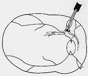
A standard three-port approach is used to carry out a complete pars plana vitrectomy. The placement of the sclerotomy is critical. The surgeon should study the angiogram and decide preoperatively where the retinotomy is to be placed to avoid damaging major vessels, to provide adequate access to the subretinal membrane, and to minimize the size of the scotoma. These factors usually dictate that the retinotomy be created in a straight temporal location and thus the superotemporal sclerotomy should be made near the horizontal meridian. If a sewn-on ring system is used to hold a corneal contact lens, it is sometimes advantageous to rotate the fixation flanges superotemporally and inferonasally from the horizontal to allow a nearly horizontal placement of the temporal port. Occasionally these horizontal sclerotomy sites bleed more than when placed more superiorly, but this has not proven to be a significant complication.

Although data are lacking to prove the importance of removal of the posterior hyaloid, we attempt to remove it in every case. Our preferred technique is using a silicone-tipped extrusion needle to aspirate over the attached cortical vitreous near the optic disc.16,17 The surgical assistant or technician indicates whether infusion fluid is dripping into the collection chamber. Absence of dripping indicates that the silicone tip has engaged the posterior hyaloid, and at that point the aspiration pressure can be raised to 300 or 400 mm Hg. Once engaged, the hyaloid can usually be pulled free from the disc with a gentle stripping motion. In many cases, the surgeon can see a Weiss ring as the hyaloid detaches and is stripped approximately to the equator (Fig. 1). (If the hyaloid remains adherent, then one may proceed with the case. After the creation of the retinotomy, an angled needle tip or the subretinal pick can often be used to find the cleavage plane between posterior hyaloid and retina at the edge of the retinotomy and achieve detachment of the hyaloid.) The vitreous cutter is reintroduced and the vitrectomy is completed.

Fig. 1. The posterior hyaloid is engaged with a silicone tip extrusion cannula near the optic disc.

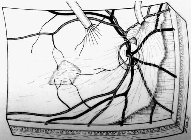
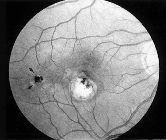
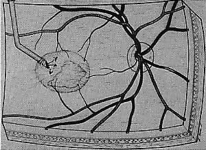
The placement of the retinotomy takes into account (1) the exact location of the membrane under the fovea; (2) the presence of presumed adhesions between the neurosensory retina and underlying tissue (previous photocoagulation scars and/or evidence of pigment migration into neurosensory retina or retinochoroidal vascular anastomoses); (3) the dimensions of the subretinal instruments (specifically the length of the angled instrument tips that determines how far away from the fovea the retinotomy can be made and still allow the tips to reach the membrane); and (4) the topographic anatomy of the neurosensory retina and nerve fiber layer. (A retinotomy made temporally in the macula disrupts less nerve fibers than does a retinotomy made nasally or superiorly that risks the creation of an arcuate visual field defect or reduced central acuity secondary to the interruption of the papillomacular bundle.) In most cases, these factors dictate a straight temporal or slightly superotemporal location for the retinotomy. However, the surgeon may choose to create a retinotomy superonasal to the fovea. With newer 33- and 36-gauge instruments, the retinotomies are small enough that no significant damage to the papillomacular bundle occurs. This approach may allow the surgeon to use his or her dominant hand for subretinal manipulation.

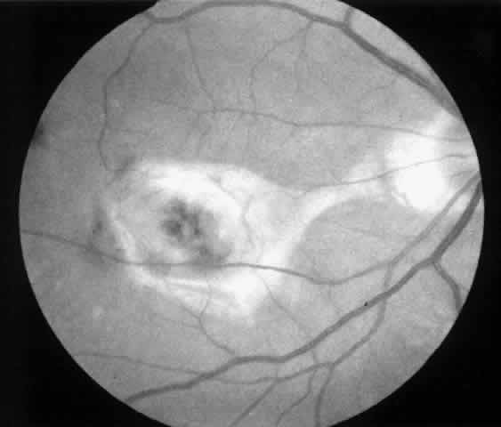
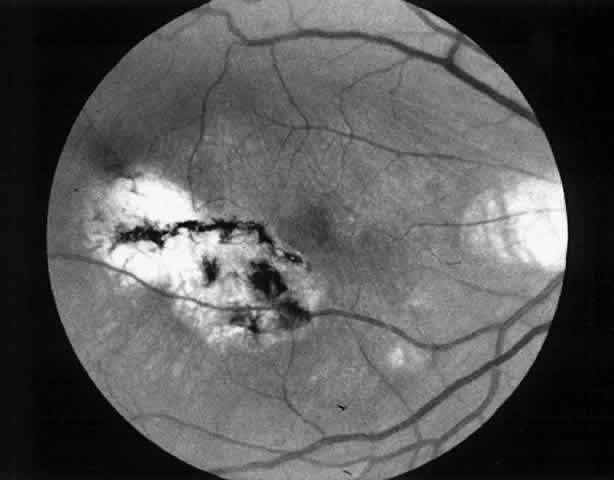
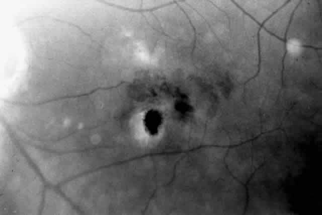
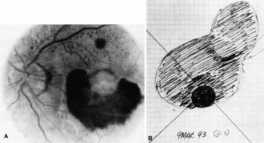
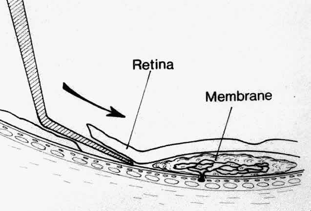
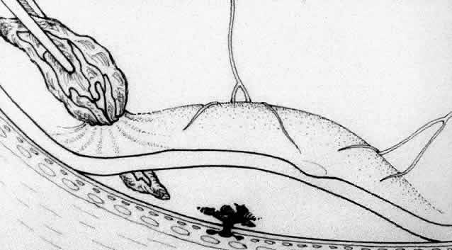
Besides being in the most advantageous location, the retinotomy should be as small as possible. Initially, we lightly diathermized the surface of the retina and then used the myringotomy blade to tease open a small hole through which an angled infusion needle was introduced.15 At the suggestion of Lambert and co-workers at Emory University, we stopped using diathermy. We now use a 120° angled, sharply pointed 36-gauge subretinal pick to pierce undiathermized neurosensory retina (Fig. 2). Occasional slight retinal hemorrhage can be controlled by transiently increasing the intraocular pressure. After the tiny hole has been made, the surgeon introduces the angled 33-gauge infusion needle beneath the retina and the assistant gently infuses balanced salt solution to elevate the neurosensory retina. This is accomplished by pushing on the plunger of a syringe that is connected to the hub of the needle by a short piece of tubing. As the fluid enters the subretinal space, attention is directed to edges of laser scars and/or adhesions to the underlying membrane (Fig. 3). Excessive infusion pressure can easily tear the retina. If areas of retina remain adherent, the infusion is stopped and the tip of an angled subretinal pick is carefully passed over the anterior surface of the membrane surface to break any residual adhesions. In a similar manner, the tip of the angled subretinal pick can be used to gently separate the thinned retina from an underlying photocoagulation scar. Occasionally, horizontal subretinal scissors are necessary to cut firm adhesions. These scissors have a similar 130° bend and blades approximately 3 mm in length to allow manipulation through an eccentric retinotomy. Trauma to foveal photoreceptors from either the pick or scissors is carefully avoided. If the retina is not mobilized over the entire photocoagulation scar, separation is achieved at least far enough into the scar to allow manipulation and extraction of the membrane without tearing the adjacent retina. The sharp tip of the angled subretinal pick is used to elevate the edge of the neovascular complex from the underlying RPE (Fig. 4). Care is taken to swing the pick in a pivoting or rotating manner to stretch or enlarge the retinotomy as little as possible. This requires close attention not only to the primary site of action at the membrane but also to the instrument shaft at the retinotomy site. In the appropriate cases, the complex dislodges easily from the underlying subfoveal RPE but remains attached to the edge of a laser scar (in recurrent cases) or to the stalk of choroidal vascular ingrowth.

Fig. 2. A small retinotomy is created with a 120° angled, sharp, 36-gauge pick. Arrow indicates direction of pick motion tangential to underlying RPE.

Fig. 3. The neurosensory retina is gently elevated by the slow infusion of balanced salt solution through a 120° angled, bevel-up 33-gauge subretinal cannula (arrow).

Fig. 4. The neovascular complex is elevated from the underlying retinal pigment epithelium with the sharp, 36-gauge subretinal pick.

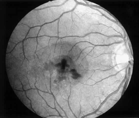
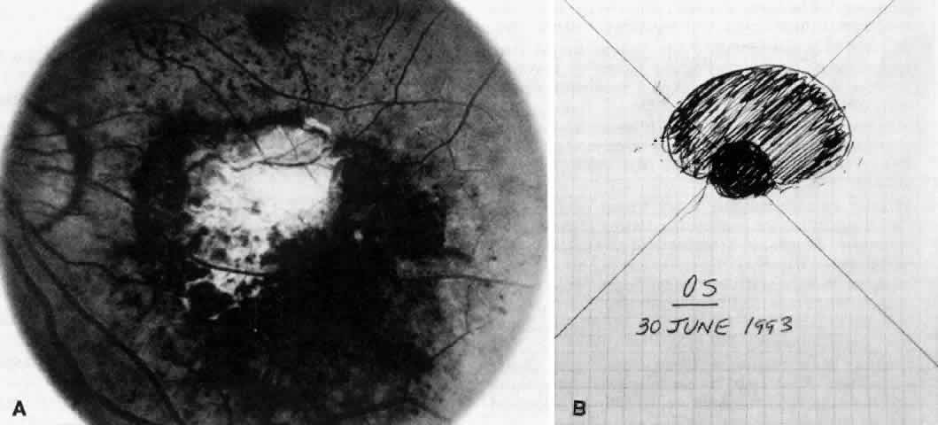
We use positive action horizontal forceps that are angled 130° and have tips 3.2 mm in length.15 The tips are introduced (closed) through the retinotomy, which has usually enlarged slightly during the subretinal manipulation. The objective is to place the opened blades around the stalk or the adhesion, with the membrane in front of the blades. Gentle traction with the blades held closed breaks the connection (Fig. 5). This step is performed slowly and carefully. If traction on the retina is seen, the membrane is released and further separation of the complex from neurosensory retina is accomplished. If excessive tugging and displacement of surrounding RPE is seen, then consideration is given to using the subretinal scissors to cut the stalk rather than breaking it with the forceps. In virtually every case, the membrane (and often the adjacent laser scar) can be removed in one piece. As pathologic examination has confirmed, the abundance of cohesive basement membrane material matrix surrounding occasional capillaries, creates a complex of significant tensile strength.14

Fig. 5. The neovascular complex is grasped with horizontal subretinal forceps and removed from the eye.

When the vascular connection from the choroid is about to be severed, the intraocular pressure is raised to approximately 80 mm/Hg. Despite this precaution, minimal hemorrhage is often countered when the membrane is removed. In one early case, while attention was directed to removing the neovascular complex through the sclerotomy, a massive hemorrhage occurred beneath the retina. In two additional cases, enough blood accumulated in the subretinal space to require removal with subretinal forceps. We now maintain the intraocular pressure elevated for at least 1 minute and watch closely for any evidence of rebleeding while the pressure is slowly lowered. If more bleeding occurs, the intraocular pressure is raised again. After the implementation of these measures, significant subretinal hemorrhage has not occurred in the last 60 cases.

Once hemostasis is achieved, the membrane is extracted through the sclerotomy for pathologic examination or it is cut and aspirated with the vitrectomy probe. Occasionally, large membranes cannot be removed through a standard size sclerotomy. We prefer dividing the membrane into smaller pieces with intraocular scissors rather than enlarging the sclerotomy. Plugs are placed and scleral depression is performed 360° to verify that no peripheral retinal tears have occurred.

A complete air-fluid exchange is next performed. We use standard extrusion needles or silicone-tipped needles for the exchange, with the aspirating tip over the optic nerve. In early cases, once the retina was flat, endolaser burns were applied around the retinotomy. The resulting laser scars were eccentric from the fovea but did occasionally produce symptomatic scotomas. In the past 60 cases, we have used no laser and have found the retina to remain attached. Our use of intraocular tamponade has also undergone evolution. Initially, we used nonexpansile concentrations of sulfur hexafluoride or perfluoropropane and encouraged facedown positioning for 1 to 2 weeks. As improved instrumentation allowed smaller retinotomies, we have questioned the need for gas tamponade. We perform fluid-air exchange and leave the eye with a one-half to two-thirds fill of filtered air. In the most recent 15 cases, we have gently reinfused balanced salt solution over the optic nerve after the vitreous cavity has been dry for a few minutes. The eye is completely filled with fluid, and no special positioning is used. In each of these cases, the posterior hyaloid has been meticulously removed. In each case the retinotomy has sealed without gas tamponade. Additional cases will be required before this modification can be widely advised.

Back to Top
POSTOPERATIVE MANAGEMENT
Patients are examined 1 and 7 to 10 days after surgery to verify that intraocular pressure is acceptable, the retina remains attached, and no infection occurs. Three weeks after surgery, the view is usually adequate for assessment of the presence or absence of subfoveal RPE. Occasionally, residual subretinal blood will obscure the underlying tissues for a longer period of time. Within the first month, angiography is repeated to evaluate for recurrence of neovascularization. Not uncommonly, the site of the original choroidal ingrowth stalk demonstrates recurrent neovascularization. Often this site is not subfoveal and therefore slit lamp laser photocoagulation can be employed to ablate the recurrence. Given the fact that membranes recur in approximately one third of cases within 6 months, close follow-up is essential.
Back to Top
EARLY CLINICAL EXPERIENCE
Results of our first 58 consecutive cases of subfoveal membranes managed with vitrectomy were presented at the 1991 annual meeting of the American Academy of Ophthalmology.18 Since the techniques and especially the indications for the surgery are still in evolution, it is not surprising that the visual results in that early group were often disappointing. To briefly summarize those results, the cases were grouped according to etiologies of the membranes as well as the surgical technique employed. In most, the neovascular complex was dislodged and removed. In some, the fibrovascular scar was disconnected from its underlying choroidal blood supply and then left in place. In 5 eyes, patches of RPE were positioned beneath the fovea after extraction of the membrane. With limited follow-up (mean, approximately 8 months), two or more Snellen lines of visual acuity improvement compared with preoperative acuity was achieved in 7 of 22 eyes with ARMD membrane removal (1 eye 20/20), none of 4 eyes with ARMD membrane removal and RPE patches, and 1 of 7 eyes with ARMD membrane disconnection. Significant improvement (two or more Snellen lines) was seen in 6 of 16 eyes with POHS membrane removal and none of 4 eyes with POHS membrane disconnection. Of 5 eyes with membranes of other etiologies, 2 improved (20/20 and 20/40). The one RPE patch placed in a case of idiopathic neovascularization failed to improve vision.
Back to Top
COMPLICATIONS
In the first 58 cases, intraoperative complications included two posterior retinal tears and two peripheral retinal tears related to sclerotomies. Postoperatively, two patients developed retinal detachments and four eyes developed macular pucker. In our subsequent 60 cases, two retinal detachments occurred owing to peripheral tears.

Berger and Kaplan19 presented their results in 34 cases of removal of subfoveal membranes and found slightly higher rates of visual improvement in POHS and slightly lower rates of improvement in ARMD. Subsequently, Lambert and colleagues20 reported 10 cases of membrane removal in ARMD. Their surgical indications required a macular neurosensory retinal detachment as well as subfoveal neovascularization. They reported improved visual function in 70% of eyes, but 70% still had acuity of only 20/200 (6/60) or worse. In reviewing these early cases, it is clear that unless subfoveal RPE is preserved after surgery, excellent central acuity is not regained. Intraoperative preservation of foveal RPE has become the single most important predictor of good visual outcome. Thus it is of critical importance to be able to select for surgery only those eyes in which RPE can be preserved.

Back to Top
CURRENT CASE SELECTION

MEMBRANES ANTERIOR TO RETINAL

PIGMENT EPITHELIUM

At the present time, we advise surgical extraction of a membrane only if the membrane appears to lie anterior to the RPE. In some cases, this is an easy determination to make preoperatively; in others, it is difficult. We study the macula with stereoscopic viewing at the slit lamp through a 60- or 78-diopter lens or with a contact lens. We also obtain color, stereo, 2× magnified views of the macula for subsequent review. The findings on clinical examination that suggest an anterior location may include the following:

  1. A well-defined edge of the membrane may be discernible.
  2. With stereoscopic viewing, the membrane may be seen to lie anterior to underlying tissues (RPE).
  3. If blood is present, it may help demonstrate a cleavage plane between the membrane and RPE by helping to “outline” the overlying membrane.
  4. In some cases, the membrane may have a pigmented border (varying between tan and dark brown) that corresponds to a rim of hypofluorescence seen angiographically.

Angiographic findings consistent with membranes anterior to the RPE include the following:

  1. The boundary between the hyperfluorescence of the membrane and background choroidal fluorescence is distinct, and often a rim of blocked fluorescence is seen between the two. (If the hyperfluorescence is immediately adjacent to background choroidal fluorescence, then in our experience, the membrane is either beneath the RPE or at least not amenable to stripping from the RPE).
  2. The membrane is more likely to be relatively compact angiographically, rather than demonstrating irregularly shaped areas of hyperfluorescence.
  3. The fluorescence in the membrane appears in a relatively homogeneous pattern.
  4. Stereoscopic viewing of the angiogram may demonstrate an anterior location of the membrane.
  5. The absence of late staining in surrounding tissues outside the membrane (as seen in the early frames) helps confirm that the membrane is discrete.

SURGICAL INTERVENTION FOR SUBRETINAL STRANDS IN PROLIFERATIVE VITREORETINOPATHY

Subretinal fibrocellular proliferation (subretinal strands or bands) may be encountered in up to 47% of eyes undergoing vitreous surgery for rhegmatogenous retinal detachment complicated by proliferative vitreoretinopathy.21 It is also known to occur after long-standing exudative and tractional retinal detachment.22,23

While the retina is detached, metaplastic RPE and glial cells proliferate on the outer retinal surface and across the subretinal space forming dots, sheets, and branching fibrocellular strands that have single or multiple foci of attachment to the overlying neurosensory retina and RPE.23–30 This results in a retroretinal scaffold that elevates and distorts the overlying neurosensory retina in a manner that resembles a bed sheet on a clothesline.

Successful reattachment of the retina is possible in 70% to 95% of eyes without removal of subretinal strands.21,31 Occasionally, however, persistent retroretinal traction will prevent intraoperative flattening of the retina and subretinal strand removal becomes necessary.

Several techniques have been described to accomplish this goal.21,32–35 The essence of these techniques can be condensed into three basic steps:

  1. A small retinotomy (intraocular diathermy needle or vitreoretinal scissors) is created in the vicinity of the subretinal strand. Likewise, the subretinal tissue can be accessed through a pre-existing retinal break.
  2. The subretinal strand is engaged with a vitreoretinal pick or forceps and peeled off the outer surface of the retina and/or RPE by gentle tangential traction (Fig. 6).
  3. If a firm adhesion to the overlying neurosensory retina or RPE precludes removal of the subretinal strand, retroretinal traction can be relieved by segmentation of the strand at a single or multiple points. Several retinotomies may be necessary, depending on the vectors of traction and the configuration of the detachment.

Fig. 6. After a retinotomy, the subretinal strand is grasped with intraocular forceps and removed from the eye.

Successful intraoperative flattening of the retina is possible in 95% of eyes requiring subretinal strand removal.21 Complications of this procedure include retinal or RPE tears, enlargement of the retinotomy, and subretinal hemorrhage.

Back to Top
SAMPLE CASES
  1. A 35-year-old patient with serpiginous choroidopathy developed a recurrent choroidal neovascular membrane extending into the fovea with a decrease in vision to 20/200 (6/60) (Fig. 7). Five months after surgery his visual acuity had improved to 20/25 (6/7.5) (Fig. 8).
  2. A 35-year-old patient developed an idiopathic subfoveal choroidal neovascular membrane with decrease in vision to 20/200 (6/60) (Fig. 9). He underwent surgical excision of the membrane. Laser was applied to the retinotomy at the conclusion of the case. One year after surgery, his visual acuity remained stable at 20/20 (6/6) (Fig. 10).
  3. A 39-year-old patient with POHS developed a recurrent choroidal neovascular membrane involving the fovea with a decrease in vision to 20/100 (6/30) (Fig. 11). He underwent surgical excision but laser was not applied to the retinotomy site at the conclusion of the case. One month after surgery his vision improved to 20/30 (6/9) and the retinotomy was not noticeable (Fig. 12).
  4. A 71-year-old patient with ARMD developed a recurrent choroidal neovascular membrane extending into the fovea with a preoperative vision of 20/320 (6/96) (Fig. 13). He had only marginal visual improvement after surgery (20/200 [6/60]) owing to a subfoveal RPE defect, but the size of the scotoma decreased after resolution of subretinal fluid (Fig. 14).

Fig. 7. Preoperative fundus photograph of a 35-year-old patient with serpiginous choroidopathy and a recurrent subfoveal neovascular membrane (case 1). Visual acuity is 20/200 (6/60).

Fig. 8. Postoperative fundus photograph (case 1). Five months after surgery, patient's visual acuity is 20/25 (6/7.5).

Fig. 9. Preoperative fundus photograph of a 35-year-old patient with idiopathic subfoveal neovascularization (case 2). Visual acuity is 20/200 (6/60).

Fig. 10. Postoperative fundus photograph (case 2). One year after surgery, visual acuity is 20/20 (6/6). Note laser scar at retinotomy site (arrows).

Fig. 11. Preoperative photograph of a 39-year-old patient with presumed ocular histoplasmosis syndrome (case 3). Visual acuity is 20/100 (6/30).

Fig. 12. Postoperative photograph (case 3). One month after surgery visual acuity is 20/30 (6/9). Retinotomy site is not noticeable.

Fig. 13. Preoperative fluorescein angiogram (A) and Amsler grid (B) of a 71-year-old patient with age-related macular degeneration (case 4).

Fig. 14. Postoperative fundus photograph (A) and Amsler grid (B) (case 4). Central retinal pigment epithelial defect accounts for minimal visual improvement. Significant improvement is noted on Amsler grid.

Surgical removal of choroidal neovascular membranes is currently undergoing evaluation in [xB]a multicenter, randomized, prospective clinical trial. Until the results of such a trial are available, these techniques cannot be advised for widespread application.

Back to Top
REFERENCES

1. Gass JDM: Stereoscopic Atlas of Macular Diseases: Diagnosis and Treatment, 3rd ed, p 18. St. Louis, CV Mosby, 1987

2. Green WR, McDonnel PJ, Yeo JH: Pathologic features of senile macular degeneration. Ophthalmology 92:615, 1985

3. Macular Photocoagulation Study Group: Argon laser photocoagulation for idiopathic neovascularization: Results of a randomized clinical trial. Arch Ophthalmol 101:1358, 1983

4. Macular Photocoagulation Study Group: Argon laser photocoagulation for histoplasmosis: Results of a randomized clinical trial. Arch Ophthalmol 101:1347, 1983

5. Macular Photocoagulation Study Group: Argon laser photocoagulation for senile macular degeneration: Results of a randomized clinical trial. Arch Ophthalmol 100:912, 1982

6. Macular Photocoagulation Study Group: Krypton laser photocoagulation for neovascular lesions of age-related macular degeneration: Results of a randomized clinical trial. Arch Ophthalmol 108:816, 1990

7. Macular Photocoagulation Study Group: Krypton laser photocoagulation for idiopathic neovascular lesions: Results of a randomized clinical trial. Arch Ophthalmol 108:832, 1990

8. Macular Photocoagulation Study Group: Krypton laser photocoagulation for neovascular lesions of ocular histoplasmosis: Results of a randomized clinical trial. Arch Ophthalmol 105:1499, 1987

9. Macular Photocoagulation Study Group: Laser photocoagulation of subfoveal neovascular lesions in age-related macular degeneration: Results of a randomized clinical trial. Arch Ophthalmol 109:1220, 1991

10. Macular Photocoagulation Study Group: Laser photocoagulation of subfoveal recurrent neovascular lesions in age-related macular degeneration: Results of a randomized clinical trial. Arch Ophthalmol 109:1232, 1991

11. DeJuan E, Machemer R: Vitreous surgery for hemorrhagic and fibrous complications of age-related macular degeneration. Am J Ophthalmol 105:25, 1988

12. Blinder KJ, Peyman GA, Paris CL et al: Submacular scar excision in age-related macular degeneration. Int Ophthalmol Clin 15:215, 1991

13. Peyman GA, Blinder KJ, Alturki W et al: A technique for retinal pigment epithelium transplantation for age-related macular degeneration secondary to extensive subfoveal scarring. Ophthalmic Surg 22:102, 1991

14. Thomas MA, Kaplan HJ: Surgical removal of subfoveal neovascularization in the presumed ocular histoplasmosis syndrome. Am J Ophthalmol 111:1, 1991

15. Thomas MA, Ibanez HE: Instruments for submacular surgery. Retina 14:84, 1994

16. Grizzard WS, Allarakhia L: New silicone tipped cannulas for subretinal fluid drainage. Br J Ophthalmol 73:838, 1989

17. Mein CE, Flynn HW: Recognition and removal of posterior cortical vitreous during vitreoretinal surgery for impending macular hole. Am J Ophthalmol 111:611, 1991

18. Thomas MA, Grand MG, Williams DF et al: Surgical management of subfoveal choroidal neovascularization. Ophthalmology 99:952, 1992

19. Berger AS, Kaplan HJ: Clinical experience with the surgical removal of subfoveal neovascular membranes. Ophthalmology 99:969, 1992

20. Lambert HM, Capone A, Aaberg TM et al: Surgical excision of subfoveal neovascular membranes in age-related macular degeneration. Am J Ophthalmol 1132:257, 1992

21. Lewis H, Aaberg TM, Abrams GW et al: Subretinal membranes in proliferative vitreoretinopathy. Ophthalmology 96:1403, 1989

22. Sternberg P, Machemer R: Subretinal proliferation. Am J Ophthalmol 98:456, 1984

23. Wilkes SR, Mansour AM, Green WR: Proliferative vitreoretinopathy: Histopathology of retroretinal membranes. Retina 7:94, 1987

24. Federman JL, Folberg R, Ridley M et al: Subretinal cellular bands. Trans Am Ophthalmol Soc 81:172, 1983

25. Machemer R: Discussion of presentation of Federman JL, Folberg R, Ridley M et al. Trans Am Ophthalmol Soc 179, 1983

26. Machemer R, Laqua H: Pigment epithelial proliferation in retinal detachment. Am J Ophthalmol 80:1, 1975

27. Laqua H, Machemer R: Glial cell proliferation in retinal detachments. Am J Ophthalmol 80:602, 1975

28. Laqua H, Machemer R: Clinical pathological correlation in massive panretinal proliferation. Am J Ophthalmol 80:913, 1975

29. Trese MT, Machemer R: Subretinal strands: Ultrastructural features. Graefes Arch Clin Exp Ophthalmol 223:35, 1985

30. Hiscott P, Morino I, Alexander R et al: Cellular components of subretinal membranes in proliferative vitreoretinopathy. Eye 3:606, 1989

31. Wallyn RH, Hilton GF: Subretinal fibrosis in retinal detachment. Arch Ophthalmol 97:2128, 1979

32. Machemer R: Surgical approaches to subretinal strands. Am J Ophthalmol 980:81, 1980

33. Machemer R: Retinotomy. Am J Ophthalmol 92:73, 1981

34. Michaels RG: Surgery of retinal detachment with proliferative vitreoretinopathy. Retina 4:63, 1984

35. Charles S: Vitreous Microsurgery, p 126. Baltimore, Williams & Wilkins, 1981

Back to Top