Chapter 60
Laser and Cryotherapy Treatment for Retinal Tears
MAGDALENA F. SHULER and WILLIAM TASMAN
Main Menu   Table Of Contents

Search

TREATMENT PURPOSE
TYPES OF RETINAL TEARS
MODALITY OF TREATMENT
PROCEDURE
COMPLICATIONS
REFERENCES

Retinal tears are commonly treated with either laser photocoagulation or cryotherapy. The goal of treatment is to form a chorioretinal seal around the retinal break to avoid the passage of fluid into the subretinal space. The treatment of a retinal tear and the modality of treatment applied are dependent on the presentation and clinical findings.
Back to Top
TREATMENT PURPOSE
Retinal tears cause rhegmatogenous retinal detachments (RRD) by allowing fluid to enter the subretinal space. When a retinal tear has significant vitreous traction, fluid may enter even more readily. Retinal tears in the superior retina can lead to RRD more rapidly because of gravitational forces. In contrast, retinal tears in the inferior retina often are more chronic and slower to advance. Evidence of chronicity consists of pigmented demarcation lines.

Both laser photocoagulation and cryotherapy are used to create a firm adhesion between the retina and retinal pigment epithelium. Chorioretinal scarring occurs in 7 or more days with cryotherapy, but an earlier retinal adhesion is noted with laser.1

Back to Top
TYPES OF RETINAL TEARS
Clinical examination to identify retinal tears requires binocular indirect ophthalmoscopy with scleral indentation.2 Not all retinal tears require treatment. A symptomatic retinal tear is defined as a full-thickness retinal break caused by vitreoretinal traction associated with symptoms of floaters and/or photopsias (Fig. 1). While not all retinal tears require intervention, symptomatic retinal tears should be treated to prevent RRD. An operculated retinal hole associated with symptoms may warrant either observation or treatment depending on its clinical appearance. The presence of pigmentation around the hole with no vitreoretinal traction or no significant fluid suggests chronicity, and observation should be considered (Fig. 2).

Fig. 1. Horseshoe retinal tear with traction.

Fig. 2. Round operculated retinal hole with pigmentation.

An asymptomatic retinal tear is a full-thickness retinal break not associated with symptoms. Significant evidence suggests that patients with asymptomatic retinal tears may be observed.3–6 In fact the effectiveness of treating asymptomatic retinal tears in preventing RRD is unproved.7,8 Retrospective analyses have shown that subsequent posterior vitreous separation in eyes with preexisting asymptomatic retinal tears does not predispose to subsequent RRD.6 However, the prophylactic treatment of some retinal tears may be considered.

Recent evidence suggests that the fellow eye of patients with RRD is at considerable risk of developing rhegmatogenous events.9 Treatment of fellow eyes of patients with RRD can be considered, but RRD can still arise despite prophylactic laser.10 Patients with RRD caused by giant retinal tears (GRT, tear encompassing greater than 3 clock hours) are a specific subgroup of patients at risk for rhegmatogenous events in the fellow eye. Some recent evidence suggests prophylactic 360-degree cryotherapy treatment in the fellow eye of patients with RRD caused by spontaneous GRT should be considered but this is not universally accepted.11

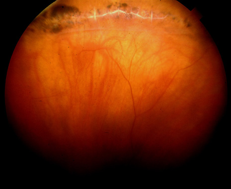
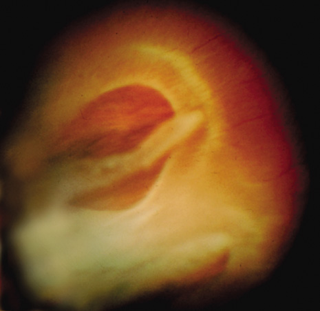
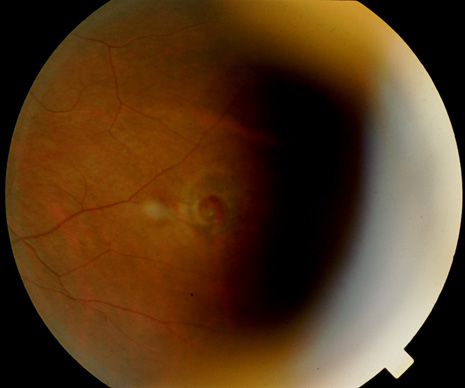
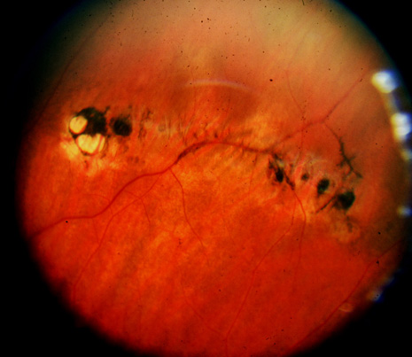
Prophylactic treatment of patients planning on undergoing other ocular procedures can be considered. Asymptomatic retinal tears or extensive lattice (Fig. 3) with traction (Fig. 4) may warrant prophylactic treatment prior to intraocular surgery such as cataract extraction with intraocular lens implant. Prophylactic treatment of myopic eyes prior to LASIK is controversial but may be considered if extensive lattice and/or traction exist. Recent evidence suggests that prophylactic treatment of asymptomatic retinal tears be considered prior to yttrium-aluminum garnet (YAG) laser posterior capsulotomy in high-risk eyes with axial myopia.12

Fig. 3. Extensive lattice degeneration in a myopic patient.

Fig. 4. Lattice degeneration with traction.

Back to Top
MODALITY OF TREATMENT
Although retinal tears can be effectively treated by either cryotherapy or laser photocoagulation, certain clinical findings factor into the decision-making process. Small retinal tears without significant subretinal fluid at the vitreous base with a clear media are usually treated by laser photocoagulation. Posteriorly located retinal tears also necessitate treatment with laser (Fig. 5) because standard cryotherapy would require a conjunctival incision to reach a posteriorly located retinal tear. In contrast, an anteriorly located retinal tear is sometimes more easily treated with cryotherapy because of the optical constraints of laser photocoagulation.

Fig. 5. A posteriorly located retinal tear (A), surrounded by laser photocoagulation (B).

Laser photocoagulation to a retinal tear can be delivered using the slit-lamp biomicroscope (Fig. 6) with a contact lens or the laser indirect ophthalmoscope (LIO). Each method has its advantages and disadvantages. With media clarity, laser applied using the slit lamp allows for a better magnified view of the lesion to be treated and no inadvertent laser applications. However, treatment of larger tears may require lengthy manipulation of the contact lens thereby reducing the clarity of the cornea for subsequent examination and/or treatment using the LIO. Lengthy treatments at the slit lamp are also not well tolerated by most patients because of positioning. Patients with significant cataracts, posterior capsule opacities and/or mild vitreous hemorrhage are more easily treated with LIO. However, LIO can be more difficult to master and may not be readily available.13

Fig. 6. Laser photocoagulation application via slit-lamp biomicroscope and contact lens (Courtesy of Iridex Corporation Presentation Slides).

Cryotherapy for retinal tears should be performed for specific indications. Because laser photocoagulation requires the retina to be in close approximation to the retinal pigment epithelium in order to produce the appropriate thermal effect, the presence of significant subretinal fluid necessitates the use of cryotherapy. Lack of media clarity has led some to treat retinal tears using ultrasound guided cryotherapy, a method not advocated by these authors.14,15

Back to Top
PROCEDURE
Treatment of retinal tears by both cryotherapy and laser has certain requirements. Informed consent is of primary importance in this and in every procedure performed in the office setting. Patients should be aware that this treatment is an attempt to seal a retinal tear and that despite adequate treatment some patients will still develop a retinal detachment as well as other new retinal tears necessitating more treatment and/or surgery. The complications covered in the following section should also be discussed so that the patient realizes that risks do exist with laser and cryotherapy. After the treatment has been performed the patient should be advised of retinal detachment precautions. The presence of an increase in photopsias, more floaters, or a black curtain (loss of peripheral visual field) should warrant an urgent reevaluation of the retina. Routine follow-up can be tailored to the clinical setting but is usually done within a week or two of treatment.

Appropriate anesthesia is patient-dependent and procedure-dependent. A patient who did not easily tolerate the examination of the peripheral retina with indirect ophthalmoscopy and scleral indentation may require retrobulbar anesthesia for treatment of the retinal tear. However, the use of retrobulbar anesthesia may make treatment of an anterior lesion difficult because the patient will not be able to assist in rotation of the globe. Other patients may only need topical or subconjunctival anesthesia. Most patients undergoing cryotherapy for a retinal tear can tolerate the treatment with subconjunctival lidocaine injected at the site of the planned cryotherapy several minutes prior to the treatment, or for a small break just topical anesthesia.

Cryotherapy to a retinal tear is performed using a cryoprobe and an indirect ophthalmoscope. Testing the cryoprobe prior to the procedure is useful to make sure the probe is freezing. The retinal tear is localized by indenting the sclera using the cryoprobe. It is imperative that only the tip of the cryoprobe be visualized when indenting, and not the shaft to avoid an inadvertent poorly localized and incorrectly placed cryoprobe application. The area of the retinal tear should be visualized during the cryotherapy to ensure appropriate retinal pigment epithelium and retinal whitening. Freezing or whitening of the retinal pigment epithelium will be noticed first, followed by delineation of the edges of the retinal tear and whitening of the retina. Excessive freezing or ice crystal formation should be avoided. The entire retinal tear should be surrounded with contiguous treatments and treatment should be carried out to the ora serrata. If the tear is within an area of lattice degeneration then the entire area of lattice should also be treated with an adequate margin.16

Laser photocoagulation to a retinal tear performed at the slit lamp usually only requires topical anesthesia for the contact lens. Sometimes patients may not tolerate the repetitive nature of the bright lights and will require retrobulbar anesthesia. LIO may be performed usually with topical anesthesia and occasionally retrobulbar anesthesia. Excessive movement of the eye may necessitate retrobulbar anesthesia for LIO. The laser parameters must be tailored to each patient according to media opacity and pigmentation. The minimum settings necessary to obtain a white thermal reaction should be determined. Treatment by encircling the entire retinal tear with two to three rows of confluent treatment and extension to the ora serrata is the goal (Fig. 7).

Fig. 7. Laser photocoagulation to a retinal tear.

After the treatment the patient may be told that pain can be ameliorated with over-the-counter analgesics. Specifically, patients who underwent cryotherapy may experience lid edema that can respond to cold and/or warm compresses. Any patient who required retrobulbar anesthesia should be patched with their eyelid closed and instructed to not remove the patch until the next day or until they are able to easily move their eye and close their eyelids. An explanation that blurry vision after this procedure is normal can avoid frantic late night phone calls.

Back to Top
COMPLICATIONS
The treatment of retinal tears with laser or cryotherapy is relatively safe; however, complications can occur.

The risks inherent with retrobulbar anesthesia are discussed elsewhere but are important considerations. Albeit rare, sometimes catastrophic complications can occur from retrobulbar anesthesia.

During the treatment, several complications can occur. The placement of inadvertent laser or cryotherapy treatment can be reduced by improving visualization of the tear and optimizing patient comfort during the procedure. Proper orientation is especially important during LIO because the optics can be disorienting. Because repetitive laser is applied using a foot pedal, physically removing the foot from the pedal during repositioning and reorienting of the eye can minimize the chance of inadvertent laser placement.

The presence of a bridging vessel over the retinal tear can be problematic (Fig. 8). During the procedure further tension on a bridging vessel can lead to vitreous hemorrhage and loss of visualization. Cryotherapy applied to the bridging vessel first can sometimes help to minimize the bleeding, but manipulation of the probe prior to the application can result in rupture of the blood vessel. Careful placement of the probe and quick cryotherapy application to the bridging vessel is advised. Laser applied to a retinal tear with a bridging vessel should be placed furthest away from the vessel and approaching the vessel last. Laser can be applied onto the distal and proximal portion of the vessel, but treatment to the bridging portion of the vessel should be avoided. Sometimes bleeding occurs despite precautions, and if visualization is dramatically reduced, careful observation is warranted. Further surgical intervention may be necessary if insufficient treatment was performed prior to the vitreous hemorrhage.

Fig. 8. Retinal tear with a small bridging vessel.

Both laser and cryotherapy treatments have postprocedure complications. Extensive cryotherapy can lead to cystoid macular edema.17 Both laser and cryotherapy can result in epiretinal membrane formation with an incidence between 5.5% and 10% of treated eyes.18 Although the presence of an epiretinal membrane does not always result in macular pucker and metamorphopsia with the need for surgical intervention, patients should be aware of this possibility.

Proliferative vitreoretinopathy (PVR) is the leading cause of retinal reattachment surgical failure. Retinal pigment epithelial (RPE) cells play a critical role in the formation of PVR. Extensive cryotherapy has been implicated in the liberation of RPE cells into the vitreous cavity.19 Surgery for both primary RRD and RRD caused by GRT, requiring extensive cryotherapy, may be a stimulating factor in the development of PVR and recurrent RRD.20,21 In an animal model, single cryotherapy lesions had no significant effect on the vitreous while multiple cryotherapy treatments led to vitreous membrane formation.22 When treating with cryotherapy, careful judicious use of the cryoprobe is warranted. Appropriate treatment is required, but excessive use should be avoided.

Back to Top
REFERENCES

1. Folk JC, Sneed SR, Folberg R, et al: Early retinal adhesion from laser photocoagulation. Ophthalmology 96:1523, 1989

2. Natkunarajah M, Goldsmith C, Goble R: Diagnostic effectiveness of noncontact slitlamp examination in the identification of retinal tears. Eye 17:607, 2003

3. Byer NE: Prognosis of asymptomatic retinal breaks. Mod Prob Ophthalmol 12:103, 1974

4. Kramer SG, Benson WE: Prophylactic therapy of retinal breaks. Surv Ophthalmol 22:41, 1977

5. Kazahaya M: Prophylaxis of retinal detachment. Semin Ophthalmol 10:79, 1995

6. Byer NE: What happens to untreated asymptomatic retinal breaks and are they affected by posterior vitreous detachment? Ophthalmology 105:1045, 1998

7. Wilkinson C: Interventions for asymptomatic retinal breaks and lattice degeneration for preventing retinal detachment. Cochrane Database Syst Rev 3:3170, 2001

8. Wilkinson CP: Evidence-based analysis of prophylactic treatment of asymptomatic retinal breaks and lattice degeneration. Ophthalmology 107:12, 2000

9. Gonzales CR, Gupta A, Schwartz SD, et al: The fellow eye of patients with rhegmatogenous retinal detachment. Ophthalmology 111:518, 2004

10. Mastropasqua L, Carpineto P, Ciancaglini M, et al: Treatment of retinal tears and lattice degeneration in fellow eye of high risk patients suffering retinal detachment: a prospective study. Br J Ophthalmol 83:1046, 1999

11. Wolfensberger TJ, Aylward GW, Leaver PK: Prophylactic 360 cryotherapy in fellow eyes of patients with spontaneous giant retinal tears. Ophthalmology 110:1175, 2003.

12. Ranta P, Tommila P, Kivela T: Retinal breaks and detachment after neodymium: YAG laser posterior capsulotomy: five-year incidence in a prospective cohort. J Cataract Refract Surg 30:58, 2004

13. Ghosh YK, Banerjee S, Tyagi AK: Effectiveness of emergency argon laser retinopexy performed by trainee doctors. Eye 19:52, 2005

14. Schenek M, Rosenthal G, Klemperer I, et al: Ultrasound guided cryotherapy for retinal tears in patients with opaque ocular media. Br J Ophthalomol 83:628, 1999

15. Kelley LM, Walker JP, Wing GL, et al: Ultrasound guided cryotherapy for retinal tears in patients with vitreous hemorrhage. Ophthalmic Surg Lasers 28:565, 1997

16. Benson WE, Morse PH, Nantawan P: Late complications following cryotherapy of lattice degeneration. Am J Ophthalmol 84:514, 1977

17. Kimball RW, Morse PH, Benson WE: Cystoid macular edema after cryotherapy. Am J Ophthalmol 86:572, 1978

18. Saran BR, Brucker AJ: Macular epiretinal membrane formation and treated retinal breaks. Am J Ophthalmol 120:480, 1995

19. Campochiaro PA, Kaden IH, Vidaurri-Leal J, et al: Cryotherapy enhanced intravitreal dispersion of viable retinal pigment epithelial cells. Arch Ophthalmol 103:434, 1985

20. Bonnet M, Fleury J, Guenoun S, et al: Cryopexy in primary rhegmatogenous retinal detachment: A risk factor for postoperative proliferative vitreoretinopathy? Graefes Arch Clin Exp Ophthalmol 234:739, 1996

21. Glaser BM, Vidaurri-Leal J, Michels RG, et al: Cryotherapy during surgery for giant retinal tears and intravitreal dispersion of viable retinal pigment epithelial cells. Ophthalmology 100:466, 1993

22. Dunker S, Faulborn J, Haller EM, et al: The effect of retinal cryoapplication on the vitreous. Retina 17:338, 1997

Back to Top