Chapter 59
Techniques of Scleral Buckling
GEORGE A. WILLIAMS and THOMAS M. AABERG, JR.
Main Menu   Table Of Contents

Search

ANESTHESIA
BASIC SURGICAL TECHNIQUE
METHODS OF SCLERAL BUCKLING
MANAGEMENT OF SUBRETINAL FLUID
CLOSURE
COMPLICATIONS
ANATOMIC AND VISUAL RESULTS AFTER SCLERAL BUCKLING
PSEUDOPHAKIC RETINAL DETACHMENT
SUMMARY
REFERENCES

Scleral buckling is an effective surgical technique for the repair of rhegmatogenous retinal detachment. The scleral indentation achieved with scleral buckling can be accomplished by a variety of techniques and materials, each of which has inherent advantages and disadvantages. By adopting a flexible approach that incorporates the advantages and benefits of different techniques, the surgeon can maximize the rate of anatomic success in reattaching the retina while minimizing potential complications. This chapter discusses the basic principles of scleral buckling surgery, with an emphasis on the techniques the authors favor. Alternative methods of retinal detachment repair, such as vitrectomy, pneumatic retinopexy, and temporary inflatable balloons, are discussed elsewhere in this text.
Back to Top
ANESTHESIA
Scleral buckling can be performed with the patient under either local or general anesthesia. Selection of anesthetic technique often becomes a function of surgeon preference, the patient's emotional and physical state, and the availability of quality general anesthesia. The increasing frequency of outpatient scleral buckling surgery has decreased the use of general anesthesia at many centers. The majority of scleral buckling procedures are performed under local anesthesia.

The advantages of local anesthesia include shorter operating time, quicker postoperative recovery, and possibly decreased morbidity and mortality in select cases. However, retrobulbar placement of local anesthetic is not without risk to the eye or the patient's general health. Perforation of the globe, particularly in myopic patients, and damage to the optic nerve may result in permanent visual loss after retrobulbar injection.1–3 Respiratory arrest and grand mal seizures have also been reported.4 Damage to rectus muscles, typically the inferior rectus, may occur after retrobulbar injection causing diplopia.5 These complications can be minimized but not eliminated by use of either a subconjunctival or peribulbar technique of anesthetic administration.6 Subconjunctival infiltration with lidocaine allows a limbal peritomy and dissection of Tenon's capsule. Additional lidocaine and bupivacaine then can be administered via retrobulbar irrigation with a blunt cannula. This avoids the passage of sharp needles and diminishes the risks of ocular perforation and intravascular or intraneural injection.7 The authors use this technique to supplement general anesthesia and diminish postoperative pain and nausea.8

A peribulbar approach may be employed for anesthesia during scleral buckling. This technique also reduces the risk of ocular perforation compared with retrobulbar injections. A 1:1 mixture of 2% lidocaine and 0.5% bupivacaine is injected at the junction of the temporal and central thirds of the inferior orbital rim beneath the globe and just medial to the supraorbital notch. No attempt is made to enter the retrobulbar space. A total of 7 to 10 mL of anesthetic is injected, with two-thirds of the mixture delivered inferiorly and one-third given superiorly.9

The major disadvantage of local anesthesia is inadequate analgesia.10 Using a retrobulbar approach, 0.45% bupivacaine and 1.6% lidocaine provides superior anesthesia as compared with lidocaine alone.11 Either retrobulbar or peribulbar anesthesia may be supplemented during the procedure with a sub-Tenon's capsule irrigation or local infiltration of additional anesthetic. This can be administered via a flexible catheter12 or with a blunt cannula.13 Intravenous sedation with propofol or narcotics such as morphine and fentanyl or neuroleptics such as droperidol may also be helpful. These agents may cause respiratory depression and therefore require close monitoring by skilled personnel.14

Back to Top
BASIC SURGICAL TECHNIQUE
After induction of either general or local anesthesia, the operative field is prepared for surgery. Careful use of antisepsis can minimize the risk of postoperative infections. The lashes and lid margins are major sources of bacterial contamination,15 so particular care is directed to this area. Preoperatively, the lashes are clipped flush with the lid margin. At the time of surgery, the lash margins are scrubbed with antiseptics. Povidone-iodine solution is an excellent antiseptic that also is nontoxic to the cornea and conjunctiva.16 The authors place a drop of 5% povidone-iodine solution in the conjunctival sac during the sterile field preparation. Other antiseptics, such as soaps, alcohol, and hexachlorophene, are also effective but are toxic to the cornea.

Copious irrigation of the conjunctival sac with sterile saline effectively washes away most debris from the conjunctival surface. Despite irrigation, the conjunctiva usually is colonized by a variety of bacteria. Staphylococcus epidermidis can be cultured in 37% to 70% of patients before scleral buckling.15 More virulent pathogens, such as Staphylococcus aureus or gram-negative bacteria, including Proteus and Pseudomonas, are present in 5% to 9% of routine preoperative cultures. The presence of these pathogens preoperatively increases significantly the incidence of postoperative buckle infections, even with preoperative topical antibiotic treatment for 1 to 2 days.15 Although some surgeons use preoperative topical antibiotics, such as fluoroquinolones, there is no compelling evidence that infection rates are diminished.

After irrigation, the skin surface is dried with sterile gauze. This is particularly important when disposable adhesive drapes are used. The goal of surgical draping is to isolate and protect the surgical field from contamination. Common sources of contamination are the oral and nasal cavities.17 When properly applied, adhesive drapes can effectively seal off these areas from the operative field.

SURGICAL ANATOMY OF CONJUNCTIVA, TENON'S CAPSULE, AND EXTRAOCULAR MUSCLES

Conjunctival opening can be performed either at the limbus or 4 to 8 mm posterior to the limbus.18,19 Because of the considerable manipulation the conjunctiva undergoes during scleral buckling, radial relaxation incisions are necessary to prevent tearing of the conjunctiva. If a lateral canthotomy is performed, the relaxing incision should be made away from the horizontal meridian to prevent symblepharon formation. The authors routinely use a limbal peritomy and believe this approach allows both better exposure of the sclera and better coverage of the buckle with less postoperative irritation than with a posterior peritomy. In patients with filtering blebs or recent limbal wounds, the peritomy can be extended posteriorly to avoid the area of concern.

Conjunctival opening at the limbus can be facilitated by spreading with scissors beneath Tenon's capsule just posterior to the limbus, thereby avoiding the fusion of conjunctiva and Tenon's capsule, which occurs at the limbus. Once the space between Tenon's capsule and the sclera is entered, conjunctiva and Tenon's capsule can be elevated from the sclera easily and then cut flush at the limbus. With a limbal incision, conjunctiva and Tenon's capsule can be retracted together, usually with less bleeding. If only one or two quadrants are to be buckled, a 360° opening of conjunctiva is not necessary. Conjunctiva and Tenon's capsule can be reflected in the required quadrants only and the appropriate muscles isolated, as described later.

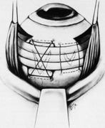
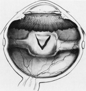
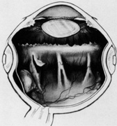
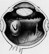
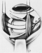
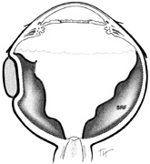
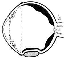
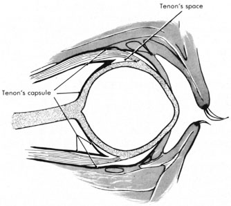
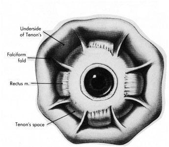
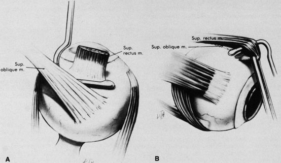
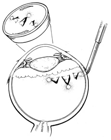
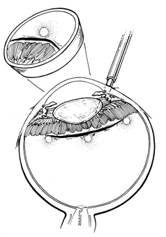
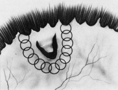
Tenon's capsule is a fascial tissue that invests both the globe and extraocular muscles. Anteriorly it fuses with conjunctiva at the limbus, and posteriorly it ends at the optic nerve sheath. Between Tenon's capsule and the sclera is the interfascial space of Tenon, or simply Tenon's space. Entering this space allows complete exposure of the scleral surface. The extraocular muscles pass through Tenon's capsule, entering Tenon's space to insert on the sclera. At the site of penetration by the individual extraocular muscles, Tenon's capsule reflects posteriorly around the muscles for 10 to 12 mm to form the muscle sheaths. The muscle sheaths are connected by the intermuscular membrane, which, in turn, is connected to the orbital wall by complex fascial arrangements. The retinal surgeon is most concerned with the extraocular muscles after they pass through Tenon's capsule (Fig. 1). At this point they do not possess a muscle sheath but, rather, are invested by episcleral tissue that fuses with the muscle. This tissue forms the falciform folds that fan out from the edges of the muscle to the overlying Tenon's capsule (Fig. 2).

Fig. 1. Anatomy of Tenon's capsule and extraocular muscles.

Fig. 2. Relationship of falciform folds to Tenon's capsule and extraocular muscles.

After the peritomy, the space between Tenon's capsule and sclera is entered in the quadrants between the rectus muscles with closed, blunt scissors. Opening the scissors lyses the episcleral fascial connections between Tenon's capsule and sclera. The muscle insertion is then engaged with a muscle hook. The muscle hook should be placed on the sclera and slid posteriorly to the muscle insertion in a circumferential direction. It is then brought anteriorly to engage the insertion. When in the proper space, the muscle hook should glide along the sclera and beneath the muscle with ease. Significant resistance to passage usually means the muscle hook is not on the scleral surface and should be repositioned. The muscle is best engaged by staying anterior to the equator. This also avoids endangering the vortex veins. Once the muscle insertion is engaged, the connections to Tenon's capsule can be identified and separated from the muscle. This is done either by stripping the muscle with a cotton-tipped applicator or with sharp dissection using scissors. When the vertical rectus muscles are isolated, care should be taken not to strip too posteriorly to avoid damaging the levator muscle superiorly or the inferior oblique muscle and Lockwood's ligament inferiorly. After isolation of the muscle is complete, a traction suture is placed around the muscle using either a fenestrated muscle hook20 or a reversed needle; 2-0 black silk is an effective traction suture. All four rectus muscles can be isolated in this manner.

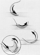
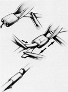
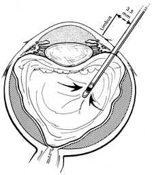
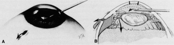
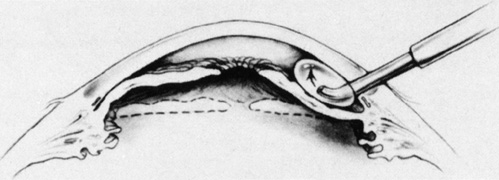
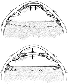
The superior rectus muscle requires additional care to avoid the superior oblique muscle tendon, which inserts 3 to 5 mm posterior to the lateral margin of the superior rectus insertion. Passing the muscle hook from the nasal side anterior to the oblique muscle insertion best avoids engaging the superior oblique muscle tendon. A second method is to rotate the eye inferiorly and grasp the superior rectus muscle insertion with a toothed forceps. The muscle hook then can be passed just posterior to the insertion, thereby avoiding the tendon of the superior oblique (Fig. 3). After the suture is placed around the superior rectus muscle, an inspection for evidence of incarceration of the oblique muscle is made by sweeping posteriorly along the lateral and medial margins of the superior rectus muscle. When incarcerated, the oblique muscle tendon appears as a white band that is pulled anteriorly by the suture.

Fig. 3. A. Correct position of muscle hook for securing superior rectus. B. Globe rotated inferiorly to facilitate placement of muscle hook behind superior rectus.

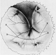
After all rectus muscles are isolated, the surface of the sclera is inspected in each quadrant for evidence of thinning, staphyloma, or anomalous vortex veins, and the location of any abnormalities is noted before scleral depression or marking of retinal breaks. Scleral thinning may appear as a gray-blue area, sometimes associated with obvious ectasia. Less prominent thinning may appear as radial gray lines. Although scleral thinning may occur anywhere, it is most common superotemporally.21

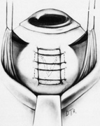
Isolation of the four rectus muscles usually allows adequate access to all areas of the sclera necessary to perform scleral buckling. Rarely, however, a muscle must be removed to adequately expose an area. This usually occurs with breaks beneath muscles or in eyes with tight or small orbits. After the muscle has been isolated, it is elevated with a muscle hook, and a double-armed absorbable synthetic 5-0 or 6-0 suture is passed through the body of the muscle parallel to the insertion and just posterior to the hook. A central knot may be placed. The suture is then passed in a single or double loop through the end of the muscle to occlude the anterior ciliary arteries. With both the suture and hook used for traction, the muscle is elevated from the sclera and cut, leaving a small portion of insertion intact on the sclera. The arms of the suture are secured, and the muscle is allowed to retract posteriorly. A 4-0 silk traction suture is placed through the insertion with at least two passes. After completion of scleral buckling, the muscle is repositioned to the insertion with the double-armed suture (Fig. 4).

Fig. 4. A to C. Technique for removal and replacement of rectus muscle.

After isolation of the rectus muscles, the sclera is exposed and accessible for suturing everywhere except beneath the superior oblique muscle and the inferior oblique muscle. Because of its anterosuperior location and wide insertion, the superior oblique muscle more commonly overlies retinal breaks than does the inferior oblique muscle. When this occurs, fibers of either oblique muscle tendon can be cut to expose the underlying sclera. Of course, care should be taken not to entirely disinsert the muscle and also to avoid any underlying vortex veins. Partial resections of the oblique muscle tendons are well tolerated and do not result in motility disorders.

LOCALIZATION

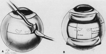
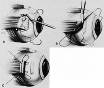
No aspect of scleral buckling is more critical than accurate placement of the buckle on the sclera. This requires precise localization of retinal breaks on the scleral surface. Several instruments for localization and for marking the sclera have been described.22–24 Other surgeons prefer to use the wooden end of a cotton-tipped applicator or a diathermy probe. When the sharper scleral markers are used for localization, it is important to inspect the sclera thoroughly. Sharp localizers and diathermy probes should be avoided if the sclera is thin. The authors prefer the O'Connor localizer, which places a 1-mm circular mark on the sclera with mild indentation (Fig. 5). 24 This instrument also has a smooth surface 90° to the marking surface to allow for exploratory depression before final positioning of the probe. Once the proper site is located, the probe is rotated 90° and the sclera marked with mild indentation pressure for approximately five seconds. The scleral mark is then enhanced with a sterile pen, cautery, or both.

Fig. 5. O'Connor localizer.

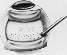
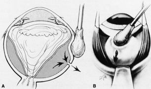
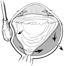
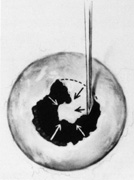
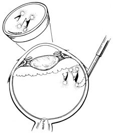
Regardless of which technique or instrument is used for localization and scleral marking, accurate placement of the marks with respect to the intraocular pathology is crucial. For small flap tears or atrophic holes, a single mark on the posterior edge of the break is sufficient. Larger flap tears and nonradial tears require localization of both the anterior and posterior extent of the break (Fig. 6). This anteroposterior orientation is particularly important when radial elements are employed. In areas with multiple, closely spaced tears, it is not necessary to mark each break. Marking the most posterior extent and the circumferential extent of the breaks is adequate (Fig. 7). This approach is also sufficient for marking a retinal dialysis. The circumferential extent of the dialysis is marked anteriorly at the edges of the dialysis, and then the most posterior extent is marked (Fig. 8).

Fig. 6. Scleral marking technique for small and larger flap retinal tears.

Fig. 7. Scleral marking technique for multiple breaks.

Fig. 8. Scleral marking technique for retinal dialysis.

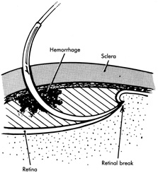
If the retina is bullously detached, accurate localization of retinal breaks is difficult. Bullously elevated breaks appear to lie more posteriorly than their true location because of parallax (Fig. 9). This can result in unnecessarily large and posterior buckles. Inaccurate localization can be minimized by first marking the break at its least elevated margin, usually anterior, and then marking the more elevated margins. Other clues, such as the presence of pigment epithelial changes underlying the break and the location of the ora serrata in relation to the break, may be helpful. Rarely, it may be necessary to drain subretinal fluid to flatten the retina before localization; however, this softens the eye, usually necessitating a saline solution injection to restore volume. It also makes further drainage difficult, presumably because of choroidal swelling secondary to hypotony.

Fig. 9. Apparent posterior location of bullously elevated retinal break caused by parallax.

TREATMENT OF RETINAL BREAKS

The rationale for the treatment of retinal breaks is to create an adhesion between the retinal pigment epithelium (RPE) and the retina. This is accomplished by inducing a thermal injury with one of three energy sources: diathermy, cryotherapy, or laser. The morphologic and cellular response of the retina and pigment epithelium to each of these energies is essentially similar.25 Two weeks after application, all three methods show comparable effects on retinal adhesive force.26 However, there are significant differences in retinal adhesion immediately after application.26 Photocoagulation increases retinal adhesion within 24 hours of application. Cryopexy reduces retinal adhesion for 1 week after application. Also, there are significant differences in the response the sclera and choroid have to these modalities and in their methods of administration.

Diathermy

Diathermy is produced by high-frequency, 13.56 MHz, alternating electrical current that generates heat because of the impedance (resistance) of the tissue through which it is passed. The heat created by the diathermy energy coagulates tissues. The histologic picture seen after diathermy depends on the intensity with which it is applied and on whether the retina is attached or detached. When the retina is attached and low-intensity diathermy applied, pigment epithelial migration into the inner retina occurs.27 If the retina is detached, however, low-intensity diathermy treatment results in pigment epithelial proliferation between Bruch's membrane and the external limiting membrane following retinal reattachment.12 High-intensity diathermy treatment in areas of detached retina causes rupture of Bruch's membrane, resulting in fibroblastic proliferation between the choroid and retina when the retina is reattached. The highest-intensity diathermy applications actually rupture the sclera, choroid, and Bruch's membrane, resulting in scar tissue formation across the entire ocular wall.27

Although diathermy produces an effective RPE adhesion, it also induces immediate scleral shrinkage and subsequent scleral necrosis.12,28 This scleral shrinkage results in increased intraocular pressure during diathermy application. The necrosis induced by diathermy weakens the strength of the sclera both immediately after application and over the long term. This weakening complicates reoperation and also may increase the incidence of scleral abscess formation.29 In addition, penetration of diathermy through intact sclera to the retina depends on the scleral thickness. Thus, variations in scleral thickness result in nonuniform and unpredictable transmission of energy to the retina, which can cause choroidal and retinal bleeding and retinal holes.12

The problems of diathermy administration associated with scleral necrosis, shrinkage, and thickness can be minimized by applying it through lamellar scleral flaps, which are discussed later. Transscleral and transconjunctival diathermy without scleral flaps may be administered with a modified diathermy electrode, which causes less scleral damage than conventional diathermy.30 However, because of the full-thickness damage that occurs with diathermy, vortex vein ampullae and long posterior ciliary arteries and nerves must be avoided, even with scleral flaps.12,31 Techniques of diathermy application are discussed in the section on implants.

Cryotherapy

Cryotherapy (cryopexy) produces an effective pigment epithelial–retinal adhesion without the scleral complications that characterize diathermy. This provides cryotherapy with significant advantages: (a) retinal pathologic conditions can be treated without the need for scleral dissection, and (b) retinal breaks can be treated regardless of their location in relation to vortex veins or long posterior ciliary vessels or nerves.

The histologic response following cryotherapy depends on whether the pigment epithelium alone or the pigment epithelium and the overlying detached retina together are frozen.32 The ability to treat detached retina is another significant advantage over both diathermy and photocoagulation. If only the pigment epithelium is frozen without freezing the overlying retina, the pigment epithelial–retinal adhesion that forms once the retina is reattached shows pigment epithelial hyperplasia and loss of retinal outer segments. Therefore the normal microvillous interdigitations seen between retina and pigment epithelium are missing. If both the pigment epithelium and overlying retina are frozen, the adhesion that results after reattachment demonstrates cellular connections between the retina and pigment epithelium consisting of desmosome formation between retinal glia and pigment epithelium or direct contact between retinal glia and Bruch's membrane.

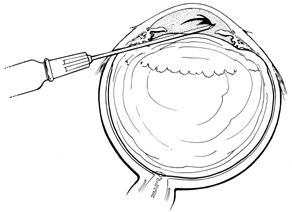
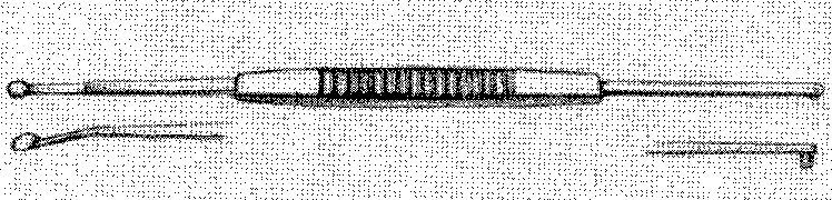
Current cryotherapy instrumentation employs expansion of high-pressure nitrous oxide at the tip of a probe generating temperatures as low as 89°C. The temperature effect is confined to the tip of the probe by an insulating sleeve. A probe 2.0 to 2.5 mm in diameter usually is used for retinal work. Treatment of retinal breaks and pathologic conditions requires accurate placement of the cryoprobe tip. The surgeon must be certain that the indentation visualized with the indirect ophthalmoscope is the tip of the probe and not the shaft. Confusion between the tip and the shaft of the cryo-probe can cause inadvertent posterior freezes.33 To minimize the possibility of this complication, the surgeon must indent only with the tip of the cryoprobe (Fig. 10). It is also helpful to perform the first freezes at the most anterior aspect of the area requiring treatment to assess both location and intensity of treatment.

Fig. 10. A. Inadvertent posterior placement of cryoprobe tip caused by visualization of cryoprobe shaft. B. Correct placement of cryoprobe tip.

The goal of treatment is to surround all retinal breaks with 1 to 2 mm of contiguous treatment. When possible, treatment should include freezing of the overlying retina, because this results in a stronger adhesion than does treatment of the pigment epithelium alone.25 To avoid the damage of refreezing, treatment should not significantly overlap. The treatment end point is retinal whitening without ice crystal formation.34 Slight whitening of the retina because of retinal edema is noted several minutes after freezing, which helps to assess the adequacy of treatment. If retinal treatment is impossible because of bullous retinal elevation, treatment of the pigment epithelium alone may be performed, or treatment can be deferred until after drainage of subretinal fluid.

For flap retinal tears, treatment is performed contiguously around the tear and then extended anteriorly to the ora serrata. Care is taken not to freeze bare RPE in the bed of the retinal break where there is no overlying retinal tissue. Small retinal breaks and atrophic retinal holes can be treated with single freezes centered on the retinal break (Fig. 11).

Fig. 11. Placement of cryotherapy around flap retinal tear.

One potential disadvantage of cryopexy is dispersion of pigment epithelial cells, which can result in subretinal pigmentary changes after reattachment.35,36 Also, dispersion of viable pigment epithelial cells capable of causing proliferative vitreoretinopathy has been demonstrated following cryopexy.37 The clinical relevance of cryopexy-induced pigment epithelial cell dispersion in the pathogenesis of proliferative vitreoretinopathy following scleral buckling is controversial. Some retrospective analyses suggest that cryopexy is a risk factor for the development of postoperative proliferative vitreoretinopathy (PVR),38,39 whereas other studies do not show an association between cryopexy and PVR.40 Accordingly, it seems prudent to minimize cryotherapy-induced pigment epithelial cell dispersion by not over treating and by avoiding unnecessary scleral depression of treated areas, which enhances dispersion of viable cells.41 Therefore localization and examination with scleral depression should be performed before cryopexy.

In addition, although cryopexy does not permanently damage the choroid, it does induce choroidal congestion and hyperemia,27 which may complicate drainage of subretinal fluid through treated areas. Finally, cryopexy causes breakdown of the blood–ocular barrier,42 and its use has been implicated as a cause of postoperative cystoid macular edema and exudative detachments after scleral buckling.43 Despite these potential problems, cryopexy remains the choice of most retinal surgeons for the intraoperative treatment of retinal breaks during scleral buckling.44

Photocoagulation

Photocoagulation is another means for creating pigment epithelial–retinal adhesion. Laser delivery systems coupled to an indirect ophthalmoscope allow retinal surgeons to use photocoagulation during scleral buckling.45 Laser photocoagulation can also be applied postoperatively after retinal reattachment.46 The pigment epithelium is the primary chromophore for energy uptake with photocoagulation. Therefore the area of retina to be treated must be either attached or placed in juxtaposition to the pigment epithelium by scleral depression. As a result, treatment of bullously elevated retinal breaks or areas with atrophic or attenuated pigment epithelium may be difficult. Intraoperative transpupillary photocoagulation also requires optimal visualization. Miosis, corneal opacification, cataract, and vitreous hemorrhage may preclude transpupillary photocoagulation.

Transscleral diode laser photocoagulation also may be used to treat retinal pathology.47 The long wavelength (810–840 nm) of the diode laser penetrates the sclera and is absorbed by the pigment epithelium. Placing the detached retina in juxtaposition to the pigment epithelium with scleral depression allows treatment of retinal pathology. The desired end point is a gray to gray-white burn. Potential complications include rupture of Bruch's membrane and the pigment epithelium, which may result in choroidal bleeding. Mild thermal effects on the sclera may be evidenced by blue-gray discoloration of the sclera. A potential advantage of transscleral diode photocoagulation over other methods is the ability to treat through preexisting scleral buckling components.48 A disadvantage of both transscleral and transpupillary photocoagulation is that photocoagulation is more time consuming and requires more individual treatment spots than cryopexy.

Photocoagulation has several desirable features. It can be delivered with great precision, both in terms of location and intensity. Compared with diathermy and cryopexy, it causes less breakdown of the blood–ocular barrier.42 The thermal effect of photocoagulation is confined predominantly to the retina and pigment epithelium, with little or no effect on the choroid or sclera.49 Finally, photocoagulation induces an adhesive effect between the retina and pigment epithelium within 24 hours.50

Back to Top
METHODS OF SCLERAL BUCKLING
A variety of buckling techniques and materials may be employed for retinal reattachment surgery.

EXPLANT TECHNIQUES

Scleral buckling, or indentation, was popularized by Ernst Custodis in 1953.51 He described placement of a polyviol explant that was sutured to the sclera overlying any retinal break. The explant indented the sclera against the retina and closed the retinal break. Although the precise mechanism by which scleral buckling works remains uncertain, it was Custodis' observations that led to current explant scleral buckling. Lincoff and colleagues further refined Custodis' procedure and introduced cryotherapy as a substitute for diathermy.52,53 Over the ensuing years, explant techniques and materials have continued to evolve.34,54

Explant techniques allow accurate and relatively easy placement of scleral buckling material to support retinal pathology. The development and refinement of cryotherapy were major steps in the evolution of explant techniques.53 The ability to treat retinal pathologic conditions effectively without the need for scleral dissection has resulted in explant surgery becoming the procedure of choice for most retinal surgeons.

Buckling Materials

Explants are made of solid silicone rubber or silicone sponges and come in a variety of sizes and shapes. Medical-grade silicone rubber, consisting of cross-linked polydimethyl Siloxane is the most common buckling material used. This solid silicone rubber material was originally described by Schepens and colleagues55 for use as scleral implants.

Solid silicone rubber is produced in a variety of sizes and shapes. Three basic shapes are available: straight, symmetric tire, and asymmetric tire. All three shapes can be grooved to accommodate placement of an encircling band. Unlike the straight implants, the tire implants have radii of curvature that approximate the shape of the globe (Fig. 12). The asymmetric tires also provide increased buckle height posteriorly. For many detachments, the authors prefer an asymmetric tire, which allows effective buckling of the ora serrata while minimizing anterior displacement of the encircling band because of its posteriorly placed groove (Fig. 13). This advantage is negated if the tire completely encircles the eye. The thinner anterior edge of this element may also minimize the chance of anterior extrusion. Solid silicone meridional implants can be used as explants. These elements can fit beneath either encircling bands or tires. When radial elements beneath circumferential buckles are indicated, the authors prefer solid silicone meridional elements (Fig. 14).

Fig. 12. Drawings and dimensions of strips and symmetric tires.

Fig. 13. Drawing of shape and dimensions of 7- and 10-mm asymmetric tires.

Fig. 14. Drawing of shape and dimensions of meridional elements.

Silicone sponges also are made of silicone rubber but have many small air-filled pockets that give the sponge great compressibility and elasticity. Unlike true sponges, however, their absorption capabilities are minimal.56 Sponges are made in varying sizes and shapes, from 3 mm circular sponges to 5 × 7 mm oblong sponges. They may be grooved or have a central tunnel for anchoring of an encircling band.57

Biologic materials, including fascia lata, preserved human sclera,58 and gelatin,59 also have been used for scleral buckles. Currently, these are rarely used and are discussed primarily for historical interest. Of these, gelatin is the most versatile. As it hydrates, gelatin gradually swells, providing increased buckle height a few days after surgery. Over 3 to 6 months, the gelatin, which is predominantly hydrolyzed collagen, is broken down, and the buckling effect is lost. Gelatin can be used as either an implant59 or explant.34

The Miragel implant (Mira, Inc., Waltham, MA) is a nonbiologic hydrogel made of a hydrophilic copolymer of methyl acrylate with 2-hydroxy ethyl acrylate cross-linked with ethylene glycodiacrylate. It was used as an explant from 1981 until 1996, when the manufacturer stopped its distribution. Although no longer available, this buckling material must be familiar to ophthalmologists because of its late-onset complications. Although Miragel implants were reported to be tolerated well in both short- and long-term evaluation,60,61 complications such as extrusion, fragmentation, limitation of ocular motility, and intrusion of the implant have been reported to occur between 7 and 11 years after implantation.62–65 Consequently, patients who received this buckle material must be monitored closely over the long term. Often the buckle material must be removed. Indications for removal include buckle exposure, infection, motility disturbance, or intrusion. Intrusion of the buckle may be heralded by recurrent vitreous hemorrhage. Removing the hydrogel is difficult because of the friability of the material. The material must be gently “milked” from the sub-Tenon's location with a muscle hook, extracted with a cryoprobe, or carefully aspirated with small-gauge suction. There have been reported cases of scleral rupture and retinal incarceration during removal of the buckle material. Consequently, extreme care should be taken when traction is applied to the rectus muscles or when exploring the subconjunctival and sub-Tenon's space for buckle fragments.62,63

Scleral Suture Technique

Explants are secured to the sclera with partial-thickness scleral sutures. When bands, tires, or sponges are used, these sutures are placed in a mattress fashion parallel to the long axis of the element being supported. To support solid silicone meridional elements beneath tires or bands, the surgeon places the mattress suture perpendicularly to the long axis of the meridional element (Fig. 15).

Fig. 15. Suture placement for both tire and meridional element.

Accurate and effective suture placement is critical to the success of explant procedures. After identification and localization of all retinal breaks and other retinal pathology requiring support, the surgeon selects an appropriate explant. For most detachments, the actual element selected is not as important as the accurate localization and proper placement of the element with respect to the retinal breaks. Proper placement of the element requires effective suturing technique. A spatula needle with a 5-0 nonabsorbable suture such as polyester, nylon, or polypropylene is used.

When suturing, the surgeon must firmly fixate the globe. This is best done by grasping a muscle insertion with a toothed forceps. Magnification with loupes or the operating microscope facilitates suture placement. The suture is passed through the sclera at one-half to three-fourths depth over a distance of 3 to 5 mm, usually in a horizontal mattress fashion. A combination of adequate depth and length is necessary for maximum suture strength.66 Once the proper scleral depth has been obtained, the suture should be passed at that level. Uneven passage of the needle induces buckling of the sclera, which may lead to perforation. After the needle has been passed through the sclera and the tip brought out, the needle is released from the needle holder and the tip is grasped. It is important to complete passage of the needle along the arc of the needle, avoiding posterior pressure or dragging on the hub of the needle, which may perforate through the remaining underlying sclera (Fig. 16).

Fig. 16. Passage of spatula needle through sclera. It is important to complete passage of the needle along the arc of the needle, avoiding posterior pressure or dragging on the hub of the needle, which may perforate through the remaining underlying sclera (inset).

Usually sutures are placed a minimum of 2 mm farther apart than the width of scleral contact for a given element (e.g., 9 mm apart for a 7-mm element). To ensure that the most posterior edge of the retinal break is supported, the surgeon places the posterior suture a minimum of 2 to 3 mm posterior to the scleral localization mark. For encircling elements, placement of the mattress suture in the same meridian as the retinal break or breaks provides additional height beneath the breaks (see Fig. 15).

When one is suturing posteriorly, the vortex veins and their tributaries must be avoided. This sometimes requires straddling a vortex, as seen in Figure 17. Thin sclera also presents problems, and sometimes long suture passes are not possible. In this case, several short bites in areas of thicker sclera may be effective. The use of cyanoacrylate adhesive to support suture bites in thin sclera has been described.67 Currently, in eyes with very thin sclera, the authors perform primary vitrectomy techniques for retinal detachment repair.

Fig. 17. Suture placement to avoid intrascleral passage of vortex vein.

Some surgeons prefer silicone sponges for explants. Sponges provide a rounded buckling contour, the height of which is easily adjusted by varying the distance between the mattress suture bites. The buckle height obtained with segmental sponge explants persists for at least 3 years.68 Sponges are easily trimmed and thus are quite adaptable. They are effective for support of radial tears and are particularly useful for posterior radial tears.69

Based on the size of the tear, an appropriate sponge is selected. Sutures are placed 2 to 3 mm beyond the circumferential and posterior extent of the tear as determined by localization (Fig. 18). Usually one or two mattress sutures are placed, and the buckling effect is carried anteriorly to the ora serrata. The anterior suture is usually a simple horizontal mattress; the posterior suture may be either a crossed or simple mattress. Often it is difficult to pass a suture from posterior to anterior when one is working posterior to the equator. A crossed mattress alleviates this problem but does not provide as effective posterior support, as does a simple mattress. Thus the buckle may need to be extended farther posteriorly. When necessary, crossed mattress sutures can be converted to simple mattress sutures (Fig. 19).

Fig. 18. Placement of sutures for radial sponge.

Fig. 19. Technique for placement of posterior mattress suture.

Segmental versus Encircling Buckles

Placement of explant material can be either segmental or encircling. Segmental buckles usually are reserved for detachments with single or closely spaced retinal breaks less than one clock hour in total extent or with posterior breaks. These breaks can be effectively closed using radial sponges or segmental placement of circumferential solid silicone elements. The primary advantage of segmental buckles is the relative ease of placement; another advantage is minimal change in refractive error induced by segmental buckles. For posterior breaks, segmental elements allow closure of the break while avoiding the side effects of large posterior encircling elements. However, for most large posterior breaks and all macular holes, the authors prefer closure with gas and vitrectomy techniques.

Although segmental buckles effectively close isolated tears, they do not provide retinal support elsewhere. Specifically, other areas of vitreoretinal traction away from the segmental element are not supported, which may result in the formation of new retinal breaks. Because of the limited support offered by segmental buckles, the authors prefer encircling procedures when possible. Encircling procedures are particularly indicated in: (a) cases with multiple breaks in different quadrants, (b) aphakia, (c) pseudophakia, (d) myopia, (e) diffuse vitreoretinal pathologic conditions, such as extensive lattice degeneration or vitreoretinal degenerations, and (f) proliferative vitreoretinopathy of grade B or greater.34,54,70 Either sponges or solid silicone rubber can be used as encircling elements. Because of their compressibility, encircling sponges tend to result in a variable undulating contour to the buckle unless multiple sutures are placed in each quadrant. The authors prefer solid silicone elements for encircling procedures.

The anteroposterior position of the encircling element depends on the location of the vitreoretinal pathology to be supported. When retinal breaks in detached retina are associated with traction, the buckle should be positioned so that the posterior edge of the break lies on the posterior crest of the buckle. The buckling effect should extend for 30° on either side of the tear and extend anteriorly to the ora serrata (Fig. 20). If the encircling element is supporting pathologic conditions in attached retina, such as a retinal break, lattice degeneration, or prominent vitreoretinal adhesions, the most posterior aspect of the condition needs to be supported by the encircling element. If no specific pathologic factor is to be supported, the encircling element should support the posterior margin of the vitreous base.

Fig. 20. Correct position of buckle for flap retinal tear.

The vitreous base eccentrically straddles the ora serrata, extending more posteriorly on the nasal side. The exact location of the vitreous base sometimes can be determined with indirect ophthalmoscopy and, when this is possible, the vitreous base can be marked on the sclera to ensure accurate placement of the encircling element. If the vitreous base cannot be identified ophthalmoscopically, it can be assumed to lie approximately 3 mm posterior to the ora serrata nasally and 2 mm posterior to the ora serrata temporally (Fig. 21).

Fig. 21. Position of vitreous base in relation to ora serrata.

A solid silicone band 2.5 mm wide often is used to support the vitreous base or vitreoretinal pathologic condition in attached retina. The encircling band is secured to the sclera with either mattress sutures or through scleral tunnels to prevent migration of the band once it is shortened. It is not necessary to place the band around the greatest curvature of the eye. The ends of the band can be secured with a clove hitch nonabsorbable suture,34 or silicone sleeve.71 The authors prefer the silicone sleeve because it allows easy adjustment of the band throughout the surgery (Fig. 22).

Fig. 22. Technique for silicone sleeve placement.

One result of circumferential encircling scleral buckles is fish-mouthing of retinal breaks.72,73 This is caused by the relative circumferential shortening of the sclera and choroid induced by the buckle in relation to the retina. This relative excess of retinal surface causes the retina to lie in radial folds over the buckle. These folds, in conjunction with vitreous traction, can result in the failure of retinal breaks to settle over the buckle. The breaks may form an elongated oval configuration in the anteroposterior meridian over the buckle, resembling the open mouth of a fish (Fig. 23). Persistent elevation of the retina resulting from the fishmouth phenomenon can result in surgical failure and therefore needs to be addressed.

Fig. 23. Fishmouth appearance after circumferential buckle.

There are three basic techniques in the management of fishmouth retinal tears. The easiest is to decrease the height of the circumferential buckle. This usually is effective only if buckle height is excessive. Placement of a radial element beneath the circumferential element effectively closes most fishmouth tears.69 The radial element increases the surface area of the pigment epithelium and choroid beneath the break, thereby decreasing the disparity between the retinal surface area and the surface area of the bed of the buckle (Fig. 24). 73 Injection of an intravitreal gas bubble, combined with appropriate positioning, also effectively closes fishmouth tears. The gas is injected through the pars plana under direct visualization with the indirect ophthalmoscope. During the injection, care must be taken to avoid formation of multiple small bubbles, which may then pass through the tear into the subretinal space. This can be prevented by injecting into the most superior aspect of the vitreous cavity. With accurate positioning, a bubble of 0.3 mL is adequate to close retinal breaks of up to one clock hour.74 The use of expansile gases, such as sulfur hexafluoride or perfluoropropane, can increase the intraocular volume of gas.

Fig. 24. Radial element beneath tear alleviating fishmouth phenomenon.

For encircling elements, buckle height can be obtained in two ways. For thin encircling elements, such as solid silicone bands, the explant can be shortened in relation to the circumference of the globe. The best method of determining how much to shorten the band is a visual assessment of the buckle height created by the band.34,75 The band is shortened only the amount necessary to create the desired buckle height. This helps to avoid the complications of excessive buckle height, such as anterior segment necrosis.76 The height and breadth of the scleral indentation obtained are a linear function of the amount the encircling element is shortened.77 However, this leads to circumferential shortening of the globe in relation to the retina, predisposing to radial folds on the buckle and the fishmouth phenomenon.59,73 Increased axial elongation of the globe also occurs.75,77

The second method of obtaining buckle height is by suture placement without shortening of the element in relation to the ocular circumference. This technique is used with wider and thicker explants such as tires and sponges. The farther apart the bites of the mattress suture are placed, the greater the height of the buckle when the sutures are tightened. This effect requires two sutures per quadrant but provides excellent buckle height by shortening the sclera in an anteroposterior direction rather than circumferentially. With this technique, axial length actually decreases.77

IMPLANT TECHNIQUES

The current era of scleral buckling using implant techniques for rhegmatogenous retinal detachment began in 1960 with Schepens and colleagues'55 description of scleral dissection, intrascleral placement of silicone buckles (implants), and diathermy. Subsequent modifications and refinement of the technique have been described.12

Scleral Dissection

After localization of all retinal breaks requiring support, a lamellar scleral undermining or scleral dissection is performed. The extent of dissection depends on the size of the intended buckle. It is recommended that the lamellar scleral dissection extend 3 mm posterior, 2 mm anterior, and 3 to 4 mm circumferentially beyond the retinal breaks.12 The dissection is begun with an incision parallel to the limbus at the posterior edge of the retinal breaks. The depth of the incision is considered appropriate if a thin gray layer of sclera remains over the choroid. The lamellar dissection is performed using a blunt dissector with traction on the reflected scleral flaps. Care is taken when dissecting posteriorly to the equator to avoid severing the vortex veins. Modifications of the posterior scleral flap often are necessary to avoid the vortex veins. The size of the scleral dissection is designed to accommodate the width plus the height of the desired implant (Fig. 25).

Fig. 25. A. Technique for scleral dissection. B. Recommended extent of scleral dissection for implant placement in relation to retinal breaks.

Diathermy

After completion of lamellar scleral dissection, diathermy is applied. The use of diathermy is the primary reason that lamellar scleral dissection is performed. When diathermy is applied to full-thickness sclera, energy transmission is irregular and unpredictable. Diathermy through full-thickness sclera also induces scleral necrosis and shrinkage.28,53 These problems are minimized by placing diathermy through the surgically thinned sclera.

Diathermy is applied for 3 to 5 seconds using a blunt conical electrode on the dried scleral surface. The intensity is assessed ophthalmoscopically and then adjusted as required. The diathermy burns leave a 1-mm mark on the thinned scleral surface and are spaced about 2 mm apart in staggered rows parallel to limbus. Treatment is started posteriorly and proceeds anteriorly (Fig. 26). If a stronger than usual chorioretinal adhesion is desired, the intensity of burn, not the number of burns, should be increased.12 However, excessive intensity may result in retinal holes if attached retina or choroidal hemorrhage are being treated. When diathermy is placed, the long posterior ciliary arteries, nerves, and vortex veins should be avoided. Transillumination viewed through the thinned scleral bed is helpful in identifying these structures.78

Fig. 26. Distribution of diathermy placement in bed of scleral dissection.

Buckle Implantation

After placement of diathermy, the silicone implant is trimmed to fit beneath the scleral flaps, and nonabsorbable 4-0 mattress sutures are placed in the scleral flaps. The flaps then are closed with temporary suture ties. An encircling band may be placed through the groove of the implant, beneath the scleral flaps and around the globe.

The band is anchored with scleral tunnels or mattress sutures. The ends of the band are joined with a tantalum clip, nonabsorbable suture, or silicone sleeve (Fig. 27). Drainage of subretinal fluid usually is required and is described later.

Fig. 27. Placement of implant in scleral dissection and closure of scleral flaps.

If 360-degree buckling with a silicone implant is required, Schepens12 recommends that this be performed as a two-step procedure. He suggests that a scleral lamellar undermining be made for 220 to 270 degrees and that the implant placed with an encircling band. If this fails to reattach the retina, the remaining sclera is undermined and buckled with a second procedure 2 to 6 weeks later. This apparently diminishes the complications associated with 360-degree scleral dissection, such as anterior segment ischemia and choroidal detachment.

Proponents of implant techniques believe that diathermy is the best means of obtaining a chorioretinal adhesion because of the strength of the adhesion obtained.12 This belief is based on a presumed correlation between the histologic appearance of diathermized eyes and the strength of the chorioretinal adhesion. Whether or not this correlation is valid is unknown. Comparisons of the tensile strength of chorioretinal adhesions produced by diathermy, cryotherapy, and photocoagulation show little difference among the three methods.26,79 However, the techniques to measure the tensile strength of chorioretinal adhesions are fraught with methodologic difficulties. Implanted buckles may also have the advantage of being less likely to become infected or extrude.

Back to Top
MANAGEMENT OF SUBRETINAL FLUID
The need for drainage of subretinal fluid during scleral buckling remains a controversial issue. Some authors believe that most cases can be managed without drainage of subretinal fluid, whereas others believe that drainage is a crucial aspect of the procedure. 19,34,54,84,91,93,96,184 Drainage of subretinal fluid is necessary in virtually all implant diathermy cases because of the increased intraocular pressure caused by diathermy-induced scleral shrinkage.

DRAINAGE TECHNIQUES

The rationale for drainage of subretinal fluid is twofold: to diminish intraocular volume so as to allow elevation of the buckle without difficulties with elevated intraocular pressure, and to allow the retina to settle on the elevated buckle by removing fluid from the subretinal space. Effective drainage of subretinal fluid places the retinal breaks in juxtaposition to the buckle, thereby facilitating closure of the breaks. Although many retinal detachments can be managed effectively without drainage as described later, the authors prefer to drain retinal detachments with one or more of the following characteristics:

  1. Bullous detachments. Drainage usually is necessary in these cases so that the retinal break can be placed in juxtaposition to the buckle. This is particularly valid if confluent retinopexy around the tear cannot be obtained because of the bullous elevation.19
  2. Inferior breaks. Inferior breaks tend to settle less readily on the buckle than do superior breaks, perhaps because of gravity.80 Also, inferior breaks are less effectively managed postoperatively with air injections.
  3. Proliferative vitreoretinopathy. Proliferative vitreoretinopathy may prevent the retina from settling, resulting in persistence of open retinal breaks. The authors drain all cases of grade B or greater that undergo scleral buckling.
  4. Highly myopic detachments and aphakic or pseudo-phakic detachments. The syneresis of the vitreous that occurs in myopia or following lens extraction may be a factor in the failure of these retinas to settle on the buckle without drainage.
  5. Chronic detachments. Subretinal fluid in chronic detachments becomes viscous, assuming the biochemical profile of plasma.81 The high osmolarity of the fluid may slow resorption by the pigment epithelium.82,83
  6. Poor RPE function. Detachments in patients with age-related macular degeneration and high myopia are characterized by prolonged resorption of subretinal fluid, presumably because of decreased ability of the RPE to remove fluid from the subretinal space.
  7. Eyes intolerant of sustained intraocular pressure rises, such as those with known glaucoma.

The selection of an external drainage site is affected by several factors. Obviously the location of subretinal fluid is a primary concern. The authors prefer to select the drainage site after placement of scleral sutures and loose placement of the buckle. This ensures that the location or amount of the subretinal fluid has not changed during the placement of sutures. It is not necessary to drain where the fluid is greatest but where there is adequate fluid to safely enter the subretinal space. When possible, it is preferable to drain just above or below the horizontal meridian, either temporally or nasally (Fig. 28). This location avoids the major choroidal vessels and vortex veins. Usually the vortex veins can be identified ophthalmoscopically and thereby avoided. However, sometimes transillumination may also be useful.78 The horizontal meridian allows relatively easy access to the sclera, although in patients with large noses or tight orbits the nasal approach sometimes can be difficult. If subretinal fluid in the horizontal meridian is inadequate, drainage must be performed elsewhere. Usually either side of the vertical rectus muscles avoids major choroidal vessels. The authors try to avoid drainage through areas that have received cryotherapy because of the choroidal hyperemia and congestion that cryotherapy induces.27

Fig. 28. Location of preferred drainage sites.

Drainage can be performed via either a radial sclerotomy and external observation of the drainage site, or external needle drainage and indirect ophthalmoscopic observation of the needle in the subretinal space. External needle drainage is technically more challenging. It is performed either with the scleral buckle left loose as originally described by Charles in 198584 or with it tightened to an appropriate height and the sutures permanently tied.85 Using the technique reported by Charles, Burton, and colleagues reported an 84% successful rhegmatogenous retinal detachment repair with one surgery.86 However, subretinal hemorrhage occurred in 22% of cases, and the blood extended subfoveally in four cases. Raising the intraocular pressure by tightening the scleral buckle is thought to mitigate intraocular bleeding. The subretinal hemorrhage rate as described by Jaffe and colleagues was 4%.85 To perform external needle drainage, the surgeon uses a 0.5-inch 25-, 26-, or 27-gauge needle attached to a tuberculin, 3- or 5-cc syringe with the plunger removed. The authors prefer the larger gauge needles because these do not bend easily when scleral depressing or while angling the needle to enter the eye. The authors also prefer the larger volume syringe to minimize the water pressure within the syringe. A tuberculin syringe holding its maximum of 1 mL of fluid has more pressure and therefore resistance to drainage when held vertically, compared to a 5-cc syringe. Consequently, more subretinal fluid can be drained with a larger volume syringe before equilibrium exists between the water pressure of the syringe and the intraocular pressure.

The bevel of the needle is directed away from the sclera. With the needle shaft tangential to the sclera to prevent premature scleral penetration, scleral depression is performed to confirm the correct location for drainage. When the correct location is found, the syringe is angled away from the eye and the needle is passed through the sclera and choroid into the subretinal space. The surgeon directly observes the needle with indirect ophthalmoscopy as it passes into the subretinal fluid. The detached retina immediately begins to undulate, signifying that subretinal fluid is leaving via the needle. Pressure must be maintained during the drainage, either by the tightened scleral buckle or by an assistant pulling on the rectus muscle bridle sutures or depressing the sclera. Pulling on the rectus muscle or scleral depression may result in movement of the globe if not done carefully. Any movement makes proper needle position difficult. Such movements are eliminated when the scleral buckle is pretightened. As the retina settles and nears the drainage needle, the needle is slow withdrawn until it is removed from the subretinal space. If the retina is inadvertently penetrated, retinopexy should be applied to the site. If the eye is hypotensive after the drainage, it can be reformed with balanced saline, air, or expansile gas.

A scleral cut down and external drainage does not require indirect ophthalmoscopic observation of the needle passing into the subretinal space. Before entrance into the subretinal space is made, a 3- to 4-mm radial sclerotomy is made with a rounded disposable blade. Care must be taken to cut the sclera perpendicular to its surface to avoid shelving of the wound. As the cutdown approaches the suprachoroidal space, magnification with loupes or with the 20-diopter lens is helpful to visualize residual scleral fibers over the choroid. It is important to remove these fibers because during drainage they may swell and prematurely close the drainage site. The anteroposterior location of the drainage site is dependent primarily on the location of the subretinal fluid and the configuration of the detachment.

When possible, the authors prefer to drain in the posterior third of the bed of the buckle. This provides adequate support of the drainage site in the event of complications such as retinal incarceration or choroidal hemorrhage and also provides immediate effective closure of the drainage site when the buckle is pulled up. If, because of the configuration of the detachment or the position of the buckle, it is not possible to drain in the bed of the buckle, a mattress suture is placed through the margins of the sclerotomy to provide closure once drainage ends. Drainage outside the bed of the buckle may allow the buckle to be pulled up as the drainage proceeds. If the drainage site is in the bed of the buckle, the authors do not close the site with a suture because of the danger of erosion of the suture through the sclera by the pressure of the overlying explant. The authors have never seen fibrous ingrowth at the drainage site.

After the suprachoroidal space is entered, diathermy can be used to shrink the margins of sclerotomy to improve choroidal exposure. The choroid then is inspected under magnification for choroidal vessels. If major choroidal vessels are seen, consideration is given to selecting another drainage site if possible. The choroid is diathermized to decrease the chance of bleeding on entry. The diathermy shrinks the choroid and closes choroidal vessels. Under magnification, diathermy can be seen to change the normal smooth, dark blue color of the choroid to a parchment gray color and texture (Fig. 29).

Fig. 29. A. Location of scleral cutdown in bed of buckle. B. Diathermy of scleral margin. C. Diathermy of choroid.

Entry through the choroid and into the subretinal space can be performed by a variety of techniques using needles, diathermy electrodes, or lasers. When a sharp diathermy electrode is used, the diathermy is activated during entry to minimize the chance of hemorrhage. A flat, insulated diathermy electrode may be used to vaporize the choroid to create a choroidotomy without entering the subretinal space.87 This technique was reported to have a low complication rate and was successful in achieving drainage in 83% of cases. The remainder required needle drainage.

Photocoagulation using either an external endolaser probe or an indirect ophthalmoscope laser delivery system can also vaporize the choroid to create a choroidotomy. The endolaser probe is placed within 1 to 2 mm of the exposed choroids, taking care to avoid touching the choroid. When using a green wavelength, a power of 0.5 to 1.5 W and a duration of 0.2 to 0.5 second usually is adequate to form a choroidotomy.88 When using an infrared diode wavelength, lower power is necessary. A randomized trial demonstrated external argon laser choroidotomy to be as safe and effective as needle choroidotomy.89 However, the use of an endolaser probe is expensive. The expense can be eliminated by using a laser indirect ophthalmoscope delivery system.90 With this technique the exposed choroid is visualized using the indirect ophthalmoscope and a 20-diopter lens. The magnification of a 20-diopter lens allows excellent visualization of the choroid and subsequent drainage through the choroidotomy. An initial power of 0.3 W and duration of 0.1 to 0.2 seconds is used. The power is increased in 0.1 W increments until drainage is achieved.

All of the preceding techniques are effective and each may have some advantages in select detachments. For example, laser or diathermy choroidotomy may be preferable in shallow detachments, because they do not require entry into the subretinal space. Charles' technique is particularly useful in eyes with a thickened choroid or posteriorly displaced subretinal fluid. However, for most detachments requiring drainage the authors prefer a simple choroidotomy with a 27- to 30-gauge needle. Entry into the subretinal drainage space can be either perpendicular or tangential to the choroid, depending on the amount and location of the subretinal fluid. Usually a single stab incision of the choroid is made; however, when highly viscous subretinal fluid is expected, a large needle or cutting entry may be used, although this larger choroidal incision may increase the chance of choroidal hemorrhage.

After the subretinal space is entered, the needle is quickly removed and the site inspected under magnification. Usually the presence of fluid around the needle signifies entry into the subretinal space (Fig. 30). As the fluid drains, it is important to maintain a relatively normal and constant intraocular pressure to prevent retinal incarceration or hemorrhage. Indentation of globe at the ora serrata in the meridian of the drainage site facilitates elevation of the retina over the site and allows movement of subretinal fluid to the drainage site (Fig. 31). As the fluid drains, the loss of intraocular volume can be compensated for by indentation with cotton-tipped applicators starting 180° away from the drainage site. The site itself is not manipulated so long as good drainage of subretinal fluid proceeds.

Fig. 30. Entry into subretinal space avoiding any obvious choroidal vessels.

Fig. 31. A. Depression anterior to drainage site to facilitate drainage of subretinal fluid. B. Correct position for depression anterior to drainage site.

As the subretinal fluid drainage slows, pigment particles may be seen in the subretinal fluid, indicating that drainage is nearing completion. When possible, it is helpful if the surgeon or assistant observes the retina over the drainage site ophthalmoscopically. As the fluid drains, the retina will be seen to flutter like a sail in the wind. If visualization of the retina is attempted, care must be taken not to compromise the external drainage site. Despite attempts to maintain constant intraocular pressure as the fluid drains, the eye may soften; this may result in formation of folds in the choroid, which can close the drainage site.

If drainage slows or stops prematurely, the sclerotomy may be manipulated with a fine-toothed forceps by pulling the sclera either tangentially or perpendicularly to the choroid. This often reopens the site and reestablishes flow. When drainage ceases, the site is closed quickly by tightening the buckle suture nearest the drain or by closing the preplaced sclerotomy suture.

Quick inspection with the indirect ophthalmoscope assesses the extent of residual fluid; during this examination, intraocular pressure must be maintained with the traction sutures. If drainage is deemed adequate, the remainder of the buckle sutures are tightened and secured with temporary ties, starting in the location of the primary retinal break. If additional drainage is necessary, the buckle suture or drain suture is released and the drainage site again manipulated to allow further drainage. If no further fluid is obtained but additional drainage is deemed necessary, the original site can be closed with a mattress suture and a second drain site selected; however, this is rarely necessary.

Drainage occasionally may be excessive, particularly in eyes with liquefied vitreous and large retinal breaks. This may result in loss of vitreous volume through the break into the subretinal space and out the drainage site. This event can be anticipated in eyes with large breaks and liquefied vitreous (e.g., in elderly patients with highly myopic eyes). By selecting a drainage site away from the break, one can indent over the break during drainage, thereby closing the break and preventing passage of vitreous contents into the subretinal space (Fig. 32).

Fig. 32. Vitreous drainage into subretinal space prevented by closure of retinal break with depression during drainage.

When the amount of drainage appears excessive in relation to the amount of subretinal fluid, loss of liquid vitreous should be suspected. If excessive drainage is obtained, volume usually can be restored by tightening the buckle. If this is inadequate or results in too high a buckle, balanced salt solution or sterile air may be carefully injected through the pars plana to restore volume.

The surgical goal of drainage should be kept in mind. Total drainage of subretinal fluid is not necessary and, in some cases, may not be desirable. The goal is for enough drainage to allow the scleral buckle to close the retinal breaks effectively without excessive elevation of intraocular pressure.

A helpful intraoperative sign to determine the adequacy of subretinal fluid drainage is to assess the contour of the retina in relation to the scleral buckle. If after drainage the retina follows the contour of the buckle, drainage is usually adequate even if there are residual subretinal fluid and retinal folds. However, if after drainage and elevation of the buckle the retina does not appear to follow the contour of the buckle or if the buckling effect is not visible beneath the retina, additional drainage usually is necessary (Fig. 33).

Fig. 33. A. Retinal tear elevated after drainage and placement of a scleral buckle. B. Retinal tear partially closed after drainage with the retina conforming to the shape of the buckle.

After successful subretinal fluid drainage and closure of the drainage site, the buckle is positioned with the preplaced scleral sutures to achieve appropriate support of the retinal pathology. The sutures overlying the retinal break or breaks are pulled up first, followed by the remainder of the sutures. The band, if present, is then adjusted with the silicone sleeve. As the sutures are tightened, they are secured with temporary ties, and the optic nerve is inspected to ascertain arterial perfusion. Once the buckle is positioned and the band adjusted, the fundus is again inspected to determine the status of the retinal breaks in relation to the position and height of the buckle and perfusion of the nerve. The temporary ties allow easy adjustment of the buckle height or position if necessary. This flexibility is a primary advantage of explant techniques.

After the buckle is positioned and the band adjusted, the perfusion of the retina is reassessed. Perfusion can be ensured by inducing pulsations or closure of the central retinal artery by indenting the globe while visualizing the optic nerve with the indirect ophthalmoscope. If pulsations cannot be induced, it must be assumed that the central retinal artery is closed. The maximum duration of central retinal artery closure during retinal detachment surgery that can safely be tolerated is unknown and probably variable among patients.

The authors do not allow the artery to remain closed for more than 5 minutes. If flow is not spontaneously re-established, the pressure is lowered first by loosening the band and then by loosening the sutures away from the retinal tears and drainage site. Usually, with time, the facility of outflow allows the eye to soften enough to reposition the buckle. If blood flow is not re-established, anterior chamber or vitreous taps, as described later, are considered. When the surgeon is satisfied with placement of the buckle and perfusion of the retina, the temporary suture ties are converted to permanent knots.

NONDRAINAGE PROCEDURES

Nondrainage procedures have been shown to reattach the retina with success rates comparable with those of drainage procedures.69,91–94 The primary advantage of a nondrainage procedure is the avoidance of the possible complications associated with transchoroidal drainage. In eyes with relatively shallow detachments, the eye may soften enough after scleral depression for examination and cryopexy to allow placement of the buckle without intraocular pressure problems. Waiting several minutes between tightening of the scleral sutures also may soften the eye. However, nondrainage techniques often require lowering the intraocular pressure by either medical or surgical means. Intravenous osmotic therapy with 20% mannitol (1–2 g/kg of body weight) can soften the eye but, because of the time lag involved, is most effective if given preoperatively when a nondrainage procedure is anticipated.

Medical attempts to lower pressure usually are effective only for relatively small buckles. For circumferential buckles extending more than one quadrant, intraocular volume must be removed surgically. In phakic eyes, anterior chamber paracentesis can remove up to 0.3 to 0.4 mL of aqueous at one time. Paracentesis is performed under magnification at the limbus with a blade, which is passed in a shelved manner through the cornea, entering the anterior chamber over the iris to protect the lens. The blade then is rotated, thereby opening the wound to allow aqueous to egress. The depth of the anterior chamber is closely monitored to prevent damage to the lens. When enough drainage has been obtained, the blade is turned back to its original entering position. This closes the corneal wound and minimizes the chance of iris incarceration as the blade is removed. The corneal wound is self-sealing (Fig. 34). This technique is effective also in pseudophakic patients with posterior chamber intraocular lenses and intact posterior capsules or capsulotomies sealed by the intraocular lens. Alternatively, a 30-gauge needle can be used to remove aqueous.

Fig. 34. Anterior chamber paracentesis technique using limbal approach.

In aphakic patients, limbal entry and drainage often results in vitreous incarceration in the corneal wound. This can be avoided by entering through the pars plana with a 27- or 30-gauge needle on a syringe. The needle is then angled anteriorly through the anterior hyaloid into the anterior chamber, from which aqueous fluid is removed (Fig. 35). If the anterior hyaloid is ruptured and the anterior chamber is filled with vitreous, this technique usually is not effective.

Fig. 35. Anterior chamber paracentesis through pars plana.

When more than 0.4 mL must be removed, the vitreous cavity must be tapped. This is most safely done through the pars plana using a single sclerotomy and a vitreous cutting instrument (Fig. 36). Using a vitreous cutting instrument alleviates the risks of vitreous traction that are inherent to any attempted needle aspiration of liquefied vitreous. The vitrectomy instrument allows removal of a controlled volume, which can be regulated as the buckle is pulled up.

Fig. 36. Vitreous tap using vitrectomy instrument.

Although the final reattachment rates for nondrainage procedures are excellent, the time for complete resorption of subretinal fluid can be prolonged. Subretinal fluid persists for more than 1 week in approximately 25% of nondrainage cases.95,96 Several factors have been implicated in the absorption of subretinal fluid after nondrainage procedures. Increased patient age has been correlated by some investigators19,93 with prolonged absorption of subretinal fluid, but others have not confirmed this association.95 Retinal detachments of greater than 2 weeks' duration require more time for subretinal fluid absorption,96 but this relationship may not hold for detachments of more than 1 month's duration.95 The relationship of the retinal tear to the buckle at the end of surgery correlates with postoperative fluid absorption.91,95 Retinal detachments with retinal tears that are closed by the buckle at the time of surgery tend to have complete absorption of subretinal fluid within 2 days. If there is a slight amount of fluid between the retina and the buckle, the residual subretinal fluid usually is absorbed within 1 week. When the retinal breaks are significantly elevated from the buckle at the end of surgery, more than one-half of patients require more than 1 week for complete absorption of subretinal fluid, and approximately 25% require up to 3 months. Nondrainage procedures are facilitated by preoperative and postoperative bed rest. The increasing trend toward outpatient scleral buckling surgery makes effective bed rest difficult.

Back to Top
CLOSURE

After final adjustments of the buckle have been made, the sutures are tied and the knots rotated to the posterior edge of the buckle. Retrobulbar irrigation of 4 mL of 0.75% bupivacaine significantly decreases postoperative pain after both general or local anesthesia.97 Tenon's capsule is then identified in all quadrants. The authors prefer a layered closure, first closing Tenon's capsule to the muscle insertions in all quadrants (Fig. 37). This ensures that the explant and nonabsorbable sutures are covered by the thick Tenon's capsule and also removes tension on the conjunctival closure, thereby minimizing the possibility of the buckle eroding. During conjunctival closure, the relaxation incisions are closed with a running 6-0 plain gut suture while an attempt is made to evert the edges of the incision to diminish the chance of conjunctival inclusion cyst formation. The conjunctiva is secured at the limbus with one or more sutures (Fig. 38).

Fig. 37. A. Closure of Tenon's capsule to muscle insertion. B. Final appearance after closure of Tenon's capsule to all rectus muscles.

Fig. 38. Conjunctival closure.

Back to Top
COMPLICATIONS
Complications of scleral buckling in the intraoperative and postoperative periods can affect the final visual outcome.

INTRAOPERATIVE COMPLICATIONS

Corneal Clouding

During scleral buckling procedures, adequate visualization throughout the case is critical to the success of the operation. Corneal clouding is a common intraoperative problem and usually is caused by epithelial edema from increased intraocular pressure, which occurs during scleral depression. The epithelium may also become damaged by desiccation or mechanical trauma during the procedure. Mild amounts of epithelial edema may be resolved with topical glycerin or by rolling the epithelium with a dry cotton-tipped applicator. Extensive epithelial edema usually requires debridement with a rounded blade. The cornea is scraped centripetally starting about 2 to 3 mm from the limbus to preserve the limbal stem cells necessary for postoperative re-epithelialization (Fig. 39). The debrided epithelial cells are removed with a cellulose sponge to prevent dispersion, which may result in epithelial inclusion cysts.

Fig. 39. Technique for debriding corneal epithelium.

Miosis

A second intraoperative problem obscuring visualization is miosis, which can occur despite adequate preoperative dilation. It may result from hypotony at the time of drainage or from surgical inflammation caused by cryotherapy or paracentesis. If miosis occurs intraoperatively, additional topical dilation with sympathomimetic and parasympatholytic agents is often helpful. If this is unsuccessful, intracameral epinephrine, 0.2 mL of 1:10,000 dilution, may be considered for aphakic or pseudophakic eyes. The safety of epinephrine in phakic eyes has not been established. Photomydriasis with the argon laser may be obtained by treating the iris stroma midway between the pupil and the angle with an endolaser probe directed through the cornea. This dilates the pupil, but the effect is often short-lived and results in considerable postoperative inflammation. Iris hooks passed through the limbus are useful in pseudophakic and aphakic eyes, but may cause cataract in phakic eyes.

Scleral Perforation

Scleral perforation during placement of scleral sutures is a potentially disastrous complication (Fig. 40). Perforation usually is noticed at the time of suture placement and is heralded by the presentation of blood, pigment, subretinal fluid, or a combination thereof through the suture tract. If perforation is recognized, the retina should be inspected immediately with an indirect ophthalmoscope to determine the depth of the perforation. If the suture is in the choroid or subretinal space without evidence of bleeding or continuing subretinal fluid drainage, nothing additional needs to be done, and placement of remaining suture may be performed if necessary.

Fig. 40. Scleral perforation during suture placement with subretinal hemorrhage and retinal break.

If subretinal fluid is lost with continuing drainage through the suture tract, drainage is allowed to proceed while an attempt is made to maintain a constant intraocular pressure. When drainage stops, the retina is inspected to determine if drainage has been adequate. If so, any remaining necessary sutures are placed and the buckle positioned.

If the perforation has created a retinal break, this should be supported by extending the buckle to support the break and treating the break with retinopexy. When scleral perforation results in subretinal hemorrhage, immediate pressure over the perforation site should be applied and the eye positioned to prevent gravitation of the blood beneath the fovea. If massive subretinal bleeding occurs, immediate vitrectomy with internal drainage of subretinal fluid and removal of subretinal blood should be considered.98 Smaller subretinal hemorrhages can be displaced from the macula by injecting 0.3 to 0.5 cc of air or gas and positioning the patient face-down postoperatively. The bubble will displace the blood from the macula to the peripheral subretinal space.99

Drainage Complications

Despite the use of proper techniques, complications occur during drainage of subretinal fluid.100 A dry tap usually results from failure to perforate the choroid completely. This can be avoided by advancing the needle steadily until fluid is present around the needle. The possibility of subretinal fluid shifting away from the drainage site can be minimized by visualizing the subretinal fluid immediately before drainage. Retinal perforation may occur if the needle strikes the retina because of shallow subretinal fluid or excessive entry of the needle. If the drainage site is in the bed of the buckle, usually no treatment is required, with the possible exception of retinopexy. If the perforation occurs outside the bed of the buckle, a new, unsupported retinal break has been created, and this requires cryotherapy and adjustment of the buckle for adequate support.

Retinal incarceration may occur despite attempts to avoid large fluctuations in intraocular pressure during drainage. Retinal incarceration is identified by the characteristic dimpled appearance of the retina over the drainage site (Fig. 41). Minimal degrees of incarceration rarely result in retinal breaks, but large amounts of incarceration require support. If incarceration occurs outside the bed of the buckle, the buckle should be modified to support the incarceration site.

Fig. 41. Appearance of retinal incarceration into drainage site.

Choroidal hemorrhage is perhaps the most feared complication of subretinal fluid drainage; it usually occurs at the time of choroidal perforation and is marked by the appearance of dark red blood at the drainage site. If this occurs, the drain site should be closed as quickly as possible with either the buckle or sclerotomy suture and the intraocular pressure elevated above the systolic perfusion pressure. Because choroidal blood flow is not autoregulated, it is dependent on ocular pressure and therefore can be diminished. This may decrease the severity of the choroidal bleeding. Indentation over the drain site may also limit the extent of the hemorrhage. If the drainage site is temporal, the eye should be positioned to place the drainage site as inferiorly as possible to prevent the subretinal blood from gravitating to the fovea. One advantage of nasal drainage sites is that hemorrhage is less likely to reach the fovea. After the drain is closed, the pressure elevated, and the eye positioned, the extent of the hemorrhage is assessed ophthalmoscopically. If a large amount of blood is in the subretinal space and beneath the fovea, a vitrectomy with internal drainage of subretinal blood or gas injection with postoperative face-down positioning should be considered.

Late complications can occur months to years after drainage. Subretinal choroidal neovascularization can occur at the drainage site.101,102 This can develop into an angiomatous mass that can decrease vision as a result of vitreous hemorrhage. Treatment of the vascular mass with photocoagulation or cryotherapy may induce resolution of the neovascularization.

POSTOPERATIVE COMPLICATIONS

Complications that follow scleral buckling procedures can occur hours to years after surgery.

Glaucoma

A variety of secondary glaucomas may occur after scleral buckling. However, in the first few days after scleral buckling, routine postoperative discomfort and inflammation may obscure the diagnosis of glaucoma unless the intraocular pressure is measured. Applanation tonometry may be difficult postoperatively; a Tono-Pen, Schiötz tonometer, or pneumotonometry may be helpful.

Angle closure after scleral buckling may occur with or without pupillary block. This is manifested by increased intraocular pressure, corneal edema, and shallowing of the peripheral and sometimes central angle. When pupillary block is present, there is accompanying iris bombé. Usually, however, pupillary block is not present. One presumed mechanism of the angle closure in these cases is shallow detachment of the ciliary body, resulting in anterior displacement of the ciliary body occluding the angle.103 The detachment of the ciliary body results from serous fluid in the suprachoroidal space, which only occasionally is associated with posterior choroidal detachments.104,105 This form of angle closure usually occurs 2 to 7 days after surgery but may occur on the first postoperative day. Accurate gonioscopy often is difficult because of corneal clouding, but slit-lamp examination confirms a shallow peripheral angle. High-frequency anterior segment ultrasound can accurately identify supraciliary choroidal effusions, angle closure, or plateau iris configuration.

Initial management consists of medical therapy to control the intraocular pressure and hourly topical corticosteroids to decrease inflammation and minimize formation of synechia. Medical therapy may be attempted for up to 7 days, but the longer the angle remains closed, the greater the chance for the formation of synechia. Corneal indentation with the four-mirror gonioscopy lens sometimes opens the angle by displacing aqueous between the cornea and iris. This is helpful in assessing the presence of synechia.

If medical therapy is unsuccessful in opening the angle, surgery is necessary. When pupillary block is present or cannot be ruled out, iridotomy with either the argon or YAG laser is performed along with the institution of the medical therapy. A patent iridotomy that fails to deepen the angle and lower the pressure confirms that mechanisms other than pupillary block are operative.

Burton and Folk106 have described a technique of laser iridoplasty that is helpful in alleviating angle closure without pupillary block. Argon laser burns of 200 μm and 0.2-second duration are placed on the iris as close as possible to the angle. This causes the iris to contract and pull away from the angle. Immediate deepening of the angle can be observed by gonioscopy as the treatment proceeds (Fig. 42).

Fig. 42. Technique of laser iridoplasty for angle closure.

If laser surgery is unsuccessful, the suprachoroidal space is drained.107 Radial sclerotomies 3 to 4 mm long are centered over the ciliary body in one to four quadrants, depending on the extent of the angle closure. As the suprachoroidal drainage proceeds, balanced salt solution is injected through a self-sealing limbal stab incision to deepen the anterior chamber (Fig. 43). The anterior chamber injection usually deepens the anterior chamber and opens the angle.

Fig. 43. A. and B. Drainage of suprachoroidal space and deepening of anterior chamber.

If the angle remains closed despite deepening of the anterior chamber and drainage of the suprachoroidal space, it is probably closed by synechia. The synechia can be lysed mechanically with a spatula,107 but the authors prefer to open the angle by injecting sodium hyaluronate between the cornea and iris through a corneal stab incision with a blunt 25- to 27-gauge cannula (Fig. 44). 108 The sclerotomies are left open to allow for further drainage and are covered with conjunctiva. Occasionally the angle closes off again as additional suprachoroidal fluid accumulates, and repeat drainage and anterior chamber deepening may be required. If this occurs, excessive height of the buckle should be considered and the possibility of loosening or cutting the encircling band entertained.

Fig. 44. Use of viscoelastic to lyse peripheral anterior synechia.

Anterior Segment Ischemia

Another cause of postoperative glaucoma is anterior segment ischemia. This should be suspected after encircling procedures with high buckles, particularly if more than one rectus muscle is disinserted. The initial clinical appearance is marked by stromal corneal edema, fibrinous anterior chamber reaction, elevated intraocular pressure and, occasionally, a shallow anterior chamber. Later changes include iris atrophy, anterior and posterior synechiae, cataract, and corneal vascularization. Distinguishing clinically between anterior segment ischemia and postoperative angle closure may be difficult. Mild cases of anterior segment necrosis respond to medical treatment with topical or systemic steroids; however, in severe cases it is necessary to cut the encircling band. Fortunately, with the current trend to use smaller buckles anterior segment ischemia is rarely seen any longer.

The pathogenesis of anterior segment ischemia is unclear. The association with removal of muscles or damage to posterior ciliary arteries suggests that it results from decreased arterial flow to the ciliary body.109 However, experimental models suggest that the clinical picture of anterior segment ischemia is caused by obstruction by the encircling band of venous drainage from the ciliary body.110 The role of venous obstruction by the encircling band is consistent with the usual beneficial clinical response after cutting the band. Apparently, compromise of either arterial inflow to the ciliary body or venous outflow from the ciliary body can result in anterior segment ischemia.

Patients with sickle cell (SC) hemoglobinopathy are at greater risk for anterior segment ischemia after scleral buckling, even when an encircling band is not placed.111 Use of explants and cryotherapy, as opposed to scleral dissection and diathermy, is recommended. Although the incidence of anterior segment ischemia is approximately 3% in the general retinal detachment population, 71% of patients with SC hemoglobinopathy develop this complication after scleral buckling if appropriate preventive measures are not taken.111 Preoperative hemoglobin electrophoresis is necessary in all black patients for whom an accurate hemoglobin analysis is not available. Again, the use of smaller buckles and primary vitrectomy has decreased the incidence of anterior segment ischemia in sickle cell patients.

Infection and Extrusion

Scleral buckling materials constitute foreign bodies and are therefore at risk for infection and extrusion. Clinical features of infected buckling materials differ between cases employing diathermy and scleral implants and those using cryotherapy and scleral explants. Infection in the presence of an implant after diathermy manifests as an acute syndrome with pain, proptosis, vitreitis, and scleral abscess, usually occurring in the first 4 to 9 days after surgery.112 The scleral necrosis induced by diathermy is the key factor in this distinct syndrome. The scleral necrosis facilitates scleral abscess formation and enhances intraocular inflammation.

Infections after cryotherapy and scleral explants do not appear immediately. Usually the infection appears 2 weeks to 2 months after surgery, but cases of infection have been reported as soon as a few days and as long as 13 years after scleral buckling surgery.112–115 Clinical signs include fistula formation, granuloma formation, purulent discharge, and subconjunctival hemorrhage. Progression of the infection may lead to abscess formation with increasing pain, chemosis, and proptosis. A creamy subretinal exudate and retinal whitening may occur overlying the buckle with an exudative retinal detachment. An accompanying vitreitis begins over the buckle. Although this vitreitis is usually a sterile response to bacterial toxins, endophthalmitis may occur.

Staphylococcus is the most common infecting organism.112–116 Coagulase-positive S. aureus species and gram-negative species appear earlier and with greater virulence. Coagulase-negative staphylococcus organisms, such as S. epidermidis and S. albus, tend to appear later (months) with a chronic granulomatous response and buckle extrusion. Explant extrusion may also occur without evidence of infection. Although chronic subclinical infection may play a role, mechanical factors involving the explant and suture placement may be involved.17

The incidence of explant infections and extrusion with modern techniques is about 1%.113,115,116 Preoperative soaking with antibiotics significantly reduces the incidence of both explant infection and extrusion when soft silicone sponges are used.56,113 Some authors recommend squeezing of sponges in the antibiotic solution or direct antibiotic injections into the sponge to enhance antibiotic penetration.113,117 The placement of multiple sponges increases the risk of both infection and extrusion.117,118 Reoperations also have a higher rate of infection and extrusion.113,116

Effective management of infected scleral buckling material usually requires removal of any soft silicone sponge or large solid silicone elements. Topical and systemic antibiotics may occasionally result in symptomatic improvement but are rarely curative. When an encircling silicone band is present, it may be left in position if all soft silicone sponges and supporting sutures are removed.113

The resistance of infected scleral buckles to appropriate antimicrobial therapy involves the formation by bacteria of a glycocalix matrix or biofilm on the silicone elements. This biofilm is an extracellular polysaccharide elaborated by bacteria that functions to enhance bacterial adherence and colonization. Within the biofilm, bacteria are protected from the host response by both mechanical and immunologic mechanisms. The biofilm also limits antimicrobial penetration. Biofilm has been demonstrated on infected scleral buckles by electron microscopy. The presence of the biofilm probably explains the necessity of removing infected scleral buckles despite antimicrobial therapy.119

Scleral buckling material extrusion without evidence of infection also usually requires removal of the offending buckle element. However, if removal of the element is deemed undesirable, scleral patch grafts or processed human donor pericardium may be attempted to cover the element and allow conjunctival reepithelialization over the element.120,121 Occasionally, extruded explants may be well tolerated for extended periods if the patient continues topical antibiotic treatment.122 Removal of scleral buckling material carries a risk of re-detachment of 4% to 33%.123 Patients with elements removed for acute infection run a greater risk of re-detachment than do those in whom elements are removed for extrusion.124 Factors associated with re-detachment after removal of scleral buckling material include the extent of vitreous traction present in the original detachment and the size of the original detachment. Eyes with buckles in place for longer duration have less chance of re-detachment after removal.116 The presence of proliferative vitreoretinopathy carries a high risk of redetachment.124 Because of the risk of re-detachment after removal of scleral buckling material, placement of photocoagulation around retinal breaks or sites of vitreoretinal traction 2 weeks before removal may be worthwhile when possible.

Choroidal Detachments (Choroidal Edema)

Accumulation of serous or serosanguineous fluid in the suprachoroidal space is a relatively common occurrence after scleral buckling and is referred to as choroidal detachment or choroidal edema (Fig. 45). Choroidal detachments occur after both implant diathermy procedures and explant cryotherapy procedures.57,125–127 The overall incidence of choroidal detachments in two separate series was about 40%.57,127

Fig. 45. Schematic of choroidal detachment.

The primary pathogenic factor in the development of choroidal detachments is vortex vein obstruction. This has been demonstrated in animal models110,125,126 and confirmed in clinical studies.57,127 Using explant and cryotherapy techniques, the incidence of choroidal detachment correlates with the circumferential length and posterior position of the scleral buckle.128 If only one quadrant is buckled or only one vortex vein obstructed, the chance of choroidal detachment is minimal. Placement of scleral sutures no further posterior than 14.0 mm from the limbus also lessens the likelihood of choroidal detachment.

Drainage of subretinal fluid is another factor that correlates with choroidal detachment formation.126 Choroidal detachments are less likely to develop with nondrainage procedures. Drainage often results in some degree of hypotony, which appears to exacerbate other factors, such as vortex vein compromise, to induce choroidal detachments.125 Patient age also correlates with choroidal detachment formation. Packer et al.128 found the incidence of choroidal detachment to be 14% in patients 50 years of age or younger, 35% in patients aged 51 to 65 years old, and 53% in patients 66 years and older. The mechanism of patient age in choroidal formation is uncertain but may reflect age-related changes in the integrity of the choroidal vasculature. The extent of retinal detachment also correlates to a limited degree with choroidal detachment formation. If less than 20% of the retina is detached, choroidal detachment is unlikely, but this may simply reflect that smaller detachments are less likely to have encircling posterior buckles and subretinal fluid drainage.

Although the incidence of choroidal detachment is relatively high, the overall effect of choroidal detachments on vision appears minimal. The presence or absence of choroidal detachment has no effect on final visual acuity or anatomic reattachment.128 However, this does not mean that choroidal detachment cannot have a detrimental effect in select cases. As previously described, angle-closure glaucoma with visual loss may occur. Severe choroidal detachment may also result in retinal apposition, and choroidal detachment may occur as part of anterior segment ischemia.

Choroidal detachment usually appears 2 to 4 days after surgery. An associated vitreous haze may be present, which presumably is an inflammatory vitreitis. If the extent of the choroidal detachment is mild to moderate, without risk of retinal apposition, and the pressure is acceptable without angle closure, the natural course is benign with resolution over a few weeks. Antiinflammatory medications such as corticosteroids have been used to treat choroidal detachments.127,128 However, treatment has not been shown to be of visual benefit. Prevention of choroidal detachment by use of routine postoperative systemic corticosteroids (prednisone, equivalent of 80 mg/day) for 2 to 4 weeks has been described in a nonrandomized study.127 Although corticosteroid use appeared to diminish the incidence of choroidal detachments, the final visual acuities were not improved.

In patients with massive choroidal detachment and retinal apposition or angle closure and elevated intraocular pressure, surgical intervention is necessary. For angle closure, choroidal drainage and anterior chamber deepening is performed, as previously described (see the section on glaucoma). When massive choroidal detachment with retinal apposition occurs, a hemorrhagic choroidal detachment should be suspected. This is heralded by a sudden increase in the size of the choroidal detachment, often associated with severe pain. Myopic and elderly eyes are predisposed to hemorrhagic choroidal detachment. Ultrasound may distinguish between hemorrhagic, serous, or combined hemorrhagic-serous detachments. Extensive choroidal detachments with retinal apposition, whether hemorrhagic or serous, are best managed with prompt surgical drainage.129

In aphakic or pseudophakic eyes, the choroidal detachments are drained with a transscleral cutdown into the suprachoroidal space while infusing air with an air pump through the limbus or anterior pars plana (Fig. 46). In phakic eyes, the anterior chamber is deepened with a balanced saline solution injection through a limbal stab incision before choroidal drainage. As drainage proceeds, additional balanced saline solution is injected either by syringe or infusion bottle into the anterior chamber. If the pupil is widely dilated, fluid should pass from the anterior chamber into the vitreous cavity as the choroidal detachment drains.

Fig. 46. A and B. Drainage of choroidal detachment using automated air pump.

Cystoid Macular Edema

Cystoid macular edema is a well-recognized response to ocular surgery. The pathogenesis of cystoid macular edema remains uncertain, but considerable experimental and clinical evidence implicates prostaglandin-mediated inflammation.130 Prostaglandins are compounds derived from cell membrane phospholipids, specifically arachidonic acid; they have multiple effects on microvascular physiology. In the eye, prostaglandins and related compounds increase microvascular permeability, resulting in breakdown of the blood–ocular barrier.130 It has been hypothesized that prostaglandins or related compounds mediate the increased permeability of perifoveal capillaries that characterize the angiographic appearance of cystoid macular edema.130 Prostaglandin production has been demonstrated in both the retina and uvea.131 In addition, rhegmatogenous subretinal fluid contains both prostacyclin and thromboxane A2 derivatives.132 These prostaglandin-related compounds have potent vasoactive properties. The levels of prostacyclin and thromboxane derivatives in subretinal fluid vary greatly among individual patients.

Regardless of which specific compounds are operative, it is clear that cystoid macular edema occurs as a response to ocular inflammation. The surgical trauma of scleral buckling, subretinal fluid drainage, cryotherapy, photocoagulation, or diathermy results in significant ocular inflammation. It is not surprising, therefore, that cystoid macular edema is a relatively common occurrence after scleral buckling. Using cryotherapy and explant techniques, the incidence of angiographic cystoid macular edema 4 to 6 weeks after surgery in phakic eyes was 25% to 28% in three separate studies.130,133,134 Aphakic eyes have a higher incidence: 40% to 60%. Age greater than 55 years correlates with cystoid macular edema development, but the preoperative status of the macula with regard to attachment and the drainage of subretinal fluid do not seem to affect cystoid macular edema.134

The role of cryotherapy in the pathogenesis of cystoid macular edema is controversial. In one study using diathermy and implant techniques, phakic eyes had a 6% incidence of cystoid macular edema 2 months postoperatively.43 It was speculated that diathermy produces less inflammation and that this accounted for the lower incidence. However, in a randomized trial comparing diathermy and implants with cryotherapy and explants, no difference in the incidence of cystoid macular edema was found in phakic, pseudophakic, or aphakic eyes.135

The significance of cystoid macular edema on final visual acuity is difficult to assess. Many additional factors, such as preoperative status of the macula, duration of detachment, and formation of macular pucker, confuse the effect of the cystoid macular edema per se in the relatively small series studied to date.134 Miyake et al.,130 however, demonstrated that eyes with cystoid macular edema after scleral buckling were statistically less likely to obtain vision of 20/40 or better than were eyes that did not develop cystoid macular edema after buckling. The authors find postoperative ocular coherence tomography is useful for the diagnosis of cystoid macular edema.

Prevention of angiographic cystoid macular edema after scleral buckling by use of topical indomethacin has been reported; in a prospective randomized trial of patients undergoing scleral buckling, the incidence of angiographic cystoid macular edema was lowered from 33% in the placebo-treated group to 13% in the indomethacin-treated group.130 However, there was no difference in final visual acuity at 12 weeks between the two groups. For the treatment of established cystoid macular edema, a variety of regimens employing combinations of systemic and topical nonsteroidal antiinflammatory agents and topical, subconjunctival, sub-Tenon's, intravitreal, and systemic corticosteroids have been described. The therapeutic rationale for these treatments is to suppress ocular inflammation. Although no treatment for cystoid macular edema after scleral buckling has been proved effective in a randomized clinical trial, treatment with antiinflammatory agents is reasonable.

Macular Pucker

Macular pucker is a major cause of decreased vision after scleral buckling. The cellular constituents of these preretinal membranes are derived from pigment epithelial cells and retinal glia.136 The incidence of macular pucker formation after scleral buckling ranges from 3% to 17%. Risk factors identified for the development of macular pucker include preoperative proliferative vitreoretinopathy of grade B or greater, age, total retinal detachment, vitreous hemorrhage, and vitreous loss during drainage.135,137,138 Myopic patients have a lower risk than emmetropic patients.137 The presence of aphakia, uveitis, cataract, or the use of implants as opposed to explants are not factors for the development of macular pucker.137

Motility Disturbances

The incidence of heterotropia in the first 6 weeks after scleral buckling surgery is as high as 80%. Fortunately, the vast majority of these deviations are transient, and the incidence of permanent postoperative diplopia is approximately 4%.139–143 Encircling elements and the placement of large elements, either implants or explants, beneath rectus muscles are correlated with diplopia. The diplopia is probably the result of orbital scarring or of myoscleral adhesions. Traumatic damage to rectus muscles during buckle placement or as a result of periocular anesthetic injections also may be a factor.5 Ocular torsion occurs in about half of patients with post scleral buckling diplopia.144 The effect of muscle removal and subsequent reattachment on postoperative diplopia is controversial. Some authors believe diplopia is more common after muscle mobilization, particularly that of vertical rectus muscles,140 whereas others believe that no relationship exists between postoperative muscle imbalance and muscle mobilization.141,145

Initial management of postoperative heterotropia is conservative, because most cases resolve spontaneously. Surgical management can be difficult because buckling elements beneath muscles can hinder muscle recession and adhesions between muscles and buckling elements tend to reform sometimes requiring buckle removal. Because of the anatomic alterations that occur after scleral buckling, standard strabismus surgery principles may not be applicable. For these reasons, prism therapy usually is preferable. If muscle surgery is necessary, an adjustable suture technique should be considered.5,139

Changes in Refractive Error after Scleral Buckling

The extent and direction of change in refractive error after scleral buckling depend on the surgical technique employed. Encircling procedures induce the greatest change in refractive error. This change is greater for phakic than aphakic eyes because of anterior displacement of the lens resulting in an increased shift.75 The amount of direction of refractive error change is related to the height of indentation induced by the encircling band. In a subjective assessment of buckle height after encircling with a 2-mm wide band, Rubin75 found that low to moderate buckle height resulted in −1.56 to 2.24 diopters of change in phakic eyes and −0.74 to 1.14 diopters of change in aphakic eyes. However, high encircling buckles of at least 5 mm indentation resulted in a hyperopic shift of 0.35 diopter for phakic eyes and 0.59 diopter for aphakic eyes. This is because low to moderate buckles induced with this band increase axial length, but higher buckles shorten axial length. Using a 2.5-mm encircling band with varying combinations of radial sponges and 7.5-mm hard silicone elements, Smiddy and colleagues,143 found an average increase in axial length of 0.99 mm and an average induced myopia of 2.75 diopters.

Segmental buckles, whether implants or explants, have little effect on refractive error.143,146 However, large radial elements, such as full-thickness sponges that extend anteriorly beyond the ora serrata, may induce an irregular astigmatism. Changes in corneal topography may also occur with segmental circumferential sponges, steepening in the meridians of the buckle. Encircling buckles tend to produce either corneal flattening with focal central steepening or flattening on one side with steepening on the opposite side. These changes may persist for up to 6 months in an irregular and asymmetric configuration.147

Changes in refractive error after scleral buckling usually stabilize within 2 to 3 months after surgery.75,146–148 When symptomatic irregular astigmatism is created by an anterior segmental buckle, a hard contact lens may provide some relief. Removal of the element entirely alleviates the astigmatism.149 Severe symptomatic aniseikonia or anisometropia may be treated with a hard contact lens or, more recently, with keratorefractive surgery.150

Failure to Reattach

Although most rhegmatogenous retinal detachments can be cured with scleral buckling, a finite number of retinas remain detached after a single scleral buckling procedure. In a retrospective review of 1,088 consecutive cases of retinal detachment undergoing scleral buckling, Rachal and Burton151 reported an initial success rate with one buckling procedure in 76% of patients compared with their final success rate of 89%. In eyes undergoing reoperation, 83% of the retinas eventually were reattached. They analyzed the reasons for failure after one operation and found that most of these were caused by proliferative vitreoretinopathy precluding closure of a known retinal break.

Presumably, proliferative vitreoretinopathy results in traction on the retinal break that is not relieved by the scleral buckle. Because 83% of their failures eventually were reattached with further buckling, they suggested that greater use of the higher and wider buckles, such as those used in the successful reoperations, may decrease failure rates in cases with preoperative signs of proliferative vitreoretinopathy. In addition to proliferative vitreoretinopathy, there are other causes of failure.152 Unsupported retinal breaks, either new or not recognized at the time of the initial surgery, may occur. Inadequate buckling also results in failure. Usually these problems can be addressed with appropriate revision of the scleral buckle.

The primary cause of permanent surgical failure is severe proliferative vitreoretinopathy, which accounts for more than 90% of all permanent surgical failures. Scleral buckling alone is inadequate to address the pathophysiology of proliferative vitreoretinopathy in advanced stages. Although a limited number of cases with proliferative vitreoretinopathy can be reattached with scleral buckling,54 the authors believe that most cases of proliferative vitreoretinopathy of grade C-1 or worse require primary vitrectomy.

Management of Failed Scleral Buckles

Successful management of failed scleral buckles depends on the cause of failure. Missed retinal breaks, new retinal breaks, and inaccurate buckle placement are obvious causes of failure that can be remedied with appropriate revision of the scleral buckle. As discussed earlier, proliferative vitreoretinopathy is the most common cause of failure and usually requires vitrectomy techniques for repair. Occasionally, despite accurate buckle placement and recognition of all retinal breaks, the retina fails to reattach. This usually results from unrelieved traction on the retinal break and may be a harbinger of severe proliferative vitreoretinopathy.

Sometimes bed rest allows the retina to settle on the buckle. Intravitreal injection of a gas bubble with appropriate positioning usually closes the retinal break and allows the retina to settle on the buckle. If wet folds are on the buckle and the break is not bullously elevated, postoperative photocoagulation around the retinal tear can reattach the retina.153 This usually is not effective, however, if there is significant traction on the breaks.

Photocoagulation apparently induces choroidal swelling, which seals the breaks. The laser burns provide some immediate adhesive effect and later mature into strong chorioretinal adhesions. In the presence of significant subretinal fluid beneath the break, gas injection and positioning, followed by photocoagulation on the buckle around the break once the retina is reattached, are often effective.

If attempts to reattach the retina with gas injection and photocoagulation are unsuccessful, formal revision of the buckle to relieve the persistent traction may be performed. This is accomplished by placement of radial elements beneath the buckle or by an increase in the height and breadth of the buckle. Generally, formal revision of the buckle is required only in cases with inferior retinal breaks. Superior breaks in the absence of proliferative vitreoretinopathy are nearly always closed with gas and photocoagulation.

Exudative Retinal Detachment

The presence of increasing subretinal fluid postoperatively does not necessarily imply surgical failure. An exudative retinal detachment after scleral buckling may occur after resolution of the rhegmatogenous fluid.126,154 This fluid usually begins to accumulate 48 to 72 hours after surgery and may or may not be associated with choroidal detachment. Exudative subretinal fluid usually is turbid, in contrast to clear rhegmatogenous fluid, and shifts markedly with changes in position. The key diagnostic feature in distinguishing exudative fluid from recurrent rhegmatogenous fluid is the relationship of the fluid to the retinal breaks. Exudative fluid is not contiguous with the retinal breaks, which usually are flat on the buckle. Exudative fluid may accumulate rapidly over a few days, resulting in total detachment even in cases that were not totally detached before scleral buckling. Photocoagulation may exacerbate exudative fluid accumulation. Exudative fluid spontaneously resolves over 2 weeks to 3 months; in select cases, systemic corticosteroids appear to speed resolution of the fluid.155

The cause of exudative detachments after scleral buckling is unknown, although vortex vein obstruction by encircling elements and cryopexy-induced inflammation have been implicated. Postoperative hypotony usually is not a factor.

Back to Top
ANATOMIC AND VISUAL RESULTS AFTER SCLERAL BUCKLING
The anatomic results after scleral buckling for rhegmatogenous retinal detachments are dependent on many preoperative and intraoperative factors. An overall reattachment rate of at least 90% is achievable. Multiple preoperative and intraoperative risk factors have been correlated with the anatomic prognosis after scleral buckling. These include the presence of PVR, vitreous hemorrhage, choroidal detachment, greater extent of detachment, larger or giant retinal tears, hypotony, failure to identify a retinal break, or injection of air, gas, or fluid.156–159 Although earlier reports found aphakia and pseudophakia to be risk factors for failure,160,161 more recent studies do not confirm this association.70,157,162

The strongest predictor of both postoperative reattachment and vision is the preoperative status of the macula. Detachments with the macula attached (macula-on) at the time of surgery have a significantly better anatomic and visual result than do detachments in which the macula is detached preoperatively (macula-off). Cumulative data from three series demonstrated successful anatomic reattachment in 376 of 380 cases of macula-on retinal detachment.163–165 However, despite this high anatomic success rate, decreased visual acuity can occur; usually this is caused by postoperative macular problems such as cystoid macular edema and macular pucker. Approximately 5% to 10% of eyes with macular-on detachments lose two lines or greater from their postoperative vision.

In eyes with successful retinal reattachment of macula-off detachment, approximately 40% to 60% of eyes have final visual acuity of 20/50 or better.160,163,166 Histologic studies of experimental retinal detachment and of human autopsy eyes suggest that visual recovery is limited by permanent photoreceptor damage, cystoid macular edema, and epiretinal membrane formation.136 The extent and duration of preoperative macular elevation correlates with visual outcome. When the macula is bullously elevated, visual recovery is less.162,166,167 This is probably caused by permanent photoreceptor degeneration, which in experimental retinal detachment increases as the distance between the pigment epithelium and the photoreceptors increases.136

There is a strong correlation between the duration of macular detachment and final visual acuity; decreasing visual acuity occurs with increasing duration of detachment. Hassan and colleagues reported that in eyes with a macula-off retinal detachment and preoperative vision of 20/200 or worse, a final vision of 20/40 or better occurred in 71% of eyes with a duration of macular detachment of 10 days or less; 27% of eyes with a duration of macular detachment of 11 days to 6 weeks; and 14% of eyes with a duration of macular detachment greater than 6 weeks.168 Other studies demonstrate a similar correlation between duration of macular detachment and final visual acuity, suggesting that macula-off retinal detachments should be repaired within 7 to 14 days when possible.169 Girard and Karpouzas correlated both the duration of macular detachment and visual recovery.162 They found that macular detachment of more than 7 days' duration significantly decreased the chance of obtaining 20/50 or better postoperative visual acuity. When the macula was detached for 7 days or less, a shallow detachment had a better visual prognosis than a bullous macular detachment. In fact, this study found that shallow macular detachments of less than 7 days' duration had a visual prognosis similar to macula-on detachments, suggesting that macular function can be restored if detachment-related damage is minimal.

These results are consistent with the anatomic changes associated with experimental retinal detachment.136 As the duration of detachment increases beyond 2 weeks, cystoid spaces extend throughout the retina. These spaces decrease in size as the retina becomes increasingly atrophic as the detachment persists. Photoreceptor damage is also related to the duration of detachment. Reattachment of the retina does result in some reversal of the histologic changes seen during detachment, but the extent of recovery depends on the duration of detachment. This work in animals is supported by human autopsy studies that demonstrate permanent photoreceptor damage in eyes after retinal reattachment.170,171 Experimental work suggests that hypoxia is the primary cause of photoreceptor damage.172

The advent of ocular coherence tomography (OCT) allows in vivo correlation of the pathologic and experimental studies of retinal detachment. OCT findings in rhegmatogenous retinal detachment demonstrate intraretinal edema and outer retinal separation within 3 days of retinal detachment. These findings are similar to the progression of cystoid spaces from the inner retina to the outer retina, which begins within 3 days of experimental retinal detachment.173 OCT findings correlate with visual results. Higher elevation of the retina and intraretinal separation with outer retinal changes as measured by OCT are associated with poor postoperative vision (Fig. 47). Persistent subclinical subretinal fluid has been documented by OCT as a cause of poor postoperative vision (Fig. 48). This fluid may persist for up to a year. Vision improves with resolution of the subretinal fluid. The etiology of this subretinal fluid in the absence of open retinal breaks is unknown, but persistent posterior vitreous traction and hyperosmolar subretinal fluid have been suggested as pathogenic factors. The authors now use OCT routinely in the postoperative evaluation of retinal detachment patients.174–176

Fig. 47. Ocular coherence tomography of chronic retinal detachment of approximately 1-year duration. Note intraretinal cysts and outer retinal disorganization despite relatively shallow elevation.

Fig. 48. Ocular coherence tomography demonstrating pockets of persistent subretinal fluid 3 months after retinal reattachment with scleral buckling.

It is apparent that the probability of anatomic success and level of postoperative visual acuity are multifactorial processes. Today, overall reattachment rates of greater than 90% are expected. In select groups of detachment, such as macula-on detachments and detachments with single breaks, anatomic success rates approach 100%.37,162,165,168,177

Back to Top
PSEUDOPHAKIC RETINAL DETACHMENT
With routine use of intraocular lens implantation after cataract extraction, pseudophakic eyes constitute approximately 30% of rhegmatogenous retinal detachments.178

Although the basic surgical principles of scleral buckling apply to pseudophakic detachments, there are considerations and problems unique to pseudophakic eyes. As techniques for cataract extraction and lens implantation have evolved, retinal detachment surgery also has evolved to address the particular consequences of the pseudophakic state.

Perhaps the greatest problem in the repair of pseudophakic detachments is the difficulty of visualizing the peripheral retina. Retinal breaks cannot be found in 9% to 20% of pseudophakic detachments, and this may be a risk factor for surgical failure.54,70,163 This can result from: (a) miosis associated with iris-fixated lenses, (b) difficulty seeing through the edge of the intraocular lens, (c) cortical remnants, and (d) capsular opacification, particularly with small capsulorrhexis incisions. Visualization tends to be more difficult in cases with anterior chamber or iris-fixated lenses. In one series, incomplete visualization of the ora serrata occurred in 44% of eyes with iris-fixated lenses and 27% with anterior chamber lenses.161 The location of the intraocular lens presents specific problems during scleral buckling. With anterior chamber lenses, care must be taken when the sclera is depressed anteriorly not to force the intraocular lens into the angle, which may result in bleeding. The mobility of iris-fixated lenses can result in corneal damage from anterior displacement of the iris and intraocular lens. This can occur at the time of subretinal fluid drainage if the eyes become hypotonus. An intravitreal gas bubble may also anteriorly displace the iris–intraocular lens plane. Placement of a gas bubble, suture, or sodium hyaluronate in the anterior chamber before intravitreal injection prevents the anterior displacement of the intraocular lens (Fig. 49). 179–181 Postoperatively, pupillary block or choroidal detachment may result in anterior displacement of the intraocular lens against the cornea, requiring reformation of the anterior chamber. Iris-fixated and posterior chamber lenses may dislocate into the vitreous cavity. Overall, posterior chamber intraocular lenses pose the fewest difficulties during scleral buckling. However, the recent trend toward small optical zones and capsulorrhexis may compromise visualization. Because of the problems with poor visualization, encircling procedures usually are indicated in pseudophakic cases.70,162,182

Fig. 49. Prevention of anterior displacement of iris-fixated intraocular lens by injection of air into anterior chamber.

More than half of pseudophakic detachments occur in the first year after surgery, with an additional 10% to 20% occurring after 2 years. In eyes that develop retinal detachment after YAG capsulotomy or secondary intraocular lens implantation, there is a strong tendency (47%–59%) for the detachment to occur within 3 months of the procedure.182 Pseudophakic detachments typically present with the macula detached. In two large series the macula was detached in 77% and 81% of cases.162,182,183

Although earlier reports suggested that anterior chamber and iris-fixated lenses had a poorer anatomic and visual prognosis,161 more recent reports show no difference between these lenses and posterior chamber lenses.70,162,182 Reported reattachment rates in these series range from 85% to 96%. As with phakic detachments, PVR is responsible for the vast majority of surgical failures.

The visual results for pseudophakic detachments depend primarily on the preoperative status of the macula. As expected, macula-on detachments have an excellent prognosis, with 94% to 96% of eyes obtaining 20/50 or better.70 Unfortunately, pseudophakic detachments typically present with the macula detached, and overall visual recovery of 20/40 or better occurs in only approximately 50% of eyes.70,162,182 Some studies suggest anterior chamber lenses have a poorer visual prognosis,161,182 whereas other studies find no difference.70,162 The most common causes for decreased vision after successful reattachment include cystoid macular edema, macular pucker, and photoreceptor dysfunction.162,182

As the refractive state of pseudophakia has become more important to patients and cataract surgeons, retina surgeons need to consider the refractive implications of retinal detachment repair with scleral buckling. The authors avoid large high scleral buckles in pseudophakic patients and prefer primary vitrectomy techniques with a low encircling band when possible. In the refractive surgery era, scleral buckle induced myopia and anisometropia should be minimized when possible and can be treated with refractive surgery techniques.184

Back to Top
SUMMARY
Over the past five decades, the materials and techniques of scleral buckling surgery have undergone continued refinement, resulting in a high rate of retinal reattachment. Further understanding of the pathobiology of retinal detachment should contribute to even greater success in the future. Recently, other techniques for the repair of rhegmatogenous retinal detachment, including pneumatic retinopexy and primary vitrectomy, have been introduced. Although such alternative approaches clearly have some advantages, scleral buckling remains the standard by which their overall rate of success in reattaching the retina and restoring vision must be judged.
Back to Top
REFERENCES

1. Duker JS, Belmont JB, Benson WE, et al.: Inadvertent globe perforation during retrobulbar and peribulbar anesthesia: patient characteristics, surgical management and visual outcome. Ophthalmology 98:519, 1991

2. Hay A, Flynn HW, Hoffman JI, et al.: Needle penetration of the globe during retrobulbar and peribulbar injections. Ophthalmology 98:1017, 1991

3. Ramsay RC, Knobloch WH: Ocular perforation following retro–bulbar anesthesia for retinal detachment surgery. Am J Ophthalmol 86:61, 1978

4. Ahn JC, Stanley JA: Subarachnoid injection as a complication of retrobulbar anesthesia. Am J Ophthalmol 103:225, 1987

5. Capo H, Guyton DL: Ipsilateral hypertropia after cataract surgery. Ophthalmology 103:721, 1996

6. Davis DB, Mandel MR: Posterior peribulbar anesthesia: an alternative to retrobulbar anesthesia. Geriatric Ophthalmol 3:27, 1987

7. Mein CE, Woodcock MG: Local anesthesia for vitreoretinal surgery. Retina 10:47, 1990

8. Mahfouz AK, Preoperative analgesia in rhegmatogenous retinal detachment surgery: Is it effective? Retina 22:602, 2002

9. Orgel IK, Williams GA: Peribulbar anesthesia for scleral buckling surgery, in Vitreoretinal Surgery and Technology. Slack: Thorofare, NJ, 1992

10. Li HK, Abooleish A, Grady J, et al.: Sub–tenons injection for local anesthesia in posterior segment surgery. Ophthalmology 107:41, 2000

11. Holekamp TLR, Arribas NP, Boniuk I: Bupivacaine anesthesia in retinal detachment surgery. Arch Ophthalmol 97:109, 1979

12. Schepens CL, Retinal Detachment and Allied Diseases. Philadelphia: WB Saunders, 1983

13. Mein CE, Fynn HW: Jr., Augmentation of local anesthesia during retinal detachment surgery. Arch Ophthalmol 107:1084, 1989

14. Ravin MB, Medications supplementing local anesthesia in ophthalmic surgery. Surv Ophthalmol 18:275, 1974

15. Ulrich RA, Burton TC: Infections following scleral buckling procedures. Arch Ophthalmol 92:213, 1974

16. MacRae SM, Brown B, Edelhauser HF: The corneal toxicity of presurgical skin antiseptics. Am J Ophthalmol 97:221, 1984

17. Findall RJ, Norton EWD, Curtin VT, et al.: Reduction of extrusion and infection following episcleral silicone implants and cryopexy in retinal detachment surgery. Am J Ophthalmol 71:835, 1971

18. King LM Jr: Schepens CL: Limbal peritomy in retinal detachment surgery. Arch Ophthalmol 91:295, 1974

19. O'Connor PR: External buckling without drainage. Int Ophthalmol Clin 16:107, 1976

20. Gass JDM: Fenestrated muscle hook for retinal detachment surgery. Arch Ophthalmol 77:676, 1967

21. Watzke RC: Scleral staphylomas and retinal detachment. Arch Ophthalmol 70:796, 1963

22. Gass JDM: Scleral marker for retinal detachment surgery. Arch Ophthalmol 76:700, 1980

23. Landers A: Localizer for retinal detachment surgery. Am J Ophthalmol 86:428, 1978

24. O'Connor PR: Scleral depression: marker for retinal detachment surgery. Am J Ophthalmol 72:1013, 1971

25. Laqua H, Machemer R: Repair and adhesion mechanisms of the cryotherapy lesion in experimental retinal detachment. Am J Ophthalmol 81:833, 1976

26. Kita M, Negi A, Kawano S, et al.: Photothermal, cryogenic and diathermic effects on retinal adhesive force in vivo. Retina 11:441, 1991

27. Oosterhuis JA, Brihaye M, DeHaan AB: A comparative study of experimental transscleral cryocoagulation by solid carbon dioxide and diathermocoagulation of the retina. Ophthalmologica 156:38, 1968

28. Norton EWD: Present status of cryotherapy in retinal detachment surgery. Trans Am Acad Ophthalmol Otolaryngol 73:1029, 1969

29. Schwartz A, Rathbun E: Scleral strength impairment and recovery after diathermy. Arch Ophthalmol 93:1173, 1975

30. Jabbour NM, McCormick SA, Gong H: Transscleral and transconjunctival diathermy. Retina 9:127, 1989

31. Bhatt N, Peyman GA, Karacorlu M: Scleral drainage: Comparison of standard and modified diathermy electrodes. Int Ophthalmol 17:255, 1993

32. Kreissig I, Lincoff H: Mechanisms of retinal attachment after cryosurgery. Trans Ophthalmol Soc UK 95:148, 1975

33. Banks WS, III, Havener WH: Unanticipated posterior freezing: a hazard in retinal cryosurgery. Trans Am Acad Ophthalmol Otolaryngol 72:80, 1970

34. Aaberg TM, Wiznia RA: The use of solid soft silicone rubber exoplants in retinal detachment surgery. Ophthalmic Surg 7:98, 1976

35. Hilton GF: Subretinal pigment migration: effects of cryosurgical retinal reattachment. Arch Ophthalmol 91:445, 1974

36. Sudarsky RD, Yannuzzi LA: Cryomarcation line and pigment migration after retinal cryosurgery. Arch Ophthalmol 83:395, 1970

37. Campochiaro PA, Kaden IH, Vidaurri–Leal J, et al.: Cryotherapy enhances intravitreal dispersion of variable pigment epithelial cells. Arch Ophthalmol 103:434, 1985

38. Bonnet M, Fleury J, Guenoun S, et al.: Cryopexy in primary rhegmatogenous retinal detachment: a risk factor for proliferative vitreoretinopathy. Graefes Arch Clin Exp Ophthalmol 234:739, 1996

39. Bonnet M, Guenoun S: Surgical risk factors for severe proliferative vitreoretinopathy in retinal detachment with grade B PVR. Graefes Arch Clin Exp Ophthalmol 233:789, 1995

40. Wilkinson CP, Rice TA (eds.): Michels Retinal Detachment, 2nd ed. Mosby: St Louis, 1997

41. Singh AK, Michels RG, Glaser BM: Scleral indentation following cryotherapy and repeat cryotherapy enhance release of viable retinal pigment epithelial cells. Retina 6:176, 1986

42. Jaccoma EH, Conway BP, Campochiaro PA: Cryotherapy causes extensive breakdown of the blood–retinal barrier: a comparison with argon laser photocoagulation. Arch Ophthalmol 103:1728, 1985

43. Ackerman AL, Topilow HW: A reduced incidence of cystoid macular edema following retinal detachment surgery using diathermy. Ophthalmology 92:1092, 1985

44. Welch RB: A survey of retinal detachment surgeons on the use of cryotherapy or diathermy. Am J Ophthalmol 69:749, 1970

45. Friberg TR: Clinical experience with a binocular indirect ophthalmoscope laser delivery system. Retina 7:28, 1987

46. Van Meurs JC, Feron E, Van Ruyven R, et al.: Postoperative laser coagulation as retinopexy in patients with rhegmatogenous retinal detachment treated with scleral buckling surgery: a prospective clinical study. Retina 22:733, 2002

47. Haller JA, Lim JI, Goldberg MF: Pilot trial of transscleral diode laser retinopexy in retinal detachment surgery. Arch Ophthalmol 111:952, 1993

48. Nanda SK, Han DP: Experimental transconjunctival diode laser retinal photocoagulation through silicone scleral exoplants. Arch Ophthalmol 113:926, 1995

49. Smiddy WE, Fine SL, Green WR, et al.: Clinicopathologic correlation of krypton red, argon blue–green, and argon green laser photocoagulation in the human fundus. Retina 4:15, 1984

50. Folk JR, Sneed SR, Folberg R, et al.: Early retinal adhesion from laser photocoagulation. Ophthalmology 96:1523, 1989

51. Custodis E: Scleral buckling without excision and with polyviol implant., in Importance of the vitreous body in retinal surgery with special emphasis on reoperations, Schepens CL, Editor. Mosby: St Louis, 1960

52. Lincoff HA, Baras I, McLean J: Modifications to the Custodis procedure for retinal detachment. Arch Ophthalmol 73:160, 1965

53. Lincoff HA, McLean JM, Nano H: Cryosurgical treatment of retinal detachment. Trans Am Acad Ophthalmol Otolaryngol 68:412, 1964

54. Grizzard WS, Hilton GF: Scleral buckling for retinal detachments complicated by periretinal proliferation. Arch Ophthalmol 100:419, 1982

55. Schepens CL, Okamura ID, Brochhurst RJ, et al.: Scleral buckling procedures, V. synthetic sutures and silicone implants. Arch Ophthalmol 64:868, 1960

56. Lincoff H: Discussion of Arribas NP, Olk RJ, Schertzer M, Okun E, Johnston GP Boniuk I, Escoffery RF, Grand MG, Burgess DB: Preoperative antibiotic soaking of silicone sponges: does it make a difference? Ophthalmology 91, 1984

57. Packer AJ: The tunnel sponge–an explant for retinal detachment surgery. Ophthalmic Surg 15:29, 1984

58. Francois J, Verbraeken H, Hanssens M: Scleral pockets and lyophilized sclera in retinal detachments. Ophthalmologica 179:153, 1979

59. Borras A, Meerhoff A: Ten years' experience with intrascleral gelatin implants in retinal detachment. Am J Ophthalmol 73:390, 1972

60. Tolentino FI, Refojo MF, Schepens CL: A hydrophilic acrylate implant for scleral buckling: technique and clinical experience. Retina 1:281, 1981

61. Tolentino FI, Roldan M, Nassif J, et al.: Hydrogel implant for scleral buckling: long–term observations. Retina 5:38, 1985

62. Oshitari K, Hida T, Okada AA, et al.: Long–term complications of hydrogel buckles. Retina 23:257, 2003

63. Le Rouic JF, Bettembourg O, D'Hermies F, et al.: Late swelling and removal of miragel buckles: a comparison with silicone indentations. Retina 23:641, 2003

64. Roldan-Pallares M, del Castillo Sanz JL, Awad-El Susi S, et al.: Long-term complications of silicone and hydrogel explants in retinal reattachment surgery. Arch Ophthalmol 117:197, 1999

65. Marin JF, Tolentino FI, Refojo MF, et al.: Long-term complications of the MIRA hydrogel intrascleral buckling implant. Arch Ophthalmol 110:86, 1992

66. Lum DMB, Thompson JT: The tensile strength of sclera in the placement of sutures for scleral exoplants. Invest Ophthalmol Vis Sci 29:303, 1988

67. Folk JR, Dreyer RF: Cyanoacrylate adhesive in retinal detachment surgery. Am J Ophthalmol 101:486, 1986

68. Stone RD, Irvine AR, Santos E: An ultrasonographic study of the persistence of buckle height three years after segmental sponge explants. Am J Ophthalmol 84:508, 1977

69. Lincoff H: Radial buckling in the repair of retinal detachment. Int Ophthalmol Clin 16:127, 1976

70. Wilkinson CP: Pseudophakic retinal detachments. Retina 5:1, 1985

71. Watzke RC: An encircling element connection for scleral buckling procedures. Am J Ophthalmol 56:989, 1963

72. Birchall CH: The fishmouth phenomenon in retinal detachment: old concepts revisited. Br J Ophthalmol 63:507, 1979

73. Goldbaum MH, Smithline M, Poole TA, et al.: Geometric analysis of radial buckling. Am J Ophthalmol 79:958, 1975

74. Parver LM, Lincoff H: Geometry of intraocular gas used in retinal surgery. Mod Probl Ophthalmol 18:338, 1977

75. Rubin ML: The induction of refractive errors by retinal detachment surgery. Trans Am Ophthalmol Soc 73:452, 1975

76. Lincoff H, Kreissig I, Parver L: Limits of constriction in the treatment of retinal detachment. Arch Ophthalmol 94:1473, 1976

77. Harris M, Blumenkranz MS, Wittpenn J, et al.: Geometric alterations produced by encircling scleral buckles: biometric and clinical considerations. Retina 7:14, 1987

78. Freeman HM, Schepens CL: Innovations in the technique for drainage of subretinal fluid: transillumination and choroidal diathermy. Trans Am Acad Ophthalmol Otolaryngol 78:829, 1974

79. Zauberman H: Tensile strength of chorioretinal lesions produced by photocoagulation, diathermy and cryopexy. Br J Ophthalmol 53:749, 1969

80. Robertson DM: Delayed absorption of subretinal fluid after scleral buckling procedures. Am J Ophthalmol 87:57, 1979

81. Kaufman PL, Podos SM: The subretinal fluid in primary rhegmatogenous retinal detachment. Surv Ophthalmol 18:100, 1973

82. Negi A, Marmor MF: Effects of subretinal and systemic osmolarity on the rate of subretinal fluid resorption. Invest Ophthalmol Vis Sci 25:616, 1984

83. Pederson JE, MacLellan HM: Experimental retinal detachment. I. Effect of subretinal fluid composition on reabsorption rate and intraocular pressure. Arch Ophthalmol 100:1150, 1983

84. Charles ST: Controlled drainage of subretinal and choroidal fluid. Retina 5:223, 1985

85. Jaffe GJ, Brownlow R, Hines J: Modified external needle drainage procedure for rhegmatogenous retinal detachment. Retina 23:80, 2003

86. Burton TC, Cairns JD, Campbell WG, et al.: Needle drainage of subretinal fluid: a randomized clinical trial. Retina 13:13, 1993

87. Saran BR, Brucker AJ, Maguire AM: Drainage of subretinal fluid in retinal detachment surgery with the El–Mofty insulated diathermy electrode. Retina 14:344, 1994

88. Bovino JA, Marcus DF, Nelsen PT: Argon laser choroidotomy for drainage of subretinal fluid. Arch Ophthalmol 103:443, 1985

89. Ibanez HE, Bloom SM, Olk RJ, et al.: External argon laser choroidotomy versus needle drainage technique in primary scleral buckle procedures: a prospective randomized study. Retina 14:348, 1994

90. Pitts JF, Schwartz SD, Wells J, et al.: Indirect argon laser drainage of subretinal fluid. Eye 10:465, 1996

91. Chignell AH: Retinal detachment surgery without drainage of subretinal fluid. Am J Ophthalmol 77:1, 1974

92. Hilton GF, Grizzard WS: The drainage of subretinal fluid: a randomized controlled clinical trial. Retina 1:271, 1981

93. Lincoff H, Kreissig I: The treatment of retinal detachment without drainage of subretinal fluid: modifications of the Custodis procedure, Part VI. Trans Am Acad Ophthalmol Otolaryngol 76:1221, 1972

94. O'Connor PR: Absorption of subretinal fluid after external scleral buckling without drainage. Am J Ophthalmol 76:30, 1973

95. Chignell AH, Talbot J: Absorption of subretinal fluid after non drainage retinal detachment surgery. Arch Ophthalmol 96:635, 1978

96. Leaver PK, Chester GH, Saunders SH: Factors influencing absorption of subretinal fluid. Br J Ophthalmol 60:557, 1976

97. Duker JS, Nielsen J, Vander JF, et al.: Retrobulbar bupivacaine irrigation for postoperative pain after scleral buckling: a prospective study. Ophthalmology 98:514, 1991

98. Rubasamen PE, Flynn HW Jr, Civantos JM, et al.: Treatment of massive subretinal hemorrhage from complications of scleral buckling procedures. Am J Ophthalmol 118:299, 1994

99. Sarrafizadeh R, Williams GA: Submacular hemorrhage during scleral buckling surgery treated with an intravitreal air bubble. Retina 20:415, 2000

100. Wilkinson CP, Bradford RH Jr: Complications of draining subretinal fluid. Retina 4:1, 1984

101. Gottlieb F, Fammartino JJ, Stratford TP, et al.: Retinal angiomatous mass: a complication of retinal detachment surgery. Retina 4:152, 1984

102. Holland PM: Postoperative subretinal neovascular membrane at the drain site of a scleral buckle. Ophthalmic Surg 16:174, 1985

103. Perez RN, Phelps CD, Burton TC: Angle–closure glaucoma following scleral buckling operations. Trans Am Acad Ophthalmol Otolaryngol 81:247, 1976

104. Paulin CJ, Rutmin SS, Devenyi R, et al.: Supraciliary effusions and ciliary body thickening after scleral buckling procedures. Ophthalmology 104:433, 1997

105. Maruyama Y, Yooki T, Kimura Y, et al.: Ciliary detachment after retinal detachment surgery. Retina 17:7, 1997

106. Burton TC, Folk JC: Laser iris retraction for angle closure glaucoma following retinal detachment. Ophthalmology 95:742, 1988

107. Simmons RJ: Angle-closure glaucoma after scleral buckling operations for detached retina. In Epstein DL (ed): Chandler and Grant's Glaucoma, 3rd ed. Lea & Febiger: Philadelphia, 1986

108. Han DP, Lewis H, Williams GA: Management of complete iridocorneal apposition after vitrectomy. Am J Ophthalmol 103:108, 1987

109. Freeman HM, Hawkins WR, Schepens CL: Anterior segment necrosis: an experimental study. Arch Ophthalmol 75:644, 1966

110. Hayreh SS, Baines JAB: Occlusion of the vortex veins: an experimental study. Br J Ophthalmol 57:217, 1973

111. Ryan SJ, Goldberg MF: Anterior segment ischemia following scleral buckling in sickle cell hemoglobinopathy. Am J Ophthalmol 72:35, 1971

112. Lincoff H, Nadel A, O'Connor P: The changing character of the infected scleral implant. Arch Ophthalmol 84:421, 1970

113. Arribas NP, Olk RJ, Shertzer M, et al.: Preoperative antibiotic soaking of silicone sponges. Does it make a difference? Ophthalmology 91:1684, 1984

114. Folk JR, Cutkomp J, Koowtz FP: Bacterial abscesses after retinal buckling operations: pathogenesis, management and laboratory investigations. Ophthalmology 94:1148, 1987

115. Smiddy WE, Miller D, Flynn HW Jr: Scleral buckle removal following retinal detachment surgery: clinical and microbiologic aspects. Ophthalmic Surg 24:440, 1993

116. Wiznia RA: Removal of solid silicone rubber exoplants after retinal detachment surgery. Am J Ophthalmol 95:495, 1983

117. Buettner H, Goldstein BG, Anhalt JP: Infection prophylaxis with silastic sponge explants in retinal detachment surgery. Dev Ophthalmol 2:71, 1981

118. Russo CE, Ruiz RS: Silicone sponge rejection: early and late complications in retinal detachment surgery. Arch Ophthalmol 85:647, 1971

119. Holland SP, Pulido JS, Miller D, et al.: Biofilm and scleral buckle associated infections: a mechanism for persistence. Ophthalmology 98:933, 1991

120. Dresner SC, Boyer DS, Feinfeld RD: Autogenous facial grafts for exposed retinal buckles. Arch Ophthalmol 109:288, 1991

121. Weissgold DJ, Millary RH, Bochow TA: Rescue of exposed scleral buckles with cadaveric pericardial patch grafts. Ophthalmology 108:753, 2001

122. Watzke RC: Scleral patch graft for exposed episcleral implants. Arch Ophthalmol 102:114, 1984

123. Hilton GF, Wallyn RH: The removal of scleral buckles. Arch Ophthalmol 96:2061, 1978

124. Lindsey PS, Pierce LH, Welch RB: Removal of scleral buckling elements: Causes and complications. Arch Ophthalmol 101:570, 1983

125. Aaberg TM: Experimental choroidal detachments. Mod Probl Ophthalmol 12:167, 1974

126. Aaberg TM, Maggiano JM: Choroidal edema associated with retinal detachment repair: experimental and clinical correlation. Mod Probl Ophthalmol 20:6, 1979

127. Valone J Jr, Moser D: Management of rhegmatogenous retinal detachment with macula detached: steroids, choroidal detachment and acuity. Ophthalmology 93:1413, 1986

128. Packer AJ, Maggiano JM, Aaberg TM, et al.: Serous choroidal detachment after retinal detachment surgery. Arch Ophthalmol 101:1221, 1983

129. Abrams GW, Thomas MA, Williams GA, et al.: Management of postoperative suprachoroidal hemorrhage with continuous infusion air pump. Arch Ophthalmol 104:1455, 1986

130. Miyake K, Miyake Y, Maekubo K, et al.: Incidence of cystoid macular edema after retinal detachment surgery and the use of topical indomethacin. Am J Ophthalmol 95:451, 1983

131. Preud'homme Y, Demolle D, Boeynaems JM: Metabolism of arachidonic acid in rabbit iris and retina. Invest Ophthalmol Vis Sci 26:1336, 1985

132. Williams GA, Reeser F, O'Brien WJ, et al.: Prostacyclin and Thromboxane A2 derivatives in rhegmatogenous subretinal fluid. Arch Ophthalmol 101:463, 1983

133. Lobes LA Jr, Grand MG: Incidence of cystoid macular edema following scleral buckling procedure. Arch Ophthalmol 98:1230, 1980

134. Meredith TA, Reeser FH, Topping TM, et al.: Cystoid macular edema after retinal detachment surgery. Ophthalmology 87:1090, 1980

135. Sabates NR, Sabates FN, Sabates R, et al.: Macular changes after retinal detachment surgery. Am J Ophthalmol 108:22, 1989

136. Aaberg TM, Machemer R: Retinal detachment. In Garner A, Klintworth GK (eds.): Pathobiology of Ocular Disease: A Dynamic Approach. Marcel Dekker: New York, 1982

137. Lobes LA Jr, Burton TC: The incidence of macular pucker after retinal detachment surgery. Am J Ophthalmol 85:72, 1978

138. Tanenbaum HL, Schepens CL, Elzeneiny I, et al.: Macular pucker following retinal detachment surgery. Arch Ophthalmol 83:286, 1970

139. Mallette RA, Kwon JY, Guyton DL: A technique for repairing strabismus after scleral buckling surgery. Am J Ophthalmol 106:364, 1988

140. Kanski JJ, Elkington AR, Davies MS: Diplopia after retinal detachment surgery. Am J Ophthalmol 76:38, 1973

141. Mets MB, Wendell ME, Gieser RG: Ocular deviation after retinal detachment surgery. Am J Ophthalmol 99:667, 1985

142. Sewell JJ, Knobloch WH, Eifrig DE: Extraocular muscle imbalance after surgical treatment for retinal detachment. Am J Ophthalmol 78:321, 1974

143. Smiddy WE, Loupe DN, Michels RG, et al.: Refractive changes after scleral buckling surgery. Arch Ophthalmol 107:1469, 1989

144. Cooper LL, Harrison S, Rosenbaum AL: Ocular torsion as a complication of scleral buckle procedures for retinal detachments. JAAPOS 2:279, 1998

145. Smiddy WE, Loupe DN, Michels RG, et al.: Extraocular muscle imbalance after scleral buckling surgery. Ophthalmology 96:1485, 1989

146. Jacklin HN: Refraction changes after surgical treatment of retinal detachment. South Med J 64:148, 1971

147. Hayaski J, Hayaski K, Nakao F, et al.: Corneal shape changes after scleral buckling surgery. Ophthalmology 104:831, 1997

148. Domniz YY, Cahana M, Auni I: Corneal surface changes after pars plana vitrectomy and scleral buckling surgery. J Cataract Refract Surg 27:868, 2001

149. Burton TC: Irregular astigmatism following episcleral buckling procedure with the use of silicone rubber sponges. Arch Ophthalmol 90:447, 1973

150. Sforza PD, Saffra NA: Laser in situ keratomileusis as a treatment for anisometropia after scleral buckling surgery. J Cataract Refract Surg 29:1042, 2003

151. Rachal WF, Burton TC: Changing concepts of failures after retinal detachment surgery. Arch Ophthalmol 97:480, 1979

152. Lincoff H, Kreissig I: Extraocular repeat surgery of retinal detachment: a minimal approach. Ophthalmology 103:1586, 1996

153. Chan CK, Olk RJ, Arribas NP, et al.: Supplemental photocoagulation on the buckle for prevention of surgical revision after scleral buckling procedures. Arch Ophthalmol 105:490, 1987

154. Topilow HW, Ackerman AL: Massive exudative retinal and choroidal detachments following scleral buckling surgery. Ophthalmology 90:143, 1983

155. Aaberg TM, Pawlowski GJ: Exudative retinal detachments following scleral buckling with cryotherapy. Am J Ophthalmol 74:245, 1972

156. Crowley M, Conway BP, Campochiaro PA, et al.: Clinical risk factors for proliferative vitreoretinopathy. Arch Ophthalmol 107:1147, 1989

157. Girard P, Mimoun G, Karpouzas I, et al.: Clinical risk factors for proliferative vitreoretinopathy after retinal detachment surgery. Retina 14:417, 1994

158. Grizzard WS, Hilton GF, Hammer ME, et al.: A multivariate analysis of anatomic success of retinal detachments treated with scleral buckling. Graefes Arch Clin Exp Ophthalmol 232:1, 1994

159. Sharma T, Challa J, Ravishankar KV, et al.: Scleral buckling for retinal detachment: predictors of anatomic failure. Retina 14:338, 1994

160. Burton TC: Preoperative factors influencing anatomic success rates following retinal detachment surgery. Trans Am Acad Ophthalmol Otolaryngol 83:499, 1977

161. Ho P, Tolentino FI: Pseudophakic retinal detachment: surgical success rate with various types of IOLs. Ophthalmology, 91:847, 1984

162. Girard P, Karpouzas I: Pseudophakic retinal detachment: anatomic and visual results. Graefes Arch Clin Exp Ophthalmol, 233:324, 1995

163. Girard P, Karpouzas I: Visual recovery after scleral buckling surgery. Ophthalmologica 209:323, 1995

164. Tani P, Robertson DM, Langworthy A: Rhegmatogenous retinal detachment without macular involvement treated with scleral buckling. Am J Ophthalmol 90:503, 1980

165. Wilkinson CP: Visual results following scleral buckling for retinal detachments sparing the macula. Retina 1:113, 1981

166. Tani P, Robertson DM, Langworthy A: Prognosis for central vision and anatomic reattachment in rhegmatogenous retinal detachment with macula detached. Am J Ophthalmol 92:611, 1981

167. Davidorf FH, Havener WH, Lang JR: Macular vision following retinal detachment surgery. Ophthalmic Surg, 6:74, 1975

168. Hassan TS, Sarrafizadeh R, Ruby AJ, et al.: The effect of duration of macular detachment on results after the scleral buckle repair of primary macula-off retinal detachment. Ophthalmology 109:146, 2002

169. Ross WH, Kozy DW: Visual recovery in macula-off rhegmatogenous retinal detachments. Ophthalmology 105:2149, 1998

170. Barr CC: The histopathology of successful retinal reattachment. Retina 10:189, 1990

171. Wilson DJ, Green WR: Histopathologic study of the effect of retinal detachment surgery on 48 eyes obtained postmortem. Am J Ophthalmol 103:167, 1987

172. Lewis GP, Talaga KC, Linberg KA, et al.: The efficacy of delayed oxygen therapy in the treatment of experimental retinal detachment. Am J Ophthalmol 137:1085, 2004

173. Machemer R: Experimental retinal detachment in the owl monkey, II. Histology of the retina and pigment epithelium. Am J Ophthalmol 66:396, 1968

174. Kaga T, Fonseca RA, Dantas MA, et al.: Optical coherence tomography of bleb–like subretinal lesions after retinal reattachment surgery. Am J Ophthalmol 132:120, 2001

175. Hagimura N, Iida T, Suto K, et al.: Persistent foveal retinal detachment after successful rhegmatogenous retinal detachment surgery. Am J Ophthalmol 133:516, 2002

176. Wolfensberger TJ, Gonvers M: Optical coherence tomograph in the evaluation of incomplete visual acuity recovery after macula-off retinal detachment. Graefes Arch Clin Exp Ophthalmol 24:85, 2002

177. Burton TC, Lambert RW Jr: A predictive model for visual recovery following retinal detachment surgery. Ophthalmology 85:619, 1978

178. Smith PW, Stark WJ, Maumenee AE, et al.: Retinal detachment after extracapsular cataract extraction with posterior chamber intraocular lens. Ophthalmology 84:495, 1987

179. Diddie KR, Smith RE: Intraocular gas injection in the pseudophakic patient. Am J Ophthalmol 89:659, 1980

180. Fuller DG, Hutton WL: Anterior chamber sutures in pseudophakic retinal detachments requiring gas. Arch Ophthalmol 98:1101, 1980

181. Swartz M, Olson RJ, Pingree GC, et al.: The use of anterior chamber Nahyaluronate in a pseudophakic patient requiring intravitreal air during retinal reattachment surgery. Ophthalmic Surg 12:98, 1981

182. Greven CM, Sanders RJ, Brown GC, et al.: Pseudophakic retinal detachments: anatomic and visual results. Ophthalmology 99:257, 1992

183. Sasoh M: The frequency of subretinal fluid drainage and the reattachment rate in retinal detachment surgery. Retina 12:113, 1992

184. Belda JI, Ruiz-Moreno JM, Perez-Santonja JJ, et al.: Laser in situ keratomileusis to correct myopia after scleral buckling for retinal detachment. J Cataract Refract Surg 29:1231, 2003

Back to Top