Chapter 50
Intrastromal Corneal Ring Segments (INTACS)
RICHARD L. ABBOTT and V. NICHOLAS BATRA
Main Menu   Table Of Contents

Search

HISTORY
SURGICAL PROTOCOL
EXCHANGE OR EXPLANTATION
RESULTS OF FDA TRIALS
DISCUSSION
REFERENCES

Refractive surgery is emerging as one of the most innovative areas in ophthalmology. Efforts to achieve a safe, efficacious, and potentially reversible method for the correction of refractive errors has led to the development of new refractive procedures. One of the new keratorefractive techniques is that involving the ICRS or intrastromal corneal ring segments, also called INTACS.
Back to Top
HISTORY
INTACS have been in development for many years. Use of materials placed in the cornea began in 1949 with Barraquer,1 who experimented with various materials for corneal implantation and refractive change. He abandoned the use of alloplastic materials because of the high incidence of extrusion in studies in cat and rabbit eyes. Other investigators continued to research the implantation of alloplastic materials, including polymethylmethacrylate (PMMA).2 In 1978, Fleming and Reynolds3 conceived the idea of a ring-shaped implant (intrastromal corneal ring [ICR]) that could be introduced through a single radial incision to alter the corneal curvature.

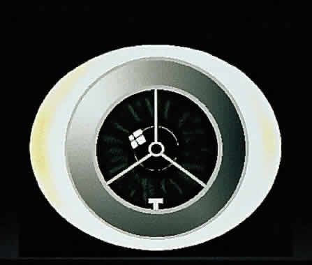
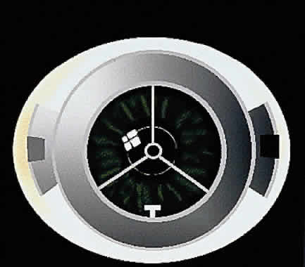
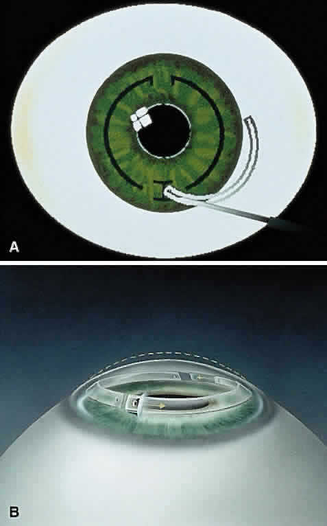
Early theoretical and animal work speculated that this ring would alter the corneal curvature through expansion or constriction of the device's diameter, causing a flattening or steeping for myopia and hyperopia, respectively.4 Further refinement of a model for the refractive effect came from Silvestrin's group5 postulating an arc shortening effect of the ring. Their model assumed that there are corneal lamellae that stretch from limbus to limbus. In that model, a spacer element such as the ICR separates corneal lamellae, resulting in a shortening of the arc length that is correlated with the device thickness (Fig. 1). Eye bank studies by T.E. Burris and coworkers then demonstrated the effect of varying implant thickness.6 By increasing the thickness of the ICR, an increased change in spherical equivalent was documented in a nearly linear fashion. Pinsky and colleagues7 then refined the model further by using finite element modeling (Fig. 2). A 360-degree ICR was used in initial studies in nonfunctional and myopic eyes in the Brazil and the United States.8 All eyes tolerated the implants well without evidence of extrusion or corneal thinning. Studies in nonfunctional eyes demonstrated the safety of the ICR (KeraVision, Fremont, CA).9 Based on these results, the US Food and Drug Administration (FDA) phase II studies of the intrastromal ring were started.

Fig. 1. A spacer element separates the corneal lamellae resulting in a shortening of the arc length and decrease in corneal curvature.

Fig. 2. Finite element model of the intrasomal corneal ring segments showing the change in corneal curvature induced by placement of the ring segments.

The original ICR was a 360-degree PMMA ring with an external diameter of 8.1 mm, internal diameter of 6.8 mm, and thicknesses of 0.25 to 0.45 mm. Uncorrected visual acuity of 20/40 or better was obtained in 85% of 66 patients observed on a followup basis for 1 year.10 The ring was well tolerated; explants were performed in ten eyes. In all cases, eyes after explantation returned to within 1 diopter (D) of their preoperative refraction, indicating the potential removability of the ICR.11

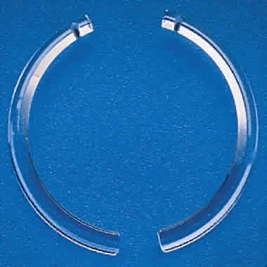
In the phase II study, wound healing problems were observed with the PMMA ring directly beneath the incision.12 To address this issue, the ring was then redesigned into two 150-degree arc segments, which simplified insertion technique and separated the implant material away from the radial incision. The ring segments were then placed through a 1.8 mm superior incision at two thirds the corneal depth. Studies showed that the ICR segments were similar in refractive effect to those of the ICR.13,14

A phase III study has been completed to show the effects of the ICR segments for mild to moderate degrees of myopia with two 150-degree arc PMMA segments.15 Three segment thicknesses have been evaluated: 0.25, 0.30, and 0.35 mm. The objectives of the study were to rule out major safety risks, to ensure an acceptable outcome in uncorrected visual acuity, to ensure predictability of refractive effect, and to ensure stability of the refractive effect.16

Back to Top
SURGICAL PROTOCOL
The current version of INTACS consists of two 150-degree arc length segments of medical grade PMMA that vary in thickness from 0.25 to 0.35 mm in 0.05-mm increments. The outer diameter of the ring segments is 8.1 mm and that of the inner diameter 6.8 mm. The segments have a hexagonal cross-section and the curvature conforms to that of the cornea. The segments are shown in Figure 3.

Fig. 3. The polymethylmethacrylate ring segments.

Before the procedure is begun, patients should be given informed consent that includes the potential risks, benefits, and complications of the INTACS. Patients are typically given an antibiotic drop the day before to surgery to help prevent infection. This procedure may be performed under topical anesthetic or with other types of anesthesia. In the FDA phase II and III trials, 77.3% of patients received topical anesthesia with oral sedation, 8% received topical anesthesia alone, 14.5% received topical anesthesia with conscious IV sedation, and 0.2% received IV conscious sedation with parabulbar anesthesia.

Patients are prepped and draped for anterior segment surgery. Care should be taken to isolate the eyelashes and remove them from the surgical field. The center of the cornea should be marked using horizontal and vertical corneal measurements. A specially designed ICR segment marker is used to identify the correct location of the radial corneal incision (Fig. 4). An ultrasonic pachymeter is used to check the corneal thickness at the incision site. A diamond knife is then set at two thirds of this depth, and a 1.8-mm radial incision is made at the marked location. Then, a lamellar stromal spreader is inserted at the base of the incision and the tissue is separated laterally (Fig. 5). A vacuum guide is centered, and the vacuum is gradually increased to about 20 mm Hg (Fig. 6). The vacuum is used to fixate the globe during lamellar dissection. Specially designed dissecting instruments are then used to create pockets in a clockwise and counterclockwise manner for 190 degrees in each direction (Fig. 7). After these channels have been made, the segments are inserted through the incision and into the intrastromal channel (Fig. 8). The segments are selected using the Keravision nomogram (Table 1). After both segments have been placed, one or two 10-0 or 11-0 nylon sutures are used to close the incision (Fig. 9). The patient is given an antibiotic-steroid combination at the end of the procedure. A shield is placed on the eye at bedtime for up to 6 to 8 weeks.17,18

Fig. 4. Incision and placement marker used to identify the radial corneal incision.

Fig. 5. A lamellar stromal spreader that is used to separate the base of the incision.

Fig. 6. Vacuum centering guide used to fixate the globe.

Fig. 7. Lamellar-dissecting instrument used to create channels for the segments.

Fig. 8. Segments are inserted one at a time into the channels.


TABLE 1. ICRS Nomogram


ICRS Thickness (mm)Predicted Change in Spherical Equivalent (D)
0.251.3
0.302.0
0.352.7

ICRS, intrasomal corneal ring segments; D, diopter.


Fig. 9. Incision closed with a suture at the end of the procedure.

Postoperatively, the patient is given a antibiotic-steroid combination for 1 week, and the steroid is tapered over 3 weeks. The sutures were cut at 6 weeks in the phase II trial and at 2 to 4 weeks in the phase III trial (Fig. 10). This duration has been significantly shortened with increasing clinical experience.

Fig. 10. Intrasomal corneal ring segments after insertion.

Back to Top
EXCHANGE OR EXPLANTATION
An exchange procedure can be done in selected patients with overcorrection or undercorrection. The procedure can be done with the patient under only a topical anesthetic. Early in the postoperative period, a Sinskey hook can be used to reopen the original incision. If healed, a radial incision is made at the original site at the same depth as the original incision. Fibrous tissue is gently loosened with blunt dissection. A Suarez spreader is used to dissect the channel opening. A Sinskey hook is then used to engage the positioning hole and rotate the segment out of its channel. In cases of exchange, a new ICR segment can then be placed into the previously made channel. The incision is closed with a suture. The same postoperative procedure is followed as with the original insertion. Six exchange procedures were performed during FDA trials, all for undercorrection. Five of six patients had satisfactory exchange results, with the last patient requiring removal for persistent undercorrection.15,17 Overall, the ICR segment was removed in 30 of 449 (6.7%) of eyes because of dissatisfaction with the procedure. No difficulties or complications occurred during segment removal.
Back to Top
RESULTS OF FDA TRIALS
In total, 449 of 452 patients underwent implantation of the ICR segment in US phase II and III trials. Three equally distributed ICR segment thicknesses were tested: 0.25, 0.30, and 0.35 mm. Phase II, which began in May 1995, enrolled a total of 91 patients at 6 investigational sites. Phase III enrolled 361 patients at 10 investigational sites, including 5 of the original phase II sites. Distribution of patients is shown in Table 2.


TABLE 2. Patient Disposition


ParameterNo.
Patients enrolled in phases II and III449
Completed 12-month analysis but missed analysis cutoff date4
Did not complete month-12 examination5
Lost to follow-up at month 125
ICRS removed (1 for safety reasons; 19 due to dissatisfaction)20
Single segment removed; single segment left in place1
Exchange procedure performed by month 124
Total patients reported on at month 12410


Patients were given a complete preoperative examination that included best spectacle-corrected visual acuity (BSCVA) on standard ETDRS charts, cycloplegic and manifest refraction, central keratometry, ultrasonic pachymetry, tonometry, videokeratography, and indirect ophthalmoscopy.

REFRACTIVE OUTCOME

This procedure was considered to be predictable in the FDA phase II and III trials. Patients enrolled had preoperative refractive errors ranging from -0.75 to -3.63 D. At 12 months, 277 of 408 (68.9%) of eyes were within ±0.5 D of intended correction and 368 of 408 (90.2%) were within ±1 D of intended correction. Of the 40 eyes with greater than 1 D deviation from the intended correction, 17 were undercorrected and 23 were overcorrected. Of those undercorrected, 11 of 17 (64.7%) had 20/40 or better visual acuity. In the overcorrected group, 14 of 23 (60.9%) had uncorrected visual acuity of 20/20 or better and 22 of 23 (95.7%) were 20/40 or better. As the intended correction increased, the predictability decreased (Fig. 11). In sum, 421 of 435 (97%) eyes had less than a 1 D shift in refraction from month 3 to month 12. This indicates a reasonably stable refractive result. Astigmatism, if present, continued to resolve over time. Another 15 of 410 (3.7%) eyes had induced astigmatism of more than 1 D at month 12. Astigmatic effects of the ring segments are not well understood, but at 12 months only 3 of 410 (0.7%) of patients had induced cylinder more than 1.50 D.

Fig. 11. Predictability of refractive effect with different ring thicknesses.

Uncorrected visual acuity improved rapidly with the ICR segment. On the first postoperative day, 150 of 448 (33.5%) of patients could see 20/20 or better and 364 of 448 (81.3%) of patients saw 20/40 or better. There was further improvement at 1 and 12 months in uncorrected visual acuity. At 1 month, 266 of 443 (60%) of patients saw 20/20 or better and 409 of 443 (92.3%) saw 20/40 or better. At 12 months, 303 of 410 saw 20/20 or better and 396 of 410 (96.6%) saw 20/40 or better. Interestingly, 216 of 410 (52.7%) saw 20/16 or better, with 88 of 410 (21.5%) experiencing an uncorrected visual acuity above preoperative BSCVA. Loss of two or more lines of BSCVA occurred in 4 of 410 patients at 12 months. Ring thickness does not appear to influence loss of BSCVA.

SECONDARY OUTCOME MEASURES

Corneal curvature changes were measured using central keratometry as well as videokeratography. Consistent with eye bank studies, the central anterior corneal surface became more prolate and retained its positive asphericity.19 Mean keratometry readings changed from 43.7 ± 1.34 D to 41.79 ± 1.61 D. Contrast sensitivity changed only modestly from baseline to month 12 as measured by Functional Activity Contrast Test (FACT) charts. Central corneal sensation loss of 20 mm or more occurred in 13 of 237 (5%) of eyes tested at month 12. No eyes developed neurotrophic keratitis.

Few adverse intraoperative events occurred in the FDA trials. In all, 449 of 454 (98.9%) of eyes had successful placement of the ICR segment. One eye had a posterior corneal perforation resulting in the ICR segment's not being placed. Three eyes had anterior corneal surface perforations and the surgeries discontinued. Two eyes had perilimbal hemorrhage. One patient had an allergic reaction to the surgical scrub. No patient had reduced BSCVA.

Postoperatively, haze occurred along the stromal channel of the ring segment medially and laterally. This grayish opacity appeared after surgery and decreased gradually over time. Lamellar channel deposits appeared as crystalline/refractile or whitish round droplets along the edge of the ring segments (Fig. 12). These deposits appeared in 68.3% of eyes in the first year and gradually decreased during that year. These are thought to be lipid deposits from stressed keratocytes.15,20 Small epithelial inclusion cysts were observed in the incision in 169 of 449 (37.6%) of eyes at some point during the FDA trial; 33 of 449 (7.0%) of patients presented with cysts at the last examination.

Fig. 12. Lamellar channel deposits.

Five ICR segments had to be repositioned because of shallow or deep lamellar dissection, incorrect depth, or decentration of the ICR segment. One ICR segment was repositioned at 2 weeks because of subjective halos and glare that were thought to result from a nasally displaced segment. One eye had segments repositioned because of contact of the ends of the segments. One patient experienced glare that was successfully resolved by repositioning. One eye required removal of the ICR segment because of findings of severe halos at night. All eyes that had repositioning maintained 20/25 uncorrected visual acuity. One patient had microbial keratitis but retained BSCVA of 20/20. The ICR segment was removed in this patient.

Overall, 30 of 449 (6.7%) of eyes had removal of ICR segment because of dissatisfaction with the procedure. There were no difficulties in removing the ring segments or complications during segment removal. In all, 87% of patients reported being satisfied with their visual outcome, experienced little or no pain during and after the procedure, and thought the procedure required a minimal time investment. Another 85% reported that they would recommend it to a friend.

Back to Top
DISCUSSION
Uncorrected visual acuity dramatically improves the first week after surgery and then stabilizes. This indicates a fairly rapid onset of action for the refractive change induced by the ICR segment and then later stability. Overall, 52.6% of patients had 20/16 vision or better, 73.9% had 20/20 or better, and 96.6% had 20/40 or better. Among these patients, 21.6% had improved BSCVA, which is thought to result from maintenance of the positive asphericity of the cornea with this procedure. Contrast sensitivity was not statistically changed from defined preoperative values. Only 0.9% of patients lost two or more lines of BSCVA, indicating the safety of the ICR segment devices. These results are comparable with findings associated with other refractive procedures.9,10 When loss of BSCVA did occur, it was reversible by explantation of the ICR segment.7,9,10

The potential reversibility of the ICR segment is based on two criteria: preservation of BSCVA with removal of the segments and a return to within ±1 D of the preoperative refraction by 3 months after removal. All eyes had BSCVA of 20/20 or better postremoval and none lost any lines of BSCVA from levels registered during the preoperative examination. Of these patients, 13 of 20 experienced a gain of BSCVA postremoval.

ICR segments seem to be very well tolerated. None of the segments extruded or eroded. Epithelial cysts at the suture site persisted in 7.3% of patients. A faint haze surrounded the ring segments in all patients, but this did not appear to be visually significant. Focal deposition of material adjacent to the ring segments appeared during the first 6 months and some decreased over time. Deep neovascularization occurred in 2.2% of patients. The ICR segment seems to be biocompatible.

The KeraVision nomogram accurately predicts 90.2% of the patients within ±1 D of their attempted correction. Consistent undercorrection seems to occur at each ring segment thickness. Furthermore, as the attempted correction is increased the amount of undercorrection seems to increase as well (see Fig. 11). This finding is similar to other keratorefractive procedures.10,12 Clearly, there are other factors involved in predicting refractive outcome with the ICR segment. Possible variables include age, wound healing, preoperative corneal curvature, centration, calibration of instruments, and depth of ring placement.

Complications with the ICR segment were limited. Cases of keratitis and perforations have been reported with the ICR segment.15 These cases resolved with appropriate antimicrobial therapy and explantation of the ICR segment. There have been no reported cases of loss of BSCVA after removal of the ICR segment. This indicates the potential removability of the procedure with preservation of BSCVA. Although the number of patients who have had exchanges is small, the ICR segment appears to have adjustable capabilities.

The ICR segment has some advantages and disadvantages when compared with photorefractive keratectomy, laser in situ keratomileusis, and radial keratotomy. The ICR segment is potentially reversible and adjustable, the central cornea is left largely undisturbed, and there is a low capital cost for the equipment. However, the ICR segment has limitations, particularly only correcting for low degrees of spherical myopia. Photorefractive keratectomy and laser in situ keratomileusis are predictable, stable, and have a wider range of correction than the ICR segment. However, there is a high cost for the equipment and potential for scarring of the visual axis. In the case of photorefractive keratectomy, slow patient recovery after surgery is also a problem. Radial keratotomy has a quick recovery of visual function and the results are known for a longer duration of time; however, there is a high reoperation rate and there seems to be instability in the refractive outcome.10,13

The ICR segment can be implanted with a stable refractive outcome and a low incidence of complications. The predictability of the nomogram will require further refinement to increase the accuracy of the refractive outcome. In conclusion, the ICR segment is an effective device for the correction of low degrees of spherical myopia with the advantage of removability and potential adjustability, which clearly warrants further investigation and refinement.

Back to Top
REFERENCES

1. Barraquer JI: Queratoplasia refractiva, estudios e informaciones. Oftalmologicas 2:10–30, 1949

2. Stone W, Herbert E: Experimental study of plastic material as replacement for the cornea: A preliminary report. Part 2. Am J Ophthalmol 36:168–173, 1953

3. Fleming JF, Reynolds AE et al: The intrastromal corneal ring: two cases in rabbits. J Refract Surg 3:227–232, 1987

4. Fleming JF, Wan WL et al: The theory of corneal curvature change with the intrastromal corneal ring. CLAO J 5:179–193, 1989

5. Silvestrini T et al: Optical modeling of the intrastromal corneal ring [letter]. J Refract Surg 12:456–457, 1996

6. Burris TE, Baker PC et al: Flattening of the central corneal curvature with intrastromal rings of increasing thickness: An eye bank study. J Cataract Refract Surg 19(Suppl):182–187, 1993

7. Pinsky et al: Invest Ophthalmol Vis Sci 36(Suppl):S309, 1995

8. Nose W, Neves RA et al: The intrastromal corneal ring: One year results of first implants in humans: A preliminary nonfunctional eye study. Refract Corneal Surg 9:452–458, 1993

9. Assil KK, Barrett AM et al: One year results of the intrastromal ring in nonfunctional human eyes. Arch Ophthalmol 113:159–167, 1995

10. Nose W, Neves RA et al: Intrastromal corneal ring: 12 month sighted myopic eyes. Refract Surg 12:20–28, 1996

11. Quantock A et al: Stromal healing following explanation of an ICR from a nonfunctional human eye. Arch Ophthalmol 113:159–167, 1995

12. Hwang DG: The intrastromal corneal ring segment: Current status and review of US Phase II clinical results. Ophthalmol Clinic 10:591–597, 1997

13. Schanzlin DJ, Asbell PA et al: The intrastromal corneal ring segments: Phase II results for the correction of myopia. Ophthalmology 104:1067–1078, 1997

14. Waring GO, Abbott RL et al: One year outcomes of Intrastromal Corneal Ring segments for the correction of -1.0 to -3.5 Diopters of myopia. Presented at American Academy of Ophthalmology annual meeting, New Orleans, November 1998, (submitted).

15. KeraVision Inc, Phase III protocol summary, KeraVision, Fremont, CA

16. Schanzlin DJ: Keravision INTACS: History, Theory, and Surgical Technique. Refrac Eyecare Ophthalmol (Suppl):6–10, 1999

17. Keravision Surgical Protocol, KeraVision, Fremont, CA

18. Burris TE, Holmes-Higgin MS: Corneal asphericity in eye back eyes implanted with the intrastromal corneal ring. J Refract Surg 13:556–567, 1997

19. Rodrigues MM, McCarey BE et al: Lipid deposits posterior to impermeable intracorneal lenses in rhesus monkeys: Clinical, histochemical and ultrastructural studies. Refract Corneal Surg 6:32–37, 1990

20. Krachmer JH, Mannis MJ et al (eds): Cornea, Vol III, Surgery of the Cornea and Conjunctiva. St. Louis: Mosby, 1997

Back to Top