Chapter 48
Surface Ablation: Photorefractive Keratectomy, Lasek, and Epi-LASEK
ROGER F. STEINERT and ANN Z. MCCOLGIN
Main Menu   Table Of Contents

Search

BACKGROUND
PREOPERATIVE EVALUATION
SURGICAL TECHNIQUE
POSTOPERATIVE CARE
CLINICAL RESULTS
COMPLICATIONS
CONCLUSION
REFERENCES

The development of excimer laser ablation represented a breakthrough in the correction of ametropia. Photorefractive keratectomy (PRK) employs a 193-nm argon fluoride excimer laser to ablate the anterior corneal stroma to a new radius of curvature to decrease refractive error. In the early 1990s, PRK became a common technique worldwide for treating low to moderate myopia because it offers a wider treatment range and more predictable and stable results than incisional keratotomy.1–3 PRK has been the subject of extensive investigations, with ongoing improvements in hardware and technique. Laser in situ keratomileusis (LASIK) utilizes PRK technology, but performs the procedure under a lamellar flap formed with a microkeratome. LASIK is discussed in detail in the next chapter. In this section we review the background, preoperative evaluation, surgical technique, clinical results, and complications of PRK and its more recent variants, LASEK and Epi-LASEK. The current techniques for these surface treatments are sometimes collectively termed advanced surface ablation (ASA).
Back to Top
BACKGROUND
Trokel et al4 and Srinivasan5 demonstrated a new form of laser–tissue interaction, termed photoablation, in 1983. Srinivasan, an IBM engineer, was studying the far-ultraviolet (UV; 193 nm) argon fluoride excimer (a contraction of “excited dimer,” the photochemical process that emits the laser photons) laser for photoetching of computer chips when Trokel, an ophthalmologist, postulated and subsequently proved that a similar process could also remove corneal tissue discretely and precisely with minimal damage to the adjacent cornea. The original observation of the effect of ultrashort UV light on the cornea is attributed to a civilian contractor, John Taboada, who was studying potential laser toxicity for the military. Trokel rapidly recognized the potential of the excimer laser for refractive and therapeutic corneal surgery.

Photoablation occurs because the cornea has an extremely high absorption coefficient at 193 nm. A single 193 nm photon has sufficient energy to directly break the carbon–carbon and carbon–nitrogen bonds that form the peptide backbone of the corneal collagen molecules. Consequently, excimer laser radiation ruptures the collagen polymer into small fragments, and a discrete volume of corneal tissue is expelled from the surface with each pulse of the laser.6–8

Initially, investigators studied whether the excimer laser could be used as a “laser scalpel” for corneal surgery in procedures such as astigmatic and radial keratotomy (Fig. 1).9,10 However, the excimer laser is a poor replacement for a cutting scalpel because the laser removes tissue, creating a “kerf”—it does not incise the tissue.11

Fig. 1. Two linear deep incisions created by the excimer laser in an ex vivo human cornea.

The more successful application of the excimer laser for correcting ametropia is the sculpting or reshaping of the outer de-epithelialized surface of the cornea to alter its refractive power. This surgical procedure, termed photorefractive keratectomy (PRK) by Marshall and Trokel, was the focus of extensive preclinical investigations before it was applied to sighted human eyes.12–16 The results of early animal studies provided evidence for normal wound healing in laser-ablated corneas,17–22 and as the laser and delivery system technology matured,23–26 confidence grew that sighted ametropic human eyes could successfully undergo PRK. McDonald and co-workers27 treated the first sighted human eye in 1988, and now millions of PRK procedures have been performed worldwide. The popularity of PRK faded rapidly when LASIK was popularized in the late 1990s, primarily because LASIK offered a faster visual recovery and less postoperative discomfort. While more LASIK procedures continue to be performed than PRK and its variants, surface ablation has returned as an attractive alternative in specific indications such as very low corrections and thin corneas. Moreover, current data suggest that the LASIK flap is associated with increased postoperative higher-order optical aberrations than PRK, and the postoperative quality of vision may be higher with wavefront-guided PRK compared to LASIK. As a result, some surgeons are advocating a complete return to surface ablation, and that LASIK should be avoided altogether for standard corneal refractive surgery.28

Back to Top
PREOPERATIVE EVALUATION
When a patient is considering refractive surgery, several issues must be evaluated preoperatively. First, it is important to determine pre-existing ocular or systemic conditions that could interfere with healing or the predictability of the procedure. In addition, the patient's refractive status, including stability, degree of refractive error, and astigmatism, must be determined. Finally, and perhaps most importantly, the patient's goals and expectations must be evaluated.

It is important to obtain the patient's full medical history, review the systems, and perform a complete ocular examination to rule out the presence of any conditions in which refractive surgery is contraindicated. A history of collagen-vascular and autoimmune diseases, such as rheumatoid arthritis, systemic lupus erythematosis, and Sjögren's syndrome, are considered contraindications to PRK due to unpredictable corneal wound healing with the potential for corneal melting.29–31 A history of keloid scar formation and prior herpes simplex keratitis are relative contraindications. However, systemic antiviral prophylaxis begun preoperatively and continued for several months postoperatively may make PRK safe in patients with a history of herpes simplex keratitis. Diabetes must be well controlled preoperatively because it involves instability of refraction and potentially poor wound healing. Dense amblyopia is a relative contraindication because the refractive procedure puts the patient's better eye at risk. Patients with this disorder should wear protective spectacles rather than undergo surgery to reduce spectacle use.

A full ocular examination is mandatory. An external examination should consider the orbital anatomy. A deep-set globe, high brow, and/or a narrow palpebral fissure are features that may favor PRK and LASEK over LASIK or Epi-LASEK, because with the latter techniques it may be difficult or impossible to access the microkeratome and apply the suction ring. A thorough examination of extraocular movements is important because the presence of a phoria or tropia could result in postoperative diplopia. Pupil size should be measured because a large pupil may contribute to glare and haloes postoperatively, although recent improvements in our understanding of dim light vision symptoms suggests that corneal aberrations (not the pupil size itself) are the principal issue. A commercial pupillometer can more accurately assess pupil size under mesopic or scotopic conditions. Eye dominance is tested in case the patient desires monovision (in monovision corrections, typically the dominant eye is corrected for distance vision). The eyelids and tear film should be evaluated for blepharitis or dry eyes that should be treated preoperatively. A careful examination of the cornea should reveal the presence of any corneal scars, vessels, or evidence of previous inflammation. Patients with epithelial basement membrane dystrophy (EBMD, often termed map-dot-fingerprint dystrophy) are better candidates for PRK than for LASIK because PRK may be therapeutic, by enhancing epithelial adhesion, whereas the microkeratome friction in LASIK may cause a frank epithelial defect.

Preoperative corneal pachymetry is mandatory for all patients.32 The central corneal thickness is determined by ultrasonic pachymetry or with the Orbscan scanning slit topographer. In addition to being a screening test for possible preexisting keratoconus or other types of corneal ectasia (see below), corneal pachymetry can determine the amount of corneal tissue that will be available for excimer tissue ablation without risking weakening the cornea to the point where postoperative ectasia and irregular astigmatism become significant risks. The current generally employed standard is that the cornea must have at least 250 μ or 50% of the original thickness (whichever is greater) of the residual stromal bed under a LASIK flap. If the flap thickness plus tissue ablation depth are calculated to leave inadequate residual stromal thickness, LASIK is contraindicated and surface ablation is preferable.

The lens must be examined closely to rule out the presence of an early cataract, and a dilated-fundus examination should be performed to rule out preexisting peripheral retinal degeneration or peripheral holes that should be treated preoperatively.

Computed corneal topography is vital in the preoperative evaluation of refractive surgery patients. Corneal topography is particularly useful in the evaluation of irregular astigmatism. Corneal warpage can occur with the use of contact lenses, and its presence will interfere with the accuracy of preoperative refractive measurements. For this reason, soft contacts should not be worn for a minimum of 3 days, and hard contacts should be avoided for at least 2 weeks prior to the preoperative evaluation. If corneal warpage is suspected, the patient should not wear contact lenses until the topography is stabilized (Fig. 2).

Fig. 2. Left: Corneal topography of a patient with corneal warpage from hard contact lens wear. Right: One month after the contact lenses were discontinued, the warpage has resolved.

The presence of frank or subclinical (forme fruste) keratoconus can also be detected by corneal topography (Fig. 3). Some series have reported successful PRK in keratoconus suspects.33 However, the presence of keratoconus is generally considered a contraindication to PRK because the removal of additional tissue from an already ectatic cornea may lead to further ectasia and unpredictable results. When keratoconus is suspected on topography but is not visible on examination, two tests are helpful. First, the topography should be repeated with the patient looking slightly superiorly (one or two rings on the placido disc). If the patient has true keratoconus, the abnormality of eccentric inferior steepening will persist. If the patient has a slightly inferiorly shifted corneal apex rather than a true keratoconus, the pattern will become symmetric with a slight upgaze. The second test is corneal pachymetry. In true keratoconus, the central cornea will be thinner than average and will thin slightly more when the pachymetry is repeated 1–2 mm inferior to the corneal center. The Orbscan topography device includes a readout of the posterior corneal topography. An abnormal amount of anterior curvature of the posterior cornea lends further support to a suspicion of corneal ectasia. The clinician should also use a retinoscope to determine whether a “scissoring” light reflex is present, which would increase the likelihood of the presence of keratoconus.

Fig. 3. Corneal topography of a forme fruste keratoconus. Note the inferior corneal steepening.

Careful evaluation of the patient's refractive status is critical. In general, patients should be 18 years of age or older, and the refraction should be stable to within ±0.5 diopter (D) over the previous year. Visual acuity should be checked without correction, as well as with the patient's current spectacles. Both manifest refraction and cycloplegic refraction must be performed. Corneal topography is a useful adjunct in evaluating astigmatism. In addition, the surgeon must evaluate any clinically significant disparity between the manifest and cycloplegic refractions. Most patients will have a cycloplegic refraction between 0.25 and 1.0 D shifted in the hyperopic direction. Because some or all of this disparity can be attributed to the optics of the peripheral cornea and a small posterior shift of the crystalline lens with cycloplegia, many surgeons will plan their laser input based on the manifest refraction if it has been performed with a careful defogging technique. A disparity between the manifest and cycloplegic refractions of more than 1 D of sphere warrants re-evaluation, often including a post-cycloplegic manifest refraction “pushing plus” to overcome accommodative spasm. Accommodative spasm is common in both myopes who have become “over-minused” in their contact lenses or spectacles (extra minus causes a slight minification of the image, which causes a perception of higher contrast) and in hyperopes who have a lifelong habit of accommodation to improve distance vision. In middle-aged patients, a cycloplegic refraction is still mandatory because surprising amounts of accommodative spasm may remain. Tropicamide 1% is usually strong enough to relax accommodation in patients over 40, but cyclogyl 1% may be advisable in younger patients, especially hyperopes. Several clinical tests are helpful for resolving disparities between the manifest and cycloplegic refractions. In addition to a “defogging” technique that “pushes plus,” a time-honored technique is the duochrome chart (one half of the chart is green and the other half is red). Because of chromatic aberration, the longer red wavelengths are focused slightly behind the shorter green wavelengths. An over-minused refraction will cause the focal point to be behind the retina. Red wavelengths will be further retrofocused and therefore letters on the red half of the chart will appear more blurred than the letters on the green half of the chart. Other helpful clinical strategies include placing a 4-mm aperture in the trial frame during the cycloplegic refraction to restrict vision to the more central cornea, and comparing the refraction to the readings from an autorefractor with a defogging device system or a wavefront analyzer.

If there is a discrepancy between the patient's refractive and corneal cylinders, the refraction should be reevaluated. If possible, it is desirable to perform a wavefront analysis to identify patients with a high level of preoperative aberration that may become more symptomatic postoperatively, as well as to guide a customized ablation if indicated.

Finally, proper patient education is the key to a successful refractive surgery procedure. The goal of refractive surgery is to decrease the patient's dependence on glasses and contact lenses. Achieving this goal depends on the patient's daily visual needs. The patient must understand that the technique is still not as accurate or as predictable as correcting myopia with contact lenses or spectacles, which has a virtually 100% chance of achieving 20/20 or better vision in a healthy eye. The extra time taken with patients to ensure that they fully understand the goal of the procedure can save much frustration for both surgeons and patients postoperatively.

Informed consent should include a discussion of other surgical as well as nonsurgical options for the patient. Likely outcomes, the potential need for enhancements, and potential complications must also be discussed. In addition, presbyopia should be explained, and monovision (in which one eye is intentionally left mildly myopic for near vision) should be offered as an option to middle-aged patients. If the patient is interested, a monovision simulation with contact lenses should be performed prior to the procedure.

Back to Top
SURGICAL TECHNIQUE

LASER CALIBRATION

On the day of surgery, the laser should be checked for an adequate homogenous beam profile, alignment, and power output, following the manufacturer's guidelines

PREOPERATIVE PLANNING

Many surgeons employ a nomogram adjustment of the correction that is to be programmed into the laser. If possible, it is preferable to perform these calculations in advance of the moment of surgery to avoid time pressure or distractions that may lead to calculation errors. Most nomograms are individualized to the surgeon, laser, and specific technique used, as well as patient factors, which may include the amount of correction, patient age, gender, and other variables. These factors may all influence the outcome of the ablation.

Often the manifest and cycloplegic refractions differ, or the amount and axis of the topographic and refractive astigmatism differ. Consequently, it may be unclear which refraction should be entered into the laser. If it is confirmed that the refractive cylinder differs from the topographic cylinder, it is assumed that lenticular astigmatism or posterior corneal curvature is the cause. In this case, the laser is usually still programmed with the axis and amount of cylinder noted on refraction. The surgeon should take particular care to check the consistency of the axis on the refraction and topography with the value programmed into the laser. This is a common source of error, particularly when there is a conversion between plus and minus cylinder formats.

In many laser models the surgeon also must enter the size of the optic zone, and whether or not a blend of the ablation zone should be performed. If there is sufficient corneal tissue, an ablation zone larger than the scotopic pupil size is usually selected. A “blend zone” is an area of peripheral asphericity that is designed to reduce the possibly undesirable effects of an abrupt transition from the optical zone to an untreated cornea. A common approach to creating a “blend zone” is to have, for example, a −6 D correction consist of a −5 D correction at a 6-mm optical zone and a −1 D correction at an 8-mm optical zone. The larger the treatment area, the deeper the ablation.

In wavefront-guided “custom” ablations, these parameters are usually fixed by the wavefront optical measurement and the laser model.

The surgeon must calculate whether an adequate stromal bed will remain. This is rarely an issue for surface ablation, but in thinner corneas and higher dioptric treatments, there may be inadequate tissue for LASIK, prompting the surgeon to select surface ablation. In simple spherical corrections of myopia, the approximate depth of ablation can be calculated by the Munnerlyn formula:

t = S2 D / 3

where t is the thickness of the tissue ablated in microns, S is the diameter of the optical zone in millimeters, and D is the dioptric correction. For example, in a 6-mm optical zone, the formula is simply 12 × D. A 4 D myopic correction would remove about 48 μ centrally. Note, however, that aspheric peripheral blend zones and wavefront-guided corrections remove more tissue than the simple spherical correction in the Munnerlyn formula, and the surgeon is advised to program the correction into the laser to have the laser software calculate the ablation depth. In addition, the surgeon must remember to input the intended total correction value, not the nomogram-reduced value, in order to obtain the true amount of tissue removal

PATIENT PREPARATION

Before the laser treatment is performed, the patient should be instructed about the sounds and smell of the laser. Anxious patients may receive an oral sedative, such as diazepam, and the operative eye (in unilateral treatments) should be indicated with an adhesive label or some other temporary mark on the forehead.

Before or after the patient reclines under the head of the laser, the patient's skin is typically prepped with povidone iodine (Betadine) or alcohol wipes. Some surgeons also use a sterile drape over the skin and lashes. Topical tetracaine and/or proparacaine anesthetic drops are placed in the eye. A lid speculum is placed in the operative eye and a patch is placed over the fellow eye to avoid cross-fixation. A gauze pad may be taped over the temple between the operative eye and the ear to absorb any fluid run-off. The amount of desired correction, accounting for the vertex distance, is entered into the laser and checked by the surgeon. The patient is asked to fixate on the laser centration light while the surgeon focuses and centers the laser. For most patients, line-of-sight fixation by the patient during PRK produces more accurate centration than globe immobilization by the surgeon.34,35

SURGICAL TECHNIQUE

In most original techniques and in U.S. phase III investigations, the epithelium is removed by a sharp blade or a blunt spatula (Fig. 4). In this technique, the surgeon defines the outer limit of de-epithelialization with an optical zone marker and then debrides the periphery followed by the center. To avoid dessication and resultant variability in ablation rates, the surgeon must perform this procedure efficiently. Skill is required to avoid nicking Bowman's layer. An ophthalmic surgical cellulose sponge lightly moistened with an artificial tear lubricant, such as carboxymethylcellulose 0.5%, is brushed over the surface of the cornea to remove any residual epithelium and provide a smooth surface (Fig. 5). Subsequently developed alternative methods for removing the epithelium include the use of a rotating corneal brush,36 application of diluted absolute alcohol (typically around 20% concentration) to the corneal surface to loosen the epithelium,37–39 and transepithelial ablation by the excimer laser itself.40 The epithelium should be removed efficiently and consistently to prevent hydration changes in the stroma, because the rate of excimer laser ablation may be increased by excessive corneal stromal dehydration, resulting in an overcorrection.41

Fig. 4. De-epithelialization with a blade before PRK.

Fig. 5. Final cleaning of the surface before beginning the laser exposure.

In the LASEK variant of PRK, which was first described by Camellin,42 the goal is to preserve the patient's epithelium. Instead of debriding and discarding the epithelium, or ablating the epithelium with the excimer laser, the surgeon employs a technique to remove an intact sheet of epithelium. First, the surgeon can place a radial mark of gentian violet ink to assist in later realignment of the epithelial flap. Most commonly, a solution of approximately 20% diluted absolute alcohol is applied for 20 to 30 seconds. The alcohol is restricted to the area to be de-epithelialized with a ring (usually an optical zone marker) pressed onto the corneal surface, or by means of a round surgical sponge soaked with the alcohol solution. After the desired exposure time has passed, the alcohol is removed from the “well” of the optical zone marker by absorption into a microsurgical spear sponge. After the alcohol is fully absorbed and the optical zone marker is removed, the ocular surface is copiously irrigated with balanced salt solution (BSS) to minimize toxicity to the limbal germinal epithelium. The surgeon then uses an instrument, which often has a hoe or spatula configuration, to carefully separate a flap of full-thickness epithelium from the underlying Bowman's layer. The epithelium is delicately folded back on itself until all of the epithelium has been removed except for a small “hinge,” which is usually located superiorly. Figure 6 shows the key steps involved in performing LASEK.

   
Fig. 6. Technique for LASEK. A: After the gentian violet radial orientation marks are placed, the epithelial flap is delineated with an optical zone marker or similar instrument. B: Alcohol to loosen the epithelial flap is carefully contained within a trephine-shaped well pressed on the cornea. C: The separation of the flap edge is begun with a “microhoe.” D: The separation of the epithelial flap continues as a “micro hockey stick” strokes the epithelium back, with care taken to not perforate the epithelium. E: The epithelium is fully retracted off the area to be ablated by the laser, leaving a “hinge” of epithelium for about one clock hour in the superior periphery. G: After the laser exposure is completed, the epithelial sheet is gently replaced by floating it and stroking it with a BSS cannula. H: The epithelial flap is realigned using the radial marks. I: The epithelial sheet is briefly dried with filtered air through a microtip to begin adhesion to the stroma. J: After a BSCL is placed, the procedure is complete. (All photographs courtesy of Daniel Durrie, M.D.)

The goal of LASEK is to reduce postoperative pain and achieve a speedier return of vision compared to PRK, and perhaps to reduce postoperative haze formation.43 The success of LASEK in achieving these goals has not been demonstrated in a number of controlled studies, however. With LASEK, vision may be slighter better on the first postoperative day compared to PRK, but some reports indicate more discomfort and a delay in recovery of vision with LASEK after the first day.

Recently, efforts have been made to develop a variant of the microkeratome used in LASIK. The microkeratome is modified with a dull blade and thin applanation plate to mechanically remove the epithelial flap without the use of toxic agents, such as alcohol, that are used in LASEK. The goal is to create an epithelial flap that may remain viable and more likely to successfully readhere postoperatively. These techniques are most commonly termed Epi-LASEK.

LASER ABLATION

After the epithelium is out of the treatment zone and the stroma has been exposed with one of these techniques, the laser is centered and focused according to the manufacturer's recommendations. Improved PRK centration occurs when the aiming beams or reticule are centered on the entrance pupil instead of the corneal apical light reflex.44,45 The patient is instructed to maintain good fixation during the stromal ablation. Small microsaccades should not adversely affect the outcome of the procedure. If the patient begins to lose fixation, however, the surgeon should immediately stop the treatment until adequate refixation is achieved. If the laser includes a tracking mechanism, it is still important for the surgeon to monitor for excessive eye roll, which can result in decentration despite the tracking device.

To correct myopia, the surgeon flattens the cornea by placing the largest number of laser pulses centrally, and progressively fewer pulses toward the periphery of the optical zone (Fig. 7). To correct hyperopia, the cornea must be steepened. No pulses are placed in the precise center, the maximum number of pulses is placed in the peripheral optical zone, and then a blend zone creates a smooth transition from the edge of the optical zone back to the peripheral corneal surface (Fig. 8). Although the excimer laser beam at 193 nm is invisible to the human eye, a faint fluorescence of deep blue light is sometimes visible during stromal ablation. The sound of the laser firing is the main feedback signal to the surgeon, along with an alteration in the light reflex as the stromal ablation progresses. If topical mitomycin C (MMC, usually 0.2 mg/ml) is to be employed (usually in cases of high myopic correction) when the PRK is applied to a LASIK flap, or to enhance a previous PRK hazy scar, most surgeons will apply it as a soaked pledget placed on the ablated surface for 1 minute or less at the end of the laser exposure.46 Animal studies have shown a reduced keratocyte population and less haze in eyes that undergo excimer laser treatment followed by a single topical application of low-dose MMC. Vision-threatening complications from the use of MMC, including glaucoma and corneal perforation, have been reported. The search continues for the ideal topical wound-healing modulator that has a high specificity for collagen synthesis without toxic side effects.

Fig. 7. Schematic representation of the excimer laser flattening the central cornea for the treatment of myopia.

Fig. 8. Schematic representation of the pattern of corneal ablation necessary to correct hyperopia with the excimer laser. The greatest amount of tissue is removed from the periphery of the optical zone with a gradual transition toward the anterior corneal surface.

IMMEDIATE POSTOPERATIVE TREATMENT

After the procedure is completed, antibiotic, steroid, and nonsteroidal anti-inflammatory (NSAID) drops are usually placed in the eye, followed by a bandage soft contact lens (BSCL). If the LASEK or Epi-LASEK variants have been performed, the surgeon carefully floats and repositions the epithelial sheet back into position with BSS before he or she applies the medications and the BSCL. Some surgeons also apply chilled BSS before and/or after the PRK procedure in the belief that cooling reduces pain and haze formation, although the advantage of this practice has not been substantiated in a controlled study. If the patient cannot tolerate a BSCL, a pressure patch may be used.

Back to Top
POSTOPERATIVE CARE
During the first 24 hours, the patient may experience a large amount of pain, which may be relieved by an oral narcotic pain medication. Topical NSAID drops have been shown to significantly reduce postoperative pain,47–51 although they may also slow the rate of re-epithelialization and promote sterile infiltrates. Studies have also demonstrated a reduction in pain with the use of topical anesthetic drops.52–54 Topical anesthetic drops must be used cautiously because they may cause severe corneal complications when used excessively over a prolonged period. However, topical tetracaine used ad libitum in conjunction with a BSCL for 1 or 2 days after PRK does not appear to delay re-epithelialization or cause keratopathy.

The patient should be followed closely until the epithelium is intact, usually within 72 hours. At this point, the BSCL, antibiotic, and NSAID drops (if used) may be discontinued. If they have been used at all, the topical anesthetic drops must be confiscated from the patient to prevent prolonged use. Following re-epithelialization, a decision is made to continue or discontinue the topical steroid drops. The administration of topical steroids to modulate postoperative wound healing and reduce anterior stromal haze and regression of the refractive effect remains a controversial subject. Some investigators have demonstrated that corticosteroids have no significant long-term effect on corneal haze or visual outcome after PRK.55–58 Other studies demonstrated that steroids were effective in limiting haze and myopic regression after PRK, particularly after higher myopic corrections.59,60 Currently, surgeons who advocate the use of topical steroids after removal of the BSCL generally believe that only patients with higher levels of myopia (e.g., greater than about 4.0 D) require long-term topical steroids. If they are employed, steroid drops are usually tapered over a 3- to 4-month period, depending on the patient's corneal haze and refractive outcome. Keratocyte healing activity is maximal at 1 to 2 months. Abrupt termination of steroid drops at this time may trigger an excess healing reaction with haze formation and regression of the correction.

Back to Top
CLINICAL RESULTS

Impact of Evolving Technology

The effectiveness of PRK improved markedly during the pre–market-approval clinical trials conducted in the United States from 1990 to 1994, because the broad-beam excimer laser systems were updated several times and the surgeons became more familiar with the PRK technique. Also, the size of the ablation zone increased as experience revealed that small ablation zones, which were originally believed to be desirable to limit the depth of tissue removal, produced more haze and regression, as well as subjective glare and haloes. Larger treatment zones, including larger true optical zones and aspheric peripheral blend zones, are now commonly employed to improve both optical quality and refractive stability in both myopic and hyperopic treatments.

Subsequent improvements in beam quality and the development of scanning slit and spot lasers have been associated with better outcomes, including less postoperative haze and less irregularities (especially fewer “central island” elevations).

Many surgeons now employ outcome analysis programs that refine the success rates. Scanning spot laser technology necessitated the development of tracking technology to ensure proper registration of the shot pattern. The potential for tracking technology to improve centration led all of the manufacturers of such devices to introduce tracker technology.

The most common types of tracker technology employ one of two strategies. In so-called closed-loop trackers, high-speed oscillating infrared beams scan across the edge of a fixed dilated pupil. These beams detect the abrupt change in reflected light at the edge of the pupil. This signal then directs rapidly responding mirrors to create a space-stabilized image, and the laser treatment is located on the cornea based on that image.

The second type of tracker is based on an infrared image of the pupil, and uses video technology to monitor the location of the pupil image and shift the laser beam accordingly, in a so-called open-loop system.

Most recently, trials of wavefront analysis-guided treatments generally reported improvements in both visual acuity and quality of vision.61

CLINICAL OUTCOMES

In a study of the first laser to gain FDA approval in the United States (the Summit Phase III PRK study62), 701 eyes with myopia ranging between −1.5 to −6.0 D were enrolled, with 2-year follow-up data available on 612 of the eyes. In that trial, 92.5% of the eyes had an uncorrected visual acuity (UCVA) of 20/40 or better, and 66.5% had a UCVA of 20/20 or better. In addition, 77.8% of the eyes had a spherical equivalent manifest refraction within 1.0 D from emmetropia, and 6.9% had lost two lines of best corrected visual acuity (BCVA). In the VISX Phase III Study for low myopia,63 691 eyes with myopia ranging from −1.0 to −6.0 D were followed for 24 months. In that study, 85% of the eyes had a UCVA of 20/40 or better, with 79% of the eyes within 1.0 D of emmetropia, and 1% of the eyes lost two lines of BCVA.

Since that time, the general trend has been for improved outcomes compared to the initial PRK studies. The trend toward reduced accuracy with higher myopic corrections persists, however.64–66 The long-term safety and stability of PRK were recently confirmed by a report of 12-year follow-up of patients who were in one of the initial clinical trials of PRK.67 Overall, the refractive outcome was stable after the first year, and haze diminished slightly.

Most studies indicate that when higher degrees of myopia are treated, there is an increased severity of haze and loss of BCVA. Greater amounts of attempted correction are also associated with decreased predictability, increased regression, and less likelihood of obtaining 20/40 or better UCVA. A consensus about the upper limit of PRK has not been achieved, but most agree that the results are unacceptable for myopia of more than −10.0 D, because of the combination of an increased risk of haze and scar formation, less accuracy, and degradation of the optical performance when the cornea is excessively flattened. Progress in the successful treatment of higher levels of myopia with PRK may depend on pharmacologic mediators of wound healing, including but not limited to intraoperative topical MCC.68 Technical improvements in the excimer laser, particularly the adoption of a peripheral transition zone, appear to result in better predictability. For example, in one study of myopic eyes between −7 and −17 D, 25 of 47 eyes (53%) were within 1 D of attempted correction at the 2-year follow-up.69

TORIC PRK

To correct astigmatism, the excimer laser flattens the steep meridian of the cornea to match the flatter meridian. In general, toric PRK was not as predictable as spherical PRK, and tended to undercorrect the cylinder.70,71 The trend toward undercorrection of the cylinder in earlier studies is probably attributable to conservative nomograms rather than axis error, since the postoperative axis was usually within 10 to 15 degrees of preoperative values.

Scanning lasers allow more direct recontouring of the toric corneal surface into a sphere, in conjunction with simultaneous correction of hyperopia or myopia. In addition, less tissue is usually removed during toric PRK with a scanning laser as compared to a broad beam laser.72–74 A recent report of PRK using wavefront-guided ablation patterns in 23 eyes with myopia less than −8 D and cylinder less than −3.5 D, UCVA of 20/16 or better was achieved in 25 eyes (83%) at 1 year, with no loss of high-contrast conventional BCVA in any of the eyes, and no loss of BCVA with glare in 25 (83%) of the eyes.75

HYPEROPIC PRK

In contrast to myopic PRK, in which the central cornea is flattened (Fig. 6), in hyperopic PRK more tissue is removed from the midperiphery than the central cornea, which results in an effective steepening (Fig. 7). A sharp transition between treated and untreated cornea in the periphery can cause significant haze and regression. For this reason, additional pulses are applied to blend the maximally ablated midperipheral area with the untreated peripheral cornea. Hyperopic PRK therefore requires a large ablation area in order to maintain an adequate size of central hyperopic treatment.

Initial studies using hyperopic treatment zones of 4.0 mm combined with a total blended area out to 7.0 mm revealed an unacceptable regression effect. In addition, a significant number of eyes lost best corrected vision, largely secondary to mild decentration combined with the small optical zones.76,77

Later studies were performed using larger hyperopic treatment zones with transition zones out to 9.5 mm. These studies generally demonstrated that with at least 12 months of follow-up after treatment for low hyperopia (up to +4.0 to +5.0 D) 65% to 92% of the eyes were within 1.0 D of the intended correction.78 The period from surgery to postoperative stabilization for the same quantity of correction is longer for hyperopic than myopic corrections. Treatment of higher degrees of hyperopia results in poorer predictability and stability.79,80

HYPEROPIC ASTIGMATIC PRK

Most studies have reported that approximately 50% of patients with hyperopic astigmatism achieve 20/20 UCVA at 6 months, and 95% have 20/40 or better UCVA at 1 year. LASIK is employed much more often than PRK for this indication.

ENHANCEMENTS

After the UCVA and refraction are stabilized (typically about 3 to 6 months after the PRK), the patient and surgeon assess their satisfaction with the results of the PRK. If the patient is dissatisfied with the UCVA, and the surgeon agrees it is prudent, reoperation or “enhancement” can be performed to treat the residual refractive error. The surgeon should avoid performing enhancements for a patient with unrealistic expectations. The patient who wants enhancement to improve the UCVA of 20/20 in his “bad eye” to the UCVA of “20/15” that his “good eye” has achieved should instead be re-educated about the realistic results. The appropriate goal of refractive surgery is improvement in the UCVA, not perfect vision. When enhancement of myopic PRK is contemplated, the surgeon must be aware that the epithelium is often thickened (hyperplastic) centrally as a reaction to the flattened corneal profile created by myopic ablation. Initial undercorrection of the laser ablation must be differentiated from regression of the initial regression. Regression may be due to stromal collagen healing, epithelial hyperplasia, or a combination of the two. A PRK enhancement may result in overcorrection if the regression due to epithelial hyperplasia is included in the enhancement and an equal amount of epithelial hyperplasia does not reoccur.

When debriding the epithelium for PRK enhancement, the surgeon will usually recognize that the exposed previously ablated stroma is rougher than the normal smooth appearance of Bowman's layer.

COMPARISONS OF ASA TECHNIQUES

Recent investigations have shifted away from reports of basic outcomes to comparisons of competitive techniques. After the initial wave of enthusiasm for LASEK as a method to reduce pain and speed visual recovery compared to PRK subsided, objective studies showed less benefit from LASEK. Pirouzian and co-workers81 randomized both eyes of 30 military patients between LASEK and PRK. No difference in pain between the two techniques was demonstrated, and epithelial defects were smaller on day 1 but larger on day 3 with LASEK. Similarly, Hashemi et al82 found no difference in pain and vision recovery, or in re-epithelialization between PRK and LASEK.

In comparison with earlier experience with PRK and LASIK for low myopia, in which the outcomes did not differ in the long term, Kaya and co-workers83 found similar outcomes at 6 months for LASIK and LASEK for myopia under −6.0 D. LASEK has not been proven to induce fewer high-order optical aberrations than LASIK.84

In one series of 470 eyes with greater than −6 D of myopia, the overall results for UCVA and predictability with LASIK were superior to those obtained with LASEK.85

Back to Top
COMPLICATIONS
Because refractive surgery performed with the excimer laser is elective, and the patient's preoperative BCVA is excellent, PRK must provide a very high standard of safety. Although severe complications after PRK are rare, patients need to be informed regarding the potential of such complications.

CENTRAL ISLANDS

Central islands are disclosed by computerized videokeratography as an area of central corneal elevation surrounded by an area of flattening corresponding to the myopic treatment zone in the paracentral region (Fig. 9). Different studies employ varying thresholds to define a central island; at a minimum, an elevation of at least 1 D with a diameter of >1 mm compared to the paracentral flattened area should be present. Islands generally occur with the use of wide-beam laser systems, rather than scanning delivery systems. They have been reported more frequently in ablations larger than 5.0 mm in diameter and with greater attempted corrections.86 Central topographic islands may be associated with decreased visual acuity, monocular diplopia and multiplopia, ghost images, and decreased contrast sensitivity. Reports have indicated that they may be present in 29% to 71% of patients treated with broad-beam lasers in the early postoperative period.87–89

Fig. 9. Corneal topography of a central island 1 month after PRK. Note the central steepening within the flattened ablation zone. The patient complained of mild “ghosting” of images.

The etiology of central islands is unknown, but several theories about their origin have been developed. The plume theory90,91 states that a plume of gaseous and particulate debris92 is emitted from the corneal surface whenever the energy delivered by the laser strikes the cornea (Fig. 10). The gas or debris tends to block the laser energy from reaching the central cornea during succeeding pulses. Initially, the VISX system utilized a flow of nitrogen gas across the corneal surface to remove this plume of material.93,94 This gas flow was subsequently discontinued because it led to corneal drying and surface irregularities. After the gas flow was discontinued, a markedly greater incidence of central island formation occurred. A variant of the plume concept is the vortex theory. During the ejection of corneal particles from the surface, a pressure gradient creates a vortex that may deposit ablation debris in the center of the ablation zone. This debris acts as a masking agent, reducing the central tissue removal with each successive laser pulse.95

Fig. 10. Sequential images of the ablation plume obtained at various times after a 7-mm circular zone was ablated, demonstrating central vortex currents. (Reprinted from Ref. 95.)

Another theory relates to the lack of homogeneity of the laser beam. Poor homogeneity may lead to less laser energy being delivered to the central cornea. Certain laser systems may have a beam profile that causes less energy to be delivered in the center, thus contributing to the formation of central islands. A decrease in the beam homogeneity may also result from degradation of beam optics over time within the laser delivery system. This theory appears less plausible since most surgeons find that the frequency of central islands does not correlate with the maintenance cycle of the laser.

Another potential cause of central island formation is uneven corneal surface hydration.96 One proposed explanation for differential corneal hydration is the “shock wave” theory, which postulates that as laser energy strikes the cornea during ablation, the laser pressure wave may force water from the underlying stroma to the center of the cornea. This fluid then acts as a masking agent, reducing the central corneal ablation rate. With the nitrogen gas flow, a uniform, dry, “frosted” appearance of the cornea is observed that is no longer present after the gas flow is discontinued. Lin proposed another mechanism in addition to the differential corneal hydration theory97: the cornea acts like a “sponge” and is continuously hydrated from the endothelial surface. Since the cornea is thinner in the center compared to the periphery, the central cornea may become moist more quickly than the periphery as the ablation proceeds. This added moisture centrally may serve to limit the amount of laser energy that reaches the corneal surface.

A final theory implicates the wound-healing properties of the cornea after PRK, rather than intraoperative problems, in the formation of central islands. This theory proposes that a differential amount of epithelial hyperplasia occurs after PRK, with more epithelium being formed in the central cornea than in the peripheral cornea. This results in a steeper area being present centrally on the topography map. However, retreatment of persistent central islands generally discloses hypoplastic, thin epithelium over the elevated subepithelial substance.

Applying extra laser pulses (usually calculated as a fixed percentage of the total number of treatment pulses) to the central cornea reduces the incidence of central islands in broad-beam lasers. In addition, multizone and multipass techniques have been proposed to result in a smoother ablation. If the islands do not resolve spontaneously by 6–12 months, they may be retreated directly with the excimer laser.

OPTICAL ABERRATIONS

Some patients report optical aberrations, including glare, ghost images, and haloes after PRK.98–100 These symptoms are most prevalent after treatment with smaller ablation zones101–103 and after a higher attempted correction.104,105 These complaints seem to be exacerbated at night, and are most prevalent in young, myopic patients with large pupillary diameters due to an optical zone that is smaller than the entrance pupil under conditions of dim illumination. In general, a larger, more uniform, and well-centered optical zone will provide a better quality of vision, especially at night.

Sophisticated analyses of the wavefront optics of the eye, which can now be performed with newly developed technologies, indicate that night vision complaints are often attributable to spherical aberration, although other higher-order aberrations may also contribute to distortions. In general, spherical aberration increases as a function of the amount of induced change in corneal curvature, particularly in the midperipheral cornea, as well as the amount of total spherical aberration present in the cornea and lens prior to laser treatment. Larger pupil size correlates with the frequency of complaints about aberrations because spherical aberrations increase when the midperipheral corneal optics contribute to the light energy that passes to the retina. Wavefront-guided, customized corneal treatment patterns are designed to reduce both existing aberrations and the creation of increased aberrations, and thus promise a better quality of vision after laser ablation.

DECENTERED ABLATION

It is important to achieve accurate centration during the PRK procedure to optimize a patient's visual potential. Centration is more critical for hyperopic than myopic treatments. A decentered stromal ablation may occur if the patient's eye slowly begins to drift and loses fixation, or if the surgeon improperly positions the patient's head (Fig. 11). Decentrations are associated with greater attempted correction, probably secondary to an increased duration of fixation time required for higher refractive corrections.106,107 A greater number of decentrations are also seen with inexperienced surgeons.

Fig. 11. Corneal topography of a horizontally decentered ablation. The patient has a best spectacle-corrected vision of 20/30 associated with glare and haloes.

Decentration may result in glare, haloes, and decreased visual acuity. The patient may experience more symptoms if the decentration is more than 1 mm in severity, but may not necessarily show any symptoms if it is less than 0.5 mm.108 Patients with larger pupils may experience symptoms with smaller amounts of decentration, because induced aberration (principally coma) worsens in the periphery. Decentrations may be prevented through proper stabilization of the patient's head and alertness by the surgeon to stop the ablation if the patient begins to lose fixation. If the patient is symptomatic from a decentration, the most promising remedy is a wavefront-guided enhancement with the goal of reducing the aberrations induced by the decentration. The surgeon must be mindful that wavefront treatments have been developed and approved for primary treatments, not therapeutic retreatments, and experience with wavefront-guided enhancements is preliminary and “off-label” in the United States. See the discussion in the chapter on LASIK.

CORNEAL HAZE

Subepithelial corneal haze typically appears several weeks after PRK, peaks in intensity at 1–2 months, and gradually disappears during the next 6–12 months (Fig. 12). Several histologic animal studies have shown that the subepithelial corneal haze resulting from PRK is most likely due to abnormal glycosaminoglycans and/or nonlamellar collagen deposited in the anterior stroma as a consequence of epithelial-stromal wound healing. Histologic specimens obtained from patients who have undergone PRK reveal a continuous acellular collagen layer underlying the epithelium.109 Other human studies indicate that the subepithelial haze is composed primarily of glycosaminoglycans.110,111 Most histologic studies from animals and humans treated with PRK have demonstrated an increase in the number and activity of stromal keratocytes,112,113 which suggests that increased keratocyte activity may be the source of the extracellular deposits.

Fig. 12. Fine reticular corneal haze 2 months after PRK. The patient is asymptomatic.

Confocal microscopy has been used to demonstrate the occurrence and time course of the corneal haze.114,115 This technique allows thin, optical sections of tissue to be viewed under high magnification in vivo. In a study of this technique, stromal keratocyte numbers and size were found to peak at 1 month postoperatively and return to almost normal levels by 6 months. The time course of the keratocyte activity corresponded to the early phase of the subepithelial haze. Confocal microscopy also demonstrated a bright subepithelial deposit that appeared by 2 weeks in the majority of patients. The level of subepithelial deposit peaked between 1 and 3 months, and gradually declined after that point.

The subepithelial deposits may contribute to the loss of BCVA that is frequently seen during the first 3 months following PRK. With the aid of confocal microscopy, one study116 found that keratocyte disturbance within the first 1 to 2 months after PRK was associated with marked loss of contrast sensitivity. Between 2 and 6 months, the subepithelial deposition of new tissue was correlated with significant loss of low-frequency contrast sensitivity and visual dysfunction. Most studies have found that the corneal transparency improves after 3 months, and haze that is graded 2 or higher in the ranking scale is rare at 6 months.117–120

Persistent severe haze is usually associated with greater amounts of myopic correction.121 Seiler and co-authors122 found the presence of scar tissue in 1% of eyes that did not exceed −6.00 D, and in 17% of eyes with myopia that exceeded −6.00 D. Using a scatterometer, which is an objective means of measuring of corneal light scattering, Braunstein et al123 found that ablations with depths greater than 80 μ produced higher levels of haze with decreased visual acuity than ablations less than 80 μ. Corneal subepithelial haze is more severe in patients who have been treated with smaller-diameter ablation.124 Animal studies have demonstrated that UV-B exposure after PRK prolongs the stromal healing process and results in augmentation of subepithelial haze.125 Clinical case reports of haze after high UV exposure, such as at high altitudes, corroborate these studies. A late-onset corneal haze has been described that occurs several months postoperatively after a period of relatively clear cornea.126–128

Haze sometimes responds to an increase in the frequency and intensity of corticosteroids.129 If clinically unacceptable haze persists, treatment with topical MCC alone, a superficial keratectomy, or phototherapeutic keratectomy (PTK) with or without MCC may be performed. Reablation should therefore be delayed for at least 6 months. The refraction is often inaccurate and overestimates the amount of myopia in the presence of haze.

A recent study demonstrated that in eyes with mild to moderate haze after PRK, the deterioration in low-contrast visual acuity is mainly attributable to increases in the wavefront aberration, rather than the corneal haze.130

OVERCORRECTION

Overcorrections may occur if substantial stromal dehydration develops before the laser treatment is initiated, because more stromal tissue will be ablated per pulse. Overcorrection tends to occur more often in older individuals131,132 because they do not have as strong a wound-healing response. Loewenstein and co-authors133 demonstrated that older patients (35 to 54 years old) with moderate to high myopia displayed a greater response to the same amount of dioptric correction compared to younger patients. Overcorrections may be treated by inducing a myopic regression through wound healing by abruptly discontinuing corticosteroids. If the overcorrection persists, the wound-healing response may be stimulated by epithelial scraping.134 The resteepening of the cornea is accomplished by the laying down of tissue through the stimulation of subepithelial haze. In a study by Cherry, 12 of 19 eyes (63%) responded to scraping with an improvement of over 1.00 D135. No significant change in the refraction occurred in the first month post-scraping, but the hyperopia was much improved at 3 months and remained stable after that point. Alternatively, our experience indicates that the wound-healing response sometimes may be stimulated, or the epithelial regularity enhanced, by placement of an extended-wear contact lens for 1 or more months. If regression does not occur within 1 month, we further stimulate regression by having the patient use a topical nonsteroidal agent four times a day for 1 to 2 months, while at the same time we monitor the refractive status and perform corneal examinations.

UNDERCORRECTION

Undercorrections occur after PRK most often when a significant myopic regression occurs. The incidence and amount of myopic regression depend on the degree of correction that is attempted.136–138 In addition, regression is markedly increased with optical zones less than 6.0 mm in diameter.139–144 Individuals that experience regression after treatment of their first eye have an increased likelihood of regression in their second eye.145 Sometimes the regression may be reversed with aggressive use of topical corticosteroids. Alternatively, the patient may undergo a retreatment after the refraction has remained stable for 3 to 6 months.145–154 In general, retreatments are less predictable than primary procedures, but appear to effectively reduce residual myopia that is present after the initial procedure. Patients who have significant corneal haze associated with regression are at a higher risk for further regression after retreatment, as well as for redevelopment of significant haze with loss of BCVA.155,156 However, MCC administered at the time of retreatment can be used to modulate the response. For best results, it is recommended that the patient wait at least 6 or even 12 months for the haze to improve before the PRK is repeated.

DRY EYE

Dry eye conditions may occur after PRK as a result of denervation. This is similar to conditions after LASIK, but generally with PRK the results are less severe and of shorter duration.

EPITHELIAL DEFECTS AND INFILTRATES

The epithelial defect created during PRK usually heals within 3 to 4 days with the aid of a BSCL or pressure patch. A frequent cause of delayed re-epithelialization is keratoconjunctivitis sicca, which may be treated with increased lubrication and temporary punctal occlusion. Patients with undiagnosed autoimmune connective tissue disease or diabetes mellitus may also have poor epithelial healing. The importance of closely monitoring patients until re-epithelialization occurs cannot be overemphasized.

INFILTRATES

In the early postoperative period, subepithelial infiltrates may occur. In a survey conducted by Teal and co-authors,157 the incidence of these infiltrates was found to be approximately one in 300 cases. These infiltrates were first noted when surgeons began to switch to a combination of NSAID drops with BSCLs.158 These infiltrates are usually sterile and secondary to an immune reaction. None of the cultures of these infiltrates revealed a bacterial etiology, and the final visual outcome was the same irrespective of antibiotic therapy. Rarely, cases of infectious corneal infiltrates may occur that must be treated with appropriate topical antibiotics.159

ELEVATED INTRAOCULAR PRESSURE

Some patients may experience an increase in intraocular pressure after PRK. Most cases of elevated pressure occur secondary to prolonged therapy with corticosteroids. Depending on the criteria used to define elevated pressure, the incidence of increased pressure may range from 11%160 to 25%.161 Occasionally, the elevated pressure may be quite high. In one study, 2% of the patients had a pressure greater than 40 mm Hg.162 The elevated pressures are most frequently controlled with topical intraocular pressure-lowering medications, and typically normalize after the steroids are decreased or discontinued. Of note, the intraocular pressure measured by applanation tonometry may be artifactually less than the actual pressure by several mm Hg after PRK.163,164

ENDOTHELIAL EFFECTS

Unlike radial keratotomy, no significant change occurs in central endothelial cell density after PRK.165–170 In fact, the polymegethism associated with contact lens use typically improves after PRK, as demonstrated by the significant decrease in the peripheral coefficient of variation of cell size 2 years postoperatively.171

Back to Top
CONCLUSION
PRK and LASEK, and possibly Epi-LASEK after it evolves further, are reasonably safe, effective, and predictable techniques for correcting low to moderate myopia, astigmatism, and low hyperopia. The primary disadvantages of PRK and LASEK are the degree of postoperative discomfort, length of time required for visual recovery, and increased corneal haze with treatment of higher refractive errors. However, wound-modulating agents, such as MCC, are expanding the range of treatable refractive errors. Surface ablation may be preferable to LASIK in patients with epithelial basement membrane disease and thin corneas. Although the presence of the LASIK flap increases higher-order aberrations, this problem can be avoided with the use of surface laser ablation. Consequently, with the advent of wavefront-guided laser ablation, surface ablation may return as the procedure of choice.
Back to Top
REFERENCES

1. Salz JJ: Radial keratotomy versus photorefractive keratectomy. In: Thompson FB, McDonnell PJ (eds). Color Atlas/Text of Excimer Laser Surgery: The Cornea. New York: Igako-Shoin 1993:63–75

2. Thompson KP: Photorefractive keratectomy. Contemporary refractive surgery. Ophthalmol Clin North Am 5:745–751, 1992

3. Brancato R, Tavola A, Carones , et al: Excimer laser photorefractive keratectomy for myopia: results in 1165 eyes. Refract Corneal Surg 9:S95–S104, 1993

4. Trokel SL, Srinivasan R, Braren B: Excimer laser surgery of the cornea. Am J Ophthalmol 96:710–715, 1983

5. Srinivasan R: Ablation of polymers and biological tissue by ultraviolet lasers. Science 234:559–565, 1983

6. Puliafito CA, Wong K, Steinert RF: Quantitative and ultrastructural studies of excimer laser ablation of the cornea at 193 and 248 nanometers. Lasers Surg Med 7:155–159, 1987

7. Ren QS, Gailitis R, Thompson KP, Lin JT: Ablation of the cornea and synthetic polymers with a UV (213 nm) solid state laser. IEEE J Quant Electron (Special Issue Lasers Biol Med) 26:2284–2288, 1990

8. Srinivasan R, Sutcliffe E: Dynamics of the ultraviolet laser ablation of corneal tissue. Am J Ophthalmol 103:470–471, 1987

9. Tenner A, Neuhann T, Schroeder E: Excimer laser radial keratotomy in the living human eye. A preliminary report. J Refract Surg 4:5–8, 1988

10. Cotliar AM, Schubert HD, Mandel ER, et al: Excimer laser radial keratotomy. Ophthalmology 92:206, 1985

11. Marshall I, Trokel SL, Rothery S, Schubert H: An ultrastructural study of corneal incisions induced by an excimer laser at 193 nm. Ophthalmology 92:749–758, 1985

12. McDonald MB, Frantz JM, Klyce SD, et al: Central photorefractive keratectomy for myopia: The blind eye study. Arch Ophthalmol 108:799–808, 1990

13. Seiler T, Kahle G, Kriegerowski M: Excimer laser (193 nm) myopic keratomileusis in sighted and blind human eyes. Refract Corneal Surg 6:165–173, 1990

14. McDonald MB, Liu JC, Andrade H, et al: Clinical results of 193 nm excimer laser central photorefractive keratectomy for myopia: The partially sighted and sighted eye studies. Invest Ophthalmol Vis Sci 31:S245, 1990

15. Taylor DM, L'Esperance RA, Del Pero RA, el al.: Human excimer laser lamellar keratectomy: A clinical study. Ophthalmology 96:654–664, 1989

16. Liu JC, McDonald MB, Vernell R, et al: Myopic excimer laser photorefractive keratectomy: An analysis of clinical correlations. Refract Corneal Surg 6:321–328, 1990

17. Fantes FE, Hanna KD, Waring GO, et al: Wound healing after excimer laser keratomileusis (photorefractive keratectomy) in monkeys. Arch Ophthalmol 108:665–675, 1990

18. Malley DS, Steinert RF, Puliafito CA, Dobi ET: Immunofluorescence study of corneal wound healing after excimer laser anterior keratectomy in the monkey eye. Arch Ophthalmol 108:1316–1322, 1990

19. Hanna KD, Pouliquen YM, Waring GO, et al: Corneal wound healing in monkeys after repeated excimer laser photorefractive keratectomy. Arch Ophthalmol 109:1291–1296, 1992

20. Hanna KD, Pouliquen YM, Salvodelli M, et al: Corneal wound healing in monkeys 18 months after excimer laser photorefractive keratectomy. Refract Corneal Surg 6:340–345, 1990

21. Serdarevic ON: Excimer laser therapy for experimental candida keratitis. Am J Ophthalmol 99:534, 1985

22. SunderRaj N, Geiss MJ, Fantes F, et al: Healing of excimer laser ablated monkey corneas: An immunohistochemical evaluation. Arch Ophthalmol 108:1604–1610, 1990

23. Hanna KD, Chastang JC, Asfar L, et al: Scanning slit delivery system. J Cataract Refract Surg 15:390–396, 1989

24. Caro RG, Muller DF: A medical excimer laser system for corneal surgery and laser angioplasty. Lasers Med SPIE 712:95–98, 1986

25. Yoder PR, Telfair WB, Warner JW, Martin CA: Application of the excimer laser to area recontouring of the cornea. Excimer Lasers Appl SPIE 1023:260–267, 1988

26. Munnerlyn CR, Koons SJ, Marshall L: Photorefractive keratectomy: a technique for laser refractive surgery. J Cataract Refract Surg 14:46–52, 1988

27. McDonald MB, Kaufman HE, Frantz JM, et al: Excimer laser ablation in a human eye. Arch Ophthalmol 107:641–642, 1989

28. Duffey RJ, Leaming D: Trends in refractive surgery in the United States. J Cataract Refract Surg 30:1781–1785, 2004

29. Seiler T, Wollensak J: Complications of laser keratomileusis with the excimer laser (193nm). Klin Monatsbl Augenheilkd 200:648–653, 1992

30. Seiler T, Holschbach A, Derse M, et al: Complications of myopic photorefractive keratectomy with the excimer laser. Ophthalmology 101:153–160, 1994

31. Cua IY, Pepose JS: Late corneal scarring after photorefractive keratectomy concurrent with development of systemic lupus erythematosis. J Refract Surg 18:750–752, 2002

32. Ambrosio R Jr, Klyce SD, Wilson SE: Corneal topographic and pachymetric screening of keratorefractive patients. J Refract Surg 19:24–29, 2003

33. Sun R, Gimbel HV, Kaye GB: Photorefractive keratectomy in keratoconus suspects. J Cataract Refract Surg 25:1461–1466, 1999

34. Terrell J, Bechara SJ, Nesburn A, et al: The effect of globe fixation on ablation zone centration in photorefractive keratectomy. Am J Ophthalmol 119:612–619, 1995

35. Campos M, Hertzog L, Wand XW, et al: Corneal surface after deepithelialization using a sharp and a dull instrument. Ophthalmic Surg 23:618–621, 1992

36. Pallikaris IG, Karoutis AD, Lydataki SE, Siganos DS: Rotating brush for fast removal of corneal epithelium. J Refract Corneal Surg 10:439–442, 1994

37. Shah S, Doyle SJ, Chatterjee A, et al: Comparison of 18% ethanol and mechanical debridement for epithelial removal before photorefractive keratectomy. J Refract Surg 14:S212– S214, 1998

38. Carones F, Fiore T, Brancato R: Mechanical vs alcohol epithelial removal during photorefractive keratectomy. J Refract Surg 15:556–562, 1999

39. Abad JC, An B, Power WJ, et al: A prospective evaluation of alcohol-assisted versus mechanical epithelial removal before photorefractive keratectomy. Ophthalmology 104:1566–1574, 1997

40. Kapadia MS, Meisler DM, Wilson SE: Epithelial removal with the excimer laser (laser-scrape) in photorefractive keratectomy retreatment. Ophthalmology 106:29–34, 1999

41. Dougherty PJ, Wellish KL, Maloney RK: Excimer laser ablation rate and corneal hydration. Am J Ophthalmol 118:169–176, 1994

42. Camellin M: Laser epithelial keratomileusis for myopia. J Refract Surg 19:666–667, 2003

43. Taneri S, Zieske JD, Azar DT: Evolution, techniques, clinical outcomes, and pathophysiology of LASEK: a review of the literature. Surv Ophthalmol 49:576–560, 2004

44. Jozato H, Guyton DL: Centering corneal surgical procedures. Am J Ophthalmol 103:263–275, 1987

45. Cavanaugh TB, Durrie DS, Riedel SM, et al: Topographical analysis of the centration of excimer laser photorefractive keratectomy. J Cataract Refract Surg 19:136–143, 1993

46. Chalita MR, Roth AS, Krueger RR: Wavefront guided surface ablation with prophylactic use of mitomycin C after a buttonhole laser in situ keratomileusis flap. J Refract Surg 20:176–178, 2004

47. Sher NA, Frantz JM, Talley A, et al: Topical diclofenac in the treatment of ocular pain after excimer photorefractive keratectomy. Refract Corneal Surg 9:425–436, 1993

48. Arshinoff S, D'Addario D, Sadler C, et al: Use of nonsteroidal anti-inflammatory drugs in excimer laser photorefractive keratectomy. J Cataract Refract Surg 20:S216– S222, 1994

49. Tutton MK, Cherry PM, Raj PS, Fsadni MG: Efficacy and safety of topical diclofenac in reducing ocular pain after excimer photorefractive keratectomy. J Cataract Refract Surg 22:536–541, 1996

50. Assouline M, Renard G, Arne JL, et al: A prospective randomized trial of topical soluble 0.1% indomethacin versus 0.1% diclofenac versus placebo for the control of pain following excimer laser photorefractive keratectomy. Ophthalmic Surg Lasers 29:365–374, 1998

51. Forster W, Ratkay I, Krueger R, Busse H: Topical diclofenac sodium after excimer laser phototherapeutic keratectomy. J Refract Surg 13:311–313, 1997

52. Verma S, Marshall J: Control of pain after photorefractive keratectomy. J Refract Surg 12:358–364, 1996

53. Cherry PM: The treatment of pain following excimer laser photorefractive keratectomy: Additive effect of local anesthetic drops, topical diclofenac, and bandage soft contact. Ophthalmic Surg Lasers 27:S477– S480, 1996

54. Shahinian L Jr, Jain S, Jager RD, et al: Dilute topical proparacaine for pain relief after photorefractive keratectomy. Ophthalmology 104:1327–1332, 1997

55. Ar Ozdamar A, Aktunc R, Ercikan C: The effects of topical steroids on refractive outcome and corneal haze, thickness, and curvature after photorefractive keratectomy with a 6.0-mm ablation diameter. Ophthalmic Surg Lasers 29:621–627, 1998

56. Gartry DS, Kerr-Muir MG, Marshall J: The effect of topical corticosteroids on refraction and corneal haze following excimer laser treatment of myopia: an update. A prospective, randomized, double-masked study. Eye 7:584–590, 1993

57. O'Brart DP, Lohmann CP, Klonos G, et al: The effects of topical corticosteroids and plasmin inhibitors on refractive outcome, haze, and visual performance after photorefractive keratectomy. A prospective, randomized, observer-masked study. Ophthalmology 101:1565–1574, 1994

58. Corbett MC, O'Brart DP, Marshall J: Do topical corticosteroids have a role following excimer laser photorefractive keratectomy? J Refract Surg 11:380–387, 1995

59. Baek SH, Chang JH, Choi SY, Ki Lee JH: The effect of topical corticosteroids on refractive outcome and corneal haze after photorefractive keratectomy. J Refract Surg 13:644–652, 1997

60. Choi SY, Kim HY, Kim JY, et al: Two-year follow-up of eyes without topical corticosteroid treatment after photorefractive keratectomy (PRK). Korean J Ophthalmol 12:25–29, 1998

61. Chalita MR, Roth AS, Krueger RR: Wavefront guided surface ablation with prophylactic use of mitomycin C after a buttonhole laser in situ keratomileusis flap. J Refract Surg 20:176–178, 2004

62. Hersh PS, Stulting RD, Steinert RF, et al: Results of phase III excimer laser photorefractive keratectomy for myopia. Ophthalmology 104:1535–1553, 1997

63. Seiler T, McDonnell PJ: Excimer laser photorefractive keratectomy. Surv Ophthamol 40:89–118, 1995

64. Dutt S, Steinert RF, Raizman MB, et al: One-year results of excimer laser photorefractive keratectomy for low to moderate myopia. Arch Ophthalmol 112:1427–1436, 1994

65. McCarty C, Alfred G, Taylor H, et al: Comparison of results of excimer laser correction of all degrees of myopia at 12 months postoperatively. Am J Ophthalmol 121:372–383, 1996

66. Tuunanen TH, Tervo TT: Results of photorefractive keratectomy for low, moderate, and high myopia. J Refract Surg 14:437–446, 1998

67. Rajan MS, Jaycock P, O'Brart D, et al: A long-term study of photorefractive keratectomy 12-year follow-up. Ophthalmology 111:1799–1800, 2004

68. Hashemi H, Taheri SM, Fotouhi A, Kheiltash A: Evaluation of the prophylactic use of mitomycin-C to inhibit haze formation after photorefractive keratectomy in high myopia: A prospective clinical study. BMC Ophthalmol 4:12, 2004

69. Cennamo G, Rosa N, Breve MA, di Grazia M: Technical improvements in photorefractive keratectomy for the correction of high myopia. J Refract Surg 19:438–442, 2003

70. Steinert RF, Hersh PS: Spherical and aspherical photorefractive keratectomy and laser in-situ keratomileusis for moderate to high myopia: two prospective, randomized clinical trials. Summit Technology PRK-LASIK Study Group. Trans Am Ophthalmol Soc 96:197–221, 1998

71. Schipper I, Senn P, Wienecke L, Oyo-Szerenyi KD: Photoastigmatic refractive keratectomy for primary treatment and revision of myopic astigmatism. J Cataract Refract Surg 23:1465–1471, 1997

72. Goggin MJ, Kenna PF, Lavery FL: Photoastigmatic refractive keratectomy for compound myopic astigmatism with a Nidek laser. J Refract Surg 13:162–166, 1997

73. Zad Haviv D, Vishnevskia-Dai V, Morad Y, et al: Excimer laser photoastigmatic refractive keratectomy: eighteen-month follow-up. Ophthalmology 105:620–623, 1998

74. Febbraro JL, Aron-Rosa D, Gross M, et al: One year clinical results of photoastigmatic refractive keratectomy for compound myopic astigmatism. J Cataract Refract Surg 25:911–920, 1999

75. Dausch D, Dausch S, Schroder E: Wavefront-supported photorefractive keratectomy: 12 month follow-up. J Refract Surg 19:405–411, 2003

76. Anshutz T: Laser correction of hyperopia and presbyopia. Int Ophthalmol Clin 34:107–137, 1994

77. Dausch D, Klein R, Schroder E: Excimer laser photorefractive keratectomy for hyperopia. J Refract Corneal Surg 9:20–28, 1993

78. Jackson WB, Casson E, Hodge WG, et al: Laser vision correction for low hyperopia. An 18-month assessment of safety and efficacy. Ophthalmology 105:1727–1738, 1998

79. Sener B, Ozdamar A, Aras C, Yanyali A: Photorefractive keratotectomy for hyperopia and aphakia with a scanning spot excimer laser. J Refract Surg 13:620–623, 1997

80. Vinciguerra P, Epstein D, Radice P, Azzolini M: Long-term results of photorefractive keratectomy for hyperopia and hyperopic astigmatism. J Refract Surg 14:S183– S185, 1998

81. Pirouzian A, Thornton JA, Ngo S: A randomized prosective clinical trial comparing laser subepithelial keratomileusis and photorefractive keratectomy. Arch Ophthalmol 122:11–16, 2004

82. Hashemi H, Fotouhi A, Foudazi H, et al: prospective, randomized, paired comparison of laser epithelial keratomileusis and photorefractive keratectomy for myopia less than –6.50 diopters. J Refract Surg 20:217–212, 2004

83. Kaya V, Oncel B, Sivrikaya H, Yilmaz OF: Prosepective, paired comparison of laser in situ keratomileusis and laser pithelial keratomileusis for myopia less than –6.00 diopters. J Refract Surg 20:223–228, 2004

84. Buzzonetti L, Iarossi G, Valente P, et al: Comparison of wavefront aberration changes in the anterior corneal surface after laser-assisted subepithelial keratectomy and laser in situ keratomileusis: A preliminary study. J Cataract Refract Surg 30:1929–1923, 2004

85. Kim JK, Kim SS, Lee HK, et al: Laser in situ keratomileusis versus laser-assisted subepithelial keratectomy for the correction of high myopia. J Cataract Refract Surg 30:1405–1411, 2004

86. McGhee CNJ, Bryce IG: Natural history of central topographic islands following excimer laser photorefractive keratectomy. J Cataract Refract Surg 22:1151–1158, 1996

87. Krueger RR, Saedy NF, McDonnell PJ: Clinical analysis of steep central islands after excimer laser photorefractive keratectomy. Arch Ophthalmol 114:377–381, 1996

88. Levin S, Carson CA, Garrett SK, Taylor HR: Prevalence of central islands after excimer laser refractive surgery. J Cataract Refract Surg 21:21–26, 1995

89. Abbas UL, Hersh PS: Early corneal topography patterns after excimer laser photorefractive keratectomy 
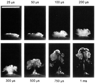
for myopia. J Refract Surg 15:124–31, 1999

90. Bor Z, Hopp B, Racz B, et al: Plume emission, shock wave and surface wave formation during excimer laser ablation of the cornea. Refract Corneal Surg 9:S111–S115, 1993

91. Kreuger RR, Krasinski JS, Radzewicz C, et al: Photography of shock waves during excimer laser ablation of the cornea: Effect of helium gas on propagation velocity. Cornea 12:330–334, 1993

92. Puliafito CA, Stern D, Krueger RR, Mandel ER: High-speed photography of excimer laser ablation of the cornea. Arch Ophthalmol 105:1255–1259, 1987

93. Campos M, Cuevas K, Garbus J, et al: Corneal wound healing after excimer laser ablation: Effects of nitrogen gas blower. Ophthalmology 99:893–897, 1992

94. Itoi M, Ryan R, Depaolis M, Aquavella JV: Clinical effect of blowing nitrogen gas across the cornea during photorefractive keratectomy. J Refract Surg 13:69–73, 1997

95. Noack J, Tonnies R, Hohla K, et al: Influence of ablation plume dynamics on the formation of central islands in excimer laser photorefractive keratectomy. Ophthalmology 104:823–830, 1997

96. Oshika T, Klyce SD, Smolek MK, McDonald MB: Corneal hydration and central islands after excimer laser photorefractive keratectomy. J Cataract Refract Surg 24:1575–1580, 1998

97. Lin DT: Corneal topographic analysis after excimer photorefractive keratectomy. Ophthalmology 101:1432–1439, 1994

98. Ambrosio G, Cennamo G, De Marco R, et al: Visual function before and after photorefractive keratectomy for myopia. J Refract Corneal Surg 10:129–136, 1994

99. O'Brart DP, Lohmann CP, Fitzke FW, et al: Night vision after excimer laser photorefractive keratectomy: Haze and halos. Eur J Ophthalmol 4:43–51, 1994

100. Verdon W, Bullimore M, Maloney RK: Visual performance after photorefractive keratectomy. A prospective study. Arch Ophthalmol 114:1465–1472, 1996

101. O'Brart DP, Corbett MC, Lohmann CP, et al: The effects of ablation diameter on the outcome of excimer laser photorefractive keratectomy. A prospective, randomized, double-blind study. Arch Ophthalmol 113:438–443, 1995

102. O'Brart DP, Corbett MC, Verma S, et al: Effects of ablation diameter, depth, and edge contour on the outcome of photorefractive keratectomy. J Refract Surg 12:50–60, 1996

103. O'Brart DP, Gartry DS, Lohmann CP, et al: Excimer laser photorefractive keratectomy for myopia: Comparison of 4.00- and 5.00-millimeter ablation zones. J Refract Corneal Surg 10:87–94, 1994

104. Seiler T, Holschbach A, Derse M, et al: Complications of myopic photorefractive keratectomy with the excimer laser. Ophthalmology 101:153–160, 1994

105. Niesen U, Businger U, Hartmann P, et al: Glare sensitivity and visual acuity after excimer laser photorefractive keratectomy for myopia. Br J Ophthalmol 81:136–140, 1997

106. Schwartz-Goldstein BH, Hersh PS: Corneal topography of phase III excimer laser photorefractive keratectomy: Optical zone centration analysis. Ophthalmology 102:951–962, 1995

107. Cantera E, Cantera I, Olivieri L: Corneal topographic analysis of photorefractive keratectomy in 175 myopic eyes. Refract Corneal Surg 9:S19–S22, 1993

108. Schwartz-Goldstein BH, Hersh PS: Corneal topography of phase III excimer laser photorefractive keratectomy: Optical zone centration analysis. Ophthalmology 102:951–962, 1995

109. Balestrazzi E, De Mofetta V, Spadea L, et al: Histological, immunohistochemical, and ultrastructural findings in human corneas after photorefractive keratectomy. J Refract Surg 11:181–187, 1995

110. Lohmann CP, MacRobert I, Patmore A, et al: A histopathological study of photorefractive keratectomy. Lasers Light Ophthalmol 6:149–158, 1994

111. Fagerholm P, Hamberg-Nystrom H, Tengroth B: Wound healing and myopic regression following photorefractive keratectomy. Acta Ophthalmol 72:229–223, 1994

112. L'Esperance FA, Taylor DM, Warner JW: Human excimer laser keratectomy. Short-term histopathology. J Refract Surg 4:118–112, 1988

113. Lohmann C, Gartry D, Kerr Muir M, et al: ‘Haze’ in photorefractive keratectomy: Its origins and consequences. Lasers Light Ophthalmol 4:15–13, 1991

114. Corbett MC, Prydall JI, Verma S, et al: An in vivo investigation of the structures responsible for corneal haze after photorefractive keratectomy and their effect on visual function. Ophthalmology 103:1366–1368, 1996

115. Moller-Pedersen T, Vogel M, Li HF, et al: Quantification of stromal thinning, epithelial thickness, and corneal haze after photorefractive keratectomy using in vivo confocal microscopy. Ophthalmology 104:360–336, 1997

116. Corbett MC, Prydall JI, Verma S, et al: An in vivo investigation of the structures responsible for corneal haze after photorefractive keratectomy and their effect on visual function. Ophthalmology 103:1366–1368, 1996

117. Tengroth B, Epstein D, Fagerholm P, et al: Excimer laser photorefractive keratectomy for myopia: Clinical results in sighted eyes. Ophthalmology 100:739–745, 1993

118. Salz JI, Maguen E, Nesburn AB, et al: A two-year experience with excimer laser photorefractive keratectomy for myopia. Ophthalmology 100:873–882, 1993

119. Gartry DS, Ker Muir MG, Marshall J: Photorefractive keratectomy with an argon fluoride excimer laser: A clinical study. Refract Corneal Surg 7:420–343, 1991

120. Caubet E: Cause of subepithelial corneal haze over 18 months after photorefractive keratectomy for myopia. Refract Corneal Surg 9:S65–S67, 1993

121. Taylor HR, McCarty CA, Aldred GR: Predictability of excimer laser treatment of myopia. Arch Ophthalmol 114:248–225, 1996

122. Seiler T, Holschbach A, Derse M, et al: Complications of myopic photorefractive keratectomy with the excimer laser. Ophthalmology 101:153–160, 1994

123. Braunstein RE, Jain S, McCally RL, et al: Objective measurement of corneal light scattering after excimer laser keratectomy. Ophthalmology 103:439–444, 1996

124. Corbett MC, Verma S, O'Brart DPS, et al: The effect of ablation profile on wound healing and visual performance one year after excimer laser PRK. Br J Ophthalmol 80:224–223, 1996

125. Nagy ZZ, Hiscott P, Seitz B, et al: Ultraviolet-B enhances corneal stromal response to 193-nm excimer laser treatment. Ophthalmology 104:375–338, 1997

126. Meyer JC, Stulting RD, Thompson KP, Durrie DS: Late onset of corneal scar after excimer laser photorefractive keratectomy. Am J Ophthalmol 121:529–523, 1996

127. Lipshitz I, Loewenstein A, Varssano D, Lazar M: Late onset corneal haze after photorefractive keratectomy for moderate and high myopia. Ophthalmology 104:369–367, 1997

128. Kuo IC, Lee SM, Hwang DG: Late-onset corneal haze and myopic regression after photorefractive keratectomy (PRK). Cornea 23:350–355, 2004

129. Marques EF, Leite EB, Cunha-Vaz JG: Corticosteroids for reversal of myopic regression after photorefractive keratectomy. J Refract Surg 11:S30, 1995

130. Tanabe T, Miyata K, Samejima T, et al: Influence of wavefront aberration and corneal subepithelial haze on low-contrast visual acuity after photorefractive keratectomy. Am J Ophthalmol 138:620–624, 2004

131. Dutt S, Steinert RF, Raizman MB, et al: One-year results of excimer laser photorefractive keratectomy for low to moderate myopia. Arch Ophthalmol 112:1427–1436, 1994

132. Hersh PS, Schein OD, Steinert R: Characteristics influencing outcomes of excimer laser photorefractive keratectomy. Summit Photorefractive Keratectomy Phase III Study Group. Ophthalmology 103:1962–1969, 1996

133. Loewenstein A, Lipshitz I, Levanon D, et al: Influence of patient age on photorefractive keratectomy for myopia. J Refract Surg 13:23–22, 1997

134. Durrie DS, Lesher MP, Hunkeler JD: Treatment of overcorrection after myopic photorefractive keratectomy. J Refract Corneal Surg 10:S29, 1994

135. Cherry PM: Removal of epithelium and scraping the underlying stroma as treatment for photorefractive keratectomy overcorrection or undercorrection of myopia. Ophthalmic Surg Lasers 27:S487–S4879, 1996

136. Kim JH, Sah WJ, Park CK, et al: Myopic regression after photorefractive keratectomy. Ophthalmic Surg Lasers 27:S435–S439, 1996

137. Corbett MC, O'Brart DP, Warburton FG, Marshall J: Biologic and environmental risk factors for regression after photorefractive keratectomy. Ophthalmology 103:1381–1391, 1996

138. Taylor HR, McCarty CA, Aldred GF: Predictability of excimer laser treatment of myopia. Arch Ophthalmol 114:248–251, 1996

139. O'Brart DP, Corbett MC, Lohmann CP, et al: The effects of ablation diameter on the outcome of excimer laser photorefractive keratectomy. A prospective, randomized, double-blind study. Arch Ophthalmol 113:438–443, 1995

140. O'Brart DP, Corbett MC, Verma S, et al: Effects of ablation diameter, depth, and edge contour on the outcome of photorefractive keratectomy. J Refract Surg 12:50–60, 1996

141. O'Brart DP, Gartry DS, Lohmann CP, et al: Excimer laser photorefractive keratectomy for myopia: Comparison of 4.00- and 5.00-millimeter ablation zones. J Refract Corneal Surg 10:87–94, 1994

142. Seiler T, Holschbach A, Derse M, et al: Complications of myopic photorefractive keratectomy with the excimer laser. Ophthalmology 101:153–160, 1994

143. Shah SI, Hersh PS: Photorefractive keratectomy for myopia with a 6-mm beam diameter. J Refract Surg 12:341–346, 1996

144. Maguire LJ, Bechara S: Epithelial distortions at the ablation zone margin after excimer laser photorefractive keratectomy for myopia. Am J Ophthalmol 117:809–810, 1994

145. Epstein D, Tengroth B, Fagerholm P, Hamberg-Nystrom H: Excimer retreatment of regression after photorefractive keratectomy. Am J Ophthalmol 117:456–461, 1994

146. Sutton G, Kalski RS, Lawless MA, Rogers C: Excimer retreatment for scarring and regression after photorefractive keratectomy for myopia. Br J Ophthalmol 79:756–759, 1995

147. Snibson GR, McCarty CA, Aldred GR, et al: Retreatment after excimer laser photorefractive keratectomy. Am J Ophthalmol 121:250–257, 1996

148. Matta CA, Piebenga LW, Deitz MR, Tauber J: Excimer retreatment for myopic photorefractive keratectomy failures. Six to 18-month follow-up. Ophthalmology 103:444–451, 1996

149. Hefetz L, Krakowski D, Haviv D, et al: Results of a repeated excimer laser photorefractive keratectomy for myopia. Ophthalmic Surg Lasers 28:8–13, 1997

150. Loewenstein A, Lipshitz I, Varssano D, Lazar M: Excimer laser reablation. Ophthalmic Surg Lasers 28:282–287, 1997

151. Pop M, Aras M: Photorefractive keratectomy retreatments for regression. One year follow up. Ophthalmology 103:1979–1984, 1996

152. Rozsival P, Feuermannova A: Retreatment after photorefractive keratectomy for low myopia. Ophthalmology 105:1189–1193, 1998

153. Chatterjee A, Shah S, Bessant DA, Doyle SJ: Results of excimer laser retreatment of residual myopia after previous photorefractive keratectomy. Ophthalmology 104:1321–1326, 1997

154. Gartry DS, Larkin FP, Hill AR, et al: Retreatment for significant regression after excimer laser photorefractive keratectomy. Ophthalmology 105:131–141, 1998

155. Pop M, Aras M: Photorefractive keratectomy retreatments for regression. One year follow up. Ophthalmology 103:1979–1984, 1996

156. Gartry DS, Larkin FP, Hill AR, et al: Retreatment for significant regression after excimer laser photorefractive keratectomy. Ophthalmology 105:131–141, 1998

157. Teal P, Breslin C, Arshinoff S, Edmison D: Corneal subepithelial infiltrates following excimer laser photorefractive keratectomy. J Cataract Refract Surg 21:516–518, 1995

158. Sher NA, Frantz JM, Talley A, et al: Topical diclofenac in the treatment of ocular pain after excimer photorefractive keratectomy. Refract Corneal Surg 9: 425–436, 1993

159. Sampath R, Ridgway AE, Leatherbarrow B: Bacterial keratitis following excimer laser photorefractive keratectomy: A case report. Eye 8:481–482, 1994

160. Gartry DS, Ker Muir MG, Marshall J: Photorefractive keratectomy with an argon fluoride excimer laser: A clinical study. Refract Corneal Surg 7:420–443, 1991

161. Brancato R, Tavola A, Carones F, et al: Excimer laser photorefractive keratectomy for myopia: Results in 1165 eyes. Refract Corneal Surg 9:95–104, 1993

162. Gartry DS, Kerr-Muir MG, Marshall J: The effect of topical corticosteroids on refraction and corneal haze following excimer laser treatment of myopia: An update. A prospective, randomized, double-masked study. Eye 7:584–590, 1993

163. Chatterjee A, Shah S, Bessant DA, et al: Reduction in intraocular pressure after excimer laser photorefractive keratectomy. Correlation with pretreatment myopia. Ophthalmology 104:355–359, 1997

164. Abbasoglu OE, Bowman RW, Cavanagh HD, McCulley JP: Reliability of intraocular pressure measurements after myopic excimer photorefractive keratectomy. Ophthalmology 105:2193–2196, 1998

165. Mardelli PG, Piebenga LW, Matta CS, et al: Corneal endothelial status 12 to 55 months after excimer laser photorefractive keratectomy. Ophthalmology 102:544–549, 1995

166. Carones F, Brancato R, Venturi E, Morico A: The corneal endothelium after myopic excimer laser photorefractive keratectomy. Arch Ophthalmol 112:920–924, 1994

167. Stulting RD, Thompson KP, Waring GO III, Lynn M: The effect of photorefractive keratectomy on the corneal endothelium. Ophthalmology 103:1357–1365, 1996

168. Trocme SD, Mack KA, Gill KS, et al: Central and peripheral endothelial cell changes after excimer laser photorefractive keratectomy for myopia. Arch Ophthalmol 114:925–928, 1996

169. Cennamo G, Rosa N, Del Prete A, et al: The corneal endothelium 12 months after photorefractive keratectomy in high myopia. Acta Ophthalmol Scand 75:128–130, 1997

170. Kent DG, Solomon KD, Peng Q, et al: Effect of surface photorefractive keratectomy and laser in situ keratomileusis on the corneal endothelium. J Cataract Refract Surg 23:386–397, 1997

171. Stulting RD, Thompson KP, Waring GO III, Lynn M: The effect of photorefractive keratectomy on the corneal endothelium. Ophthalmology 103:1357–1365, 1996

Back to Top