Chapter 46
Keratomileusis
CASIMIR A. SWINGER
Main Menu   Table Of Contents

Search

BACKGROUND
THEORY
INDICATIONS AND CONTRAINDICATIONS
INSTRUMENTATION
PREOPERATIVE EVALUATION
SURGICAL PROCEDURES
RESULTS
COMPLICATIONS
FUTURE DIRECTIONS
CONCLUSIONS
REFERENCES

As the shortcomings of radial keratotomy (RK), such as long-term instability, have manifested themselves, there has been an increased interest in lamellar refractive keratoplasty in general, and keratomileusis in particular, for the correction of all types of refractive errors, and especially for the correction of myopia.1–2021–40In this section, the various types of keratomileusis surgery that are currently being performed for the correction of myopia, along with new developments, will be discussed.
Back to Top
BACKGROUND
It has been more than 30 years since Barraquer began developing his techniques of keratophakia and keratomileusis, and it is primarily through his genius that the field of corneal surgery is what it is today. Barraquer5 first reported clinical results with autoplastic myopic keratomileusis (MKM) in 1964, and he has continued to perform it on a regular basis. The techniques were introduced in the United States by Troutman and Swinger38 in 1977.
Back to Top
THEORY
Myopic keratomileusis alters only the anterior corneal curvature, unlike RK, where both corneal surfaces have a new shape after surgery.

The refractive power of the anterior corneal surface is given by the following formula:

Refractive power (D) = 376/radius of curvature (mm)

To correct myopia, one must decrease the anterior corneal refractive power. From the above equation, this means that the radius of curvature of the cornea must be increased; that is, the cornea must be flattened. One can easily calculate the required final radius of anterior corneal curvature from the above equation by simply subtracting the spectacle refraction, calculated at the corneal vertex, from the initial corneal refractive power (keratometry). Such calculations are the basis of nomograms made available at training courses.

One may flatten the cornea in a variety of ways. As pioneered by Barraquer (Fig. 1), MKM is accomplished by performing a lamellar keratectomy on the patient's cornea, freezing the disc on a cryolathe (Steinway Instrument Co, San Diego, CA) (Fig. 2), resecting stromal tissue primarily in the center, and replacing the carved disc on the bed, resulting in a compensatory flattening of the central cornea.

Fig. 1. Classic myopic keratomileusis. A. Lamellar keratectomy on patient's cornea. B. Tissue resected (shaded area) primarily in center. C. Lenticule with reduced optical power. D. Lenticule placed on stromal bed, flattening the cornea.

Fig. 2. Barraquer cryolathe.

Swinger and co-workers35 developed planar nonfreeze MKM, whereby the freeze damage due to the cryolathe was eliminated. In this technique, called the BKS (Barraquer-Krumeich-Swinger) procedure, the resected anterior lamellar disc of the patient's cornea is inverted over one of a series of dies (Fig. 3) whose curvature is designed such that when a second planar cut is made with the microkeratome, (Fig. 4) the desired optical power is imposed onto the lamellar disc, which is then replaced.

Fig. 3. BKS keratomileusis. The lamellar disc is placed, epithelial side down, over a die of appropriate curvature such that a planar cut with the microkeratome alters the optical power of the disc, allowing for the correction of myopia or hyperopia.

Fig. 4. Barraquer-Krumeich-Swinger device.

Alternatively, nonfreeze MKM may be performed by making the second resection on the stromal bed, rather than on the back of the cap, in which case the procedure is called in situ keratomileusis (Fig. 5). When an ultraviolet laser is used to perform the optical resection on the bed (Fig. 6), the technique is called laser in situ keratomileusis; sometimes this is referred to by the acronym LASIK.

Fig. 5. In situ keratomileusis. A. Hinged flap made with microkeratome. B. Disc of tissue (shaded area) excised by microkeratome. C. Flattened cornea after replacement of flap.

Fig. 6. Laser in situ keratomileusis. A. Hinged flap made with microkeratome. B. Laser ablation (shaded area destroyed) of bed to alter curvature. C. Flattened cornea after replacement of flap.

Back to Top
INDICATIONS AND CONTRAINDICATIONS

INDICATIONS

The primary medical indication for MKM is high anisometropia with contact lens failure. In addition, patients may elect to undergo MKM for improvement of their uncorrected and/or best-corrected visual acuity. Many patients desire good uncorrected acuity for various occupations, professional or psychologic reasons, sports, and so forth. Because it has been documented that the best-corrected visual acuity can also be improved after MKM, it is indicated in patients who have subnormal vision with spectacles or contact lenses.32

Keratomileusis can also be used in myopic patients who have undergone prior surgery, such as RK or penetrating keratoplasty.33 Homoplastic MKM, whereby a donor lamellar corneal disc with optical power replaces the resected anterior cap from the patient's eye, has been used to provide very high corrections (up to 28D) and to rehabilitate patients with anterior corneal scars and myopia by simultaneously eliminating both.37

Until recently, MKM had been reserved for myopia of 6D or more, and RK for myopia of less than 6D. With the advent of laser technology, however, laser in situ MKM is being performed investigationally for myopia as low as 1 D.

CONTRAINDICATIONS

Refractive surgery should not be performed on patients whose myopia is progressing and unstable, unless there are good reasons. Autoplastic MKM should probably be reserved for myopic errors of less than 15 to 18D and should not be performed on severely dry eyes, eyes with poorly controlled glaucoma, diseased corneas (although homoplastic MKM may be used in some cases), and eyes with treatable retinal pathology that could lead to detachment. Refractive surgery of any type should be applied most cautiously to patients with good vision in only one eye.

Back to Top
INSTRUMENTATION

MICROKERATOME SET

The lamellar keratectomy is performed with an electromechanical microkeratome. The classic Barraquer (Steinway Instrument Co, San Diego, CA) and BKS (Barraquer-Krumeich-Swinger) (Eyetech-M.V.A. AG, Balzers, Liechtenstein) devices consist of a detachable head powered by a high-speed DC motor (Fig. 7) that oscillates a commercial razor blade, although newer models may be gas turbine-driven.18 The microkeratomes have a series of interchangeable base plates that allow one to vary the depth of the keratectomy in steps of 0.05 mm. The microkeratome sets also come with a set of perilimbal suction rings that vary in height above the limbus. These allow for variable corneal protrusion through the central aperture and serve to adjust the diameter of the keratectomy. To predict the diameter prior to the actual cut, an applanation lens with a reticle on its lower surface is used. Also, a smooth keratectomy is best ensured when the intraocular pressure is elevated to 65 mm Hg. An applanation tonometer is used to ensure this.

Fig. 7. Barraquer or BKS microkeratome.

In the past few years, many surgeons have ceased to use the classic microkeratome and instead use one of the second-generation devices, such as an automated microkeratome (Fig. 8), which is propelled via a series of gears (Chiron Vision, Irvine, CA). Rather than use a series of perilimbal suction rings, only a single ring is used, one that can be raised or lowered on a thread to provide a range of resection diameters. Another automated microkeratome developed by Draeger (Storz Instruments GMBH, Heidelberg, Germany) uses a circular rotating blade that is advanced automatically, rather than a linear oscillating blade as in the above devices.18,31

Fig. 8. Ruiz automated microkeratome. (Courtesy of Steinway Instrument Co., San Diego, CA)

COMPUTERS

Although a computer is used to determine the settings of the cryolathe and for laser surgery, nomograms are invariably used for all forms of mechanical nonfreeze keratomileusis, providing the surgeon with the diameter and depth of resection and the base plate to be used to achieve the desired optical result.

LASERS

Any laser capable of surface ablation may be used to remove corneal tissue. Until now, most studies have used an excimer laser. The Novatec ultraviolet laser (Novatec Laser Systems, Carlsbad, CA) may also be used.

Back to Top
PREOPERATIVE EVALUATION
In addition to a complete ophthalmologic examination, other investigations should be performed before surgery, including keratometry and topography to look for any irregularities, which may be the only sign of keratoconus. The palpebral aperture should be determined to be of sufficient size to accommodate the suction ring, and any abnormalities or restrictions of the conjunctiva should be noted, because these can interfere with suction ring placement. Other studies include ultrasonic pachymetry, a good dilated examination, and possibly an axial length determination. One may also consider an endothelial cell count when performing MKM on a patient who has undergone prior surgery (such as penetrating keratoplasty, where the endothelium may be compromised) and when evaluating new techniques such as laser in situ keratomileusis. In patients whose vision is not correctable to 20/20, we consider laser interferometry or potential acuity measurements to determine the visual potential. The preoperative evaluation concludes with a detailed informed consent wherein the risks and benefits are completely outlined, other modalities of correction are adequately explained, and the typical postoperative course is described.

TIMING

In the United States, MKM is usually performed on one eye at a time. This has the obvious advantage of safety and also allows the patient to function normally with the unoperated eye until the operated eye is rehabilitated. However, if the surgery was uncomplicated and the topography appears satisfactory, one may anticipate a full recovery of vision and perform surgery on the second eye within days to weeks. Despite some advantages in waiting before operating on the fellow eye, there is currently interest in performing simultaneous bilateral surgery using laser in situ MKM.

Back to Top
SURGICAL PROCEDURES
Only the procedures of mechanical and laser in situ keratomileusis, currently the most popular forms of MKM, will be described. In countries where laser technology is readily available, in situ MKM, performed totally with a microkeratome, has been replaced by laser in situ keratomileusis. All MKM procedures are performed on an outpatient basis, usually using only topical anesthesia and intravenous sedation.

IN SITU MYOPIC KERATOMILEUSIS (IN SITU MKM)

Initially described by Krawicz20 as lamellar stromectomy, in situ MKM has been further developed by Ruiz.30 In this procedure, a microkeratome is used to make a superficial lamellar keratectomy, approximately 0.16 mm in depth and 7.25 to 7.75 mm in diameter. Initially, the keratectomy was a complete resection of an anterior lamellar cap, and dislocation or loss of the cap was observed in about 5% of cases. More recently, it has been shown to be efficacious to produce a hinged flap, such that the distal 10% of the resection is not completed.26 Thus, the lamellar disc is fixated by a tag of tissue, and the anterior lamellar cap is reflected over the nasal aspect of the globe, allowing access to the lamellar bed. When accomplished by an automated microkeratome, this technique is frequently referred to by the nonspecific marketing name of ALK (automated lamellar keratoplasty), which is a less desirable name than in situ MKM, because any automated microkeratome can be used for a number of applications.

After the suction ring is placed, the diameter of the proposed resection is determined by an applanation lens. When placed on the ring, the flat, lower surface of the lens lies in the same plane as the cutting blade of the microkeratome. The surgeon compares the diameter of the applanated corneal reflex with the reticle, thereby estimating the resection diameter. To obtain a specific diameter, the ring is adjusted up or down until the desired diameter is applanated. After this, an applanation tonometer is allowed to rest on the corneal dome. If the intraocular pressure is 65 mm Hg or greater, the diameter of the applanated area will be equal to or less than the diameter of the reticle on the lower surface of the tonometer, which corresponds to an intraocular pressure of 65 mm Hg.

When the parameters of diameter and intraocular pressure have been satisfied, the microkeratome is engaged. The microkeratome first passes from the temporal cornea toward the nasal cornea, and it is then withdrawn in the opposite direction, leaving a hinged flap. Immediately after the keratectomy, the bed is examined for any irregularities. Pachymetry is used to calculate the thickness of the anterior cap.

After the hinged flap is reflected nasally, the ring of the microkeratome is adjusted upward to accommodate a smaller-diameter resection, typically 4.2 mm. After the base plate of the microkeratome is adjusted to accommodate the desired depth of resection, according to a nomogram, a second microkeratome resection is performed concentric to and within the confines of the first. This second resection produces a depression that is of constant thickness centrally but tapers peripherally. The diameter and depth of this second resection determine the optic correction. The anterior flap is then replaced on top of the bed, and its circumference is dried so that it adheres to the bed without sutures, because keratomileusis is typically performed in a sutureless fashion.

LASER IN SITU KERATOMILEUSIS

Mechanical microkeratomes have provided only limited accuracy with respect to tissue resection. For this reason, the substitution of ultraviolet laser ablation for mechanical cutting is generally preferred, because it also offers the possibility for correction of associated astigmatism and less postoperative haze than with surface photorefractive keratectomy (PRK). The use of tangential laser energy to ablate the back of the corneal cap was first proposed by Swinger41 and the use of perpendicular laser energy to ablate the back of the corneal cap or the stromal bed after a keratectomy was first proposed by Peyman.28 As initially used clinically, a complete anterior lamellar keratectomy, approximately 0.3 mm in thickness and 8 mm in diameter, was performed.12 The resected disc is then inverted on a plastic base, and its bare stromal surface is exposed to the perpendicular laser beam. The requisite laser energy to produce the desired refractive change is delivered. Two disadvantages of this approach are that a hinged flap cannot be easily used to accommodate the laser ablation, and an initial keratectomy of considerable thickness, at least 0.3 mm, is needed, which may be associated with some instability.

An alternative approach has been to use the hinged flap method, whereby a thin resection of approximately 0.16 mm and 7.25 to 9 mm in diameter is performed, and the flap is reflected nasally.26 The stromal lamellar bed is then exposed to the laser energy such that the desired dioptric correction is imposed. The flap is then placed back into position (LASIK technique). In both cases, the computer programs differ from those for surface PRK and are still undergoing refinement.

In general, there is a strong trend toward this latter approach, using the hinged flap, to increase the safety of the sutureless technique. A few surgeons are advocating this technique for all levels of myopia, regardless of how low the levels are, and some are investigating simultaneous bilateral surgery. A disadvantage of ablating the stromal bed is that the laser energy is delivered deeper into the cornea than it is for surface PRK. Whether there will be any adverse effects on the endothelium or the weakened retina of a high myope, especially in high corrections or with lasers using a high fluence, remains to be determined.

POSTOPERATIVE CARE

The postoperative course of MKM is straightforward. Little pain is associated with MKM, unlike RK and PRK. It is unusual to see an epithelial defect the day after surgery. The patient is treated with topical antibiotics and steroids several times a day for 1 to 2 weeks.

Visual recovery is rapid after MKM, and patients will have a marked improvement in their uncorrected vision immediately after surgery.

Back to Top
RESULTS
Although MKM has been performed for many years, there are few published reports of clinical results with the various techniques, and there is only one prospective clinical evaluation.32

REFRACTIVE CORRECTION

The mean refractive change in a given series depends on patient selection. The maximal corrections reported in the literature following cryolathe MKM are in the range of 20D. Eyes with such large corrections may have either small optic zones, causing subjective symptoms, or postoperative instability due to excessive corneal thinning. In general, most surgeons presently agree that the upper limit of correction safely attainable with cryolathe MKM is approximately 15D. Although BKS, in situ, and laser in situ forms of MKM can provide even higher corrections, the published data are insufficient to establish that this is accomplished without symptoms and with good stability.

A peculiarity of MKM and surgery for myopia in general is that there is an underestimation of the optical correction when measured by keratometry, as compared with the change measured by refraction.

ACCURACY

Although the mean percent correction obtainable with MKM can be brought close to 100%, significant inaccuracy remains with mechanical techniques, as evidenced by the standard deviations reported, which typically range between 20% and 30%. The reasons for residual inaccuracy in MKM are many, because the procedure involves many steps--contact of tissue with air and fluid, complex instrumentation, and wound healing. Considering the complexity of MKM and the fact that one is operating for very high myopia, the reported accuracies of all forms of mechanical MKM may be satisfactory, with 65% to 85% of patients corrected to within 2D of the desired goal. The accuracies reported in early studies of laser MKM appear at least as good as those of the mechanical forms, with the potential for far greater accuracy.27 Frequently, any residual myopia or astigmatism is rectified secondarily with a keratotomy procedure.

ASTIGMATISM

Myopic keratomileusis can induce both regular and irregular astigmatism, although regular astigmatism appears to be minimal. In a prospective series, there was a mean increase in keratometric astigmatism of 0.50D with no mean increase in refractive cylinder.32

Whereas regular astigmatism is of little concern after MKM, irregular astigmatism is. Although minor irregularities can be seen on the topography of many eyes after MKM, irregular astigmatism may be a cause for a reduction in best-corrected visual acuity. Mechanical nonfreeze MKM may be associated with a higher risk of irregular astigmatism than cryolathe MKM and laser in situ MKM.10,11,27

VISUAL RESULTS

Visual recovery after MKM is rapid. In the prospective series of cryolathe MKM there was an average reduction of only 2.3 lines of Snellen acuity at 1 week after surgery, and the mean preoperative spectacle acuity was attained by 1 month.32 In another study, the mean preoperative acuity of 20/49 was attained by 1 month and improved further to 20/33 at 1 year.8 Visual recovery after BKS, in situ, or laser in situ MKM is very rapid, with patients frequently seeing 20/40 or better on the first postoperative day, because there is little surgical damage and wound healing in the central cornea.

It has been well documented that the best-corrected visual acuity may also be improved after MKM when performed on eyes with subnormal vision. In one study, the best-corrected visual acuity improved from a mean of 20/94 to a mean of 20/49 for 31 patients with 3 months of follow-up.32 Sixty-three percent of patients in this series had an improvement in their best-corrected vision.

Usually, normal 20/20 or better than preoperative vision will be regained after MKM, although in up to 5% to 10% of cases the vision may be reduced by two lines or more. In general, if the surgery was uncomplicated, one can expect the vision to return to its preoperative level. The major causes of minor reduction in visual acuity after MKM are irregular astigmatism and scarring after debridement of the interface for epithelialization.

STABILITY

The optical correction after MKM can be unstable for some time after surgery. Instability can be observed by both keratometry and refraction for as long as 6 months after surgery, if not longer.29

Studies of long-term results are available for only cryolathe MKM.8 Barraquer reported an average loss of correction during the first year of 15.42%. When based on refraction, there was a mean loss of 1.43D between months 1 and 12, and 0.45D between years 1 and 9, when compared with the control fellow eye. Thus, it appears that although the refractive correction obtained may be unstable for months, the long-term stability is satisfactory. One might expect good long-term stability after in situ forms of MKM, because the resected anterior cap is thinner than after cryolathe MKM, resulting in less anteriorly directed force from the flattened Bowman's membrane acting against the greater mass of the posterior bed in such cases.

Back to Top
COMPLICATIONS
Considering the complexity of MKM, the reported complications, although varied, usually do not compromise visual acuity. The known complications of MKM are given in Table 1.


TABLE 1. Known Complications of Keratomileusis

  Intraoperative complications

  Microkeratome resection problems

  Perforation into anterior chamber, with or without rupture of crystalline lens


  Disc too thin or thick, wrong diameter, decentered, irregular, notched
  Complications during optical modification

  Disc detachment from cryolathe or BKS device, perforation of disc, irregular resection, resection or ablation of tissue of wrong dimensions, perforation into anterior chamber


  Postoperative complications

  Subjective symptoms

  Glare, loss of contrast, halos, diplopia


  Optical

  Undercorrection or overcorrection
  Induced astigmatism
  Irregular astigmatism
  Reduced acuity


  Corneal

  Contact lens intolerance
  Delayed epithelialization
  Foreign bodies in the interface
  Epithelialization of the interface
  Keratitis
  Local or diffuse lenticule necrosis
  Scarring
  Ectasia
  Neovascularization
  Infection


  Intraocular

  Central retinal artery occlusion


Patients may experience a variety of symptoms after MKM. These include halos and multiple images (especially after in situ MKM), loss of contrast, and reduction in night vision. Despite excellent acuity, patients may sometimes experience their vision as less than desirable and revert to contact lens wear. These symptoms are in part due to small or decentered optic zones and irregular astigmatism.

Although the known complications after MKM are many, most either require no treatment or can be resolved with further corneal surgery, except for the disastrous complication of central retinal artery occlusion. Common complications of concern are epithelialization of the interface and irregular astigmatism. Corneal perforation during surgery occurs only rarely, and its incidence has been reduced with improvements in technique.

Epithelialization of the interface can occur in 5% to 10% of cases. Frequently, small buds of epithelium can be seen in the periphery, and are of no concern. However, if the epithelium is central and decreases vision directly or by producing irregular astigmatism, it should be debrided. This is easily accomplished by simple reflection of the cap and debridement of the bed. Epithelialization of the interface should be treated early to avoid permanent mild opacification of the stroma and, if so treated, rarely results in a reduction of visual acuity.

The major common concern with MKM is irregular astigmatism. Although some minor irregularity is common, only in a small percentage of cases will it lead to a reduction in visual acuity. In reported series, there is an incidence of 7% to 14% where the visual reduction was attributable to irregular astigmatism. When it occurs, it is usually mild, and the reported reductions in acuity are at most several lines. However, irregular astigmatism may cause subjective symptoms, which can be more bothersome to the patient than the minor reduction in acuity. If the irregular astigmatism arises from an irregular keratectomy, one may need to perform a very deep lamellar keratoplasty to restore the cornea to its normal thickness and regularize the anterior contour. Refractive surgery can then be repeated at a later date.

Back to Top
FUTURE DIRECTIONS

INTRASTROMAL PHOTODISRUPTION

Another approach to keratomileusis is to use a laser with a transmissive wavelength that can be focused within the anterior corneal stroma to ablate tissue by photodisruption, producing a plasma cavity; when the plasma cavity collapses, the anterior cornea will assume a new curvature.36 The shape of the cavity to be ablated is designed such that the desired deformation of the anterior corneal curvature is produced upon collapse of the cavity.

Using animal and bank eyes, the author has demonstrated the ability to create fine layers of cavities, with the desired degree of spacing or overlap, in a controlled fashion (Fig. 9). Electron micrographic studies demonstrate little collateral damage at the ablation surfaces. Other investigators have also demonstrated the ability to produce ablation cavities that lead to a secondary deformation of the anterior cornea, although unpredictably. However, there is no evidence to date that one can achieve a desired deformation of a primate cornea with Bowman's membrane, let alone with any desired degree of accuracy. However, the novelty of this approach is exciting, because the anterior cornea is left undisturbed and a mechanical microkeratome is not needed. This would be a procedure with no pain, rapid visual recovery, and possibly reduced complications. Concerns remain over the effect of the shock waves produced on various aspects of the eye, especially the endothelium, although studies to date demonstrate no endothelial damage if the cavities are kept in the anterior stroma.

Fig. 9. Intrastromal photodisruption by Novatec laser.

LASER MICROKERATOME

The author and colleagues36 have developed a new solid-state laser (Novatec) and have demonstrated the possibility of producing a laser lamellar keratectomy of any desired shape. By focusing a transmissive wavelength laser appropriately, a lamellar cap of any size can be removed intact from the cornea (Fig. 10). This approach, which is still experimental, could eventually lead to a keratomileusis procedure performed only by laser, thereby avoiding mechanical microkeratomes and their limitations.

Fig. 10. Stromal bed after experimental use of Novatec laser microkeratome.

Back to Top
CONCLUSIONS
Keratomileusis has undergone a long period of development. Interest in and innovations of this surgical approach are increasing in an attempt to refine its applications and improve its accuracy and safety. A variety of approaches are now possible, and further clinical evaluations will be necessary to determine which techniques are most efficacious and applicable in a given setting.
Back to Top
REFERENCES

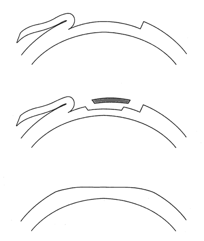
1. Ainslie D: The surgical correction of refractive errors by keratomileusis and keratophakia. Am J Ophthalmol 8:349, 1976

2. Arenas-Archila E, Sanchez-Thorin JC, Naranjo-Uribe JP et al: Myopic keratomileusis in situ: A preliminary report. J Cataract Refract Surg 17:424, 1991

3. Barker BA, Swinger CA: Keratophakia and keratomileusis. Int Ophthalmol Clin 28:126, 1988

4. Barraquer C, Gutierrez AM, Espinosa A: Myopic keratomileusis: Short-term results. Refract Corneal Surg 5:307, 1989

5. Barraquer JI: Queratomileusis para la correccion de la myopia. Arch Soc Am Oftalmol Optom 5:25, 1964

6. Barraquer JI: Keratomileusis for myopia and aphakia. Ophthalmology 88:701, 1981

7. Barraquer JI: Keratomileusis for the correction of myopia. Arch Soc Am Oftalmol Optom 16:221, 1982

8. Barraquer JI: Long term results of myopic keratomileusis--1982. Arch Soc Am Oftalmol Optom 19:137, 1983

9. Barraquer JI, Viteri E: Results of myopic keratomileusis. J Refractive Surg 3:98, 1987

10. Bas AM, Nano HD Jr: In situ myopic keratomileusis results in 30 eyes at 15 months. Refract Corneal Surg 7:223, 1991

11. Bosc JM, Montard M, Delbosc B et al: Non-freeze myopic keratomileusis. Retrospective study of 27 consecutive operations. J Fr Ophthalmol 13:10, 1990

12. Buratto L, Ferrari M, Genisi C: Myopic keratomileusis with the excimer laser: One-year follow up. Refract Corneal Surg 9:12, 1993

13. Casebeer JC, Slade SG, Dybbs A et al: Intraoperative pachometry during automated lamellar keratoplasty: A preliminary report. Refract Corneal Surg 10:41, 1994

14. Dossi F, Bosio P: Myopic keratomileusis: Results with a follow-up over one year. J Cataract Refract Surg 13:417, 1987

15. Durand L, Burillon C, Resal R: Refractive surgery and the non-freeze BKS set. Reliability of our Lyon methods. Bull Soc Ophtalmol Fr 90:441, 1990

16. Ganem S, Aron-Rosa D, Gross M et al: Myopic keratomileusis by excimer laser on a lathe. J Refract Corneal Surg 10:575, 1994

17. Gomes M: Keratomileusis-in-situ using manual dissection of corneal flap for high myopia. J Refract Corneal Surg 10(suppl):S255, 1994

18. Hoffman RF, Bechara SJ: An independent evaluation of second generation suction microkeratomes. Refract Corneal Surg 8:348, 1992

19. Kornmehl EW, Swinger CA, Pugh W et al: Corneascope evaluation of myopic keratomileusis (ARVO abstracts). Invest Ophthalmol Vis Sci 26(suppl):283, 1985

20. Krawicz T: Lamellar corneal stromectomy. Am J Ophthalmol 57:828, 1964

21. Krumeich JH: Indications, techniques, and complications of myopic keratomileusis. Int Ophthalmol Clin 23:75, 1983

22. Maxwell WA: Myopic keratomileusis: Initial results and myopic keratomileusis combined with other procedures. J Cataract Refract Surg 13:518, 1987

23. Maxwell WA, Nordan LT: Myopic keratomileusis: Early experience. J Refract Surg 1:124, 1986

24. Neumann AC, McCarty G, Sanders DR: Delayed regression of effect in myopic epikeratophakia vs myopic keratomileusis for high myopia. Refract Corneal Surg 5:161, 1989

25. Nordan LT, Fallor MK: Myopic keratomileusis: 74 consecutive non-amblyopic cases with one year follow-up. J Refract Surg 2:124, 1986

26. Pallikaris IG, Papatzanaki ME, Siganos DS et al: A corneal flap technique for laser in situ keratomileusis. Human studies. Arch Ophthalmol 109:1699, 1991

27. Pallikaris IG, Siganos DS: Excimer laser in situ keratomileusis and photorefractive keratectomy for correction of high myopia. J Refract Corneal Surg 10:498, 1994

28. Peyman GA: A method for modifying corneal curvature. U.S Patent 4,840,175, June 20, 1989

29. Polit F, Ibrahim O, El-Maghraby A et al: Cryolathe keratomileusis for correction of myopia of 4.00 to 8.00 diopters. Refract Corneal Surg 9:259, 1993

30. Ruiz LA, Rowsey JJ: A new refractive surgical approach: In situ keratomileusis for myopia and lamellar keratoplasty for hyperopia. Ophthalmology 95(suppl):145, 1988

31. Stonecipher KG, Parmeley VC, Rowsey JJ et al: Refractive corneal surgery with the Draeger rotary microkeratome in human cadaver eyes. J Refract Corneal Surg 10:49, 1994

32. Swinger CA, Barker BA: Prospective evaluation of myopic keratomileusis. Ophthalmology 91:785, 1984

33. Swinger CA, Barker BA: Myopic keratomileusis following radial keratotomy. J Refract Surg 1:53, 1985

34. Swinger CA, Barraquer JI: Keratophakia and keratomileusis: Clinical results. Ophthalmology 88:709, 1981

35. Swinger CA, Krumeich J, Cassiday D: Planar lamellar refractive keratoplasty. J Refract Surg 2:17, 1986

36. Swinger CA, Lai ST: Solid state photoablative decomposition--The Novatec laser. In Salz JJ, McDonnell PJ, McDonald MB (eds): Corneal Laser Surgery, p 269. St. Louis, CV Mosby, 1995

37. Swinger CA, Villasenor RA: Homoplastic keratomileusis for the correction of myopia. J Refractive Surg 1:219, 1985

38. Troutman RC, Swinger CA: Refractive keratoplasty--Keratophakia and keratomileusis. Trans Am Ophthalmol Soc 76:329, 1978

39. Tucker DN, Barraquer JI: Refractive keratoplasty: Clinical results in sixty-seven cases. Am J Ophthalmol 5:335, 1973

40. Zavala EY, Krumeich J, Binder PS: Laboratory evaluation of freeze vs nonfreeze lamellar refractive keratoplasty. Arch Ophthalmol 105:1125, 1987

41. Swinger C, Cassidy D: Method and Apparatus for Modifying Corneal Buttons. U.S. patent 4,660,556, April 28, 1987

Back to Top