|
| Chapter 45 Epikeratoplasty: A Historical Perspective MARK P. LESHER and DANIEL S. DURRIE Table Of Contents |
|
HISTORY SURGICAL TECHNIQUE INDICATIONS CONCLUSION REFERENCES |
| The concept of using a surgical procedure to change the refractive power of the anterior corneal surface was developed by Barraquer in the 1960s and 1970s.1 The precursors of epikeratoplasty, keratomileusis and keratophakia, proved to be technically challenging and demanding of the surgeon because they required expertise in manufacturing a donor lenticule using the cryolathe. In the hands of experienced surgeons, the results were quite good. However, for these procedures to be performed by many surgeons, they needed to be simplified. |
| HISTORY |
| Kaufman2 and Werblin3 proposed epikeratoplasty as a simple procedure for the correction of aphakia
in 1980. In epikeratoplasty, a donor corneal lenticule is frozen, shaped
with a cryolathe, and sutured to the anterior corneal surface
to provide the appropriate optical correction (myopic, plano, or hyperopic). Alternatively, the button may be lyophilized and stored in a
vacuum container for later use. The advantage of epikeratoplasty over
earlier procedures was that the lenticules could be prepared in a centralized
location by a company (e.g., Allergan Medical Optics [AMO], Cryo-Optics) dedicated to lenticule
production. This meant that the surgeon needed to master only the
surgical procedure and not the use of the cryolathe. During the 1980s, several multicenter national studies were sponsored by AMO to assess the efficacy of epikeratoplasty for several conditions: pediatric aphakia, adult aphakia, myopia, and keratoconus.4–7 The procedure was promoted as a fairly simple surgery that could be performed by most general ophthalmologists as an alternative to aphakic spectacles in the adult or for pediatric aphakia, or in lieu of penetrating keratoplasty for keratoconus. It was also promoted as a good technique for performing lamellar corneal patch grafts in cases of corneal thinning or perforation. Because the lenticules are not an economically viable product, they are no longer commercially available from AMO. |
| SURGICAL TECHNIQUE | |
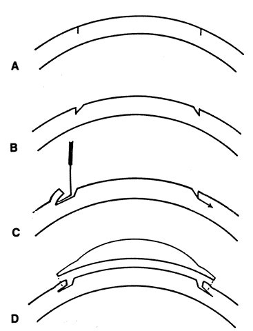
The surgeon provided two pieces of information to the central tissue processing
center to facilitate preparation of the donor lenticule: the
patient's spherical equivalent of refraction at the corneal plane
and the average central keratometry readings. The tissue was custom lathed
for each patient and delivered to the operating surgeon. The tissue
lens was removed from the vacuum container and rehydrated in balanced
salt solution with antibiotic (gentamicin). The patient's optical
center was located and marked with a needle or Sinsky hook. Centration
was extremely important, especially in the cases of myopic epikeratoplasty. A
donut of central corneal epithelium was then removed by
a combination of mechanical and chemical debridement. The central 2 to 3 mm
of corneal epithelium containing the centering mark were left intact
temporarily. A Hessburg-Barron vacuum trephine was centered on the
optical axis, and a 0.25-mm partial-thickness trephination was performed
(Fig. 1) . An annular keratectomy was performed, in cases of keratoconus, to improve
adherence of the epigraft to the host corneal stroma. The vertical
trephination was undermined approximately 1 mm peripherally with a
Suarez spreader or comparable instrument to allow the edge of the graft
to seat nicely in the keratectomy. The residual central host epithelium
was then removed mechanically and the graft bed was sometimes treated
with absolute alcohol or 4% cocaine to remove any residual epithelial
cells. The cornea was irrigated copiously with balanced salt solution, and
the rehydrated epikeratoplasty lenticule was sutured into position
with 16 to 24 interrupted 10-0 nylon sutures. Careful attention
was paid to making equal bites on the donor and recipient sides. The
ideal suture tension should be equal in every direction. The knots were
trimmed and buried just beneath the surface of the recipient to make
suture removal easier. The graft edges were then tucked into the peripheral
keratectomy, and subconjunctival antibiotic and/or steroids were
administered.
|
| INDICATIONS |
PEDIATRIC APHAKIA Epikeratoplasty was indicated for aphakia in children young enough to realize benefits from occlusion therapy (i.e., younger than 9 years of age). In most cases it was used as an alternative to an aphakic contact lens in a child with a traumatic or congenital cataract. Results in the pediatric population were reasonable, with the majority of patients in several studies achieving “useful” vision.8–10 The main difficulties with the procedure included mild graft haze, which increased the difficulty of amblyopia therapy, difficulties with centration, and inaccuracy in selecting lenticule powers in younger children. Advantages of the procedure included the ability to remove and replace the lenticules if the refractive error changed with age. The early success of intraocular lens (IOL) implants in children has resulted in a shift toward using IOLs rather than epikeratoplasty lenticules in contact lens-intolerant aphakic children. ADULT APHAKIA Posterior chamber IOL implantation is the preferred method of aphakia correction in the adult population. This is normally done in conjunction with cataract extraction at the time of lens removal. Certain situations exist in which lens implantation is not possible or advisable. Such conditions include lack of posterior capsule support, a disorganized anterior chamber that will not support an IOL, and compromised endothelial function secondary to aging or dystrophic processes. Aphakic contact lens wear is another alternative to IOL implantation. However, contact lens wear is frequently less successful in the typical cataract population because of tear film deficiency and patient difficulties with lens insertion and removal. Epikeratoplasty was proposed as a way to avoid an intraocular procedure in situations where lens implantation was difficult or where aphakic contact lens wear was not well tolerated.3,11,12 The development and improvement in secondary lens implant techniques (e.g., iris-fixated posterior chamber intraocular lenses, scleral-fixated intraocular lenses) have made these the preferred surgical techniques for correcting adult aphakia, and epikeratoplasty is rarely indicated. MYOPIA Prior to the acceptance of newer refractive procedures, such as radial keratotomy and the excimer laser, epikeratoplasty was promoted as an alternative to contact lenses or spectacles in patients with more than 6.0 diopters of myopia. The refractive results in myopic patients were variable and highly dependent on the expertise of the surgeon.6,13 Irregular astigmatism resulted in a loss of best-corrected visual acuity after surgery. The use of epikeratoplasty in the myopic population added to knowledge in the areas of graft centration, postoperative glare, and corneal topography. However, the development of the excimer laser and the increasing interest in radial keratotomy have resulted in epikeratoplasty being performed less and less frequently for myopia. Computer-controlled excimer laser ablation of the anterior corneal surface appears to offer a much more predictable way to flatten the central cornea than did epikeratoplasty. KERATOCONUS Epikeratoplasty was used as an intermediate step between contact lens wear and penetrating keratoplasty in the management of keratoconus. Indications included patients with mild to moderate keratoconus without apical scarring who were having difficulties with contact lens wear. A plano graft was sutured tightly over the central cornea to flatten the cone and reduce myopia.14,15 This procedure should theoretically have no risk of immune rejection because no donor antigens come into contact with the host's immune system. Most surgeons have abandoned epikeratoplasty for the treatment of keratoconus in favor of full-thickness keratoplasty. Improvements in keratoconus contact lens fitting have allowed surgical treatment to be delayed in most cases. Lamellar keratoplasty with use of non-commercially prepared tissue remains an intermediate option in the treatment of keratoconus. PATCH GRAFT The Keratopatch (AMO, Irvine, CA) was a lyophilized plano-corneal lenticule that could be used to perform a lamellar wound “reinforcement” in an area of corneal thinning or perforation. It offered a readily available tissue patch that did not require hand carving, was predictable in thickness, and was surgically easy to use. Other techniques for dealing with small corneal perforations, such as tissue adhesive, are only stop-gap measures. The lack of availability of freeze-dried prepared corneal tissue is certainly a loss for the ophthalmic surgeon. Hand-carved donor grafts can be used for this purpose, but such tissue is often in high demand for penetrating keratoplasty. The eye-banking system may want to address the needs of the corneal surgeon in this area by developing a suitable replacement for the Keratopatch. |
| CONCLUSION |
| Because of the lack of economic viability, epikeratoplasty lenticules are no longer commercially available from AMO in the United States. Epikeratoplasty was a relatively simple ophthalmic surgical procedure that was used for the correction of pediatric and adult aphakia, keratoconus, and myopia during the 1980s. The results varied and were frequently related to the surgeons' experience with the procedure. Improvements in intraocular lenses and secondary IOL fixation techniques have allowed secondary lens implantation to replace epikeratoplasty as the procedure of choice for correcting aphakia. The excimer laser now offers a technically simpler and more reliable method for correcting myopia. Lamellar keratoplasty remains a useful procedure in cases of corneal thinning or perforation and for selected keratoconus patients. The need for commercially prepared lyophilized corneal tissue still exists and may be addressed by the national eye-banking community in the future. |