Chapter 44
Keratoprosthesis
WILLIAM J. POWER
Main Menu   Table Of Contents

Search

HISTORY
KERATOPROSTHESIS DESIGN
INDICATIONS
SURGICAL PROCEDURE AND EARLY POSTOPERATIVE CARE
COMPLICATIONS
RESULTS
CURRENT RESEARCH
CONCLUSIONS
REFERENCES

Keratoprosthesis surgery is indicated in cases of corneal blindness for which penetrating keratoplasty is almost certain to fail. Such cases include ocular cicatricial pemphigoid, Stevens-Johnson syndrome, severe chemical burns, severely vascularized corneas, and recurrent graft failure. Keratoprosthesis surgery can be associated with significant complications, however, and requires intensive follow-up. Recent advances aimed at preventing complications and treating early complications have improved the prognosis, and there is now reason for more optimism with this technique.
Back to Top
HISTORY
Pellier de Quengsy1 first suggested the concept of an artificial cornea in the treatment of corneal blindness in 1789. In 1855, Nussbaum2 performed experimental animal work and also published human trials using a quartz crystal implant, which he implanted into the cornea. During the next 50 years, further efforts in keratoprosthesis design and insertion were continued by Heusser,3 Dimmer,4 Salzer,5 and von Hippel.6 Unfortunately, these keratoprostheses were associated with extremely high incidences of complications and typically failed early because of tissue necrosis around the device with subsequent leak, infection, and finally extrusion of the keratoprosthesis.

After 1906, when the first human-to-human corneal graft was performed, there was a loss of interest in keratoprosthesis development and surgery. In 1920, Verhoeff reported on a single case of insertion of a quartz button into a patient's cornea; however, it had to be removed shortly afterward.7 Similarly, Filatov,8 in 1935, implanted a full penetrating glass device into a patient's opacified cornea and covered it with a double conjunctival flap after surgery. He left the flap in place, and after 6 months it had thinned sufficiently to give the patient an ambulatory vision of 1/200. More details on the history of keratoprosthesis surgery around this period have been reported previously in reviews by Day,9 Cardona,10 and Lund.11

During World War II, Wunsche,12 Stone,13 and others noticed that polymethylmethacrylate (PMMA) splinters embedded in the cornea of pilots were well tolerated. This led to their experiments showing that PMMA discs could be retained in the cornea of rabbits. Soon human applications followed, and many ophthalmologists attempted to refine their procedure using these new inert plastics, primarily PMMA. However, once again, many of these cases were associated with severe complications, and the procedure lost favor. Some surgeons did persevere in their techniques and refined them over the years. However, the combined experiences of their keratoprosthesis surgeries probably did not amount to more than four or five thousand cases during the past half century, which is quite small compared with the number of penetrating keratoplasties carried out on a worldwide basis.

Back to Top
KERATOPROSTHESIS DESIGN
Keratoprosthesis designs consist of two principal types. The first and most commonly used type is made with a central stem of transparent material that is held in place in the cornea by a peripheral skirt, which is usually placed intralamellarly in the stroma and is sometimes covered by transplanted autologous tissue or lid skin. The second, less common type is a so-called collar button-shaped device, which consists of two plates joined by a central clear optical stem. This is inserted such that the plates sandwich the cornea between them. The advantages of the latter design include a short optical stem, thus providing a good view at the slit lamp, a generous visual field, and good stability, because the wide plates prevent tilting of the device off the visual axis. The design also facilitates repair should necrosis of tissue occur around the stem.

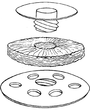
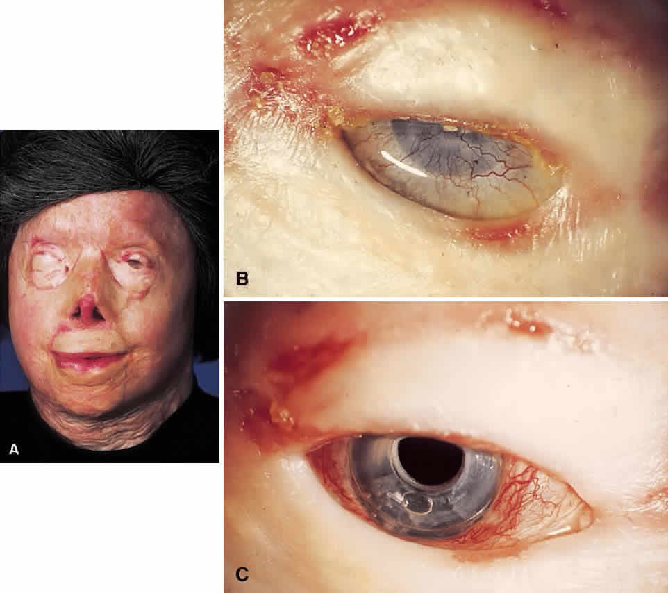
The material most frequently used for the central optical portion of the keratoprosthesis has been medical-grade PMMA. PMMA is completely transparent and biologically inert, and there is now extensive experience with its use in intraocular lenses. However, many different types of material have been used for the skirts or plates used to secure the optical stem in the cornea, such as perforated grids of PMMA, Teflon, nylon, and ceramics. More recently, attention has been directed to porous designs using materials such as Proplast,14 polytetrafluorethylene,15 hydrogels,16,17 collagen,18 and various copolymers.19,20 These materials have been manufactured and developed with the idea that colonization with natural tissue elements would subsequently hold and anchor the device more securely in place, thus decreasing the likelihood of future extrusion. In the 1960s, Strampelli21 introduced a novel idea of inserting the PMMA stem into a slice of dental bone from the patient's jaw, with the hope that the bone would subsequently heal better into the patient's cornea. This is, however, a formidable multistage procedure. DeVoe and Cardona22 made an important observation that in an extremely dry eye, if the prosthesis is allowed to protrude through the skin rather than between the lids, safety is enhanced and the extrusion rate is subsequently reduced. Considerable modifications to various keratoprosthesis designs have occurred over the years, but whether an ideal keratoprosthesis can ever be designed that will integrate into the human cornea without the risk of extrusion or necrosis has yet to be determined.

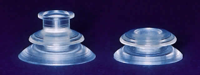
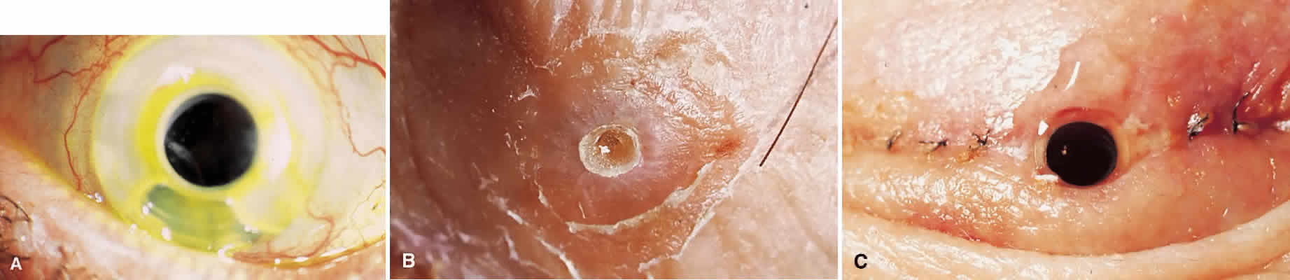
My practice is to use an all-PMMA device like that shown in Figure 1 (Dohlman-Doane Keratoprosthesis types I and II). The standard type I device is used in all but extremely dry eyes, where a “through-the-lid” approach with a type II device is preferred. In the latter case, the anterior nub is designed to protrude through an opening in the lid skin. The dimensions described for the two devices allow satisfactory visual fields. Thus, the type I permits a peripheral field of approximately 60 degrees; type II, with a longer stem, can give a visual field of approximately 40 degrees. The manufacture of these devices has previously been described.23 The Strampelli and Cardona devices are the other most frequently used models.

Fig. 1. Dohlman-Doane keratoprosthesis designs used by the author. Type I (left) is for the wet eye. Type II (right) has an added anterior nub for through-the-lid placement in the end-stage dry eye. (Courtesy of Dr. Claes H. Dohlman, Boston.)

Back to Top
INDICATIONS
It has become clear that the outcome of keratoprosthesis surgery differs markedly in various corneal disease causes, so the indications for surgery will have to be categorized accordingly. However, in general terms, certain criteria first must be fulfilled before qualifying for the procedure. Obviously, known end-stage retinal or optic nerve disease usually constitutes a contraindication. Also, young age of the patient or poor general health should be taken into consideration. One other very important consideration is the interest, commitment, and time of the surgeon, because frequent follow-ups and readiness to intervene early in threatening complications are mandatory. In addition, keratoprosthesis evaluation, surgery, and follow-up are technically difficult; therefore, it is best undertaken by corneal surgeons with a special interest in the subject.

Of the patients in whom keratoprosthesis could be considered, the patients with the poorest outlook are those with Stevens-Johnson syndrome. These patients have binocular disease and are sometimes monocular, because they frequently have undergone numerous unsuccessful surgical procedures in the past. A further difficulty with this group is that they are young and require the keratoprosthesis to remain complication-free for many years. As already mentioned, they often have ongoing ocular inflammation, which increases the postoperative complication rate. Accordingly, at this time I advise against keratoprosthesis surgery in young patients with Stevens-Johnson syndrome; I would prefer waiting until techniques have improved.

Patients with ocular cicatricial pemphigoid tend to be older, and this is in their favor. They also appear to have a more favorable outcome long term. However, they are prone to postoperative skin retraction and glaucoma.

Chemical burn patients can have good results after keratoprosthesis surgery. A simultaneous glaucoma shunt (Ahmed valve) is nearly always placed in these inflammatory conditions: the incidence of postoperative long-term glaucoma without such a shunt is extremely high.

Perhaps the most controversial category of patients are those with previous graft failures after noninflammatory edema, previous dystrophies, or trauma. The frequency of postoperative uveitis and uncontrolled glaucoma in these patients is quite low, and vision is usually restored fairly rapidly, in most cases more rapidly than a successful regraft would permit. Many of these are elderly patients with graft failure after bullous keratopathy. In certain instances, these patients may have a better chance of seeing well during their remaining days with a keratoprosthesis rather than with another corneal graft.24

Back to Top
SURGICAL PROCEDURE AND EARLY POSTOPERATIVE CARE
The goal of keratoprosthesis surgery is to keep the surgical procedure as simple as possible and to eliminate the need for nonocular support tissue. The insertion of the keratoprosthesis into a corneal graft is shown in Figure 2. Another important development has been to incorporate measures that will diminish future complications. To this end, once the prosthesis is inserted, it is temporarily covered with tissue (a total conjunctival flap of the Gundersen type, buccal mucosa, or lid skin). This reduces the incidence of postoperative necrosis of tissue surrounding the device, particularly in the early postoperative period. The threat of subsequent uncontrolled glaucoma often necessitates the simultaneous implantation of a drainage shunt. This is particularly true in conditions associated with past long-term inflammation (e.g., cicatricial pemphigoid, Stevens-Johnson syndrome, alkali burn). The Ahmed valve can be used with good results. This device can be implanted relatively easily and is associated with a lower incidence of hypotony in the postoperative period when compared with some of the other glaucoma devices. The lens, if present, is usually removed using an extracapsular technique, preserving the integrity of the posterior capsule when possible. Usually the central iris is also removed. A deep vitrectomy is standard in cases without an intact posterior capsule. Details on our surgical technique have been previously described.25

Fig. 2. Assembly of a type I keratoprosthesis. A large (10 mm) corneal graft with a central 3-mm hole is slid over the stem. The posterior plate is then screwed on tight and the entire unit is sutured in place with interrupted 10-0 nylon sutures. (Courtesy of Dr. Claes H. Dohlman, Boston.)

In the early postoperative period, it is desirable that the conjunctival flap does not retract, because attempts at repair are usually futile. Extensive surgery, such as is done in keratoprosthesis, is associated with considerable postoperative inflammation. Accordingly, we have found that small doses of corticosteroids are needed, especially in eyes with a previous history of severe inflammation (e.g., Stevens-Johnson syndrome, pemphigoid, chemical burns). Routinely, 10 mg dexamethasone is given by peribulbar injection through the lower lid at the completion of surgery. After 2 to 3 weeks, 20 to 40 mg triamcinolone may be administered by peribulbar injection if inflammation persists. The total dose of corticosteroids may be difficult to judge: too much may jeopardize wound healing, leading to necrosis, fistulas, or leak of tears. Once the flap has been opened, it is easier to monitor the anterior chamber and vitreous reaction. If postoperative uveitis is still present, peribulbar steroids may have to be continued. Occasionally, in patients with Stevens-Johnson syndrome or ocular cicatricial pemphigoid, systemic immunosuppression in the form of oral steroid treatment or other immunosuppressive agents is indicated.

Intravenous antibiotics are administered at the time of surgery. After surgery, patients are given topical antibiotic drops and a 14-day course of oral antibiotics. We usually prescribe topical antibiotic drops for a prolonged period. These are rotated every 2 to 3 months to prevent the development of resistant organisms. Patients with Stevens-Johnson syndrome who are particularly at risk for infection need to be monitored closely and should probably take topical antibiotics for life. After approximately 2 months, when the conjunctival flap or lid skin is opened, the patient is given topical anticollagenase medication. We use 1% medroxyprogesterone, which reduces collagenase synthesis, and a 1% tetracycline solution, which is a potent direct enzyme inhibitor. Although the efficacy of these agents in reducing corneal necrosis has not been established in humans, their beneficial effects have been well documented in experimental animals.26,27 Unfortunately, these suspensions are not commercially available and have to be made up in a hospital pharmacy.

Patients are seen frequently in the early postoperative period and are advised to report any symptoms of pain, lid edema, blurred vision, or discharge from the eye. The goal should be to achieve a noninflamed eye with intact tissue around the device (Figs. 3 and 4).

Fig. 3. Successful result with type I. (Courtesy of Dr. Claes H. Dohlman, Boston.)

Fig. 4. Successful result with type II. (Courtesy of Dr. Claes H. Dohlman, Boston.)

Back to Top
COMPLICATIONS
As mentioned previously, keratoprosthesis surgery is associated with a significant complication rate. In most instances, the severe complications are seen in the first year after surgery. However, the patient is never safe from potential complications and therefore requires frequent and close monitoring. If potential complications can be identified at an early stage and treated appropriately, then a more favorable outcome is likely. Clearly, care of the keratoprosthesis patient imposes a significant time burden on the corneal surgeon. Because of this, keratoprosthesis surgery in its current form is not yet suitable for developing countries, where the prevalence of corneal blindness is high but the number of physicians is low. Some attempts at reducing the complication rate have recently been described.28

The major complication is necrosis of tissue around the keratoprosthesis (Fig. 5). Unless treated, this can lead to subsequent tissue breakdown (melt), aqueous leak, infection, or retinal detachment. Certain measures reduce the risk of such calamities. Thus, the type I keratoprosthesis in the wet and adequately blinking eye is usually covered with a total conjunctival flap. This is left in place for 2 months before opening, which reduces the incidence of subsequent necrosis. The use of collagenase enzyme-suppressing medication also lowers the incidence of this complication. However, in some cases necrosis still occurs. It typically begins around the edge of the cornea adjacent to the optical stem. If identified early, various repair mechanisms can be attempted to halt the process. A thin slice of corneal tissue with a central 3-mm trephined hole and a radial cut can be slipped under the anterior plate and sutured in place.25 In many instances, this reinforces the base of the keratoprosthesis and allows further stabilization of the device. In some cases, this new donor cornea material becomes vascularized early; this is a favorable sign because it generally is associated with long-term retention of the added tissue. Once repaired, the patients need to be followed up closely. In some of these patients, more melting will occur, and they may require a further procedure. However, with the passage of time, the need for subsequent repair decreases markedly.

Fig. 5. Examples of complications. A. Significant necrosis and melt of corneal tissue between the plates. If not repaired, this can rapidly lead to aqueous leak and subsequent endophthalmitis. B. Skin overgrowth around a type II keratoprosthesis. The excess skin can be trimmed away quite easily. C. Tear fistula in a patient with a type II keratoprosthesis. (Courtesy of Dr. Claes H. Dohlman, Boston.)

Skin retraction away from the plastic stem may occur with the type II keratoprosthesis. If allowed to proceed, it can again lead to subsequent leakage, infection, and extrusion. In the early stages, retraction can be repaired by mobilizing the surrounding skin and suturing it to keep the skin snug against the optical stem. Repeated skin revisions may be necessary in the initial postoperative period (see Fig. 5), but, once again, with the passage of time the situation tends to stabilize, with fibrosis occurring around the base of the keratoprosthesis stem.

Postoperative uveitis occurs frequently after keratoprosthesis surgery. It can be associated with subsequent secondary problems, such as the formation of retroprosthetic membranes and vitreous changes. This usually abates after a month or two. Long-term uveitis is rare but can result in persistent vitreous haze and membrane formation, retinal pathology, and even detachments. As mentioned previously, the use of corticosteroids by the peribulbar route, in particular in the early postoperative period, has reduced the incidence of uveitis significantly and has also reduced the incidence of secondary retroprosthesis membranes.

With the more liberal use of corticosteroids in the postoperative period, retroprosthetic membranes are less common. When they do occur, it is usually possible to open the membranes using the YAG laser. The back plate of the keratoprosthesis has a laser ridge that will keep the membrane away from the stem. In a few patients, a dense retroprosthesis membrane that is not amenable to opening with the YAG laser may require surgical opening with a Zeigler knife or a vitrectomy cutting instrument.

Glaucoma is a common complication after keratoprosthesis surgery and is one of the more common direct causes of eventual blindness. Its pathogenesis is probably multifactorial. It may be associated with prolonged uveitis, giving rise to trabecular meshwork blockage by inflammatory cells or the development of peripheral anterior synechiae. One of the major difficulties with these patients is monitoring intraocular pressure after surgery. Obviously, tonometers are useless in this setting, and digital palpation of the globe is the only method available to obtain a rough estimate of intraocular pressure. Disc appearance can be assessed using the 90 diopter lens or direct ophthalmoscopy, and disc photographs can be taken at repeated intervals. Visual fields should be monitored on a frequent basis and can also be used to assess glaucoma damage. Treatment is limited because the topical absorption of antiglaucoma medications is impossible in the type II (through-the-lid) patients. In the type I setting, topical medication has some penetration, as can be demonstrated by the action of topically applied phenylephrine on the pupil. Systemic carbonic anhydrase inhibitors are the mainstay of treatment. Bearing these difficulties in mind, in both the postoperative monitoring and treatment of glaucoma, most of our patients now receive an Ahmed valve glaucoma shunt, which is implanted together with the prosthesis.29

Retinal detachment is fortunately not a common complication. It may be rhegmatogenous in nature and is probably associated with peripheral retinal breaks. In some patients, tractional retinal detachment occurs, particularly after chemical burns. Presumably this is secondary to the development of inflammatory vitreoretinal membranes stimulated by prolonged postoperative uveitis. Unfortunately, once a retinal detachment has occurred, treatment is rarely successful.

Infection is uncommon in the early postoperative period, but endophthalmitis can occur at any stage after surgery. Early detection and treatment of tissue melts can help reduce this complication. Nonetheless, patients sometimes present with a fulminant endophthalmitis with no obvious source of leakage around the keratoprosthesis. The outcome is usually devastating. It is our impression that patients with Stevens-Johnson syndrome are particularly prone to infection; this may be due to the fact that their tissue damage is so profound from chronic inflammation. Accordingly, we treat these patients with prophylactic antibiotics, both during and for prolonged periods after surgery.

Back to Top
RESULTS
It is difficult to present results of world-wide keratoprosthesis surgery for several reasons. Most groups use only one prosthesis design, namely their own. This makes it difficult to compare various types of prostheses. Another problem is that prognostic categories of the patients have usually not been well identified. Also, long-term follow-up is sometimes insufficient. The following results summarize an experience with the Dohlman-Doane device in a recent series of 60 patient eyes carried out at the Massachusetts Eye and Ear Infirmary between 1990 and 1997.

The need for frequent follow-up and for repair of threatening complications should not be minimized. After type II surgery, skin revision is often indicated. After type I surgery, insertion of repair tissue behind the plate may be necessary many times. Also, YAG laser opening of membranes has frequently been necessary in both types of surgery. Thus, in 36 of 60 eyes, some surgical or laser repair had to be done, in many cases repeatedly. In 6 cases, the keratoprosthesis operation had to be repeated. After the first year or several years, the situation tends to stabilize and the need for repair becomes much less frequent.

The various causes of corneal blindness differ markedly in prognosis (Table 1). This difference is related to the degree and chronicity of previous inflammation in the eye. Thus, patients who have had repeated graft failures after edema, dystrophy, and so forth, with little inflammation, have had relatively little trouble after keratoprosthesis. At the other end of the spectrum, Stevens-Johnson syndrome carries the worst prognosis. Of seven such cases followed for at least 3 years, only one patient now sees well. It appears that the chronic mucous membrane inflammation makes the tissue around the prosthesis vulnerable to aseptic necrosis and melt, which can then lead to sudden and devastating endophthalmitis years after the initial surgery. Ocular pemphigoid and severe chemical burns occupy a middle ground in terms of outcome.


TABLE 1. Prognostic Categories in Keratoprosthesis

  Noninflammatory conditions such as graft failure and dystrophies
  Graft failure after inflammatory disease such as herpes simplex and zoster keratitis
  Ocular cicatricial pemphigoid
  Chemical burns
  Stevens---Johnson syndrome


The most serious complications seen have been seven cases of endophthalmitis and four cases of retinal detachment. These were mostly in patients with Stevens-Johnson syndrome. To put these complications into context, most of the eyes in the whole patient group had very severe disease in the past that frequently involved ocular tissues beyond the cornea itself

As mentioned earlier, glaucoma is often already present in cases requiring keratoprosthesis, and it is described as a frequent postoperative cause of long-term failure and blindness (Fig. 6). Glaucoma valve shunts, implanted at the same time as the keratoprosthesis or at a later date, have substantial prophylactic value. Of shunts implanted in 40 cases, there have been few complications, and in only 5 of these cases (4 of them with preexisting end-stage glaucoma) was further deterioration of the fields observed.29 Glaucoma shunt implantation constitutes a major advance in keratoprosthesis surgery.

Fig. 6. Preoperative (A, B) and postoperative (C) examples of a type I keratoprosthesis in a patient with severe thermal burns. Unfortunately, vision did not improve substantially secondary to previous glaucomatous optic nerve damage.

On the positive side, if complications have been minimal or absent and if the posterior segment has been normal, the vision obtained has often been spectacular. Thus, if the Stevens-Johnson syndrome cases are excluded, two thirds of the patients have ended up with vision between 20/200 and 20/20. Even including Stevens-Johnson syndrome patients, the percentage of substantial visual improvement is more than 50%. Length of follow-up is clearly a factor in evaluating success: it is always uncertain for a given patient how long successful vision can be retained.

Not all cases that end up with a disastrous complication such as blindness from glaucoma, endophthalmitis, or retinal detachment can be dismissed as total failures. In the case of a young patient, bilaterally blind from Stevens-Johnson syndrome, who receives a keratoprosthesis and excellent vision but after a year loses the eye from infection, the exercise must be deemed a cruel failure. However, an elderly person with pemphigoid who gets 5 years of good vision from the surgery before losing it again is, in the end, usually still grateful for what was achieved.

Back to Top
CURRENT RESEARCH
The ideal keratoprosthesis would be an epithelialized artificial corneal button that could be implanted in a similar manner to a penetrating keratoplasty. The central transparent optical core would be surrounded by a skirt of similar composition but with greater flexibility. The skirt should allow fibroblast ingrowth and collagen deposition and should be strong enough for suture placement. The keratoprosthesis should be suitable for implantation into eyes with relatively normal ocular surfaces and, with modification, into severely dry eyes with hostile ocular surfaces. One such material that has been investigated since the early 1990s is poly(2-hydroxy-ethyl methacrylate) (PHEMA). Hicks and colleagues30,31 have implanted such a device in rabbits with relatively good success, with a minimum follow-up of 16 months. Legais and colleagues32 have reported on their results using a central PMMA optic surrounded by a flexible skirt made of biocolonizable microporous fluorocarbon. With an average follow-up of 15.7 months, 70.8% of patients had visual acuity improvements, and the extrusion rate was 12.5%.
Back to Top
CONCLUSIONS
Keratoprosthesis surgery is indicated in cases of corneal blindness not amenable to conventional treatment. Interest among corneal surgeons is slowly increasing, and at present more than a dozen groups around the world are actively pursuing new prosthesis designs and better surgical techniques. The prognosis has clearly improved. Of the two major long-term complications of keratoprosthesis surgery (tissue necrosis/melt around the device and glaucoma), the latter problem has been substantially improved with the addition of aqueous shunts. The former biologic aspect remains despite some new methods of prevention and repair. The application of biomaterials science has led to significant gains, and it is hoped that future developments will help achieve our aim of developing a truly artificial cornea.
Back to Top
REFERENCES

1. Pellier de Quengsy G: Precis au Cours d'Operations sur la Chirurgie des Yeux. Paris: Ditot, 1789

2. Nussbaum N: Cornea Artificialis, ein Substitut fur die Transplantatio Cornea. Deutsche Klinik 34:367, 1853

3. Heusser J: Die Einheilung einer Cornea artificialis. Oesterr Ztschr Pract Med 26:424, 1860

4. Dimmer F: Zwei Falle von Celluoidplatten der Hornhaut, Kim. Monatsbl Augenh 29:104, 1981

5. Salzer F: Uber den kunstlichen Hornhautersatz. Wiesbaden, 1898

6. von Hippel A: Uber die operative Behandlung totaler stationarer Hornhaut-Trubungen. Graefes Arch Clin Exp Ophthalm 23:79, 1887

7. Verhoeff FH: Cited in Cardona H: Keratoprosthesis. Am J Ophthalmol 54:284, 1962

8. Filatov VP: Alloplastik bei vollstandig “hoffnungslosen” Leukomen. Sov Viest Opht 9:400, 1936

9. Day R: Artificial corneal implants. Trans Am Ophthalmol Soc 55:455, 1957

10. Cardona H: Keratoprosthesis. Am J Ophthalmol 54:284, 1962

11. Lund O-E: Die Keratoprosthese. Berichte Deutsche Ophthalmologische Gesellschaft, p 173. Munich: Bergmann, 1977

12. Wunsche G: Versuche zur totalen Keratoplastie und zur Cornea arteficialis. Arztliche Forschung 1:345, 1947

13. Stone W Jr, Herbert E: Experimental study of plastic material and replacement for the cornea: Preliminary report. Am J Ophthalmol 36:168, 1953

14. Barber JC, Feaster F, Priour D: The acceptance of a vitreous carbon alloplastic material, Proplast, in the rabbit eye. Invest Ophthalmol Vis Sci 19:182, 1980

15. Legais J-M, Rossi C, Renard G et al: A new flurorocarbon for keratoprosthesis. Cornea 11:538, 1992

16. Trinkaus-Randall V, Capecchi J, Newton A et al: Development of a biopolymeric keratoprosthesis material. Invest Ophthalmol Vis Sci 29:393, 1988

17. Crawford GJ, Constable IJ, Chirila TV et al: Tissue interaction with hydrogel sponges implanted in the rabbit cornea. Cornea 12:348, 1993

18. DeVore D, Kelman CD, Stark W et al: Evaluation of collagen-based corneal grafts in the rabbit model. J Refract Corneal Surg 9:208, 1993

19. Kain HL: The silicon-carbon keratoprosthesis. Design and implant procedure [abstract]. Invest Ophthalmol Vis Sci 33:992, 1992

20. Kirkham SM, Dangel ME: Keratoprosthesis: Improved biocompatibility through design and surface modification. Ophthalmic Surg 22:455, 1991

21. Strampelli B: Osteo-odonto-keratoprosthesis. Ann Ottalmol Clin Ocul 89:1039, 1963

22. Cardona H, DeVoe AG: Prosthokeratoplasty. Trans Am Acad Ophthalmol Otolaryngol 83:271, 1977

23. Doane MG, Dohlman CH, Bearse G: Fabrication of a keratoprosthesis. Cornea 15:179, 1996

24. Donn A: Aphakic bullous keratopathy treated with prosthokeratoplasty. Arch Ophthalmol 94:270, 1976

25. Dohlman CH, Waller SG, Netland PA: Keratoprosthesis surgery. In Lindquist TD, Lindstrom RL (eds): Ophthalmic Surgery Update 4. Chicago: Mosby Year-Book, 1997

26. Newsome DA, Gross J: Prevention by medroxyprogesterone of perforation in the alkali-burned rabbit cornea: Inhibition of collagenolytic activity. Invest Ophthalmol Vis Sci 16:21, 1977

27. Seedor JA, Perry HD, McNamara TF et al: Systemic treatment of alkali-induced corneal ulceration in rabbits. Arch Ophthalmol 105:268, 1987

28. Dohlman CH, Doane MG: Some factors influencing outcome after keratoprosthesis surgery. Cornea 13:214, 1994

29. Netland PA, Terada H, Dohlman CH: Glaucoma associated with keratoprosthesis. Ophthalmology 105;751, 1998

30. Hicks CR, Chirila TV, Dalton PD et al: Development of an artificial corneal button for penetrating keratoplasty; design, biocompatibility and results in animals [abstract]. Invest Ophthalmol Vis Sci (Suppl) 37:2524, 1996

31. Hicks CR, Chirila TV, Clayton AB et al: Clinical results of implantation of the Chirila keratoprosthesis in rabbits. Br J Ophthalmol 82:18, 1998

32. Legais et al: Keratoprosthesis with biocolonizable microporous fluorocarbon haptic. Arch Ophthalmol 113:757, 1995

Back to Top