Chapter 42
Astigmatic Keratotomy
Y. RALPH CHU, DAVID R. HARDTEN, THOMAS D. LINDQUIST and RICHARD L. LINDSTROM
Main Menu   Table Of Contents

Search

HISTORICAL PERSPECTIVE ON THE SURGICAL CORRECTION OF ASTIGMATISM
PRINCIPLES OF ASTIGMATIC KERATOTOMY
PATIENT SELECTION
STRAIGHT OR ARCUATE KERATOTOMY
TRANSVERSE INCISIONS COUPLED WITH RADIAL KERATOTOMY INCISIONS
WOUND REVISION VERSUS ASTIGMATIC KERATOTOMY FOR POSTCATARACT ASTIGMATISM
SEMIRADIAL INCISIONS
ASYMMETRIC RADIAL INCISIONS
TRAPEZOIDAL ASTIGMATIC KERATOTOMY
INCISIONS FOR POSTKERATOPLASTY ASTIGMATISM
COMPLICATIONS
CONCLUSION
REFERENCES

About 10% of the population can be expected to have visually significant astigmatism of greater than 1.00 diopter (D).1 Techniques for correcting astigmatic refractive errors were among the first refractive corneal procedures performed. This chapter provides an overview of incisional astigmatic keratotomy, describes current techniques for performing this procedure, and discusses some of the common complications associated with incisional astigmatic keratotomy.
Back to Top
HISTORICAL PERSPECTIVE ON THE SURGICAL CORRECTION OF ASTIGMATISM
Astigmatic keratotomy appears to have been the first refractive corneal surgical procedure performed by ophthalmologists. In 1885, Schiötz,2 a Norwegian ophthalmologist, reported the case history of a patient who developed 19.50 D of astigmatism after cataract surgery. Four months postoperatively, he used a von Graefe knife to make a 3.5-mm penetrating incision at the limbus in the steep meridian, which reduced the astigmatism to 7.00 D. Ten years later, a Dutch ophthalmologist, Faber,3 performed perforating anterior transverse incisions in a 19-year-old patient. This reduced the patient's idiopathic astigmatism from 1.50 D to 0.75 D and allowed him to pursue a career in the Royal Military Academy. Lucciola of Terrin, Italy, was the first surgeon to report using nonperforating corneal incisions to correct astigmatism.4 In 1894, Bates,5 of New York City, described six patients who developed flattening of the cornea in the meridian that intersected a surgical or traumatic scar. He postulated that incisions of the cornea, made at right angles to the steeper meridian, might be used to correct astigmatism.

A Dutch ophthalmologist, Lans,6 performed one of the first systematic studies of refractive surgery while at the University of Leiden in 1896. His doctoral thesis titled “Experimental Studies of the Treatment of Astigmatism with Non-perforating Corneal Incisions” described carefully planned experimentation in rabbits to evaluate patterns of keratotomy, keratectomy, and thermokeratoplasty. These studies also defined some basic principles of astigmatic keratotomy. Lans showed that flattening in the meridian perpendicular to a transverse incision was associated with steepening in the opposite meridian and that deeper and longer incisions had a greater effect.

Sato,7–10 of Tokyo, investigated both radial and astigmatic keratotomy incisions between 1940 and 1950. He discussed the concept of coupling, in which transverse incisions might be expected to flatten the steep meridian while simultaneously steepening the flatter meridian. Sato reportedly decreased the astigmatism in a series of 15 eyes an average of 2.50 D by performing tangential posterior corneal incisions. He also studied perforating tangential incisions near the limbus and in 18 eyes achieved an average astigmatic reduction of 4.20 D.

In the 1970s, several surgeons in the Soviet Union studied myopic and astigmatic incisions. The first major publication in the English literature was by Fyodorov11 in 1981. In this article he discussed using several nonperforating anterior keratotomy patterns to correct myopic astigmatism.

Back to Top
PRINCIPLES OF ASTIGMATIC KERATOTOMY
Many investigators have contributed to our understanding of astigmatic keratotomy. The cadaver eye model has played a major role in predicting qualitative and quantitative results achieved with incisional refractive surgery.12–18 Several basic incisional patterns have been investigated, including nonperforating, straight transverse, and arcuate keratotomy incisions, for the reduction of astigmatism. The following principles of astigmatic keratotomy19 apply to all types of astigmatism:
  1. Astigmatic keratotomy incisions are placed in the steep corneal meridian. This is the meridian that is parallel to the plus-cylinder refraction axis and has the greatest power on central keratometry (Fig. 1).
  2. A plan must be made for astigmatic correction. The refraction can be converted to the minus-cylinder notation to allow the surgeon to more readily consider the effect of the keratotomy incisions on the spherical component of the refraction. The incisions are placed in the axis of the steeper meridian, which is the axis of the plus cylinder.
  3. Coupling refers to changes in corneal curvature occurring in the incised meridian and in the unincised orthogonal meridian 90° away. Therefore, transverse incisions placed across the steep meridian will flatten that meridian while also steepening the unincised meridian 90° away. The coupling ratio or flattening/steepening ratio is defined by the amount of flattening in the steep meridian caused by a transverse incision versus the compensatory steepening seen in the unincised meridian 90° away. When the amount of flattening in the steep meridian is equivalent to the amount of steepening in the flat meridian, the coupling ratio is 1, and there is no change in the spherical equivalent refraction. The coupling ratio depends on the length of the incision, its location, and its depth. Relatively short transverse incisions measuring 1 to 1.5 mm or arcuate incisions approximating 20° tend to flatten the steep meridian more than steepen the unincised meridian 90° away, thereby creating a coupling ratio of 1.5 to 2. Transverse incisions measuring 2 to 5 mm in length or arcuate incisions of 30° to 90° create a coupling ratio that approximates 1. Longer transverse incisions measuring 5 to 6 mm or arcuate incisions of 90° to 120° tend to induce more steepening in the orthogonal meridian than flattening in the incised meridian, thereby creating a coupling ratio of less than 1.
  4. The maximum efficacy per unit length incision for a straight or arcuate keratotomy incision occurs when the incision is placed coincident with a 5- to 8-mm-diameter zone. In general, transverse incisions closer to the 5-mm clear optical zone produce greater astigmatic correction.
  5. Incisions at an optical zone that is less than 5 mm are contraindicated because of irregular astigmatism and glare. Most of the effect of transverse or arcuate incisions is achieved with the first symmetric pair of keratotomy incisions; this accounts for about two thirds of the effect. Placement of an additional pair of incisions will increase efficacy 25% to 33%. No significant additional effect is achieved by placing more than two paired keratotomy incisions within the cornea.
  6. Semiradial incisions induce overall corneal flattening and reduce astigmatism. These incisions are described as incisions placed halfway between a true radial line drawn from the optical zone and a line drawn perpendicular to a transverse incision. In general, paired semiradial incisions flatten the incised meridian twice as much as the orthogonal meridian 90° away. Varying the amount of the optical clear zone will affect the degree of myopic correction as well as the degree of astigmatic correction.
  7. Transverse or arcuate incisions should not cross radial or semiradial incisions. Previous clinical studies have shown that when two incisions intersect, the point of intersection undergoes poor wound healing, which often leads to epithelial inclusion cysts, exaggerated scar formation,20,21 and early and late wound dehiscence.

Fig. 1. A. With-the-rule astigmatism. Graphical depiction of keratometric measurement of corneal astigmatism. The steep and flat axes are shown. B. Qualitative keratoscopy depicts central elliptic rings. The steep axis is marked by the arrow.

Back to Top
PATIENT SELECTION
Astigmatism greater than 0.50 to 1.00 D generally requires some form of optical correction. An astigmatic refractive error in the range of 1.00 to 2.00 D might be expected to reduce uncorrected visual acuity to the 20/30 to 20/50 range, and 2.00 to 3.00 D should reduce uncorrected visual acuity to the 20/70 to 20/100 range.22 Naturally occurring astigmatism is very common, and up to 95% of eyes may have some clinically detectable astigmatism in their refractive error. The incidence of clinically significant astigmatism in the general population has been reported to be in the 7.5% to 75% range, depending on the study and the definition of astigmatic error.1,23 Astigmatism greater than 0.50 D is seen in 44.4% of the population; 8.44% of the total population has astigmatism of 1.50 D or greater.1 Generally, about 10% of the population can be expected to have astigmatism greater than 1.00 D. At this level, the quality of uncorrected visual acuity might be considered unsatisfactory.

Visually significant astigmatism is also quite common after surgery. After extracapsular cataract extraction, astigmatism greater than 1.00 D is extremely common,24 with astigmatism greater than 3.00 D present in as many as 20% of cases. High astigmatism after penetrating keratoplasty is even more common, with astigmatism greater than 1.00 D basically being the norm.25 Troutman and Swinger26 estimated that nearly 10% of all clear penetrating keratoplasties are complicated by high postoperative astigmatism. Several surgeons have reported on the use of intraincisional relaxing incisions to correct this common postoperative problem.27–33 The surgical correction of idiopathic astigmatism is considerably more predictable than the surgical correction of postkeratoplasty or postcataract astigmatism, in which individual wound healing may affect the outcome.

Patients considering astigmatic refractive keratotomy should have stable refractions. This must be confirmed by careful preoperative refractions. Keratometry is often a better measure of the true corneal astigmatism than is refraction. Computer-assisted corneal topography instruments are sensitive enough to help ascertain which patients might be poor surgical candidates because of preclinical keratoconus, irregular astigmatism, or some other corneal abnormality. Computer-assisted videokeratography maps can be helpful in preoperative assessment, surgical planning, and postoperative monitoring of patients undergoing incisional keratotomy.

The patient's age is an important criterion in screening for incisional keratotomy. Simple and intermediate myopes have the greatest rates of myopic progression during the first and second decades of life. Therefore, patients younger than 21 years old may not have a stable refraction. Younger patients must be aware that older people receive more surgical correction for the same amount of surgery. Thus, smaller optical zones and more incisions may be necessary in the eyes of younger patients, which increases the risk of possible side effects.

Back to Top
STRAIGHT OR ARCUATE KERATOTOMY
Careful manifest refraction is performed preoperatively. In most cases, the keratometric and refractive cylinder power and axis will be compatible. In case of significant disparity, remeasurement and re-evaluation with more sophisticated methods of corneal topographic analysis, including computer-assisted videokeratography, are recommended. Surgery for postcataract and naturally occurring astigmatism is based on the refractive cylinder and axis. For postkeratoplasty astigmatism, keratometric values are generally used in conjunction with the refractive cylinder and axis. Straight transverse keratotomies can be placed coincident with a 5- to 7-mm optical zone, whereas arcuate keratotomies are placed coincident with a 6- or 7-mm optical zone. Transverse or arcuate keratotomy corrections are shown in Figures 2 through 4 and Appendix 1 (Nomograms). Younger patients tend to achieve a lesser result than older patients; therefore, the nomogram must be adjusted according to the patient's age. The expected result should be decreased by 2% per year for patients younger than age 30 and increased by 2% per year for patients older than age 30.

Fig. 2. Straight transverse keratotomy nomogram. Length of transverse incision is 3 mm. Knife setting is 100% of thinnest pachymetry at the 5- or 7-mm optical zone in the axis of astigmatism. If the patient is older than or younger than 30 years of age, efficacy is increased or decreased 2% per year: age 20 = 80% of age 30; age 55 = 150% of age 30; age 80 = 200% of age 30.

Fig. 3. Six-millimeter arcuate keratotomy nomogram. Optical zone is 6 mm. Knife setting is 100% of thinnest pachymetry at the 6-mm optical zone in the axis of astigmatism. If the patient is older than or younger than 30 years of age, efficacy is increased or decreased 2% per year: age 20 = 80% of age 30; age 55 = 150% of age 30; age 80 = 200% of age 30.

Fig. 4. Seven-millimeter arcuate keratotomy nomogram. Optical zone is 7 mm. Knife setting is 100% of thinnest pachymetry at the 7-mm optical zone in the axis of astigmatism. If the patient is older than or younger than 30 years of age, efficacy is increased or decreased 2% per year: age 20 = 80% of age 30; age 55 = 150% of age 30; age 80 = 200% of age 30.

When using these nomograms, it is assumed that the transverse incisions flatten the steeper meridian the same amount as they steepen the flatter meridian; therefore, the net effect is no change in the spherical equivalent. Transverse incisions placed coincident with a 5-mm optical zone achieve the maximal effect. Incisions placed more central than a 5-mm optical zone are to be avoided because they are less effective, induce more glare secondary to irregular astigmatism, and carry a greater risk of inadvertently violating the visual axis. Arcuate incisions less than 20° have a coupling ratio greater than 1, and arcuate incisions greater than 100° have a coupling ratio less than 1. Arcuate incisions between 30° and 90° have a coupling ratio of approximately 1. Therefore, the average paired arcuate incision can be expected to effect no change in the spherical equivalent, similar to 3-mm-long paired transverse incisions.

The recommended equipment includes the following:

  Operating microscope
  3-, 5-, and 7-mm optical zone markers
  8-, 12- and 16-cut radial keratotomy (RK) incision markers or special arcuate keratotomy markers
  Skin-marking pen
  High-quality, front-cutting, diamond micrometer knife
  Ultrasonic pachymeter

A bottle of balanced salt solution and an irrigation cannula are used to wet the cornea. Topical anesthesia using 0.5% proparacaine hydrochloride or tetracaine hydrochloride every 5 minutes for three applications is generally adequate. Peribulbar or retrobulbar anesthesia is usually unnecessary and eliminates the possibility of using patient fixation to assist in centration. A surgical keratometer is useful for intraoperative monitoring in complex cases but is not required. No pilocarpine drops are given because of undesired displacement of the pupil.

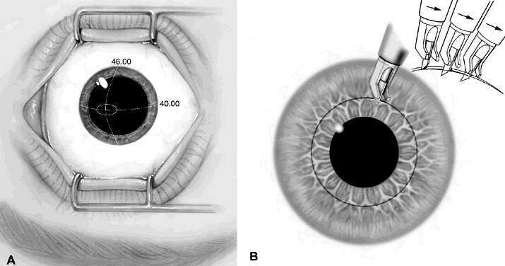
The patient is centered under the operating microscope and the eye is positioned perpendicular to the microscope. The eye is anesthetized with 0.5% proparacaine hydrochloride three times. The patient fixates on the operating microscope light, which is turned to a low level, or a red fixation light mounted on the microscope. The optical zone is then marked with a 6- or 7-mm optical zone marker, depending on the amount of astigmatism to be corrected. The steeper meridian is marked with a skin-marking pen using preoperative landmarks and an axis marker. Intraoperative keratometry and computerized topography can also be used to confirm the astigmatic axis (Fig. 5).

Fig. 5. Marking the steeper meridian.

If a 3-mm straight transverse keratotomy is selected, a 3-mm zone marker can be placed over the 6- or 7-mm zone marker in the steeper axis to delineate the incision length (Fig. 6). Alternatively, 3-mm transverse markers are also available. If an arcuate keratotomy is preferred, a 16-cut radial marker can be used to delineate an arc of 45°, a 12-cut radial marker can be used to delineate an arc of 60°, and either an 8- or 16-cut marker can be used for 90° (Fig. 7). Special arcuate keratotomy markers are available that imprint both the desired optical zone and a graduated scale (Fig. 8). Arcuate incisions greater than 90° are not recommended because the coupling ratio diverges from 1, and late wound dehiscence is more common. The marker blades can be coated with a skin-marking pen or an ink pad for better visualization of the radial lines.

Fig. 6. Use of a 3-mm zone marker to delineate the incision length.

Fig. 7. Sixteen-cut RK marker is useful to delineate 45° arcuate keratotomy. Twelve-cut RK marker is useful to delineate 60° arcuate keratotomy. Eight-cut RK is useful to delineate 90° arcuate keratotomy.

Fig. 8. A special arcuate keratotomy marker (A) delineates the desired optical zone as well as a graduated scale (B).

The corneal thickness coincident with the 6- or 7-mm optical zone in the steepest meridian is measured on both sides of the cornea intraoperatively with ultrasonic pachymetry (Fig. 9). A high-quality diamond knife is calibrated with a microscope capable of calibration to the micrometer level. The blade is set at 50 μm deeper than the thinnest paracentral pachymetry measurement or at 100% of the thinnest pachymetry reading at the desired optical zone.

Fig. 9. Pachymetry is performed either adjacent to the central optical zone mark or directly over the area to be incised.

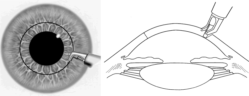
The patient is asked to fixate on the operating microscope light or the fixation light. The knife is set into the cornea, and allowed to seat for a full second. This is followed by a slow, steady guidance of the knife through the incisions (Fig. 10). A front-cutting knife allows the surgeon good visibility while pushing through the length of the keratotomy incision, thus improving accuracy. A square blade may provide better tracking. Several drops of topical antibiotic are applied to the eye. The eye is not routinely patched, nor is cycloplegia necessary. If a significant perforation occurs, a subconjunctival antibiotic, topical cycloplegia, and a pressure patch are applied.

Fig. 10. Making the incision.

The patient is generally seen 1 day, 1 week, and 1 month postoperatively. Topical antibiotics are continued until the corneal epithelium is re-epithelialized. The 1-month result correlates well statistically with the 1-year result, but additional astigmatic enhancement should not be considered until the refraction and keratometry measurements are stable. If significant undercorrection is noted on the first postoperative day, a topical corticosteroid can be used four times daily for 1 to 3 months in an attempt to delay wound healing and increase incision gape. Incisions can be reopened and extended if significant undercorrection persists after 1 month. In patients with significant overcorrection, topical hypertonic drops such as 5% sodium chloride four times daily for 1 to 3 months can be useful.

Back to Top
TRANSVERSE INCISIONS COUPLED WITH RADIAL KERATOTOMY INCISIONS
When transverse incisions are placed in conjunction with RK incisions, they can be placed between radial incisions or they can straddle radial incisions. Four-incision RK cases, if planned properly, allow for placement of transverse incisions on axis but between radial incisions. In eight-incision RK cases, the transverse incisions can either straddle the radial incisions (jump-T cuts) or be placed between radial incisions. In mini-RK, the incisions can simply stop short of the transverse incision. Experience has shown that intersecting transverse incisions with radial or semiradial incisions is to be strictly avoided. Astigmatic incisions coupled with RK incisions give a substantially greater effect than astigmatic incisions placed alone (Figs. 11 and 12).

Fig. 11. Combined RK and straight transverse keratotomy nomogram. Optical zone is 5, 6, or 7 mm. Knife setting is 100% of thinnest pachymetry at the optical zone in the axis of astigmatism. If the patient is older than or younger than 30 years of age, efficacy is increased or decreased 2% per year: age 20 = 80% of age 30; age 55 = 150% of age 30; age 80 = 200% of age 30.

Fig. 12. Combined RK and arcuate keratotomy nomogram. Optical zone is 6 or 7 mm. Knife setting is 100% of thinnest pachymetry at the optical zone in the axis of astigmatism. If the patient is older than or younger than 30 years of age, efficacy is increased or decreased 2% per year: age 20 = 80% of age 30; age 55 = 150% of age 30; age 80 = 200% of age 30.

Back to Top
WOUND REVISION VERSUS ASTIGMATIC KERATOTOMY FOR POSTCATARACT ASTIGMATISM
After cataract surgery there is a considerable difference of opinion among surgeons regarding whether induced against-the-rule astigmatism from wound slippage should be managed surgically by wound revision or by astigmatic keratotomy. If the wound dehiscence is a structural threat to the eye, then an anatomic wound revision is preferable. However, if the globe is structurally intact, the challenge then becomes one of refractive rehabilitation with the reduction of astigmatic anisometropia. Wound revision with resuturing restores the spherical equivalent of the eye. In contrast, astigmatic keratotomy may correct the astigmatic component but effectively flattens the eye because the wound gape adds tissue to the eye. The effect on the spherical equivalent must be carefully considered before an effective means of reducing against-the-rule postcataract astigmatism is chosen.
Back to Top
SEMIRADIAL INCISIONS
Paired semiradial incisions induce overall corneal flattening, resulting in a less myopic spherical equivalent. Semiradial incisions can be delineated by marking two lines drawn from the edge of a 3-mm optical zone centrally toward two points at the limbus that straddle the steep axis and are separated by 5.5 mm. Effectively, semiradial incisions flatten the steep meridian approximately twice as much as they steepen the flatter meridian 90° away. The coupling ratio is greater than 1. Therefore, semiradial incisions alone may correct moderate amounts of myopia while also correcting modest amounts of astigmatism. In cadaver eyes,15 semiradial incisions extended to a 3-mm optical zone corrected 1.4 D of astigmatism and 3.9 D of myopia, whereas at a 5-mm optical zone they corrected 2.25 D of astigmatism and 1.6 D of myopia (Fig. 13). The effect of refractive keratotomy in cadaver eyes has been shown to be equivalent to the effect achieved in an 80-year-old patient. Thus, the same values depicted for semiradial incisions in cadaver eyes can be expected to be achieved in an 80-year-old patient, and roughly 50% of this correction should be expected in a 30-year-old patient.

Fig. 13. A graphical depiction of the effect of paired semiradial incisions in a cadaver eye model at 3-, 4-, and 5-mm optical zone sizes.

Back to Top
ASYMMETRIC RADIAL INCISIONS
The principles of asymmetric radial incisions are similar to those of semiradial incisions (Fig. 14). By placing the incisions closer together, more flattening is achieved in the steep meridian. When four incisions are used, they are placed 60° apart on the steep axis rather than placed symmetrically 90° apart, as would be performed in a patient with no astigmatism. When six incisions are used, they are bunched together on the steeper axis with 45° in between each incision.

Fig. 14. Schematic representation of asymmetric incisions made at a 3-mm optical zone to correct against-the-rule astigmatism. A six-cut marker (with marks 60° apart) could be used to create the template for these incisions. The 90° and 270° marks are not cut.

Back to Top
TRAPEZOIDAL ASTIGMATIC KERATOTOMY
Semiradial incisions combined with the transverse incisions describe the trapezoidal astigmatic keratotomy popularized by Ruiz.15 The initial description of this procedure included two sets of semiradial incisions and five pairs of equally spaced transverse incisions. Several studies have now conclusively shown that the maximal astigmatic correction from trapezoidal astigmatic keratotomy can be attained with a single pair of transverse incisions placed 5 mm apart between two sets of semiradial incisions made from a 3-mm optical zone. Such studies have shown that the multiple pairs of transverse incisions initially described by Ruiz are unnecessary.13,15,21

In predicting the effect of trapezoidal astigmatic keratotomy, one can think of it as the addition of semiradial incisions to transverse incisions. The overall net effect is about a threefold flattening in the steeper meridian, with no change in the meridian 90° away. As a guide, the trapezoidal astigmatic keratotomy can be expected to correct 5.5 D of astigmatism in a 30-year-old patient and up to 11 D in an 80-year-old patient. If one converts the refraction to minus-cylinder form, a trapezoidal keratotomy will eliminate the minus cylinder.

In a clinical study of trapezoidal astigmatic keratotomy for the correction of naturally occurring astigmatism, Ibrahim and co-workers34 confirmed the values presented above. Of the 64 eyes studied, 90% were corrected between 60% and 150% of the desired change, which indicated only fair predictability. Trapezoidal astigmatic keratotomy for the correction of postkeratoplasty astigmatism has been associated with poor predictability and therefore is rarely used for the correction of postkeratoplasty astigmatism.

Back to Top
INCISIONS FOR POSTKERATOPLASTY ASTIGMATISM
Other incisional techniques available for the correction of postkeratoplasty astigmatism include relaxing incisions, arcuate keratotomy incisions with or without the use of compression sutures, and wedge resections.

RELAXING INCISIONS

A cadaver study of the effect of relaxing incisions on corneal topography demonstrated a wide range of effects on corneal astigmatism, ranging from 0.58 D for a single 30° incision to 5.93 D for a single 90° incision.33 Two symmetric relaxing incisions placed 180° apart produced 0.78 D of astigmatic change for paired 30° incisions and 13.97 D of change for paired 90° incisions. A marked disparity in the magnitude of change after symmetric 60° to 90° incisions in eye bank eyes indicated a narrow surgical “safe” zone. Clinically, marked undercorrections and overcorrections have been achieved in postkeratoplasty astigmatism with the use of relaxing incisions, making the determination of a suitable endpoint somewhat difficult. Relaxing incisions effectively flatten the steeper meridian an amount that is equivalent to the steepening of the flatter meridian. The change in spherical equivalent is, therefore, negligible.

The operative procedure involves careful dissection of the graft-host interface in a graded fashion (Fig. 15). One of the attractive features of relaxing incisions is that they can be performed at the slit lamp. Topical proparacaine hydrochloride anesthesia is generally adequate and is followed by a povidone-iodine periocular skin preparation. The axis of the steepest meridian is marked on each side of the graft-host interface. An appropriate incision length should never exceed 90° of the circumference of the graft.

Fig. 15. A. The steeper axis is marked before making relaxing incisions. Preoperative refraction: -3.00 + 6.00 × 90° (spherical equivalent = plano). B. Making a relaxing incision involves careful dissection of the graft-host interface for 90° in a graded fashion.

A gradual and careful deepening of the incision in the graft-host interface with use of a metal knife appears to be more controlled than with use of a diamond knife. In some cases, blunt dissection with the noncutting edge of the knife may actually be helpful.

Progressive deepening and lengthening of the incision is performed initially on one side of the graft-host interface. The use of a surgical keratometer or intraoperative computerized topography such as the PAR system (PAR Technologies, New York) is helpful for assessing the endpoint. Alternatively, the patient's keratometric values can be obtained intermittently with use of a conventional keratometer until the desired endpoint is obtained. We prefer the endpoint of a spherical cornea or slight (up to 33%) overcorrection in most cases. Longer incisions, however, tend to progressively gape as they heal, and in 75° to 90° incisions it may be desirable to leave a small amount of undercorrection.

COMPRESSION SUTURES

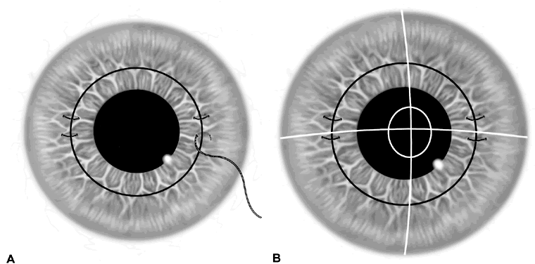
The addition of compression sutures to either relaxing incisions or arcuate keratotomy incisions can markedly increase the net effect. Compression sutures can be placed on each side of the graft-host interface 90° away from the astigmatic incisions (Fig. 16A). Suture depth should be about 75%. The sutures are tied with a slipknot and adjusted under keratometric or intraoperative computerized topographic control until the eye is spherical or an overcorrection of 33% is achieved (Fig. 16B). The knots are cut short and then buried. The use of 11-0 polyester suture has some advantages. If perfect surgical correction of astigmatism is achieved, it may be desirable to leave the sutures in place for extended periods of time. Nylon will hydrolyze in 1 to 2 years, whereas polyester suture will last longer than 7 to 10 years.

Fig. 16. A. Compression sutures are placed at 75% corneal depth on each side of the graft-host interface 90° away from relaxing incisions. B. Sutures are adjusted with a slip knot until an overcorrection of 33% is achieved.

WEDGE RESECTION

The wedge resection technique is reserved for correction of large degrees of postkeratoplasty astigmatism. In general, resection of 0.10 mm of tissue results in about 2 D of astigmatic correction. This operation is reserved for patients with greater than 10 D of astigmatism; it is capable of correcting up to 20 D of astigmatism. Wedge resection, however, requires prolonged postoperative rehabilitation because of the placement of multiple sutures, which induce a significant amount of irregular astigmatism. In general, a minimum of 6 months must be allowed for adequate wound healing before selective suture removal can be undertaken. Nevertheless, the alternative to wedge resection is repeating penetrating keratoplasty. Therefore, an attempt at wedge resection may be preferable to immediate repeat keratoplasty.

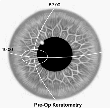
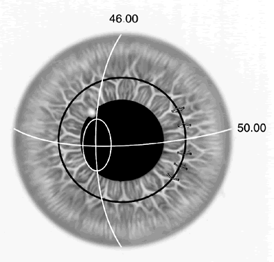
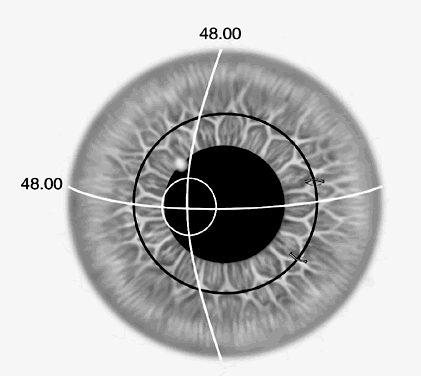
The overall effect of wedge resection is to steepen the flatter meridian about twice as much as it flattens the steeper meridian. The net effect is an increase in myopia or a decrease in hyperopia. For example, if the preoperative refraction was -6.00 + 12.00 × 90° and keratometry was 40.00/52.00 × 90° (Fig. 17), a perfect result should give postoperative keratometry measuring 48.00/48.00 with a refraction of -2 D. This would result in a 2 D myopic shift.

Fig. 17. The flatter axis is marked for a wedge resection. Preoperative refraction: -6.00 + 12.00 × 90° (spherical equivalent = plano).

Retrobulbar anesthesia is often helpful in wedge resection because the dissection and placement of multiple sutures demands excellent akinesia. Initially, the axis of the flatter meridian is marked, preferably after confirmation of the axis with a surgical keratometer. Intraoperative ultrasonic pachymetry readings are performed adjacent to the graft-host interface in the proper axis where the wedge resection is to be performed. The diamond blade is set at 100% of the thinnest pachymetry reading. A front-cutting diamond blade is preferred because it allows one to better visualize the wedge of tissue as it is excised with a free-hand dissection technique. Ninety degrees of the keratoplasty wound is incised nearly to the level of Descemet's membrane, and the wedge of tissue is then removed (Fig. 18).

Fig. 18. A 0.6-mm wedge of tissue is excised from the recipient cornea by free-hand dissection to the level of Descemet's membrane.

Five to seven deep and evenly spaced interrupted 10-0 nylon or 11-0 polyester sutures are then placed across the wound (Fig. 19). Polyester suture is preferred because it allows the sutures to be left in place indefinitely if the desired effect is achieved. Each suture is tied with a slipknot, and the sutures are then tightened under keratometric control until an overcorrection of one third to one half of the preoperative cylinder is achieved (Fig. 20). Sutures are tied with an additional square knot, cut short, and buried. For example, if the preoperative astigmatism is 12 D, sutures are tightened until a 4- to 6 D overcorrection is achieved in the axis opposite the preoperative cylinder.

Fig. 19. Five to seven sutures are placed to Descemet's membrane to close the wound.

Fig. 20. Overcorrection of suture tension to a level of 33% to 50% of the preoperative cylinder is attempted.

Postoperative care involves the use of topical antibiotics for 1 week and topical corticosteroids as necessary to maintain graft clarity. Sutures are left in place for a minimum of 8 weeks. Thereafter, one or two sutures can be removed selectively every 3 to 4 weeks in the axis of the steepest residual astigmatism. Once a satisfactory result is achieved, the remaining sutures can be left indefinitely (Fig. 21).

Fig. 21. Sutures can be removed selectively until a satisfactory result (-2.00 spherical equivalent 6 months postoperatively) is achieved.

Back to Top
COMPLICATIONS
Preoperative planning is essential to minimize complications of astigmatic keratotomy. Marking the steep meridian at the slit lamp with a marking pen preoperatively can help minimize incorrect placement of astigmatic incisions.

Transverse, arcuate, and semiradial incisions share a common problem, namely, some degree of unpredictability. Unpredictability is even greater in eyes that have had prior keratoplasty because of the variability of wound healing in the graft-host interface. Undercorrection is better tolerated than overcorrection, because additional surgery can be performed for undercorrections to achieve the desired result.

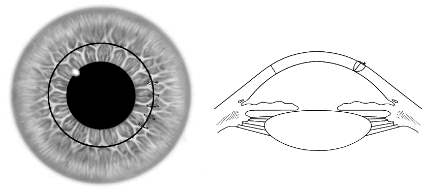
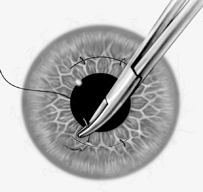
For the management of overcorrections after astigmatic keratotomy, one can reopen previously placed keratotomy incisions and then close with interrupted sutures instead of placing additional astigmatic incisions.35 Previously placed incisions can be bluntly opened with a Sinskey hook and then copiously irrigated with balanced salt solution to the depth of the incision. The incisions can then be closed with two or three 11-0 Mersilene sutures (Fig. 22). Intraoperative control of suture tension with a surgical keratometer or intraoperative computerized topography and the use of slipknots are helpful. The sutures are used to obtain a spherical result or a slight overcorrection of up to one-half of the preoperative cylinder in the appropriate direction. Selective suture removal can begin 8 weeks after suture placement as needed. The use of Mersilene sutures allows the compression sutures to be left in place indefinitely should the desired result be obtained.

Fig. 22. Use of 11-0 Mersilene sutures to close incisions for treatment of overcorrection after arcuate keratotomy.

INTERSECTING INCISIONS

One of the important lessons regarding incisional keratotomy is that intersecting incisions produce severe wound gape and significant degrees of scarring.20,36 Avoiding the intersection of keratotomy incisions should be the priority of all refractive corneal surgeons. Intersecting radial and transverse incisions creates a wound gape at the area of intersection that fills with stromal scarring and is frequently accompanied by extensive subepithelial opacification spreading out from the wound (Fig. 23). Such scars induce irregular astigmatism but also substantially increase light-scattering and glare. When transverse incisions intersect two radial incisions, a block of cornea is created that may actually protrude forward. This may take many months to heal, producing variable degrees of corneal opacification and irregular astigmatism.

Fig. 23. A. Clinical depiction of the effect of transverse incisions crossing radial incisions with exaggerated scar formation and poor wound healing. B. Clinical case of astigmatic keratotomy in which transverse incisions intersected radial incisions with resultant scar formation and epithelial inclusion cyst formation.

Back to Top
CONCLUSION
Current incisional surgical techniques have proved safe and effective in correcting wide ranges of astigmatism. Recently, the Excimer laser was introduced in a procedure known as photoastigmatic refractive keratectomy, or PARK, to reduce astigmatism. Early results appear promising. Whether this technique with its potential for more accurate correction will supplant incisional astigmatic keratotomy remains to be seen. In either case, a thorough understanding of the corneal response to incisional keratotomy will be helpful to future refractive surgeons.
Back to Top
REFERENCES

1. Guyton DL: Prescribing cylinders: the problem of distortion. Surv Ophthalmol 22:177, 1977

2. Schiotz LJ: Hin Fall von hochgradigem Horn-hautstastig-ma-tis-mus nach Staarextraction: Bessergung auf operativem Wege. Arch Augenheilkd 15:178, 1885

3. Faber E: Operative Behandeling van Astigmatisme. Ned Tijdschr Geneeskd 2:495, 1895

4. Waring GO: History of radial keratotomy. In Sanders DR, Hofmann RF, Salz JJ (eds): Refractive Corneal Surgery, pp 3–14. Thorofare, NJ, Slack, 1985

5. Bates WH: A suggestion of an operation to correct astigmatism. Arch Ophthalmol 23:9, 1894

6. Lans LJ: Experimentelle Untersuchungen uber Entstehung von Astigmatismus durch nich-perforirende Corneawunden. Graefes Arch Ophthalmol 45:117, 1898

7. Sato T: Treatment of conical cornea by incision of Descemet's membrane. Acta Soc Ophthalmol Jpn 43:541, 1939

8. Sato T: Experimental study on surgical correction of astigmatism. Juntendo Kenkyukai-zasshi 589:37, 1943

9. Sato T: Posterior incision of the cornea: surgical treatment for conical cornea and astigmatism. Am J Ophthalmol 33:943, 1950

10. Sato T: Die operative Behandlung des Astigmatismus. Klin Monatsbl Augenheilkd 126:16, 1955

11. Fyodorov SN, Durner VV: Surgical correction of complicated myopic astigmatism by means of dissection of circular ligament of cornea. Ann Ophthalmol 13:115, 1981

12. Salz JJ, Lee J, Jester J et al: Radial keratotomy in fresh human cadaver eyes. Ophthalmology 88:742, 1981

13. Lavery GW, Lindstrom RL: Trapezoidal astigmatic keratotomy in human cadaver eyes. J Refract Surg 1:18, 1985

14. Franks JB, Binder PS: Keratotomy procedures for the correction of astigmatism. J Refract Surg 1:11, 1985

15. Lindquist TD, Rubenstein JB, Rice SW et al: Trapezoidal astigmatic keratotomy: quantification in human cadaver eyes. Arch Ophthalmol 104:1534, 1986

16. Duffey RJ, Tchah H, Lindstrom RL: Spoke keratotomy in the human cadaver eye. J Refract Surg 4:9, 1988

17. Duffey RJ, Jain VN, Tchah H et al: Paired arcuate keratotomy: a surgical approach to mixed and myopic astigmatism. Arch Ophthalmol 106:1130, 1988

18. Lindstrom RL: The surgical correction of astigmatism: a clinician's perspective. Refract Corneal Surg 6:441, 1990

19. Binder PS, Waring GO III: Keratotomy for astigmatism. In Waring GO III (ed): Refractive Keratotomy for Myopia and Astigmatism, pp 1085–1198. St. Louis, Mosby-Year Book, 1992

20. Lindquist TD: Complications of ocular surgery. Int Ophthalmol Clin 32:97, 1992

21. Lindstrom RL, Lindquist TD: Surgical correction of post-operative astigmatism. Cornea 7:138, 1988

22. Duke-Elder S, Stewart S, Abrams D (eds): Ophthalmic optics and refraction. In Duke-Elder S (ed): System of Ophthalmology, Vol 5, pp 274–295. St. Louis, CV Mosby, 1970

23. Donders RL, Moore WD (trans): On the Anomalies of Accommodation and Refraction of the Eye, pp 415–417. London, New Sydenham Society, 1864

24. Jaffe NS, Claynan HM: The pathophysiology of corneal astigmatism after cataract extraction. Ophthalmology 79:615, 1975

25. Troutman RC: Microsurgery of the Anterior Segment of the Eye, p 286. St. Louis, CV Mosby, 1987

26. Troutman RC, Swinger C: Relaxing incision for control of postoperative astigmatism following keratoplasty. Ophthalmic Surg 11:117, 1980

27. Barner SS: Surgical treatment of corneal astigmatism. Ophthalmic Surg 7:43, 1976

28. Krachmer JH, Fenzel RE: Surgical correction of high post-keratoplasty astigmatism. Arch Ophthalmol 98:1400, 1980

29. Troutman RC, Swinger C: Relaxing incision for control of postoperative astigmatism following keratoplasty. Ophthalmic Surg 11:117, 1980

30. Sugar J, Kick AK: Relaxing keratotomy for post-keratoplasty high astigmatism. Ophthalmic Surg 14:156, 1983

31. Mandel MR, Shaoui MB, Krachmer JH: Relaxing incisions with augmentation sutures for the correction of post-keratoplasty astigmatism. Am J Ophthalmol 103:441, 1987

32. Lavery GW, Lindstrom RL, Hofer LA et al: The surgical management of corneal astigmatism after penetrating keratoplasty. Ophthalmic Surg 16:168, 1988

33. Lundergan MK, Rowsey JJ: Relaxing incisions. Ophthalmology 92:1226, 1985

34. Ibrahim O, Hussein HA, El-Sahn MF et al: Trapezoidal keratotomy for the correction of naturally occurring astigmatism. Arch Ophthalmol 109:1374, 1991

35. Lindquist TD, Williams PA, Lindstrom RL: Management of overcorrection following astigmatic keratotomy. J Refract Surg 4:218, 1988

36. Rashid ER, Waring GO III: Complications of refractive keratotomy. In Waring GO III (ed): Refractive Keratotomy for Myopia and Astigmatism, pp 863–936. St. Louis, Mosby-Year Book, 1992

Back to Top
Appendix 1

ARC-T 6 mm Nomogram

HOW TO USE THIS NOMOGRAM

  Identify the patient's age and the diopters of refractive cylinder that you wish to correct.
  Find the patient's age in the first column on the left.
  Move to right until you reach the surgery results closest to the refractive cylinder of the patient. In order to avoid overcorrection, it is suggested that you select a surgical goal somewhat less than the actual refractive cylinder. The column heading then tells you which surgery is needed to achieve this result.

EXAMPLE

  A 45-year old patient with a refractive cylinder of 3.75.
  Moving to the right on the Age 45 line, you see that 3.75 falls between 2.93 and 3.90. Looking at the column headings, you see that a Single 60° ARC-T will correct to 2.93 diopters of cylinder. A Paired 45° ARC-T or a Single 90° ARC-T will correct 3.90 diopters of cylinder.
  The recommendation in this case is to do a Single 60° ARC-T procedure.
  Expected surgical results were calculated using Lindstrom's Formula:


ΔD = [100 + (Age - 30) × 2] × [ΔD at age 30] × 0.01

ARC-T 7 mm Nomogram

HOW TO USE THIS NOMOGRAM

  Identify the patient's age and the diopters of refractive cylinder that you wish to correct.
  Find the patient's age in the first column on the left.
  Move to right until you reach the surgery results closest to the refractive cylinder of the patient. In order to avoid overcorrection, it is suggested that you select a surgical goal somewhat less than the actual refractive cylinder. The column heading then tells you which surgery is needed to achieve this result.

EXAMPLE

  A 45-year old patient with a refractive cylinder of 3.75.
  Moving to the right on the Age 45 line, you see that 3.75 falls between 2.60 and 3.90. Looking at the column headings, you see that a Single 90° or a Paired 45° ARC-T will correct to 2.60 diopters of cylinder. A Paired 60° ARC-T will correct 3.90 diopters of cylinder.
  The recommendation in this case is to do a Single 90° or a Paired 45° ARC-T procedure.
  Expected surgical results were calculated using Lindstrom's Formula:


ΔD = [100 + (Age - 30) × 2] × [ΔD at age 30] × 0.01

ARC-T 8–9 mm Nomogram

HOW TO USE THIS NOMOGRAM

  Identify the patient's age and the diopters of refractive cylinder that you wish to correct.
  Find the patient's age in the first column on the left.
  Move to right until you reach the surgery results closest to the refractive cylinder of the patient. In order to avoid overcorrection, it is suggested that you select a surgical goal somewhat less than the actual refractive cylinder. The column heading then tells you which surgery is needed to achieve this result.

EXAMPLE

  A 45-year old patient with a refractive cylinder of 2.25.
  Moving to the right on the Age 45 line, you see that 2.25 falls between 1.95 and 2.60. Looking at the column headings, you see that a Paired 60° ARC-T will correct 1.95 diopters of cylinder. A Paired 90° ARC-T will correct 2.60 diopters of cylinder.
  The recommendation in this case is to do a Paired 60° ARC-T procedure.
  Expected surgical results were calculated using Lindstrom's Formula:


ΔD = [100 + (Age - 30) × 2] × [ΔD at age 30] × 0.01

ARC-T Nomogram for Males (Revised Using New Formulas)

HOW TO USE THIS NOMOGRAM

  Identify the patient's age and the diopters of refractive cylinder that you wish to correct.
  Find the patient's age in the first column on the left.
  Move to right until you reach the surgery results closest to the refractive cylinder of the patient. In order to avoid overcorrection, it is suggested that you select a surgical goal somewhat less than the actual refractive cylinder. The column heading then tells you which surgery is needed to achieve this result.
  FOR FEMALES: Subtract 0.37 from each predicted value.

EXAMPLE

  A 45-year old patient with a refractive cylinder of 3.75.
  Moving to the right on the Age 45 line, you see that 3.75 falls between 3.62 and 4.72. Looking at the column headings, you see that a 2 × 60° ARC-T will correct to 3.62 diopters of cylinder.
  The recommendation in this case is to do a 2 × 60° ARC-T procedure.
  Expected surgical results were calculated using Lindstrom's Formula:


ΔD = [100 + (Age - 30) × 2] × [ΔD at age 30] × 0.01

Back to Top