Chapter 33
Conjunctival Flaps for Corneal Disease
SHAHZAD MIAN and ALAN SUGAR
Main Menu   Table Of Contents

Search

INDICATIONS
SURGICAL TECHNIQUE
RESULTS AND COMPLICATIONS
KERATOPLASTY AFTER CONJUNCTIVAL FLAPS
ADVANTAGES AND DISADVANTAGES OF CONJUNCTIVAL FLAPS
REFERENCES

The popularity of conjunctival flaps for treatment of corneal disease has diminished with the increasing success of medical therapy for corneal infections, the improvement in ocular lubrication systems, the use of bandage contact lenses for corneal surface diseases, the use of tissue adhesives for threatened perforations, the use of amniotic membrane grafts, and the improved techniques for corneal, conjunctival, and lid surgery. Nonetheless, conjunctival flaps are useful in occasional cases and should be a part of the operative repertoire of most anterior segment surgeons. Conjunctival grafting for corneal disease has a long history, especially for treatment of corneal trauma prior to the availability of modern suturing techniques. Initial procedures used full-thickness conjunctiva, including Tenon's capsule; however, this resulted in early failure and short duration of coverage of the cornea.1 Current use of conjunctival flaps is primarily based on the technique described by Gundersen in 1958, along with more recent variations.2
Back to Top
INDICATIONS
In general, the indications for conjunctival flap procedures are treatment of corneal infections resistant to medical therapy, control of resistant surface corneal disease, control of corneal pain, and structural support. Only rarely is the use of a conjunctival flap the best choice for primary treatment. A few large series of cases are reported in the literature, and it is likely that indications have changed over time. The use of conjunctival flaps for primary treatment of corneal lacerations, a frequent indication in the past, is now almost never indicated when modern microsurgical treatment is available.3

Gundersen and Pearlson presented a series of 177 cases in 1969.4 Sixty (34%) had herpes simplex keratitis, 44 (25%) had bullous keratopathy, and 11 (6%) suffered trauma. Paton and Milauskas presented 122 cases in 1970: 36 (30%) with herpetic keratitis, 33 (27%) with bullous keratopathy, 23 (19%) with nonherpetic ulcers, 12 (10%) with descemetocele, and seven (6%) with alkali burns.5 Insler and Pechous reported 33 cases 17 years later.6 Eight (25%) had herpes simplex, seven (22%) had bacterial ulcers, and three (9%) had herpes zoster. The most recent series confirm a predominance of herpes simplex keratitis as an indication for conjunctival flaps.7 Alino and colleagues presented 48 patients in 1998 with total conjunctival flaps and 13 with partial conjunctival flaps. In this series, 12 (20%) had nonherpetic corneal ulcers, nine (15%) had herpes simplex keratitis, nine (15%) had bullous keratopathy, and seven (11%) had herpes zoster keratitis.8 Khoudadoust and Quinter reported 50 cases of conjunctival flaps in 2003 with 14 (28%) secondary to herpes simplex keratitis, 11 (22%) secondary to bacterial ulcers, and eight (16%) secondary to rheumatoid arthritis related corneal melt.9 The cases series for indications for conjunctival flaps are summarized in Table 1.

TABLE 1. Conjunctival flap case series: indications


 Gundersen2Paton5Insler6Alino8Khoudadoust9
 (1969)(1970)(1988)(1998)(2003)
Disease(%)(%)(%)(%)(%)
Herpes3530362638
(HSV/HZV)252731512
Bullous keratopathy 19222022
Ulcer  3188
Graft-related    16
Rheumatoid arthritis6637
Trauma55 5
Neuroparalytic   3 
Neurotrophic     


INFECTIONS

The development and refinement of effective antimicrobial agents over the last 50 years has greatly improved the treatment of corneal infections. However, resistant corneal ulcers rarely may need to be treated with a conjunctival flap. The flap brings in blood supply that promotes healing by increasing corneal access to humoral and cellular immune mechanisms. Exposure, drying, and neurotrophic factors in delayed healing may be moderated as well. In all cases of necrotic ulcers treated with conjuctival flaps it is mandatory that appropriate drug therapy be tried first, and that remaining areas of necrosis be debrided, often with lamellar keratectomy. However, penetrating keratoplasty may have limited benefit because of the degree of inflammation and vascularization of the cornea resulting in high risk for graft rejection. Therefore, a conjunctival flap may allow stabilization of the infection and persistent inflammation in preparation for corneal grafting and visual rehabilitation.

Conjunctival flaps have been used to halt progression of a refractory pseudomonas corneal abscess.10 This is a useful technique in eyes with poor visual potential and a delayed healing rate after both infectious and sterile corneal ulcers. A conjunctival flap was used to treat such keratitis occurring in the region of the corneal incisions in a patient after radial keratotomy.11 Flaps have been promoted for treatment of fungal keratitis resistant to medical treatment.12 This approach may reflect the limited success of topical ophthalmic antifungals. Fungal keratitis, as any infection, can still progress beneath the conjunctival flap in some cases, and the flap itself may limit drug penetration postoperatively. Treatment of advanced acanthamoeba keratitis with deep lamellar keratectomy and conjunctival flap to control the infection and help relieve pain also has been reported.13

The use of flaps to treat herpes simplex keratitis has remained helpful despite the availability of antivirals.4–7 Persistent epithelial disease may rarely benefit from treatment with a flap. More often, chronic or painful herpetic stromal ulceration and keratouveitis requiring frequent examinations and medical therapy benefit from a conjunctival flap. Brown and colleagues found a marked decrease in the need for medications and office visits in such patients.7 They noted no recurrence of active viral keratitis, although perforation beneath the flap for herpetic keratitis may occur. However, Lesher and colleagues reported recurrence of herpetic stromal keratitis after a conjunctival flap.14

BULLOUS KERATOPATHY

Conjunctival flaps were more widely used for treatment of bullous keratopathy when the success rate of penetrating keratoplasty was poor.15,16 Now, conjunctival flaps are used only to treat the pain of bullous keratopathy in eyes in which a soft contact lens or anterior stromal puncture has failed and the patient refuses penetrating keratoplasty or has poor visual potential.17 Cautery of Bowman's membrane has also been used in these patients, either alone or combined with a conjunctival flap.18 Flaps may be total for diffuse bullous keratopathy or partial for peripheral localized edema.15,16

NONINFECTIOUS ULCERATIONS

Corneal ulceration from exposure or corneal anesthesia may not respond to lubricants, patching, or soft contact lenses in many cases. If these measures or appropriate lid surgery is ineffective, a conjunctival flap almost always is effective. In many cases, a thin conjunctival flap is cosmetically superior to a permanent tarsorrhaphy.19

Peripheral corneal melting from immune or inflammatory processes such as rheumatoid arthritis and Mooren's ulcer often has been treated with conjunctival resection in the involved area to limit access of immune mediators and proteases from the conjunctiva.20,21 Occasionally, conjunctival flaps are helpful in such patients, especially when the process has been controlled by systemic drug therapy.

Corneal perforation should not be treated by a conjunctival flap alone. The conjunctiva does not provide adequate support and often allows continued aqueous leakage or bleb formation.22 Flaps are helpful in protecting and promoting the healing of peripheral lamellar patch keratoplasties, especially when the limbus and sclera are included in the graft area.23 Descemetoceles with threatened perforation are better treated by patch keratoplasty or the application of tissue glue and a bandage soft contact lens.24

PREPARATION FOR A COSMETIC SHELL

In some patients with a blind eye and opaque cornea, or phthisis, who require a cosmetic prosthesis or shell, the corneal surface is irritated by the shell. A conjunctival flap over the cornea improves comfort and eliminates corneal abrasions. Putterman has followed the conjunctival flap procedure by ptosis repair and finally cosmetic shell fitting.25 Such a procedure may be a reasonable alternative to enucleation for some patients.4

Back to Top
SURGICAL TECHNIQUE

TOTAL CONJUNCTIVAL FLAP

The basic technique for total conjunctival flap surgery is that described by Gundersen in 1958.2 This method stresses dissection of the conjunctiva from Tenon's capsule to form a thin flap. Gundersen and others also stressed the importance of releasing tension on the flap and removing corneal epithelium so that the tendency toward flap retraction is minimized.2,5

A total flap, by the Gundersen technique, may be done easily with retrobulbar anesthesia, although subconjunctival anesthetic injection may be sufficient. The corneal epithelium should be removed in all areas that are to be covered by the flap. Topical cocaine or absolute alcohol may be useful to loosen adherent epithelium.26 Residual epithelium may lead to cyst formation beneath the flap. The flap also does not adhere in areas with intact epithelium. A silk traction suture then may be placed in the peripheral cornea at the 12 o'clock position, or in the superior fornix (Fig. 1A). The conjunctiva may be ballooned with local anesthetic with epinephrine to help separate Tenon's capsule from conjunctiva. The injection should be placed so that a hole is not made in the area of conjunctiva that will cover the cornea. Maguire and Shearer described a variation of anesthetic injection beneath the superior peritomy in a series of radial needle passes followed by scissors dissection.27 Another variation, to increase the area of conjunctiva available, is to evert the upper lid over a Desmarres retractor and inject anesthetic prior to incising along the superior edge of the tarsus.28

Fig. 1. A. Limbal traction suture, superior incision site. B. Dissection of conjunctiva from Tenon's capsule. C. Complete conjunctival peritomy. D. Flap should be free of traction. E. The flap is sutured into place above and below.

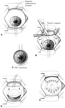
The second step may be either a peritomy or a superior conjunctival incision (Fig. 1B). The superior incision is preferred because the limbal attachments help stabilize the conjunctiva during dissection. The superior incision should be made 14 to 18 mm above the superior limbus to allow for conjunctival shrinkage.26,27 The area of the cornea is less than 1.5 cm2, and that of the whole conjunctival sac is estimated to be 16 cm2.29 Lauring and Wergeland estimate the superior conjunctiva to provide up to 6 cm2 for corneal coverage.28 Although this may appear initially to be an abundance of tissue, the conjunctiva often “shrinks” after dissection and may be diminished by scarring. The area of dissection must be considerably greater than the area to be covered to allow movement of the flap into position without tension causing it to retract. The superior incision is made horizontally for about 2 cm. Dissection is then carried to the limbus, carefully separating conjunctiva from Tenon's capsule (see Fig. 1B). The conjunctiva should be handled gently with blunt forceps. Blunt-tipped scissors also help to avoid conjunctival perforation. Dissection is carried to both sides of the cornea to free the conjunctiva at the 3 and 9 o'clock positions (Fig. 1C). When the limbus is reached, a 360-degree peritomy is performed. In addition, the inferior conjunctiva may be freed from Tenon's capsule. Relaxing incisions may be necessary to allow the conjunctiva to be pulled down easily.

After the conjunctiva is free enough to set in place over the cornea without traction, it is secured by a row of interrupted or mattress sutures inferiorly and superiorly (Figs. 1D and 1E). Suture bites should include underlying limbal or episcleral tissue. The superior edge is secured to the episclera, leaving the donor bed uncovered. Sutures of 8-0 silk, 6-0 to 8-0 chromic gut, 8-0 Vicryl, and 10-0 nylon have been used. Nonabsorbable sutures may be difficult to remove.

Among the variations of a total conjunctival flap is combination with lamellar keratectomy. This may be done to remove ulcerated necrotic tissue, improve adhesion of the conjunctiva, or “inlay” the conjunctiva. The use of a lamellar keratectomy to allow a flat conjunctival surface is most helpful for peripheral partial flaps.16 It is also possible to pull the conjunctiva over the cornea without dissection by placing sutures in the superior and inferior fornix, drawing the conjunctival fornices together over the center of the cornea, and suturing these “everted” flaps over the cornea.30 Such flaps retract within a few weeks but may be helpful in situations where more refined techniques are not feasible.

ADVANCEMENT FLAP

A partial flap may be performed in the same manner as a total flap, but covering only a portion of the cornea. If the area to be covered is peripheral and small, the peritomy alone may be used to incise the conjunctiva, sliding the conjunctiva as a “hood” flap. However, simple advancement flaps often retract with time.

PEDICLE FLAP

Pedicle flaps may be fashioned from perilimbal or superior conjunctiva to cover localized defects.31 The area to be covered is measured and the conjunctival dissection made about one-third larger. Because these flaps are sutured directly to the cornea, 10-0 nylon sutures are used. When the corneal defect is peripheral a single-pedicle (racket) flap is used, whereas a bipedicle (bucket-handle) flap is used for central or paracentral lesions.32

Back to Top
RESULTS AND COMPLICATIONS
The result of a successful total conjunctival flap is a quiet, comfortable eye with a thin vascularized flap and visible underlying cornea and anterior segment details. A successful flap remains in place without significant buttonholes or traction. Vision is usually 20/200 or worse. Data are not often reported on flap success; rather, data are reported on complication rates.

FLAP RETRACTION

This results from poor mobilization of the conjunctiva, inadequate removal of corneal epithelium, and buttonholes. Gundersen noted 10 retractions in 133 cases,4 Insler two in 33 cases,6 and Paton 13 in 122 cases.5 If treated early, it may be possible to replace the flap, making certain that no further traction or corneal epithelium remains. However, the tissue tends to contract rapidly, making a repeat flap difficult or impossible.

BUTTONHOLES

Buttonholes noted during dissection or suturing of the flap should be repaired to avoid enlargement. Buttonholes may be sutured in a bite that includes underlying corneal stroma. A tapered-point needle, such as fine blood vessel needles (e.g., BV4), should be used to avoid causing additional buttonholes by needle passage.33 Suture material of 11-0 nylon and 9-0 Vicryl has been suggested.26,34

Epithelial inclusion cyst

Conjunctival or corneal epithelial inclusion cysts may occur beneath the flap, usually near the limbus, in less than 5% of cases.4,6 They may resolve spontaneously or, if large, require excision.

HEMORRHAGE AND NEOVASCULARIZATION

Postoperatively, hemorrhage or fluid may collect beneath the flap. This resolves spontaneously without usually affecting the outcome. Corneal neovascularization and opacification also may increase over time, which may increase risk of subsequent corneal graft failure.

RECURRENCE OF DISEASE

The underlying disease process may persist or progress to erode the conjunctival flap, as in six of 122 cases reported by Paton,5 or perforate the underlying cornea, as in four of 133 cases reported by Gundersen.4 Herpes simplex stromal keratitis may recur beneath the conjunctival flap or rarely lead to perforation.14

Ptosis

Ptosis after a conjunctival flap is common, probably resulting from traction on the superior fornix. Many patients have a pre-existing ptosis related to chronic keratitis. Additional ptosis from the flap procedure can be minimized by freeing the flap tissue as completely as possible from the superior fornix, particularly lysing superior lateral traction through Tenon's adhesions.

Back to Top
KERATOPLASTY AFTER CONJUNCTIVAL FLAPS
When a conjunctival flap has been performed to stop a corneal inflammatory process (such as a bacterial ulcer or herpetic keratitis) in an eye with visual potential, later visual rehabilitation may be worth considering. Conjunctival flaps can be peeled from the corneal surface, although the underlying corneal scarring from the original process often limits visual improvement. Penetrating keratoplasty can be performed at the time of flap removal or as a separate procedure. Insler and Pechous performed keratoplasty in nine of 31 flap patients, with good results in eight, an average of 15 months after the flaps had been performed.6 It is also reasonable to trephine directly through the conjunctival flap without removal at time of keratoplasty. Geria and colleagues performed keratoplasty in eight of 47 eyes 12 to 43 months after conjunctival flap placement.35 All patients preoperatively had infectious ulcers and all patients reached a postoperative vision between 20/70 and 20/30 with an average follow-up of 9.9 years.
Back to Top
ADVANTAGES AND DISADVANTAGES OF CONJUNCTIVAL FLAPS
The advantages of conjunctival flaps include the certainty with which they control corneal inflammation and pain. They are usually cosmetically superior to tarsorrhaphy and can be done by most ophthalmologists without sophisticated instrumentation or a need for donor tissue.

The disadvantages include poor cosmesis, in comparison to keratoplasty. Vision is usually not good, and visualization of the underlying corneal process and anterior segment details is difficult, especially during the first several postoperative weeks. Intraocular pressure measurement also may be more difficult.

The indications for conjunctival flaps have been decreasing because of improvements in medical and surgical techniques but conjunctival flap procedures continue to be useful for select patients.

Back to Top
REFERENCES

1. Townsend WM: Conjunctival flaps. In The Cornea. New York: Churchill Livingstone, 1988:669

2. Gundersen T: Conjunctival flaps in the treatment of corneal disease with reference to a new technique of application. Arch Ophthalmol 60:880, 1958

3. Haik GM: A fornix conjunctival flap as a substitute for the dissected conjunctival flap: A clinical and experimental study. Trans Am Ophthalmol Soc 52:497, 1954

4. Gundersen T, Pearlson HR: Conjunctival flaps for corneal disease: Their usefulness and complications. Trans Am Ophthalmol Soc 67:78, 1969

5. Paton D, Milauskas AT: Indications, surgical technique, and results of thin conjunctival flaps on the cornea. Int Ophthalmol Clin 10:329, 1970

6. Insler MS, Pechous B: Conjunctival flaps revisited. Ophthalmic Surg 18:455, 1987

7. Brown DD, McCulley JP, Bowman RW, Halsted MA: The use of conjunctival flaps for the treatment of herpes keratouveitis. Cornea 11:44, 1992

8. Alino AM, Perry HD, Kanellopoulos AJ, Donnenfeld ED, Rahn EK: Conjunctival flaps. Ophthalmology 105:1120, 1998

9. Khodadoust A, Quinter AP: Microsurgical approach to the conjunctival flap. Arch Ophthalmol 121:1189, 2003.

10. Buxton JN, Fox MN: Conjunctival flaps in the treatment of refractory Pseudomonas corneal abscess. Ann Ophthalmol 18:315, 1986

11. Mackman G: Delayed sterile keratitis following radial keratotomy successfully treated with conjunctival flap. Refract Corneal Surg 8:122, 1992

12. Sanitato JJ, Kelley CG, Kaufman HE: Surgical management of peripheral fungal keratitis (keratomycosis). Arch Ophthalmol 102:1506, 1984

13. Cremona G, Carrasco MA, Tytiun A, Cosentino MJ: Treatment of advanced acanthamoeba keratitis with deep lamellar keratectomy and conjunctival flap. Cornea 21:705, 2002

14. Lesher MP, Lohman LE, Yeakley W, Lass J: Recurrence of herpetic stromal keratitis after a conjunctival flap surgical procedure. Am J Ophthalmol 114:231, 1992

15. Gundersen T: Surgical treatment of bullous keratopathy. Arch Ophthalmol 64:260, 1960

16. Sugar HS: The use of Gundersen flaps in the treatment of bullous keratopathy. Am J Ophthalmol 57:977, 1964

17. Hsu JKW, Rubinfield RS, Barry P, Jester JV: Anterior stromal puncture, immunohistochemical studies in human cornea. Arch Ophthalmol 111:1057, 1993

18. DeVoe AG: Electrocautery of Bowman's membrane. Trans Am Ophthalmol Soc 64:110, 1966

19. Lugo M, Arentsen JJ: Treatment of neurotrophic ulcers with conjunctival flaps. Am J Ophthalmol 103:711, 1987

20. Brown SI, Mondino BJ: Therapy of Mooren's ulcer. Am J Ophthalmol 98:1, 1984

21. Feder RS, Krachmer JH: Conjunctival resection for the treatment of rheumatoid corneal ulceration. Ophthalmology 91:111, 1984

22. Saini JS, Sharma A, Grewal SPS: Chronic corneal perforations. Ophthalmic Surg 23:399, 1992

23. Portnoy SL, Insler MS, Kaufman HE: Surgical management of corneal ulceration and perforation. Surv Ophthalmol 34:47, 1989

24. Leahey AB, Gottsch JD, Stark WJ: Clinical experience with N-butyl cyanoacrylate (Nexacryl) tissue adhesive. Ophthalmology 100:173, 1993

25. Putterman AM: Conjunctival flap–cosmetic shell–ptosis procedure, treatment of blepharoptosis in severe keratopathy. Arch Ophthalmol 107:1816, 1989

26. Mannis MJ: Conjunctival flaps. Int Ophthalmol Clin 28:165, 1988

27. Maguire LJ, Shearer DR: A simple method of conjunctival dissection for Gundersen flaps. Arch Ophthalmol 109:1168, 1991

28. Lauring L, Wergeland FL: Total conjunctival flap with a modification of the Gundersen method. Am J Ophthalmol 76:953, 1973

29. Ehlers N: On the size of the conjunctival sac. Acta Ophthalmol (Copenh) 43:205, 1965

30. Taylor RP: Fornix-based conjunctival flap in the treatment of corneal ulcers. Am J Ophthalmol 67:754, 1969

31. Cies WA, Odeh-Nasrala N: The racquet conjunctival flap. Ophthalmic Surg 7:31, 1976

32. Reinhart WJ: Conjunctival flap surgery. In Manual of Corneal Surgery. New York: Churchill Livingstone, 1987

33. Petursson GJ, Fraunfelder FT: Repair of an inadvertent buttonhole or leaking filtering bleb. Arch Ophthalmol 97:926, 1979

34. Cammarosano CA, Thoft RA: Complications of conjunctival surgery. Int Ophthalmol Clin 32:41, 1992

35. Geria RC, Zarate J, Geria MA: Penetrating keratoplasty in eyes treated with conjunctival flaps. Cornea 20:345, 2001

Back to Top