Chapter 23
Surgery for Pediatric Glaucoma
JAMES D. BRANDT
Main Menu   Table Of Contents

Search

PREOPERATIVE CONSIDERATIONS
CHOOSING THE SURGICAL APPROACH
POSTOPERATIVE CARE
LONG-TERM MANAGEMENT
FUTURE TRENDS
REFERENCES

Primary congenital glaucoma is a rare disease, occurring in approximately 1 in 15,000 live births.1–4 Because of its rarity, the general ophthalmologist may make the initial diagnosis of this disease only once in a life-long career. Nonetheless, every ophthalmologist should be familiar with the various surgical approaches to this disease because they are applicable to the management of the various secondary glaucomas seen in infancy and childhood that, although not common, may be seen in a busy general ophthalmic practice.

The purpose of this chapter is to outline a therapeutic and surgical approach to the management of congenital and pediatric glaucoma. This chapter does not substitute for experience in handling these sometimes complicated surgical problems. Because the initial surgery for these problems is often the procedure that has the highest likelihood of success, and because the infant or child faces the prospect of a lifetime of impaired vision should the surgery be unsuccessful, the management of these patients is best left to those experienced with these procedures. The postoperative management of congenital glaucoma in reality lasts a decade or more (with periodic examinations under anesthesia, amblyopia therapy, contact lens fittings, and so forth). These complicated problems should be managed using a team approach for the patient's benefit.

Back to Top
PREOPERATIVE CONSIDERATIONS
The embryology, epidemiology, and differential diagnosis of primary infantile glaucoma are covered in these volumes. In many cases, particularly where the glaucoma is unilateral, the diagnosis is relatively straightforward (Fig. 1). In bilateral cases in young infants, however, the diagnosis may be less certain and the ophthalmologist will find it useful to perform a brief examination in the office before subjecting the child to a general anesthetic.

Fig. 1. This 4-month-old girl came to the University of California, Davis, eye clinic with her older sister, who was returning for a strabismus examination. The child's mother had noted no symptoms of photophobia or tearing. The resident on the strabismus service noted her asymmetric eye size and initiated an evaluation. The intraocular pressure was 30 mmHg in the larger right eye. An axial length disparity of 3.01 mm was found at presentation (right eye, 22.58 mm; left eye, 19.57 mm). Trabeculotomy ab externo was performed 2 days after diagnosis and intraocular pressure was normalized. The axial length disparity had decreased to 0.2 mm 18 months after surgery.

In infants less than 3 or 4 months of age, it is usually possible to perform tonometry using either a hand-held Goldmann applanation tonometer or an electronic tonometer, the Tono-Pen (Mentor Ophthalmics, Norwell, MA). The Schiøtz tonometer is relatively inaccurate when used to measure the intraocular pressure (IOP) of an infant eye and should be used only to gauge a general range of IOP. The young infant should be swaddled and held in its mother's arms while the eyelids are gently pried apart to perform tonometry. The Tono-Pen device, which has a very small (about 2 mm) measuring surface, is well suited to the small intrapalpebral fissures of an infant5 and can measure IOP without placing undue pressure on the lids; the use of a lid speculum can often be avoided. The observer's head does not need to be right next to the patient's, which can alarm some children. The Tono-Pen has been found to be accurate compared with both cannulated cadaver eyes and Goldmann tonometry,6,7 but no studies exist to assess its accuracy in infant eyes.

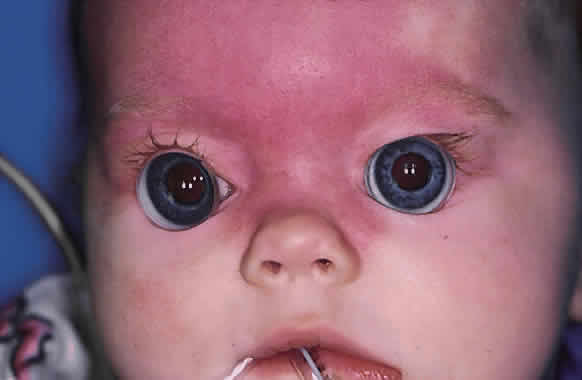
The office measurement of an IOP much greater than 20 mmHg in a calm, resting infant is suspicious for glaucoma when other signs and symptoms suggest the disease,8 as is an asymmetry of more than 5 mmHg in suspected unilateral or asymmetric cases. IOP measurements taken when the child is crying usually cannot be interpreted, because a nonglaucomatous infant can have an IOP in the forties or fifties when crying vigorously and squeezing the eyes shut.

Funduscopy is at the heart of the diagnosis of congenital glaucoma. It can be greatly facilitated by using a Richardson or Koeppe (Ocular Instruments, Bellevue, WA) diagnostic infant gonioscopy lens that has no central depression. These lenses permit good visualization of the disc, even through a small pupil.

When the diagnosis of glaucoma is strongly suspected on the basis of the office evaluation, an examination under anesthesia should be recommended to the child's parents. Once the diagnosis is firmly established, the parents will be in a state of shock, unwilling to believe that their young child has a potentially blinding eye disease. The ophthalmologist should devote a significant amount of time to the counseling and education of the child's parents regarding the diagnostic and surgical plan that will be recommended. It must be emphasized to the parents at the outset that although the surgery is usually highly successful, a decade or more of follow-up will be necessary. Frequent examinations under anesthesia, amblyopia therapy, and other interventions are part of this long follow-up care, particularly in early childhood. Because some of the infantile and pediatric glaucomas represent genetic diseases,9–11 with subsequent siblings at increased risk for the disease, appropriate counseling by a clinical geneticist familiar with ophthalmic disease should be arranged.

Congenital and pediatric glaucomas are ophthalmic emergencies, and in most cases an examination under anesthesia and probable surgery should be scheduled within 24 to 72 hours of the initial diagnosis. When for logistical or medical reasons this proves impossible, the IOP can be lowered somewhat using aqueous suppressant medications. Topical β-adrenergic antagonists can often be safely used on an every-12-hour dosage schedule, but because the potential for systemic side effects is increased in a small infant, the lowest dose should be used initially, and the infant must be carefully monitored.12–14 Carbonic anhydrase inhibitors can also be used. Acetazolamide can be administered parenterally or orally (5 to 10 mg/kg every 6 hours). For outpatient use, the pharmacist will usually find it easiest to compound an elixir based on the parenteral powder; as much as 500 mg can be made up in 5 ml of strongly flavored elixir, but 250 mg/5 ml is more palatable. The elixir is stable for about 1 week at room temperature; refrigeration improves the taste but does not lengthen stability. In adults, the topical carbonic anhydrase inhibitors dorzolamide (Trusopt) and brinzolamide (Azopt) achieve IOP-lowering comparable to the systemic use of acetazolamide and additive to β-blockers; there are no published reports regarding their efficacy in infants, however.

The α2-agonist brimonidine (Alphagan) should be avoided in infants. After several reports of near-fatal apnea in young infants after dosing with brimonidine, the US Food and Drug Administration ordered relabeling of the drug to indicate its contraindication in small children. Although similar problems have not been reported with apraclonidine (Iopidine), because it is in the same class of medications it too should probably not be used in young children.

Latanoprost (Xalatan), a prostaglandin F2α analog, has proved extremely useful in the management of glaucoma in adults; in children, however, it appears less useful. Enyedi and coworkers15 found a clinically relevant IOP response in only a small portion of their patients, an experience mirrored in my practice. Altuna and colleagues16 found similar disappointing results in patients with glaucoma associated with Sturge-Weber syndrome.

Medical therapy of congenital glaucoma is not recommended for routine use and should be used only if an examination under anesthesia and surgery must be deferred. It is far better to proceed promptly with the definitive evaluation and treatment.

EXAMINATION UNDER ANESTHESIA

General anesthetics are known to have a potent effect on IOP in both normal and glaucomatous persons. The anesthesiologist responsible for administering the general anesthetic in these cases should be familiar with the issues involved and willing to work with the surgeon to obtain the most useful information possible during the crucial first few minutes of anesthesia.

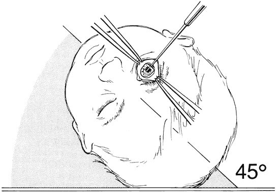
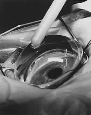
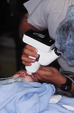
Virtually all of the inhalational anesthetics are known to depress the IOP within minutes of administration.17 Ketamine HCl slowly elevates the IOP as deeper anesthesia is attained.18,19 The benzodiazepines do not appear to have a significant effect on IOP when used in preoperative doses. Midazolam HCl (Versed) is often used as a preoperative sedative in children, and in many cases the child is sufficiently sedated with this medication to perform a quick IOP measurement before the administration of an inhalational general anesthetic. Such a measurement, with the child resting comfortably but not under the influence of the potent inhalational anesthetics, is probably the most accurate. If such a situation cannot be achieved, as in the case of a highly agitated infant or toddler, the anesthesiologist should be aware that it is preferable for the surgeon to measure the IOP as early as possible during the induction of general anesthesia (Fig. 2). At this point in the anesthetic induction, however, airway management is of paramount importance, and the surgeon must defer to the judgment of the anesthesiologist as to when measurements can safely be taken. In most cases, endotracheal intubation is appropriate.

Fig. 2. The Tono-Pen hand-held tonometer (A) and Bio-Pen biometric ruler (B) are ideally suited to the examination of young children in the operating room or office. Young children who have become accustomed to having pressure measurements taken with the Tono-Pen usually accept the Bio-Pen as well, permitting A-scan biometry to become a routine part of follow-up examinations in the office.

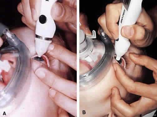
Table 1 lists the information that should be obtained during an initial or follow-up examination under anesthesia. Once the initial IOP measurements are made, the surgeon should tell the anesthesiologist what will remain to be done during the rest of the examination if surgery is not planned. Portions of the examination that require a clear media, such as ophthalmoscopy, retinoscopy, and fundus photography, should be performed first, followed by axial length measurement, gonioscopy, and corneal diameter measurements.


TABLE 1. Essential Information for the Examination Under Anesthesia

  Intraocular pressure
  Optic nerve examination (drawing, cup-to-disc estimation, photography if possible)
  Fundus examination
  Cycloplegic retinoscopy
  Axial length determination
  Corneal diameter (horizontal and vertical)
  Gonioscopy


Preoperative dilation should be avoided if the iris and anterior chamber angle appearance will be important factors in making the initial diagnosis, or if corneal opacities prevent any view of the fundus. However, ophthalmoscopy, retinoscopy, and fundus photography are important parts of the initial and subsequent evaluations of a child with glaucoma, and preoperative dilation with a short-acting mydriatic should be performed before entering the operating room to avoid prolonging anesthesia while waiting for dilation. If necessary, intracameral acetylcholine can be instilled during surgery to constrict the pupil to allow safe intraocular maneuvers.

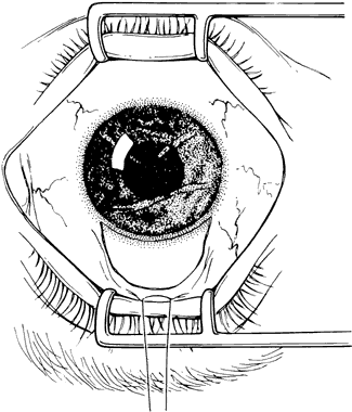
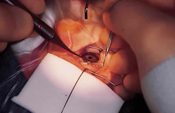
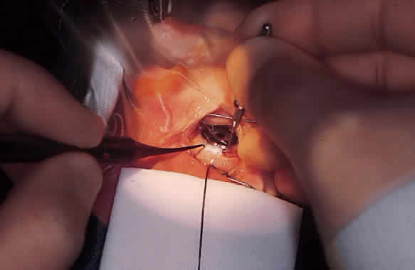
Funduscopy is essential, with both a direct and an indirect ophthalmoscope. The Richardson or Koeppe diagnostic gonioscopy lenses can be of great assistance (Fig. 3). The lens is placed, and then direct ophthalmoscopy is performed through the lens. After this, the periphery should be carefully evaluated with an indirect ophthalmoscope.

Fig. 3. The Koeppe diagnostic gonioscopy lens permits the simultaneous examination of the anterior chamber angle in both eyes, facilitating the comparison of the angles in patients with asymmetric or unilateral disease. Direct ophthalmoscopy is facilitated by the lens, which does not have a central dimple to obscure the view. It is often possible to obtain an excellent view of the optic nerve through a small pupil. In this 6-month-old with Sturge-Weber syndrome, a unilateral angle appearance similar to trabeculodysgenesis was noted in the right eye, which was also buphthalmic with an intraocular pressure of 36 mmHg.

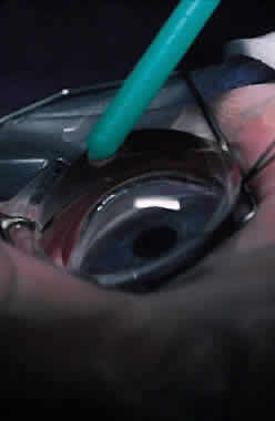
Fundus photography of the optic nerve allows the surgeon to document the extent of progression of the disease (Fig. 4). It is well known that the optic nerve cupping seen in congenital and juvenile glaucoma can to some extent reverse itself,20–22 and this can be used in some cases to gauge the success of therapy. Similarly, axial length measurements by ultrasonic biometry can help establish the degree of disparity between the two eyes, particularly in unilateral cases.23–25 In many cases the disparity decreases after successful therapy,23 and thus a constant or increasing disparity between the two eyes may signal a worsening clinical situation. The Bio-Pen hand-held ultrasonic biometric ruler (see Fig. 2) is a portable device that can be brought to the operating room for intraoperative biometry; because it is similar in size, operation, and appearance to the Tono-Pen, it can also be used in the office setting in young children who have become accustomed to IOP measurements being taken with that device.

Fig. 4. Fundus photography of the optic nerve is an essential part of the congenital glaucoma examination under anesthesia. Until the patient is old enough to cooperate with photography in the office, the optic nerve should be photographed in the operating room during an examination under anesthesia.

Gonioscopy is a mandatory part of the examination under anesthesia. It allows the surgeon to identify the underlying congenital or juvenile glaucoma diagnosis and permits appropriate surgical planning. The Koeppe gonioscopic lens, introduced in 1919,26 has significant advantages over the Zeiss four-mirror or Goldmann three-mirror gonioprism. Chief among these is the ability to compare simultaneously the gonioscopic appearance of two eyes in the same patient (see Fig. 3). The surgeon should be familiar with the gonioscopic findings in primary congenital glaucoma as well as the various secondary glaucomas seen in childhood. Gonioscopic photography can help document the appearance of the anterior chamber angle before and after treatment.

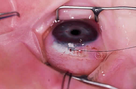
The cornea should be measured both horizontally and vertically, and the presence or absence of Haab's striae or other corneal abnormalities should be noted. Enlargement of the corneal diameter over time is worrisome for progressive buphthalmos, and some clinicians believe these measurements to be more useful than axial length determinations.27 In many cases, however, these measurements are difficult to make accurately or consistently because of uncertain landmarks at the corneoscleral limbus.

Cycloplegic retinoscopy during follow-up examinations (both under anesthesia and in the office) are used to monitor for progressive axial myopia28 and to provide the spectacle correction to be used in conjunction with patching or other amblyopia therapy.

Back to Top
CHOOSING THE SURGICAL APPROACH
The surgical options in the management of congenital and pediatric glaucomas, along with their relative indications, advantages, and disadvantages, are outlined in Table 2.


TABLE 2. Surgical Options in Surgery for Pediatric Glaucoma


ProcedureIndicationsRelative ContraindicationsAdvantagesDisadvantages
GoniotomyPrimary infantile glaucomaCorneal opacities or poor visualization of anterior chamberAvoids manipulation of conjunctivaRequires clear cornea and excellent visibility
ab internoSturge---Weber glaucoma QuickRequires skilled surgical assistant
 Juvenile-onset glaucoma  Treats only 120 degrees of angle
 Glaucoma 2º to aphakia   
 AniridiaExtensive PAS  
TrabeculotomyPrimary infantile glaucomaExtensive PASDoes not require clear corneaRequires intraocular manipulation
ab externoSturge---Weber glaucomaExtreme buphthalmosTreats 180 degrees or 360 degrees at one sitting 
 Juvenile-onset glaucoma   
 Glaucoma 2º to aphakia   
 Aniridia   
TrabeculodialysisInflammatory glaucomaExtensive PASAvoids manipulation of conjunctivaLow success rate (~50%)
   High success rate with antimetabolites 
Guarded filtration procedures (trabeculectomy)Juvenile-onset open-angle glaucomaAphakia requiring chronic contact lens useFamiliar techniqueLong-term risk of endophthalmitis due to thin filtering blebs
 Failed angle surgery High success rate with antimetabolites 
 Inflammatory glaucoma   
 Sturge---Weber syndrome   
 Aniridia   
Drainage implantsFailed conventional surgeryExtreme buphthalmos with scleral thinningCan be used when conjunctiva is badly scarredCataract if tube touches crystalline lens
 Aniridia Safe contact lens use remains possibleEarly hypotony common
 Aphakia  Strabismus possible
 Combined with other procedures (e.g., penetrating keratoplasty, lensectomy, pars plana vitrectomy)  Long-term tolerance, biocompatibility of artificial device unknown
Cyclodestructive procedures (cyclocryotherapy, cyclophotocoagulation)End-stage glaucomaGood central visual acuity in phakic eyeMinimally invasiveRequires general anesthesia in children
   RepeatableCan be painful for several days after treatment
    High incidence of phthysis bulb
    Repeat treatment often necessary

PAS, peripheral anterior synechiae.


In cases of primary infantile glaucoma, several studies have demonstrated that goniotomy ab interno and trabeculotomy ab externo are essentially comparable, with surgical success rates approaching 80% to 90%.29–31 McPherson and Berry30 found that with multiple operations, both procedures controlled IOP in most patients, but more operations were required after goniotomy. Goniotomy ab interno is a simple and elegant procedure, but it requires excellent visualization of the anterior chamber angle. Trabeculotomy ab externo is no less elegant and can be performed in virtually all cases. Both procedures have been made much safer during the past decade by the introduction of viscoelastic materials. The surgeon who anticipates performing a significant amount of congenital glaucoma surgery should be familiar with both techniques.

If the clinical situation permits the use of either approach, the surgeon's personal preference and logistical considerations should dictate the choice. A goniotomy is greatly facilitated by a surgical assistant who is familiar with the procedure and understands what the surgeon is trying to visualize and achieve. A trabeculotomy, however, can be performed with a scrub nurse as the only surgical assistant.

The choice of surgical approach in the case of secondary pediatric glaucomas is more complicated. As ophthalmologists have become more aggressive about treating congenital cataract in early infancy, perhaps the most common secondary glaucoma seen in children today is that associated with surgical aphakia.32,33 In this form of glaucoma, both open-angle and secondary angle-closure variants are recognized; it is thought that both may be the result of chronic, low-grade inflammation caused by residual lens material. In these cases, the surgeon is confronted with the dual challenge of lowering the IOP and maintaining aphakic correction to prevent the development of amblyopia. Careful preoperative gonioscopy is mandatory to determine whether (and where) a goniotomy should be attempted or whether the peripheral anterior synechiae present will lend themselves to goniosynechialysis (performed in a manner similar to goniotomy). Trabeculotomy ab externo should be avoided if extensive, broad peripheral anterior synechiae are present. Lysis of these adhesions by the trabeculotome can be associated with significant intraocular bleeding and can result in a slowly clearing vitreous hemorrhage that can be profoundly amblyogenic. In such cases, the implantation of an aqueous drainage device can be considered as a primary procedure. As will be discussed below, even though guarded filtration surgery with antimetabolite use has a reasonable chance of success in lowering pressure, it should probably be avoided because of the problems associated with extended-wear contact lens use in the presence of a filtration bleb.

In cases of inflammatory glaucoma in children and young adults where significant peripheral anterior synechiae are absent, trabeculodialysis has proven to be useful in some hands,34,35 with success rates of 50% to 60%. This approach is attractive because it avoids the creation of a filtering bleb prone to endophthalmitis in patients taking long-term topical steroids.

Another secondary glaucoma seen in children is that associated with Sturge-Weber syndrome. Glaucoma presenting with this and related syndromes (e.g., Klippel-Trenaunay-Weber syndrome) at or shortly after birth is usually associated with an angle appearance similar to that of primary infantile glaucoma. Later presentation of glaucoma in this group of syndromes is probably associated with a combination of abnormal angle structure and elevated episcleral venous pressure. A variety of surgical approaches have been used in the management of this glaucoma.36–40 In my experience, trabeculotomy ab externo, performed inferiorly or laterally to preserve the superior conjunctiva for subsequent filtering surgery, has been successful in most cases presenting in infancy.

Congenital aniridia is another syndrome associated with glaucoma for which many procedures have been attempted, with varying success.41,42 In this syndrome, true agenesis of the iris is rare; in most cases, a tiny, hypoplastic iris can be identified on gonioscopy.43 Although angle surgery can be successful in cases presenting in infancy,42 it is common for this iris stump to rotate anteriorly, closing off what little open angle remains, leading to late failures. Chen and Walton44 have reported promising results using goniotomy to prevent the development of glaucoma in these children. Their findings highlight the importance of periodic examinations under anesthesia and gonioscopy in children with aniridia to monitor them for the early gonioscopic, IOP, and nerve changes associated with glaucoma. Aqueous drainage devices have proven extremely helpful in these difficult cases, and I have found it useful in several cases to perform a staged implantation of a drainage device at the same time as angle surgery in eyes at high risk of failure of the primary surgery. The staged device can be introduced into the anterior chamber with a low risk of hypotony or choroidal hemorrhage if it is needed at a later time.

More unusual syndromes associated with glaucoma, including Peter's anomaly, sclerocornea, and microcornea, are handled on a case-by-case basis. The results of surgery for these problems are dismal, particularly in unilateral cases, but in bilateral cases it is difficult for the surgeon to stand back and do nothing. These eyes usually must be tackled using a team approach, often combining a penetrating keratoplasty with a cataract extraction, vitrectomy, and aqueous drainage device. Such an aggressive approach may provide the patient with some ambulatory vision.

GONIOTOMY AB INTERNO

When Otto Barkan introduced goniotomy ab interno in the late 1930s,45–47 the management of congenital glaucoma was revolutionized. Until then an incurable disease, congenital glaucoma was among the leading causes for admission to schools for the blind. The basic principles of the procedure have changed little since it was first described, but goniotomy has been made safer and more successful with the introduction of better instrumentation, viscoelastics, and the operating microscope.

Excellent visualization of the anterior chamber angle is needed to perform the procedure safely and accurately. Mild corneal edema can sometimes be overcome by denuding the cornea of its epithelium, either by blunt scraping or by topical alcohol and glycerin. Unfortunately, corneal clarity often worsens during the course of surgery. If the surgeon anticipates that visualization may become a problem during surgery, or if large Haab's striae or other corneal opacities prevent adequate visualization of the anterior angle structures, a trabeculotomy ab externo should be performed instead.

When a goniotomy is planned, the anesthesiologist should be informed that the surgeon and assistant will be manipulating the patient's head back and forth during the procedure, and that the endotracheal tube should be secured with this in mind. I have found a Rae-style endotracheal tube to be particularly useful in this respect.

It is technically easiest to perform a goniotomy over the nasal 120 degrees of the anterior chamber angle, using a temporal approach. This is the usual site of an initial goniotomy. The patient's head is turned away from the surgeon approximately 45 degrees from vertical and is secured firmly in place (Fig. 5). If the surgeon uses an operating microscope rather than loupes, the microscope head should be tilted to permit adequate visualization of the nasal anterior chamber angle once the gonioscopic prism is placed on the eye.

Fig. 5. Goniotomy ab interno. To give the surgeon clear access to the temporal limbal area, the patient is positioned with the head tilted 45 degrees away from the eye being treated. The surgeon sits at the side of the operating table, with the assistant at the head of the table.

The assistant, who is seated across from the surgeon or at the head of the table, grasps the eye at the 6 and 12 o'clock positions with toothed forceps to rotate the eye nasally or temporally at will. Bridle sutures are usually not necessary. The surgeon then creates a limbal stab incision through the cornea just anterior to the corneoscleral limbus and injects a viscoelastic substance into the anterior chamber to deepen it.

Various operative gonioscopic prisms are available. The Hoskins-Barkan operating gonioprism (Ocular Instruments, Bellevue, WA) permits excellent visualization of the anterior chamber angle while permitting the assistant to maintain excellent control of the eye and the surgeon to introduce the goniotomy knife through the stab incision that has already been created. The lens is placed on the eye with balanced salt solution as the gonioscopic coupling agent.

Various goniotomy knives are available to perform a goniotomy ab interno; a sharp knife with a slight bevel to the handle is preferable. The knives that permit irrigation can be more difficult to manipulate and are not preferred. Some surgeons find them helpful to prevent collapse of the anterior chamber; however, when goniotomy is properly performed with minimal distortion of the corneal wound, collapse of the anterior chamber should not be a problem. If necessary, the chamber can be well maintained using a viscoelastic material.

The stab incision should be made small enough that the anterior chamber remains formed during the procedure. The surgeon then advances the knife across the anterior chamber angle, taking great care to avoid the crystalline lens (Fig. 6). Anterior movement of the lens indicates that the stab incision is gaping, permitting egress of the anterior chamber fluids. The surgeon should assess whether the knife handle is distorting the stab incision or whether the incision is too large; it can be partially closed with a temporary suture.

Fig. 6. Goniotomy ab interno. The goniotomy knife is advanced across the anterior chamber under the protection of a generous amount of viscoelastic. The goniotomy incision is placed in the anterior aspect of the middle third of the trabecular meshwork. Inset. The incision can be carried out over approximately 60 degrees on either side of a point directly opposite the corneal incision.

The abnormal mesodermal tissue that creates the restriction to aqueous outflow in infantile glaucoma has an almost translucent appearance, blurring and distorting the anterior chamber angle anatomy. Most infants have light blue or very transparent iris stroma, making the landmarks somewhat difficult to identify; In infants with primary congenital glaucoma, the iris root usually has a scalloped, almost serrated anterior edge that provides an excellent landmark for the surgeon. The surgeon uses the goniotomy knife to place an incision at the anterior aspect of the middle third of the trabecular meshwork, behind Schwalbe's line, starting at a point 180 degrees away from the corneal incision and rotating either clockwise or counterclockwise as appropriate (see Fig. 6). The assistant can rotate the eye slightly to permit the surgeon to extend the goniotomy incision peripherally without distorting the corneal incision. The knife is then flipped 180 degrees and the goniotomy is completed in the opposite direction.

If the incision is placed too far anteriorly, the goniotomy will not function; placed too far posteriorly, the incision will incise only peripheral iris and iris root, causing a peripheral iridotomy, iridodialysis, or intraocular hemorrhage, making further surgery difficult at best. The surgeon should be able to visualize directly the iris root fall gently posteriorly as the abnormal mesodermal attachments are lysed. The goniotomy can be performed in this manner over approximately 120 degrees of nasal angle centered 180 degrees away from the corneal stab incision. A small amount of bleeding is common during a goniotomy, but the amount seen is usually less than occurs during a trabeculotomy.

At the conclusion of the procedure, any viscoelastic that remains in the eye that can be easily flushed from the eye and so removed. The corneal stab incision, although usually self-sealing, can be closed with a single 10-0 nylon suture. Subconjunctival injections of a steroid and antibiotic are at the discretion of the surgeon.

In some cases, a single goniotomy involving the nasal 120 degrees of the anterior chamber is not sufficient to control intraocular pressure, and a second goniotomy may be subsequently performed over the temporal or inferotemporal angle. This has raised the possibility that performing two simultaneous goniotomies might be advantageous. In a prospective, randomized study of infants with bilateral infantile glaucoma, Catalano and coworkers48 found no significant difference between eyes that underwent a single goniotomy at initial surgery versus those that underwent two simultaneous procedures.

TRABECULOTOMY AB EXTERNO

Trabeculotomy ab externo was introduced in the 1960s49–51 after it was recognized that an external approach was needed for cases of congenital glaucoma in which the cornea was too cloudy to permit the safe performance of a goniotomy ab interno. Like the goniotomy, trabeculotomy ab externo has benefited from the introduction of viscoelastics and the operating microscope, which have made the procedure highly successful and for all intents and purposes equivalent to goniotomy.

The child's head is prepared and draped in a manner similar to that for adult eye surgery. A superior rectus bridle suture is placed to depress and expose the superior limbal region. Rarely, a lateral canthotomy must be performed to provide adequate exposure in small or premature infants.

A limbal peritomy is performed straddling the 12 o'clock position (Fig. 7). A partial-thickness scleral flap (either rectangular or triangular) is then created in the 12 o'clock meridian in such a manner as to sit astride the corneoscleral junction (Fig. 8). The surgeon should be familiar with the three-dimensional anatomy of the limbal region and should recognize that Schlemm's canal will lie just below the transition from white scleral fibers to the blue corneal fibers. Using a microsurgical blade, the surgeon creates a radial incision measuring approximately 2 mm long that straddles the corneoscleral limbus. Carefully dissecting down layer by layer, Schlemm's canal is unroofed. Entry into Schlemm's canal can be confirmed by the egress of a small amount of aqueous humor without the shallowing of the anterior chamber; such a shallowing would indicate that the anterior chamber had been directly violated.

Fig. 7. Trabeculotomy ab externo. A bridle suture has been placed and a limited limbal peritomy performed. The cornea is hazy, and Haab's striae are noted.

Fig. 8. Trabeculotomy ab externo. A. The surgeon is creating a radial incision in the bed of the partial-thickness scleral flap with a microsurgical blade. B. The dissection is carried downward layer by layer until Schlemm's canal is unroofed. C. Schlemm's canal can be positively identified by cannulation with a 6-0 nylon suture. D. Intraoperative photograph demonstrating cannulation of Schlemm's canal with a 6-0 nylon suture. Note the subconjunctival hemangioma in this eye with Sturge-Weber-associated glaucoma.

A short length of 6-0 black nylon suture material should be used to cannulate Schlemm's canal. The cut end of the suture can easily be smoothed by the application of heat from a disposable battery-operated cautery. The nylon suture must not be forcibly introduced into Schlemm's canal, lest a false passage be created. The suture will pass easily if Schlemm's canal is correctly cannulated, and intraoperative gonioscopy with either a Koeppe lens (Fig. 9) or a Zeiss four-mirror gonioprism (Fig. 10) can be used.

Fig. 9. Trabeculotomy ab externo. A. Intraoperative use of a Koeppe diagnostic lens shows the correct placement of a length of 6-0 nylon suture in Schlemm's canal (arrow).

Fig. 10. Trabeculotomy ab externo. A Zeiss four-mirror gonioscopic lens can be used during surgery to confirm the cannulation of Schlemm's canal.

Once Schlemm's canal is correctly identified and cannulated in both a clockwise and counterclockwise manner, a trabeculotome with the appropriate curvature for the particular eye is chosen. The lower probe of the trabeculotome is then introduced into the previously identified opening into Schlemm's canal. It is then passed along Schlemm's canal in a curvilinear fashion, using the upper probe as a guide to confirm the location of the lower one (Fig. 11), making certain that the trabeculotome is oriented in such a way that on its rotation the trabeculotome will rupture Schlemm's canal internally and the probe will pass parallel to the iris and not impinge on the crystalline lens (Figs. 12 and 13).

Fig. 11. Trabeculotomy ab externo. The trabeculotome is designed with an upper probe that provides the surgeon with visual confirmation of the relative location and orientation of the lower probe. It is inserted into Schlemm's canal in a curvilinear fashion.

Fig. 12. Trabeculotomy ab externo. The trabeculotome is inserted into Schlemm's canal and rotated around the axis of the instrument handle to break through the trabecular meshwork into the anterior chamber.

Fig. 13. Trabeculotomy ab externo. To avoid damage to the crystalline lens or corneal endothelium, care must be taken to ensure that the trabeculotome is rotated into the eye parallel to the iris.

This maneuver is usually associated with a small amount of bleeding and egress of aqueous humor from the base of the trabeculotome probe. Care should be taken to avoid distorting the incision so that the anterior chamber does not collapse. If it does, it may be necessary to place some viscoelastic in the anterior chamber so that this maneuver can be carried out in the opposite direction without risk to the crystalline lens.

Once the trabeculotomy has been performed on either side of the incision, the overlying scleral flap can be closed with one or more 10-0 nylon sutures. The overlying conjunctiva is then tacked over the scleral flap with a fine absorbable suture. Subconjunctival injections of steroid and antibiotic are at the discretion of the surgeon.

Trabeculotomy carried out in this manner will open the angle over almost 180 degrees, straddling the incision at the 12 o'clock position. Another technique of performing a trabeculotomy ab externo over 360 degrees has recently been described,52 using a nylon suture to cannulate the entire circumference of Schlemm's canal (Figs. 14 and 15). This technique has two significant advantages: no metallic instruments are introduced into the anterior chamber, reducing the chance of lens damage, and the entire anterior chamber angle is treated, eliminating the need for additional angle surgery if the primary surgery fails. Unlike goniotomy,48 the question of whether a complete 360-degree trabeculotomy is superior to 180 degrees has not been resolved.

Fig. 14. A 360-degree trabeculotomy ab externo. A. Schlemm's canal is cannulated with a 6-0 nylon suture over 180 degrees. B. The suture is retrieved through a mirroring incision 180 degrees away from the initial one.

Fig. 15. A 360-degree trabeculotomy ab externo. A. Both 180-degree segments of Schlemm's canal are cannulated with 6-0 nylon. B. By grasping both ends of the suture, the surgeon can gently “cheesewire” the suture into the anterior chamber, performing a trabeculotomy over each 180-degree segment.

GUARDED FILTRATION PROCEDURES (TRABECULECTOMY)

Although guarded filtration procedures (trabeculectomy) have become the mainstay of glaucoma surgery in adults, their use in infantile and pediatric glaucoma is limited. The exuberant healing response of infants and children greatly reduces the success rate of filtration surgery in this patient population, and it is difficult to defend guarded filtration as a primary procedure when the success rates for trabeculotomy and goniotomy approach 90%.

The recent introduction of antimetabolites such as 5-fluorouracil53–55 and in particular mitomycin-C56 has given the surgeon additional tools to control this exuberant healing response and may very well allow the glaucoma surgeon to achieve the success rates seen in the adult population. Nonetheless, guarded filtration is a procedure fraught with problems in the pediatric age group.

The filtration bleb that develops after a guarded filtration procedure in which antimetabolites have been used is usually thin and cystic and prone to infection.57 One recent study of the adult population estimated the risk of a repeat hospital admission for filtration bleb infection or endophthalmitis to be up to 1.2%.58 In the pediatric age group, this level of risk is probably unacceptable. Al-Hazmi and colleagues59 performed either trabeculectomy or combined trabeculotomy/trabeculectomy in 254 eyes of 180 pediatric patients. They reported complications including cystic blebs, bleb leaks, flat anterior chamber, and cataract and found that children younger than 2 years had fewer complications but higher failure rates. Complications increased with time after surgery, and they cautioned that because bleb thinning increases with time after surgery, additional problems could be anticipated with longer follow-up. This mirrors the findings and conclusions of Beck and colleagues,60 who found trabeculectomy generally effective in older children but also cautioned that late-onset bleb-related endophthalmitis would become a substantial risk in this population.

Azuara-Blanco and coworkers61 described a series of 21 pediatric patients undergoing mitomycin C-augmented trabeculectomy; they reported reasonable short-term success in phakic patients but no success in aphakic eyes. Freedman and colleagues62 found that although guarded filtration procedures with mitomycin C, 5-fluorouracil, and postoperative suture lysis were viable in older children, the addition of these adjunctive measures did not provide any convincing improvement in the success of the procedures in pediatric patients and may have increased the complication rate.

A guarded filtration procedure can be considered if two or more attempts at angle surgery (e.g., trabeculotomy or goniotomy) have been unsuccessful. If significant buphthalmos is present, the surgeon must be particularly cautious when developing the partial-thickness scleral flap because the sclera in these eyes is extremely thin. The use of adjunctive antimetabolites probably increases the success rate of these procedures, albeit at the cost of higher long-term complication rates. The surgeon should take into consideration the social situation of the child. If it is unlikely that the parent or responsible guardian will take action at the earliest sign of a bleb infection or endophthalmitis, filtering surgery is inadvisable.

Guarded filtration procedures should probably be avoided altogether in cases of childhood glaucoma associated with surgical aphakia. The need for extended-wear contact lenses for aphakic correction and the prevention of amblyopia raise the risk of devastating endophthalmitis to an unacceptably high level if a filtration bleb is present, particularly given the poor hygiene of toddlers and young children. In general, a guarded filtration procedure should be considered in the older child in whom juvenile glaucoma presents anew, or in whom prior angle surgery has failed.

GLAUCOMA DRAINAGE DEVICES

The introduction of the Molteno implant in the early 1970s represented a new and promising option for eyes in which conventional angle surgery had failed.63 The Molteno implant and similar devices have saved many eyes that were otherwise destined to become blind and painful.

These procedures involve the placement of a small-caliber silicone drainage tube into the anterior chamber or, in the case of an aphakic, vitrectomized eye, into the posterior chamber through the pars plana. This tube is connected to an external bleb-maintaining device that is designed to create and maintain a potential space between the sclera and conjunctiva. Aqueous filtration then occurs through the surface of this bleb. Studies in primate eyes64 have demonstrated that the degree of pressure-lowering effect is roughly proportional to the surface area of this external potential space.

Each of the glaucoma implants has taken a different approach in terms of the design of the external portion of the device. The Molteno implant (IOP, Inc, Costa Mesa, CA) uses an acrylic disc that is sutured to the sclera in the equatorial region. The Baerveldt glaucoma implant (Pharmacia & Upjohn, Peapack, NJ) uses a flexible Silastic rubber device shaped to conform to the surface of the globe to maintain a bleb around the equatorial region of the eye. This device is available in three sizes (250, 350, and 425 mm2) and can be implanted in a single quadrant. A 350-mm2 implant with a right-angled bend in the silicone tube has recently been marketed for use in conjunction with pars plana vitrectomy;65 I have found this device useful in aphakic eyes undergoing a complete vitrectomy and removal of residual lens material. The smaller 250-mm2 implant is well suited to pediatric cases, although when an eye comes to undergo such a procedure it has often grown to almost adult size or larger, permitting the implantation of a larger device.

One of the many technical challenges in drainage implant surgery is the avoidance of profound postoperative hypotony and its attendant complications. The Ahmed Valve Implant (New World Medical, Inc, Rancho Cucamonga, CA) uses a Silastic valve to prevent early postoperative hypotony. Englert and colleagues66 reported promising results in a series of pediatric cases. For devices without a valve, the silicone tube portion is usually occluded in a temporary manner by using an absorbable suture such as 8-0 polyglactin (Vicryl). Techniques for the temporary occlusion of the tube that require later lysis by the argon laser or surgical manipulation are impractical for children. In all cases, the tube must be covered with donor sclera or other appropriate material to prevent its erosion and externalization through the conjunctiva.67,68 Although in adults some surgeons place the tube in the bed of a partial-thickness scleral flap, this technique should be avoided in infants and children; the sclera of a buphthalmic eye is extremely thin, and erosion of a silicone tube into or out of the eye through the thinned scleral tissue has been reported.69,70

If the tube is placed into the anterior chamber, care must be taken to avoid impinging the crystalline lens, because a cataract is likely to develop. The long-term safety and efficacy of these devices are not known at present, but some have been in place in adult patients for almost two decades. The placement of these devices should not be avoided in children for these reasons, but the parent or responsible adult must be advised of the many unknown aspects of these procedures.

Among the currently available aqueous drainage devices, glaucoma surgeons have the longest experience in the congenital and juvenile glaucomas with the Molteno implant.71–73 The Baerveldt implant has several attractive aspects to its design: a large surface area for aqueous filtration can be created in a single quadrant, and the soft, pliable nature of the implant facilitates its introduction in the tight surroundings of the pediatric orbit. Reports of strabismus associated with the Baerveldt implant,74 presumably related to the large equatorial filtration bleb the device creates, may limit its usefulness in eyes with good visual potential, but strabismus has been reported with the other aqueous drainage devices,75,76 and it remains to be seen whether this implant has more problems than the others in this regard. The fenestrated version of the Baerveldt device, which prevents the filtration bleb from growing too large, may partially solve this problem.

CYCLODESTRUCTIVE PROCEDURES

Long a mainstay of therapy when conventional incisional surgery has failed, cyclodestructive procedures have usually been perceived as procedures of last resort because of the high incidence of phthysis bulbi thought to be associated with cyclocryotherapy. Wagle and colleagues77 found that cyclocryotherapy yielded a success rate of 66% at 6 months, dropping to 44% at longer follow-up. The main complication encountered was phthysis, particularly in eyes with aniridia.

The introduction of other modalities of ciliary body ablation, namely Nd:YAG and infrared wavelength diode lasers, promises to give the surgeon more control over the amount of ciliary body that is ablated at each sitting. At present, it appears that laser cyclophotocoagulation, whether by contact or noncontact techniques, produces a lower rate of phthysis bulbi than cyclocryotherapy. Bock and coworkers78 found diode laser cyclophotocoagulation to be a useful treatment in pediatric cases, although many of the patients required retreatment. No prospective, randomized clinical study has been performed comparing the two modalities, and it is unlikely that one will ever be performed. Because of the pain associated with cyclodestructive procedures, they are usually performed under general anesthesia in the pediatric age group. I find it useful to place a long-acting retrobulbar block with bupivacaine (Marcaine) while the child is asleep to control postoperative pain, along with a subconjunctival injection of triamcinolone (Kenalog) to provide corticosteroid coverage during the early postoperative period. In general, these procedures are reserved for eyes with poor visual potential.

Back to Top
POSTOPERATIVE CARE
After a goniotomy or trabeculotomy, the patient is given topical steroids and antibiotics, preferably in a combination form to facilitate parent compliance. Cycloplegics should be avoided because they tend to crowd the angle and lead to development of synechiae in the area of angle surgery.

The patient should be examined during the first 24 hours after surgery and the IOP measured if possible. If a significant amount of viscoelastic was left in the eye, the IOP can be as high or higher than the preoperative level; it may be necessary to prescribe aqueous suppressant medication for several days until the viscoelastic has cleared the eye. In most cases, any viscoelastic or blood that was present in the eye at the conclusion of the surgery will have cleared within the first 24 hours.

Although in a small infant it may be possible to assess the IOP accurately in the office a few days or weeks after the surgery, it is advisable to perform a complete examination under anesthesia some 4 to 6 weeks after the initial surgery to gauge the success of the procedure more formally and to document the status of the eye. In particular, an examination under anesthesia will permit the ophthalmologist to perform a careful cycloplegic refraction and obtain color fundus photographs of the optic disc that will serve as a baseline for future examinations.

In some cases, the administration of chloral hydrate syrup (25 to 50 mg/kg) in the office can facilitate the performance of many if not all of the aspects of an examination under anesthesia outlined above.79 Although chloral hydrate sedation is quite safe, the drug should be administered only in a setting where equipment and trained staff are available for emergency airway management in the case of respiratory distress.

Back to Top
LONG-TERM MANAGEMENT
Although the success rate for surgery for congenital and pediatric glaucoma has been estimated to be as high as 80% to 90%, only about half of these patients reach adulthood with visual acuity of 20/200 or better. This is particularly so in unilateral cases, where anisometropic amblyopia develops as a result of the axial myopia of the glaucomatous eye. The surgeon must remain vigilant for the development of amblyopia and aggressively pursue patching and spectacle or contact lens correction to ensure that a dense amblyopia does not develop in the glaucomatous eye. Only when the patient reaches the age of 8 or 10 with excellent central visual acuity should the ophthalmologist consider the surgery to have been a success.
Back to Top
FUTURE TRENDS
With success rates approaching 90%, it is difficult to see how the procedures of goniotomy and trabeculotomy can be improved. Primary infantile glaucoma appears for the most part to be a genetic disease with variable penetrance. With the current explosion of molecular biology techniques, the future holds the prospect of better prenatal screening and perhaps one day therapy directed at the genetic defect itself.
Back to Top
REFERENCES

1. deLuise VP, Anderson DR: Primary infantile glaucoma (congenital glaucoma). Surv Ophthalmol 28:1, 1983

2. Shaffer RN, Weiss DI: Congenital and Pediatric Glaucomas. St Louis: CV Mosby, 1970

3. Kwitko ML: Glaucoma in Infants and Children. New York: Appleton-Century-Crofts, 1973

4. Brandt JD: Congenital Glaucoma. In Yanoff M, Duker J (eds): Ophthalmology. London: Mosby, 1999

5. Tucker SM, Enzenauer RW, Levin AV et al: Corneal diameter, axial length, and intraocular pressure in premature infants. Ophthalmology 99:1296, 1992

6. Minckler DS, Baerveldt G, Heuer DK et al: Clinical evaluation of the Oculab Tono-Pen. Am J Ophthalmol 104:168, 1987

7. Kao SF, Lichter PR, Bergstrom TJ et al: Clinical comparison of the Oculab Tono-Pen to the Goldmann applanation tonometer. Ophthalmology 94:1541, 1987

8. Dominguez A, Baños MS, Alvarez MG et al: Intraocular pressure measurement in infants under general anesthesia. Am J Ophthalmol 78:110, 1974

9. Plasilova M, Stoilov I, Sarfarazi M et al: Identification of a single ancestral CYP1B1 mutation in Slovak Gypsies (Roms) affected with primary congenital glaucoma. J Med Genet 36:290, 1999

10. Sarfarazi M, Akarsu AN, Hossain A et al: Assignment of a locus (GLC3A) for primary congenital glaucoma (buphthalmos) to 2p21 and evidence for genetic heterogeneity. Genomics 30:171, 1995

11. Jerndal T: Congenital glaucoma due to dominant goniodysgenesis. A new concept of the heredity of glaucoma. Am J Hum Genet 35:645, 1983

12. Hoskins HDJ, Hetherington JJ, Magee SD et al: Clinical experience with timolol in childhood glaucoma. Arch Ophthalmol 103:1163, 1985

13. McMahon CD, Hetherington JJ, Hoskins HDJ et al: Timolol and pediatric glaucomas. Ophthalmology 88:249, 1981

14. Olson RJ, Bromberg BB, Zimmerman TJ: Apneic spells associated with timolol therapy in a neonate. Am J Ophthalmol 88:120, 1979

15. Enyedi LB, Freedman SF, Buckley EG: The effectiveness of latanoprost for the treatment of pediatric glaucoma. J AAPOS 3:33, 1999

16. Altuna JC, Greenfield DS, Wand M et al: Latanoprost in glaucoma associated with Sturge-Weber syndrome: Benefits and side effects. J Glaucoma 8:199, 1999

17. Ausinsch B, Munson ES, Levy NS: Intraocular pressures in children with glaucoma during halothane anesthesia. Ann Ophthalmol 9:1391, 1977

18. Marynen L, Libert: Ocular tonometry in the child under general anesthesia with IM ketamine. Acta Anaesthesiol Belg 1976, 27(Suppl):29–40

19. Adams AK: Ketamine in paediatric ophthalmic practice. Anaesthesia 28:212, 1973

20. Shaffer RN: Glaucomatous cupping of the optic disc in the young. Trans Ophthalmol Soc NZ 25:77, 1973

21. Richardson KT, Shaffer RN: Optic-nerve cupping in congenital glaucoma. Am J Ophthalmol 62:507, 1966

22. Quigley HA: Childhood glaucoma: Results with trabeculotomy and study of reversible cupping. Ophthalmology 89:219, 1982

23. Tarkkanen A, Uusitalo R, Mianowicz J: Ultrasonographic biometry in congenital glaucoma. Acta Ophthalmol (Copenh) 61:618, 1983

24. Sampaolesi R: Corneal diameter and axial length in congenital glaucoma [letter]. Can J Ophthalmol 23:42, 1988

25. Reibaldi A: Biometric ultrasound in the diagnosis and follow-up of congenital glaucoma. Ann Ophthalmol 14: 707, 1982

26. Koeppe L: Die Mikroskopie des lebenden Kammerwinkels im focalen Lichte der Gullstrandschen Nernstspaltlampe. Graefe Arch Ophthalm 101:48, 1919

27. Kiskis AA, Markowitz SN, Morin JD: Corneal diameter and axial length in congenital glaucoma. Can J Ophthalmol 20:93, 1985

28. Egbert JE, Kushner BJ: Excessive loss of hyperopia. A presenting sign of juvenile aphakic glaucoma. Arch Ophthalmol 108:1257, 1990

29. Anderson DR: Trabeculotomy compared to goniotomy for glaucoma in children. Ophthalmology 90:805, 1983

30. McPherson SD Jr, Berry DP: Goniotomy vs external trabeculotomy for developmental glaucoma. Am J Ophthalmol 95:427, 1983

31. Hoskins HD Jr, Shaffer RN, Hetherington J: Goniotomy vs trabeculotomy. J Pediatr Ophthalmol Strabismus 21:153, 1984

32. Pressman SH, Crouch EJ: Pediatric aphakic glaucoma. Ann Ophthalmol 15:568, 1983

33. François J: Late results of congenital cataract surgery. Ophthalmology 86:1586, 1979

34. Kanski JJ, McAllister JA: Trabeculodialysis for inflammatory glaucoma in children and young adults. Ophthalmology 92:927, 1985

35. Williams RD, Hoskins HD, Shaffer RN: Trabeculodialysis for inflammatory glaucoma: A review of 25 cases. Ophthalm Surg 23:36, 1992

36. Iwach AG, Hoskins HJ, Hetherington JJ et al: Analysis of surgical and medical management of glaucoma in Sturge-Weber syndrome. Ophthalmology 97:904, 1990

37. Wagner RS, Caputo AR, Del Negro RG et al: Trabeculectomy with cyclocryotherapy for infantile glaucoma in the Sturge-Weber syndrome. Ann Ophthalmol 20:289, 1988

38. Azuara-Blanco A, Wilson RP, Spaeth GL et al: Filtration procedures supplemented with mitomycin C in the management of childhood glaucoma. Br J Ophthalmol 83:151, 1999

39. Mandal AK: Primary combined trabeculotomy-trabeculectomy for early-onset glaucoma in Sturge-Weber syndrome. Ophthalmology 106:1621, 1999

40. Irkec M, Kiratli H, Bilgic S: Results of trabeculotomy and guarded filtration procedure for glaucoma associated with Sturge-Weber syndrome. Eur J Ophthalmol 9:99, 1999

41. Wiggins RJ, Tomey KF: The results of glaucoma surgery in aniridia. Arch Ophthalmol 110:503, 1992

42. Walton DS: Aniridic glaucoma: The results of gonio-surgery to prevent and treat this problem. Trans Am Ophthalmol Soc 84:59, 1986

43. Grant WM, Walton DS: Progressive changes in the angle in congenital aniridia, with development of glaucoma. Am J Ophthalmol 78:842, 1974

44. Chen TC, Walton DS: Goniosurgery for prevention of aniridic glaucoma. Arch Ophthalmol 117:1144, 1999

45. Barkan O: A new operation for chronic glaucoma: Restoration of physiologic function by opening Schlemm's canal under direct magnified vision. Am J Ophthalmol 19:951, 1936

46. Barkan O: Technic of goniotomy. Arch Ophthalmol 19: 217, 1938

47. Barkan O: Glaucoma: Classification, causes and surgical control. Am J Ophthalmol 21:10, 1938

48. Catalano RA, King RA, Calhoun JH et al: One versus two simultaneous goniotomies as the initial surgical procedure for primary infantile glaucoma. J Pediatr Ophthalmol Strabismus 26:9, 1989

49. Smith R: A new technique for opening the canal of Schlemm. Br J Ophthalmol 44:370, 1960

50. Smith R: Nylon filament trabeculotomy in glaucoma. Trans Ophthalmol Soc UK 82:439, 1962

51. Allen L, Burian HM: Trabeculotomy ab externo. Am J Ophthalmol 53:19, 1962

52. Beck AD, Lynch MG: 360-degrees trabeculotomy for primary congenital glaucoma. Arch Ophthalmol 113:1200, 1995

53. Group TFFSS: Fluorouracil Filtering Surgery Study one-year follow-up. Am J Ophthalmol 108:625, 1989

54. Rockwood EJ, Parrish RK II, Heuer DK et al: Glaucoma filtering surgery with 5-fluorouracil. Ophthalmology 94: 1071, 1987

55. Zalish M, Leiba H, Oliver M: Subconjunctival injection of 5-fluorouracil following trabeculectomy for congenital and infantile glaucoma. Ophthalmic Surg 23:203, 1992

56. Skuta GL, Beeson CC, Higginbotham EJ et al: Intraoperative mitomycin versus postoperative 5-fluorouracil in high-risk glaucoma filtering surgery. Ophthalmology 99:438, 1992

57. Wolner B, Liebmann JM, Sassani JW et al: Late bleb-related endophthalmitis after trabeculectomy with adjunctive 5-fluorouracil. Ophthalmology 98:1053, 1991

58. Sastry SM, Street DA, Javitt JC: National Outcomes of Glaucoma Surgery: Complications following partial and full-thickness filtering procedures. J Glaucoma 1:137, 1992

59. al-Hazmi A, Zwaan J, Awad A et al: Effectiveness and complications of mitomycin C use during pediatric glaucoma surgery. Ophthalmology 105:1915, 1998

60. Beck AD, Wilson WR, Lynch MG et al: Trabeculectomy with adjunctive mitomycin C in pediatric glaucoma. Am J Ophthalmol 126:648, 1998

61. Azuara-Blanco A, Wilson RP, Spaeth GL et al: Filtration procedures supplemented with mitomycin C in the management of childhood glaucoma. Br J Ophthalmol 83:151, 1999

62. Freedman SF, McCormick K, Cox TA: Mitomycin Caugmented trabeculectomy with postoperative wound modulation in pediatric glaucoma. J AAPOS 3:117, 1999

63. Molteno ACB: New implant for glaucoma: Clinical trial. Br J Ophthalmol 53:606, 1969

64. Minckler DS, Shammas A, Wilcox M et al: Experimental studies of aqueous filtration using the Molteno implant. Trans Am Ophthalmol Soc 85:368, 1987

65. Luttrull JK, Avery RL: Pars plana implant and vitrectomy for treatment of neovascular glaucoma. Retina 15:379, 1995

66. Englert JA, Freedman SF, Cox TA: The Ahmed valve in refractory pediatric glaucoma. Am J Ophthalmol 127:34, 1999

67. Brandt JD: Patch grafts of dehydrated cadaveric dura mater for tube-shunt glaucoma surgery. Arch Ophthalmol 111: 1436, 1993

68. Freedman J: Scleral patch grafts with Molteno setons. Ophthalmic Surg 18:532, 1987

69. Muñoz M, Tomey KF, Traverso C et al: Clinical experience with the Molteno implant in advanced infantile glaucoma. J Pediatr Ophthalmol Strabismus 28:68, 1991

70. Melamed S, Cahane M, Gutman I et al: Postoperative complications after Molteno implant surgery. Am J Ophthalmol 111:319, 1991

71. Traverso CE, Tomey KF, al Kaff A: The long-tube single plate Molteno implant for the treatment of recalcitrant glaucoma. Int Ophthalmol 13:159, 1989

72. Billson F, Thomas R, Alward WL: The use of two-stage Molteno implants in developmental glaucoma. J Pediatr Ophthalmol Strabismus 26:3, 1989

73. Hill RA, Heuer DK, Baerveldt G et al: Molteno implantation for glaucoma in young patients. Ophthalmology 98:1042, 1991

74. Ball SF, Ellis GS, Herrington RG et al: Brown's superior oblique tendon syndrome after Baerveldt glaucoma implant [letter]. Arch Ophthalmol 110:1368, 1992

75. Muñoz M, Parrish R: Hypertropia after implantation of a Molteno drainage device [letter]. Am J Ophthalmol 113: 98, 1992

76. Christmann LM, Wilson ME: Motility disturbances after Molteno implants. J Pediatr Ophthalmol Strabismus 29:44, 1992

77. Wagle NS, Freedman SF, Buckley EG et al: Long-term outcome of cyclocryotherapy for refractory pediatric glaucoma. Ophthalmology 105:1921, 1998

78. Bock CJ, Freedman SF, Buckley EG et al: Transscleral diode laser cyclophotocoagulation for refractory pediatric glaucomas. J Pediatr Ophthalmol Strabismus 34:235, 1997

79. Judisch GF, Anderson S, Bell WE: Chloral hydrate sedation as a substitute for examination under anesthesia in pediatric ophthalmology. Am J Ophthalmol 89:560, 1980

Back to Top