Chapter 19
Laser Surgery in Glaucoma
ARTHUR L. SCHWARTZ and HOWARD S. WEISS
Main Menu   Table Of Contents

Search

LASER IRIDECTOMY
ARGON LASER TRABECULOPLASTY
OTHER LASER SURGERIES IN THE MANAGEMENT OF GLAUCOMA
REFERENCES

LASER IRIDECTOMY

HISTORY

The ability of light energy to create a hole was demonstrated by Boerhave in the 18th century. He used the thermal effect of light energy focused through a magnifying glass to burn a hole in a piece of paper.1 In 1956, Meyer-Schwickerath reported the use of the light energy of the xenon arc photocoagulator to create iridotomies.2 This technique unfortunately yielded a high incidence of corneal and lenticular opacities. Subsequently, the ruby laser was used to create iridotomies in rabbits and humans.3–5 Zweng and colleagues, L'Esperance and Kelly, and Patz,6,7 in their pioneering work with continuous-wave argon laser energy, adapted it to be delivered through a slit-lamp system, enabling precise focusing as well as the possibility of treating the iris plane.

The word laser is an acronym for light amplification by stimulated emission of radiation. The argon laser has a wavelength of 454 to 514 nm and is an intense coherent light beam of pure blue-green color. It is located in the part of the spectrum that is optimal for effective interaction with both hemoglobin and pigment. These properties, and the ability to focus the energy precisely on the plane of the iris, initially made continuous-wave argon laser energy the ideal method of creating holes in the iris. The terms iridectomy and iridotomy are used interchangeably.

Several investigators report series of patients in whom argon laser energy was used to produce iridectomies.8–11 Difficulties in achieving patent iridectomies in eyes with dark brown irides or light blue irides were overcome by modifying treatment parameters and using a special contact lens developed by Abraham.

From these early reports beginning in 1973, laser iridectomy has been performed on hundreds of thousands of patients. The relative safety of the procedure and ease of performance combined with few complications accounted for its overwhelming acceptance by ophthalmologists. By the early 1980s, argon laser iridectomy had replaced the traditional surgical iridectomy as the procedure of choice for creating a hole in the iris.

It was in 1984 that the first reports of the successful performance of an iridectomy using the neodymium:yttrium-aluminum-garnet (Nd:YAG) laser were reported.12,13 Moster and associates in 1986 reported a controlled study comparing argon and Nd:YAG iridectomies.14 The mean number of applications to produce iris penetration was 6 with the Nd:YAG laser and 73 with the argon laser. Microhyphema was more prevalent in the Nd:YAG iridectomy group, but there was less inflammation.

In 1987, Tomey and colleagues reported on 271 consecutive patients (373 eyes) in whom Nd:YAG laser iridectomy had been performed.15 They believed that the Nd:YAG laser offered the following advantages: lower energy level; lower incidence of spontaneous closure of the iridotomy; less inflammation; fewer required applications; absence of thermal injury to the cornea, lens, and retina; efficacy with opaque corneas; and effectiveness independent of iris color.

The Nd:YAG laser creates iridotomies by a different mechanical principal than the argon laser. The laser medium contains yttrium, aluminum, and garnet crystal with suspended neodymium atoms. It has a wavelength of 1064 nm, which is invisible to the naked eye and is in the infrared range, but it is paired with a low-powered continuous-wave helium neon aiming beam that produces a red light, which is used for focusing. Most Nd:YAG lasers are of the fundamental q-switched type and work by producing photodisruption of tissue. The photodisruption process releases shock waves that mechanically cause tissue disruption. Iris color and the presence of melanin pigment, which is important in argon laser iridectomy technique, is not a significant factor with the Nd:YAG laser. Some Nd:YAG lasers are capable of producing thermal effects using continuous-wave or pulsed action.

During the late 1980s the Nd:YAG laser became the laser of initial choice for performing iridectomies. This is because of the relative ease of performance, the reduced rate of closure of the iridectomies, and less postoperative inflammation.

INDICATIONS

Early attempts at argon laser iridectomy were not consistently successful; this was discouraging and limited its use. However, with improvements in laser design and the development of the Abraham iridectomy lens, now any patient requiring an iridectomy is considered a laser candidate. An iridectomy is indicated to relieve angle-closure glaucoma caused by a pupillary block mechanism. In eyes with angle closure caused by uveitis (without posterior synechiae), neovascular glaucoma, or the iridocorneal endothelial syndrome, an iridectomy is not helpful or indicated because the angle-closure glaucoma is not secondary to a pupillary block mechanism. Laser iridectomy is the procedure of choice in all forms of angle-closure glaucoma in which the pupillary block mechanism exists.

The indications for laser iridectomy have replaced most of the indications for a surgical iridectomy (Table 1). Currently, most residents do not get the opportunity to see or perform a surgical iridectomy, and this is a tribute to the development of laser technology and the improved techniques. A surgical iridectomy is done only if a laser iridectomy cannot be performed. This may occur in eyes with uncontrolled pressure and significant corneal edema that cannot be cleared by medical treatment. Eyes with flat anterior chambers or eyes with corneas that are so scarred that iris details cannot be seen also may not be able to have a laser iridectomy performed.


TABLE 1. Indications for Laser Iridectomy

  Nonperforate surgical iridectomy
  Acute angle-closure glaucoma
  Fellow eye of a patient with acute angle-closure glaucoma
  Chronic angle-closure glaucoma
  Positive provocative test result
  Aphakic or pseudophakic pupillary block
  Uveitis with 360° posterior synechiae
  Before a trabeculoplasty to open the angle approach and facilitate treatment
  Differentiating a pupillary block in aphakia or pseudophakia from ciliovitreal block


With the advent of the Nd:YAG laser and the development of the Wise lens with a + 103 D button, it is possible to achieve a patent iridectomy by using laser-directed energy in virtually 100% of patients.15

PREOPERATIVE PREPARATION

The important aspects of preparation involve the maintenance of the clearest cornea possible, a constricted pupil, and control of inflammation and intraocular pressure (IOP). If an eye has experienced an acute attack that is being treated medically but still is inflamed or has an edematous cornea that precludes safely performing an iridectomy, it may be more prudent to control the inflammation, let the corneal edema clear, and perform the iridectomy later. The patient should be placed on a miotic during this interval plus instructed to frequently use topical steroids and any other glaucoma medications necessary to control IOP. During this treatment interval, the fellow eye should have a laser iridectomy performed to eliminate the possibility of it having an acute attack. However, if it is feasible to perform the iridectomy in the acute eye on the day of presentation, it should be done, thus eliminating the possibility of a subsequent attack of angle closure.

If the patient has noncongestive narrow-angle glaucoma and is on a miotic, the miotic should be instilled 1 hour before laser surgery. If the patient has not been on a miotic, pilocarpine 2% should be instilled 1 hour before laser surgery. The miotic constricts the pupil and promotes penetration of the laser energy by placing the iris on stretch and making it thinner. It also reduces the contraction of the iris and pupil peaking, which sometimes is seen as the iris is treated with argon laser energy. A small pupil also reduces the possibility of laser energy inadvertently going through the pupil and striking the retina. The eye undergoing laser is routinely pretreated with topical apraclonidine. It constricts blood vessels and reduces the chance of a pressure spike. This assumes that a miotic and beta blocker, if not contraindicated, have already been used. It is recommended that any procedure adversely affecting corneal clarity not be performed on the day of laser treatment.

Laser Contact Lens

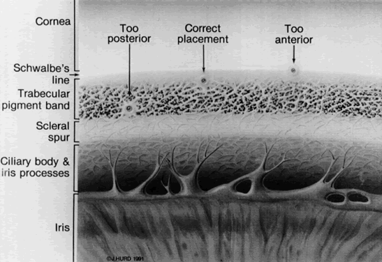
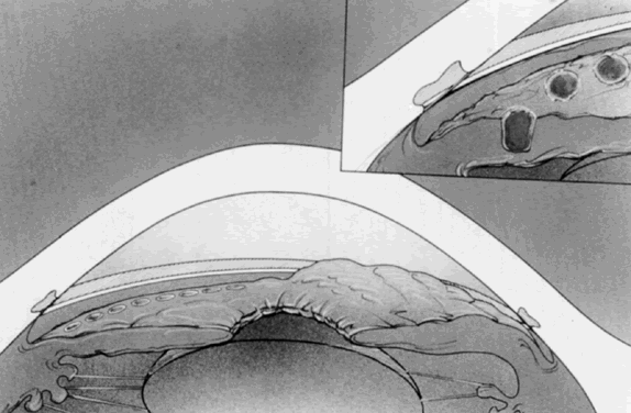
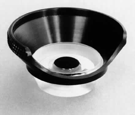
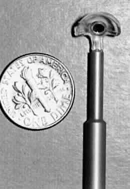
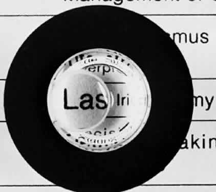
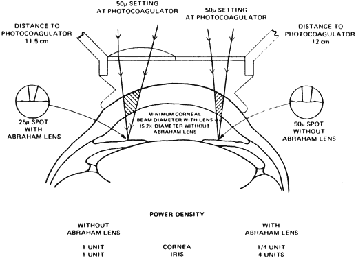
The use of an antireflection-coated lens with a strong plus button greatly facilitates the ability to penetrate the iris and has improved the success rate in achieving iridectomies. It is so important to the success of the procedure that its use is considered mandatory. The Abraham lens consists of a modified Goldmann-type fundus lens with an 8-mm hole trephined into its periphery (Fig. 1) and a 66-D plano convex button bonded into the trephine hole. The Wise lens has a 103-D button. All front surfaces are covered with an antireflective coating. The convex lens enlarges the laser beam at the corneal surface, thus reducing the chances of epithelial corneal changes and decreasing the laser beam diameter at the iris surface, which increases the power density (Fig. 2). The power density at the cornea is one fourth as great with the Abraham lens but is increased to four times as great at the iris surface, which facilitates penetration. The lens also holds the lids apart and allows the surgeon to control eye movements. The gonioscopic solution reduces heat buildup and decreases the incidence of corneal burns.

Fig. 1. Fundus lens with a high plus button eccentrically placed. The Abraham lens has a + 66 D button, and the Wise lens has a + 103 D button.

Fig. 2. Relative power density and spot size at the cornea and iris using an Abraham iridectomy lens. (Courtesy of R. Abraham)

The excellent optics of these lenses improves the visibility and magnifies the treatment site, thus reducing the depth of field and improving the precision of the laser focusing. All of these factors make these lenses an important adjunct to the successful production of iridectomies.

SURGICAL TECHNIQUE

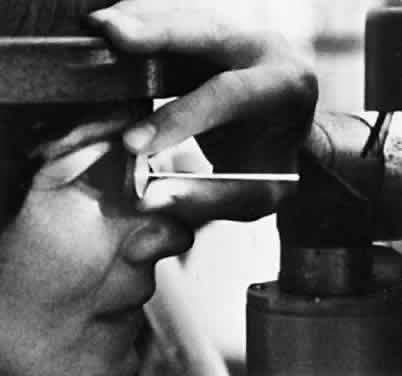
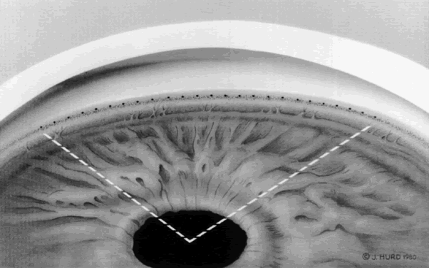
Both the surgeon and patient should be as comfortable as possible. A Velcro head strap helps to keep the patient well positioned with the forehead against the head bar. Patients tend to move backward, away from the slit lamp when the contact lens is placed on their cornea, and this makes precise focusing more difficult. An elbow rest is helpful to steady the surgeon's arm and reduce fatigue. A patient's shirt collar button may be released and tie loosened, and patients should be cautioned not to hold their breath. The laser room should be well ventilated to ensure patient comfort. A drop of topical anesthesia is used in all patients. Retrobulbar anesthesia is not necessary. The contact lens is placed on the eye with the trephined hole superiorly. The location of the iridectomy should be in the midperiphery of a constricted iris, about one third the distance from the limbus to the pupil, usually between the half past 10 and half past 1 o'clock positions (Fig. 3). By placing the iridectomy under the upper lid, the possibility of a postoperative problem with glare or diplopia is reduced. If temporal treatment is being performed, it is important to aim the argon laser beam away from the macula. The aiming beam should be perpendicular to the contact lens surface and through the center of the lens button.

Fig. 3. Superonasal location of iridectomy site at the half past 10 o'clock position in the left eye, about one third the distance from the limbus to the pupil.

Argon Iridectomy

Many authors recommend a variety of techniques to facilitate the performance of an argon iridectomy. Some of these include placing stretch burns in a circle (drumhead technique) and then placing penetrating burns in the center of the circle. Abraham and Miller recommend using stretch burns to cause a hump in the iris and then using penetrating burns to punch through the hump.9 The stretch burns contract the iris, but they are not used for penetration. Stretch burns usually are placed with a 200-μm spot size and a 0.2-second duration, using 200 to 400 mW of power.

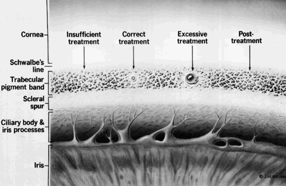
A well-accepted standard technique using the argon laser is to begin initial treatment with a 50-μm spot size and a 0.2-second duration, with about 850 mW of power. The aiming beam must be precisely focused on the surface of the iris to superimpose the burns. These two factors—precise focusing and superimposition—are the keys to success. Often, initial penetration can be achieved within 10 applications, and for any iridectomy, the tissue reaction to the initial burns often is an indicator as to the ease of the procedure. If bubble formation is seen at the site of the burn, this indicates stromal vaporization. The clinician then should focus at the base of the bubble, continuing to deliver energy until the stroma has been penetrated. This is helpful in the light blue iris.

The easiest irides to penetrate with the argon laser are hazel and light brown. The most difficult are light blue irides with minimal pigment and thick, dark brown irides. If good progress is not made at the initial treatment site, the clinician can move to another site. However, if charring is seen, which has the appearance of a black piece of coal, then switching to a chipping technique has been found to be helpful in dark brown irides. The duration is reduced to 0.02 seconds and the power increased to 1 to 1.5 W. This short-duration burn avoids charring at the base and reduces the chance of endothelial burns. However, it may take 100 to 300 applications to penetrate and complete the iridectomy using these short-duration burns. Since the development of the Nd:YAG laser, this has become less of a problem, and if good progress is not being made with the argon laser, it is prudent to switch to the Nd:YAG laser and punch through. In most cases, the Nd:YAG laser is the laser of initial choice.

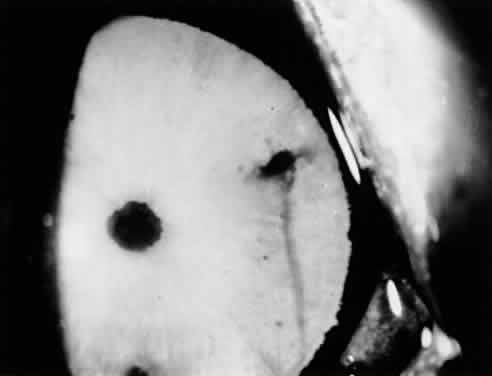
Light blue irides require a different technique. Blue eyes have little melanin pigment in the stroma, and a longer duration burn provides an increased thermal effect, which is necessary to get through both stroma and pigment epithelium in these lightly pigmented irides. A power setting of 800 to 1000 mW and duration of 0.2 to 0.5 seconds is used with a 50-μm spot size. Table 2 outlines the recommended treatment parameters for argon laser iridectomy. The clinician looks for an iris freckle or an iris crypt that would make penetration easier. The patient's eye usually will not stay still for the full 0.5 seconds, so it is necessary to be able to quickly cut off the laser energy before 0.5 seconds if eye movement is seen. A vaporization bubble sometimes is seen at these settings, and by aiming at the base of the bubble, it is often feasible to achieve penetration in two or three shots of 0.5 seconds' duration each. The signal that the laser beam has penetrated the pigment epithelium is a stream of pigment clumps carried by the posterior chamber aqueous humor into the anterior chamber in a mushroom-cloud configuration. Most of the argon laser treatment is directed toward enlarging the iridectomy and cleaning the pigment out from its edges. The edges of the initial penetration site are treated with laser energy to enlarge the actual opening and reduce the chance of subsequent closure from pigment proliferation. Care must be used because laser energy will be going through the iridectomy, possibly hitting the lens and retina.


TABLE 2. Treatment Parameters for Argon Laser Iridectomy


 Spot SizePowerDuration
Iris Color(μm)(mW)(s)
Hazel508500.2
Brown508500.2
Thick brown501000–15000.02–0.05
Blue5010000.2–0.5


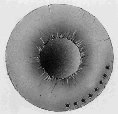
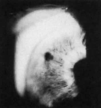
The endpoint of treatment is the direct visualization of the anterior lens capsule with the slit lamp. Transillumination is not a good technique to determine the patency of an iridectomy. There can be extensive transillumination, especially in blue eyes, whereas the iris stroma still may be intact with no through-and-through iridectomy (Fig. 4). After the iridectomy, the central anterior chamber depth usually is not affected; however, deepening of the peripheral anterior chamber is a reliable sign that the iridectomy has achieved its functional purpose.

Fig. 4. A. Extensive transillumination defect in blue iris nasally. B. Iris of same patient with iridectomy much smaller than the transillumination defect because of intact iris stroma.

In eyes with light blue irides, the Nd:YAG laser has distinct advantages and has been a major improvement.

Nd:YAG Iridectomy

Recommended settings for the Nd:YAG laser range from 3 to 7 mJ per burst and from one to three pulses per burst. It is safest to begin with a single pulse of approximately 4 to 6 mJ. As with argon iridectomy, the importance of precise focusing on the anterior stroma cannot be overemphasized. This produces maximum photodisruption and minimizes the possibility of lens injury.

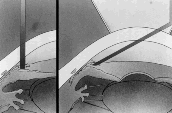
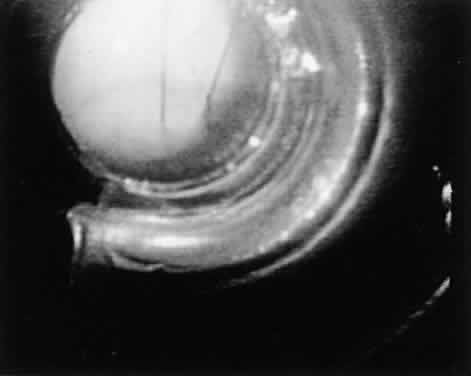
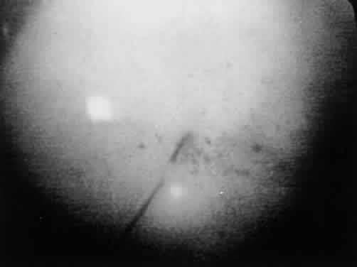
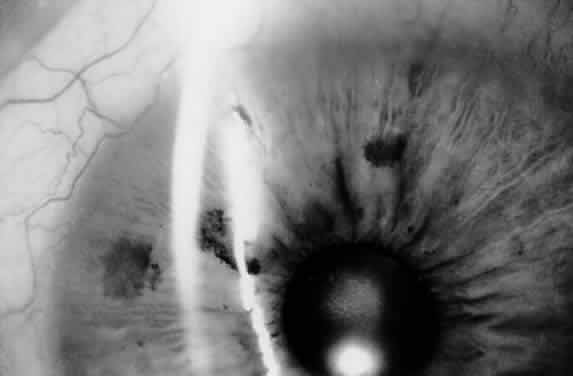
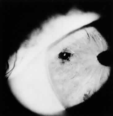
Eyes with thick brown irides are more difficult to penetrate. Sometimes, the iris stroma shreds without a through-and-through opening achieved. It can be helpful to thin out a cryptlike area with the argon laser and then punch through with the Nd:YAG laser energy. In a few cases, the reverse procedure is followed: the Nd:YAG laser is initially used, and the iridectomy opening can be completed with argon laser energy. In thick brown irides, using two to three pulses per burst often is helpful. The iridectomy typically appears slitlike (Fig. 5). In patients who are on anticoagulant therapy or have bleeding disorders, it is advisable to use argon laser energy. Bleeding from the iridectomy site is rare with the argon laser because the tissue destruction involves thermal coagulation. Bleeding at the iridectomy site is common with the use of Nd:YAG laser energy because the photodisruption process (Fig. 6) does not coagulate the iris vessels. Moster and colleagues report bleeding from the iridectomy site in 34% of eyes after Nd:YAG iridectomy but in no eyes after argon laser iridectomy.14

Fig. 5. The Nd:YAG laser iridectomy typically appears slitlike.

Fig. 6. Bleeding from a a patent Nd:YAG iridectomy site. (Pollack IP, Robin AL, Dragon DM et al: Use of the neodymium:YAG laser to create iridotomies in monkeys and humans. Trans Am Ophthalmol Soc 82:307, 1984)

POSTOPERATIVE TREATMENT

The major postoperative treatment involves monitoring and controlling IOP and the expected mild postoperative inflammation. Moster and colleagues report that 76% of patients had a maximal elevation of IOP in the first hour, whereas 16% peak at the second hour, and 8% reached maximal elevation during the third hour.14 Thirty-one percent of the Nd:YAG laser-treated eyes and 34% of the argon laser-treated eyes had an IOP rise greater than 8 mmHg above baseline. Therefore, we routinely pretreat all eyes with apraclonidine and monitor IOP after a laser iridectomy procedure. If there has been a pressure spike, the patient is seen the next day; otherwise, the patient is seen between 5 and 7 days later, at which time the second eye can undergo laser treatment if necessary.

Methods to reduce the incidence and magnitude of a postlaser iridectomy pressure spike include pretreatment with apraclonidine and miotics if they have not been used in the last 2 hours, instilling a beta blocker if not contraindicated, and considering the use of a topical carbonic anhydrase inhibitor if there is significant cupping and field loss. Robin and associates demonstrated that topical paraaminoclonidine (apraclonidine hydrochloride; Iopidine, Alcon) was effective in reducing significant pressure spikes after argon laser iridectomy.16 Forty-three percent of eyes treated with placebo and no eyes treated with apraclonidine had an IOP rise more than 10 mmHg above baseline. By reducing the incidence and magnitude of the pressure spike, there is less chance for further glaucomatous damage. No significant differences in postoperative IOP spikes have been found between the two types of lasers.

The IOP elevation may be related to the release of pigment and inflammatory debris clogging the trabecular meshwork. In eyes with poorly controlled IOP or eyes that have severely compromised outflow facilities, IOP elevations are more likely to be seen. By monitoring the IOP postoperatively if there is a significant elevation, treatment with pressure-lowering agents can be carried out. Tonographic studies done in monkeys at varying intervals after laser iridectomy have not shown decreases in outflow facility, which indicates the pigment liberated does not permanently clog the trabecular meshwork.17 Topical steroids are used to control iritis, which is seen in all patients. The usual treatment schedule is one drop of prednisolone acetate four times a day, and this usually can be discontinued by 1 week postoperatively. Miotics are continued if the patient has been on them preoperatively. Miotics also are used if there is a question about the patency of the iridectomy.

If there is a patent iridectomy, the pupil is dilated on an early postoperative visit to prevent the formation of posterior synechiae and to visualize the posterior pole and peripheral retina, which might not have been possible before. Also, dilating the pupil postoperatively serves as a mydriatic provocative test to rule out the possibility of a plateau-iris configuration. Gonioscopy always should be done on the first postoperative visit. Gonioscopy is important to assess the effect of the iridectomy on the angle configuration and to determine the extent of permanent synechiae, if present. The patient is observed over the next 6 weeks to watch for pigment proliferation leading to closure of the iridectomy. If closure occurs, the iris is retreated to open the iridectomy and enlarge it.

COMPLICATIONS OF SURGERY

Corneal Changes

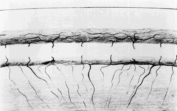
Corneal changes include epithelial and endothelial burns. They usually are transient but can impair the ability to complete an iridectomy because the laser energy cannot be delivered accurately and effectively. It helps to position the eye and aim through a noninvolved portion of the cornea. It may be necessary to go to another location if visibility is not adequate. Endothelial burns are caused by the thermal effects of treating the iris, especially in eyes with shallow chambers. They usually resolve within a few weeks but can cause focal endothelial cell loss. Investigators have not been able to demonstrate statistically significant endothelial cell loss during follow-up periods of 1 year.18,19 Nd:YAG iridectomies can cause focal endothelial cell loss if the disruption takes place less than 1 mm from the corneal endothelium.20

Schwartz and coworkers report on five eyes of three patients who developed corneal decompensation after undergoing argon laser iridectomy for angle-closure glaucoma.21 They found that a history of preexisting corneal guttata, episodes of angle-closure glaucoma with elevated IOP and inflammation, and the need for repeat treatments all seemed to predispose to this rare complication.

Lens Opacities

Focal anterior subcapsular lens opacities at the iridectomy site frequently are seen with argon laser iridectomies and also can be found after Nd:YAG iridectomy (Fig. 7). Long-term follow-up demonstrates that the opacities are nonprogressive and the visual loss from cataracts in patients who have undergone laser iridectomies is similar to an age-matched control group.22 Breaks in the anterior lens capsule in eyes undergoing Nd:YAG iridectomy have been demonstrated histopathologically in patients subsequently undergoing intracapsular cataract extraction.23 However, the widespread concern that the Nd:YAG laser may cause the rapid development of cataracts because of inadvertent rupture of the anterior lens capsule fortunately has not been shown. No differences in cataract formation were demonstrated in rabbit eyes when comparing argon and Nd:YAG laser iridectomies.24

Fig. 7. Focal anterior lens capsule opacity visible through iridectomy opening.

Treatment parameters of one or two bursts and less than 6 mJ reduce the incidence of lens opacities in monkeys.25 It also is critical to focus on the anterior iris stroma and to perform the YAG iridectomy peripherally, where the patient's convex lens is further away from the pigmented epithelium of the iris. If a capsule break should occur, animal studies show that fibrous proliferation covers the anterior capsule break.25

Closure of the Iridectomy

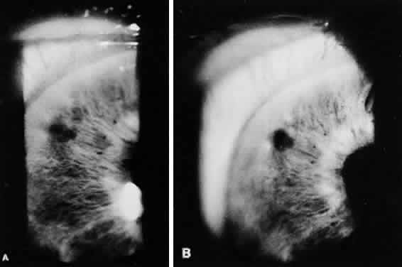
Closure of a patent laser iridectomy may be either immediate or delayed. It is important to recheck the iridectomy site at the same time that the patient's IOP is monitored postoperatively because the iridectomy site may be occluded by the landsliding of pigment epithelium into it from its edges. The opening can be enlarged, if necessary, with a few more applications of laser energy. If late closure occurs, it usually occurs by 6 weeks and is a result of pigment proliferation. Performing a large, clean initial iridectomy reduces the likelihood of an iridectomy closing and the need for retreatment. Blue irides treated with the argon laser had a 35% incidence of retreatment in our series of over 200 eyes treated between 1977 and 1982, whereas brown irides had only a 15% retreatment rate. The blue eyes had the greater need for retreatment because of the difficulty of obtaining a large iridectomy and tendency for pigment proliferation to fill in the iridectomy (Fig. 8). One of the major advantages of the Nd:YAG laser is its much reduced incidence of iridectomy closure. With photodisruption, there is not the same tendency for iris pigment proliferation, and this is a distinct advantage.

Fig. 8. A. Iridectomy site has been closed by pigment proliferation. B. Iridectomy reopened after a second laser treatment.

Diplopia and Glare

Occasionally, patients may complain of an extra image or a light reflex from the iridectomy site. By placing the iridectomy under the upper lid, these complications are significantly reduced. However, some iridectomies have been placed inferiorly with no problems. If the iridectomy is located right at the border of the upper lid margin, these symptoms seem more likely to occur.

ADVANTAGES

It is no longer necessary to compare the advantages of laser iridectomy with surgical iridectomy. Laser iridectomy has revolutionized the management of angle-closure glaucoma. It has unburdened both the patient and the ophthalmologist. In the past, one of the hardest tasks was to convince a patient who had just experienced an acute congestive attack of angle-closure glaucoma to undergo an intraocular surgical procedure in a nonsymptomatic “healthy eye” while the acute eye was quieting down.

Another difficult scenario was that of the patient with creeping angle-closure glaucoma who was totally asymptomatic and needed bilateral surgical iridectomies. In patients with combined-mechanism glaucoma, a laser iridectomy can be performed to determine if it alone will control the IOP without having to subject the patient to two intraoperative procedures if filtering surgery was to be eventually necessary. Since laser iridectomies are relatively safe and simple to perform, it is a concern that the indications can be easily abused, and that patients who do not need an iridectomy will be offered it. This may be a result of an inaccurate gonioscopic examination, a subjective examination, or one not uniformly mastered. Laser iridectomy also may be unnecessarily performed if the mechanism of primary angle closure is not truly understood. It then might be performed in eyes with peripheral anterior synechiae that are secondary to uveitis, rubeosis, or iridocorneal endothelial syndrome, disorders caused by a nonpupillary block angle-closure mechanism.

DISADVANTAGES

Compared with surgical iridectomy, there are no disadvantages to laser iridectomy. However, it may not be possible to perform a laser iridectomy if there is significant corneal edema or a flat chamber. If penetration cannot be achieved, a surgical iridectomy then must be performed. With argon laser energy there is a risk of a macular burn, and with both types of laser treatment there is the risk of a postoperative IOP spike and possible visual field loss involving fixation. Laser surgery requires the use of costly equipment that must be well maintained, and this may become more of a problem unless the cost of laser instruments decreases to match changes in reimbursement, an unlikely event.

Back to Top
ARGON LASER TRABECULOPLASTY
During the last 15 years, argon laser trabeculoplasty (ALT) has become the primary initial surgical treatment for uncontrolled open-angle glaucoma unresponsive to medical therapy. It has experienced a worldwide acceptance and has revolutionized the management of the glaucoma patient. Since its popularization in the early 1980s, the indications for ALT have been continually refined, as have the modifications in the surgical technique to reduce complications. There also has been much learned about long-term results, the role of retreatment, and ALT's role as initial therapy for patients with newly diagnosed open-angle glaucoma.

HISTORY

Krasnov first reported laser treatment of the trabecular meshwork in 1973.26 He used a high-powered ruby laser to make microscopic punctures through the trabecular meshwork and into Schlemm's canal. The openings functioned for only a few months, and retreatment was required to maintain the effect, but initial excellent IOP lowering was reported. Worthen and Wickham were the first to report on treating part of the angle (90°) with argon laser energy.27 They reported a mean IOP reduction of 9.6 mmHg in 20 patients with limited follow-up. They subsequently reported long-term follow-up with 50% of the treated eyes having an IOP of less than 25 mmHg after 4 years of follow-up.28 They were appropriately cautious in their recommendations, partially because of the report by Gaasterland and Kupfer in 1974 on the experimental production of glaucoma in monkeys by heavy repeat treatment of the angle with argon laser energy.29 This report raised the possibility that laser therapy could aggravate rather than help glaucoma in some situations, and thus dampened the enthusiasm for this treatment modality. In 1979, Wise and Witter reported a greater than 90% success rate in argon laser-treated phakic eyes in open-angle glaucoma.30 Their technique differed from the report of Worthen and Wickham in that all 360° of the angle were treated. Schwartz and coworkers reported on 35 phakic eyes with clinically uncontrolled open-angle glaucoma that underwent 360° treatment to the trabecular meshwork.31 The IOP of the untreated eye was used as a control. The mean pressure change in the treated eye at 4 months was 10 mmHg, and this was maintained throughout the initial 18-month follow-up period. At the 1980 American Academy of Ophthalmology meeting, reports by Wise,32 Wilensky and Jampol,33 and Schwartz and others31 all confirmed the efficacy of 360° argon laser treatment to the trabecular meshwork.

INDICATIONS

Initially laser trabeculoplasty was reserved for end-stage glaucomatous eyes, patients who refused filtering surgery, previous surgical failures, or patients who were poor surgical risks. The experience that has been gained has radically altered the indications. Patients with open-angle glaucoma including exfoliation syndrome and pigmentary glaucoma are being offered ALT earlier and earlier in the therapeutic sequence. Patients are no longer being subjected to the side effects of miotics and carbonic anhydrase inhibitors. ALT is not recommended for patients with juvenile glaucoma or glaucoma secondary to uveitis or angle recession. It is unlikely to provide young patients with a lifetime cure, and a filtering procedure is a more appropriate initial surgical choice for these patients. Also, if patients have an IOP that is above 35 mmHg and requires a low target pressure, filtering surgery usually is recommended, since ALT is unlikely to lower IOP enough to reach the target pressure.

Before the advent of ALT, the side effects of medical therapies had to be endured because the major alternative was filtering surgery with its known complications. ALT is not offered to patients who have elevated IOP without any signs of damage to visual function (disc or field changes), or in patients whose IOP is well controlled with tolerated medical therapy. Whereas less than 1% of patients are made worse by ALT with an IOP higher than at baseline, it is not advisable to subject a patient with normal visual function to the risks of filtering surgery because of an untoward effect from premature ALT.

Several authors suggest that ALT should be used as a primary therapy for open-angle glaucoma. These and other results spurred the initiation of the Glaucoma Laser Trial (GLT),34–38 a multicentered randomized clinical trial involving 271 patients that was designed to assess the safety and efficacy of ALT as initial treatment for newly diagnosed open-angle glaucoma patients compared with treatment using topical medications. Each patient had one eye randomly assigned to initial treatment with ALT and the other eye assigned to therapy with timolol maleate 0.5%. At 2 years, the laser-treated eyes had a lower mean IOP than the medication treated eyes, and fewer laser-treated eyes required two or more medications to control IOP (P < 0.001). Forty-four percent of the laser-treated eyes were controlled by ALT alone. With the same follow-up period, 30% of the medication-treated eyes were controlled by timolol, and only 66% were controlled within the stepped medication regimen, whereas 89% of the eyes treated with ALT plus medications were controlled. Medication was initiated or changed for either eye according to the same step regimen if the IOP was not controlled.

The long-term results of the GLT and the GLT follow-up study38 showed that at 7 years, eyes initially treated with ALT had lower eye pressures and less visual field and optic nerve damage than eyes initially treated with topical medications. The conclusion of the study was that “initial treatment with argon laser trabeculoplasty was at least as efficacious as initial treatment with topical medications.”38

PREOPERATIVE PREPARATION

Patients are instructed to continue all glaucoma medications on their regular schedule. Patients are routinely pretreated with apraclonidine to prevent a significant IOP spike that could adversely affect visual function. Apraclonidine is an alpha-2 agonist that appears to work by decreasing aqueous production. Its effect is additive to beta blockers. Robin and associates compared apraclonidine to placebo in 73 eyes receiving 360° ALT.39 None of the eyes that received the apraclonidine had IOP elevation greater than 10 mmHg, whereas 18% of the placebo-treated eyes did. The eye should not be exposed to diagnostic procedures before the treatment because this can affect corneal clarity. It had been recommended that anticholinesterase medications be discontinued before ALT, but this is no longer done.

SURGICAL TECHNIQUE

Argon laser trabeculoplasty is performed as an outpatient procedure using topical anesthesia. The gonioscopic mirror of an antireflection-coated, flanged, three-mirror lens with a knurled ring is used routinely. Other lens choices include the Ritch trabeculoplasty lens. It is a rare patient whose small palpebral aperture requires a single-mirror goniolens. Before starting treatment, it is critical to identify the angle landmarks: Schwalbe's line, the trabecular meshwork, and the scleral spur. The treatment begins with the gonioscopic mirror at the 12 o'clock position viewing the inferior angle. The inferior angle tends to be the most open part and has the most pigmentation, making angle structures more clearly defined in this area. This facilitates identification of the angle landmarks and avoids treating the wrong part of the angle. Some patients have a high iris roll and a prominent Sampaolesi's line, and it is easy to mistake Sampaolesi's line for the pigmented trabecular meshwork and treat the wrong part of the angle.

Occasionally, the initial plan may be to perform an ALT, but after more careful study of the angle, areas of peripheral anterior synechiae may be recognized or the approach may be so narrow that it may be concluded that treatment of the trabecular meshwork would be difficult. In these eyes, a laser iridectomy is performed initially to treat the narrow angle component or to facilitate future ALT. It is usually preferred to assess the effect of the iridotomy on IOP and angle configuration before later proceeding with ALT, but some glaucomatologists prefer to perform both at the same time. If only a small area of the angle is obscured because of a high iris roll, a gonioplasty can be performed.40 Treatment is directed through a goniolens at the peripheral hump of the iris, causing the iris to retract and opening the approach to the trabecular meshwork.

Typical treatment parameters for gonioplasty are 400 mW of power, 0.2-second duration, and a 100- to 200-μm spot size (see Argon Laser Gonioplasty discussed later). The purpose of the treatment is not to penetrate the iris but to cause the peripheral iris to contract. If more than 1 hour or 5 to 10 laser burns are required to open the angle, it is preferable to perform an iridectomy and reschedule the ALT. It is not recommended to perform ALT immediately after the laser iridectomy because of increased postoperative inflammation, corneal clarity changes, and possible greater problems with postlaser IOP elevations.

The use of a fixation light for the contralateral eye assists the patient in maintaining the appropriate eye position. Before the initiation of treatment, the eyepieces are focused and the argon laser is set to the standard treatment parameters: a spot size of 50 μm, a duration of 0.1 seconds, and an average power of 800 mW (range 600 to 950 mW). It is critical to check these parameters because lasers in a hospital or clinic setting are used by multiple ophthalmologists for a variety of intraocular procedures. During the treatment, the power setting required to blanch the trabecular meshwork or cause a small bubble formation is empirically determined. It is helpful to use a standardized treatment technique. This involves starting with the mirror at the 12 o'clock position and rotating it in a clockwise manner. Since the mid-1980s, many ophthalmologists initially treated only 180° of the angle. In the right eye, this always would be the temporal 180°; in the left eye, the nasal 180°. A continuouswave argon laser blue-green light is routinely used (Fig. 9), although diode laser has been reported to be equally effective.41–43

Fig. 9. A continuous-wave of argon laser energy is delivered to the trabecular meshwork. (Courtesy of I. Pollack)

The laser burns are placed at the anterior border of the pigmented trabecular meshwork (Fig. 10). Thomas and colleagues have shown that the more posterior the treatment, the greater the incidence of IOP elevation and formation of posterior synechiae.44 A power setting that produces blanching or depigmentation at the point of impact with a slight vaporization bubble is used (Fig. 11). Some investigators do not think it is necessary to gauge the tissue reaction and use a standardized treatment parameter of 800 mW in nearly all patients. We usually treat 180° at the initial setting, although 360° can be treated if clinically indicated. The spacing of the laser burns is the same whether 180° or 360° are treated, that is, 20 to 25 burns per 90° of angle treated, regardless of the area treated (Fig. 12). Previously, the laser beam was aimed at the center of the pigmented trabecular meshwork, but now it is aimed at the anterior border of the pigmented trabecular meshwork. Histologically, this corresponds to the junction between the anterior nonpigmented and posterior pigmented trabecular meshwork. This location is preferred because posterior laser treatment caused a higher incidence of IOP elevation in the immediate postoperative period. Traverso and coworkers report the development of peripheral anterior synechiae in 43% of eyes that were treated posteriorly but only in 12% of eyes treated anteriorly.45 Posteriorly placed burns also are more likely to cause patient discomfort. The procedure usually is well tolerated by patients as long as the iris and scleral spur are not struck with the laser beam energy.

Fig. 10. Optimal treatment location is at the anterior border of the pigmented trabecular meshwork.

Fig. 11. The range of treatment response to light, optimal, and heavy argon laser treatment of the trabecular meshwork. The posttreatment appearance of the burn site is shown.

Fig. 12. A 90° angle view with 25 evenly spaced laser burns in the trabecular meshwork.

Precise focusing of the aiming beam on the anterior border of the pigmented trabecular meshwork is critical. The aiming beam should be the smallest and roundest target possible. This can be achieved by mechanically directing the laser beam forward and backward. If the treating contact lens is tilted and not perpendicular to the eye, the aiming beam will appear oval. Repositioning the lens on the eye and making sure that the aiming beam is perpendicular to the lens ensures that the full power density of the treatment will be achieved. If the trabecular meshwork cannot be seen easily, having the patient move the eye in the direction of the gonioscopic mirror often opens the angle and improves visibility.

180° Versus 360° Treatment

There is a difference of opinion whether 180° or 360° of the trabecular meshwork should be treated in the initial treatment session. Some believe that by treating 360° initially, the patient's IOP is lowered maximally while completing the treatment in one session. Others have found that 180° of treatment often provides adequate IOP lowering for several years after treatment. Then the option is available of treating the second 180° at a later time if the initial ALT wears off. This is particularly important in patients with exfoliation syndrome, since exfoliation syndrome has an excellent initial response46–48 that tends to wear off 2 to 4 years after treatment. Also, if an IOP spike occurs, the magnitude of pressure rise is likely to be less than if 360° had been treated. In a typical patient, we treat 180° and wait 4 weeks before deciding whether to treat the second half. If a significant clinical response (i.e., at least a 25% lowering of IOP in patients with baselines above 20 mmHg or 20% IOP lowering in those with baselines less than 20 mmHg) is not achieved, then the second half of the treatment is performed. Some surgeons prefer to treat between 220° and 270° initially using 70 to 80 spots. They believe that this is an adequate treatment that obviates the second treatment in many patients and at the same time is associated with a lower incidence of sustained IOP elevation. If the laser effect wears off, the option still is available of placing an additional 40 applications over the untreated area.

By dividing the treatment and moving it to the anterior border of the pigmented trabecular meshwork, the problem of IOP elevation sustained beyond 24 hours has been almost eliminated. With the changes in technique, it is rare to have a patient require filtering surgery in the immediate postoperative period after an untoward response from ALT. This result also may be partly caused by the evolving selection criteria, in which patients with juvenile glaucoma, uveitis, or angle recession are not offered argon ALT, since they would have been more likely to get an untoward response.

POSTOPERATIVE MANAGEMENT

The IOP is measured 1 and 2 hours after treatment. If the pressure rises 10 mmHg above baseline or higher than 35 mmHg and the patient already has been pretreated maximally, then an osmotic drug such as oral glycerin is administered. This is rarely required. If the IOP is satisfactory, the patient is instructed to continue all glaucoma medications as usual and begin a topical steroid drop four times a day for 4 to 5 days. If the patient has had a significant IOP elevation or has severe glaucomatous damage, the IOP is measured the next day. Most patients are reexamined 2 to 5 days later and then at about 4 weeks. If at 4 weeks the pretreatment goal has not been achieved, the second half of the angle can be treated.

INTRAOPERATIVE COMPLICATIONS

Most of the complications associated with ALT are mild and transient (Table 3). The most frequent serious complication, which occurs on the day of treatment, is an IOP elevation. Since some patients undergoing ALT may have severe cupping and field loss with fixation involved, they could lose central fixation after ALT, presumably from a severe IOP elevation.44,49 By limiting treatment to 180°, aiming at the anterior border of the pigmented trabecular meshwork, and pretreating aggressively, significant pressure elevations are rare and seldom last more than 24 hours. Weinreb and coworkers50,51 report that the incidence and magnitude of the postoperative IOP rise was significantly greater in eyes that had received 100 laser burns over 360° compared with eyes receiving 50 burns over 180°. Weinreb and colleagues randomized 40 patients into 180° or 360° treatment.51 They report that the maximal IOP rise after ALT was 1.3 (±4.9) mmHg in the 180° group compared with a change of 7.35 (±9.8) mmHg in the 360° group. The GLT Research Group52 reports the acute effects of ALT on IOP in 271 eyes that received ALT as an initial intervention in two sessions spaced 4 weeks apart. Thirty-four percent of patients had a pressure rise of 5 mmHg or greater above baseline after one or both treatment sessions. Twelve percent of eyes had an increase of 10 mmHg or more. However, none of these eyes were on medications or were pretreated to prevent an IOP rise.


TABLE 3. Complications During and After Argon

  Laser Trabeculoplasty
  Pressure elevation
  Corneal epithelial opacities
  Bleeding
  Iritis
  Incorrect part of angle treated
  Fainting
  Peripheral anterior synechiae formation
  ? Encapsulation of subsequent filtering blebs


Corneal burns can occur during laser treatment, but these usually are transient and self-limiting. Inadvertent treatment of the iris, bleeding from punctures into Schlemm's canal, and a hyphema as a result of treating iris root vessels have been observed.53 Iritis can occur after ALT, which occasionally can be prolonged and severe.

Some patients experience a vasovagal reaction secondary to the ocular cardiac reflex and anxiety during the treatment. It may be necessary to abort ALT and lay the patient down with the feet elevated. It usually is possible to complete the treatment when this problem resolves.

LONG-TERM COMPLICATIONS

Long-term complications include the development of peripheral anterior synechiae.34 The incidence of peripheral anterior synechiae after ALT has been reported to be as high as 46% in the GLT. Schwartz and coworkers31 were among the first to report the complication of peripheral anterior synechiae after ALT. They found peripheral anterior synechiae in 29% of the 35 eyes in their initial report. The synechiae usually are small and localized (Fig. 13). Factors that appear to predispose to peripheral anterior synechiae formation include high power levels and posterior placement of the burns in narrow-angle eyes. Traverso and others report that peripheral anterior synechiae developed in only 12% of eyes treated on the anterior trabecular meshwork compared with 43% of those eyes that had treatment applied to the posterior meshwork.45 Treatment to the anterior border of the pigmented meshwork is strongly recommended because of this finding.

Fig. 13. Typical pattern of peripheral anterior synechiae produced by argon laser trabeculoplasty.

There is a concern that ALT might adversely affect the success rate of future intraocular filtration surgery. Richter and associates found an increased incidence of encapsulated filtering blebs in eyes that had undergone ALT.54 The Advanced Glaucoma Intervention Study has found no such association.55

LONG-TERM RESULTS

The Advanced Glaucoma Intervention Study55–59 is a National Eye Institute-sponsored study of patients with advanced field loss with uncontrolled IOP despite maximum tolerated medical therapy. In this 11-center study, patients were randomized prospectively to (1) ALT followed by trabeculectomy if needed, or (2) trabeculectomy followed by ALT if needed. Automated perimetry is being used to provide information about long-term effects on visual field after medical therapy failure. The study is designed not only to provide the ophthalmic community with long-term follow-up information about which procedure is more efficacious, but also to provide a wealth of knowledge about patients with advanced glaucoma. With 7 years of follow-up, the Advanced Glaucoma Intervention Study59 found better preservation of visual function in blacks on maximum tolerated medical treatment undergoing ALT before trabeculectomy. White patients did better if they had trabeculectomy before ALT. Retrospective long-term results reported by Schwartz and associates showed that blacks did not do as well as whites with IOP control after ALT.60 Many investigators have found similar results with filtering surgery in blacks.

Long-term results demonstrate that the efficacy of ALT depends on the clinical characteristics of the patient and the type of glaucoma treated. In general, older white patients with primary open-angle glaucoma have the best long-term results. Patients with exfoliation have the highest incidence of initial success, more than 80%, but the IOP lowering effect tends to diminish between 1.5 to 4 years postoperatively. Patients with pigmentary glaucoma have an intermediate success rate. Patients with diagnoses of glaucoma secondary to uveitis, angle recession, and juvenile glaucoma uniformly have done poorly. An ALT is of little value in these groups. Aphakic eyes with vitreous in the anterior chamber have less than a 50% incidence of success, but pseudophakic eyes with primary open-angle glaucoma and intact posterior capsules seem to do almost as well as phakic eyes.

Long-term results with ALT show less of an IOP lowering effect with time (Table 4). Schwartz and associates report a 46% incidence of success after 5 years,61 and Shingleton and colleagues found similar results.62 Wise reports that in a group of 110 eyes, 56% had an IOP of less than 21 mmHg after 10 years.63 He also found that eyes with advanced damage having cup-disc ratios greater than 0.9 at the time of ALT had a 51% incidence of later needing glaucoma surgery.


TABLE 4. Mean Changes in Pressure (mmHg) From Baseline in Eyes Treated with Argon Laser Trabeculoplasty


    IOP
TimeNo. ofMeanPReduction
(mo)Eyes(mmHg)Value(%)
273-9.7< 0.0001-38.4
678-8.9< 0.0001-35.3
1266-7.8< 0.0001-31.0
2431-7.3< 0.0001-29.8
3642-7.0< 0.0001-28.1
4019-6.8< 0.0001-28.8
6013-4.9< 0.0391-19.7

IOP, intraocular pressure.


The type of clinical response in one eye is highly predictive of what will occur in the fellow eye. If a patient does not respond to ALT in one eye, it is unlikely that a response to the procedure will occur in the fellow eye. If the IOP is above 35 mmHg in an eye with significant glaucoma damage, ALT rarely lowers the pressure enough to protect the eye from further damage. A good IOP lowering response to ALT is about a 20% to 25% decrease from baseline. It is unlikely that ALT will eliminate the need for medications in patients whose glaucoma requires multiple medications. Pollack and associates report that 82% of patients with primary open-angle glaucoma still required some of their medications.64

If the patient has demonstrated an inability to comply with medical therapy, it may be advisable to proceed directly to filtering surgery, since more than one third of patients are able to eliminate all medications after filtering surgery. Patients who disappear from follow-up care and fail to be monitored may have significant damage if the IOP lowering response to ALT wears off.

ADVANTAGES

A benefit of ALT includes the possibility of eliminating some medications. Improved compliance may result. Improved vision might be obtained if the patient's vision was being blurred by the use of a miotic. The procedure also is particularly useful in a patient with early cataract because it obviates filtering surgery with its known tendency to promote cataract progression. ALT is uniquely advantageous in a patient wearing contact lenses in whom a filtering bleb would pose a potential fitting problem and increase the risk of endophthalmitis.

DISADVANTAGES

Long-term follow-up in multiple series has shown a decreasing IOP lowering effect with time. During this period of observation, patients may be undergoing further progressive glaucoma damage before it is recognized that ALT has failed. Also, the amount of IOP lowering is less than that with filtering surgery, and in some patients it may not be enough to prevent progression of disease. A small group of patients, less than 1%, may be harmed by ALT, with an increase in IOP above baseline, and if criteria for patient selection are expanded to offer ALT to any patient with elevated pressures, some patients may be forced to undergo filtering surgery even when they have not had any signs of ocular damage. Patients still require and must comply with most medical therapy for their glaucoma after ALT.

MECHANISM OF ACTION

Much work has been done to elucidate the mechanism of action of ALT. Tonographic measurements found an improvement in aqueous outflow after ALT. Schwartz and coworkers report that the mean outflow facility in treated eyes increased from 0.10 to 0.23 μl/minute 2 months after treatment, whereas the untreated eye had no change in its outflow facility.31 It was initially thought that ALT worked by creating microperforations from the anterior chamber into Schlemm's canal. Wise and Witter suggest that the improvement in IOP may result from tissue contraction at the burn site pulling open the intratrabecular spaces.30 Van Buskirk suggests that the laser may change the biologic makeup of the trabecular endothelium, leading to a molecular chain of events within the meshwork that may improve aqueous outflow. If this is true, the improvement in IOP control after ALT may be nonspecific for laser characteristics, including waveform, burn location, number of hours treated, and laser power used.65 The trabecular cells are known to become fewer with age, and this depopulation of the trabecular meshwork may be accelerated in eyes with open-angle glaucoma.66–68 Another proposed mechanism of action of ALT is that the laser energy, when applied to the trabecular cells, may stimulate cell division.69

The exact mechanism by which ALT lowers IOP still is not known. The laser energy may have multiple effects on the trabecular tissue, and there may be both a mechanical effect from scar formation at the burn sites and biologic changes in the trabecular endothelial cells. In addition, the focal tissue destruction at the burn sites may diffusely stimulate trabecular tissue division. These issues are so important because a better understanding of the mechanism of action of ALT may lead to an optimization of the treatment parameters in the future.

RETREATMENT

Long-term follow-up in most series shows a decrease in IOP lowering effect with time. The question is whether repeat treatment will again improve outflow and provide a pressure-lowering effect for a reasonable follow-up period. Retreatment is defined as additional laser applications to eyes that have already had 360° laser treatment with a minimum of 80 applications in one or two treatment sessions. In 1982, Schwartz and Kopelman reported on six patients who were retreated after initial 360° treatment.70 None of these patients achieved long-term IOP control. Richter and associates performing a 180° retreatment achieved only a 14% success rate 21 months after retreatment.71 Brown and coworkers report that 38% of retreated patients had successful IOP control 5 months after retreatment.72 However, 12% of the retreated eyes were made worse after repeat ALT, requiring emergency filtration surgery. Starita and associates report that 53% of their 17 retreated eyes had a reduction in IOP, but again 12% of the patients had a high IOP 12 weeks after treatment.73 In view of these reports, retreatment is seldom offered to patients who have had treatment to 360° of the trabecular meshwork. A major concern is the 12% incidence of significant, sustained IOP elevation after retreatment. Since the short-term complication rate is comparable with the long-term success rate, the therapeutic index does not favor retreatment in most cases.

The only patients in whom retreatment is considered are elderly patients with exfoliation syndrome and patients with primary open-angle glaucoma who had an excellent and sustained initial response and are poor candidates for filtering surgery. The goal in these patients is to control IOP for an additional 2 to 18 months. Retreatment may be a better alternative than allowing the IOP to be elevated if a trabeculectomy cannot be performed.

Back to Top
OTHER LASER SURGERIES IN THE MANAGEMENT OF GLAUCOMA

LASER SURGERY IN NEOVASCULAR GLAUCOMA

Laser therapy has a major role in the management of neovascular glaucoma. Neovascular changes in the anterior segment may occur with diabetes mellitus, central retinal vein occlusion, other vascular insufficiencies, uveitis, and after vitrectomy. Ischemia is common to most forms of rubeosis, presumably stimulating production of a vasoproliferative factor, which stimulates the production of new vessels. These new vessels first appear at the pupillary border and then grow onto the surface of the iris and into the angle. A fibrovascular membrane develops and pulls the iris with it against the trabecular meshwork, causing a secondary angle closure. An iridectomy is not helpful in this instance because the mechanism of closure is not pupillary block. The keys to making the diagnosis of rubeosis in high-risk patients are a careful slit-lamp examination and frequent gonioscopic examination. The major factors that determine the treatment used in patients with neovascular glaucoma are the visual potential including visual acuity, the status of the angle, and the clarity of the ocular media.

Pan-Retinal Photocoagulation

The major goal of treatment in neovascular glaucoma is to eliminate the production of the vasoproliferative factor. Pan-retinal photocoagulation using argon laser energy has been shown to cause regression of the anterior segment neovascularization, presumably by destroying ischemic retina, the source of the vasoproliferative factor.74,75 The techniques and complications of panretinal photocoagulation are fully detailed elsewhere.

Goniophotocoagulation

Simmons and associates report that laser applications to vessels in the angle and on the iris surface could obliterate these new vessels and prevent synechiae formation.76,77 They named this procedure goniophotocoagulation. Its basic premise is to treat the angle neovascularization before there is synechial closure to prevent the development of neovascular glaucoma. In cases of active rubeosis with open angles, goniophotocoagulation should be combined with panretinal photocoagulation, if possible. It usually takes between 5 and 10 days before regression of the anterior segment neovascularization is seen after panretinal photocoagulation, and during that interval, it is possible that an open angle may become closed by synechiae. More than one treatment of goniophotocoagulation may be required to control active angle vascularization during this waiting phase. Patients with opaque media but good visual potential may benefit from panretinal cryo applications.

SURGICAL TECHNIQUE. The technique of laser goniophotocoagulation is similar to that of ALT. Topical anesthesia is used, and the energy is delivered through the gonioscopic mirror of a three-mirror Goldmann contact lens. Corneal clarity is important to be able to see the fine vessels in the angle. If the IOP is elevated, causing corneal edema, it should be lowered, and the eye should be pretreated with topical glycerin. The vessels are treated as they cross the scleral spur (Fig. 14). The endpoint of treatment is localized blanching of the treated vessel at the site of laser application. Typical treatment parameters are 0.10-second duration, 100-μm spot size, and power settings of 200 mW. These settings may have to be continuously modified during the procedure for maximal effectiveness.

Fig. 14. Obliteration of new vessels in the anterior chamber angle immediately after treatment. (Courtesy of R. Simmons)

Treating vessels on the trabecular meshwork should be avoided because repeated treatment could cause significant trabecular scarring, decreased outflow, and increased IOP. Multiple treatments often are required. Once the angle is closed by synechiae, laser goniophotocoagulation is useless. Panretinal photocoagulation and direct photocoagulation of the iris vessels still may be helpful. These procedures reduce the vascularity of the eye, thus minimizing the risk of intraocular bleeding and postoperative inflammation and scarring during subsequent filtering surgery, if it is necessary. By treating the iris surface in the area of the future surgical iridectomy, the risk of intraoperative bleeding can be reduced.

COMPLICATIONS OF TREATMENT. Goniophotocoagulation is a relatively benign treatment. If bleeding occurs, the vessel is sealed by increasing pressure with the goniolens or by treating the bleeding point with a larger spot size. Other complications include transient peripheral corneal edema and pain at the base of the iris, if it is treated. Postoperative topical steroids and cycloplegics are used, and IOP elevations are treated with the appropriate agents, avoiding miotics.

ARGON LASER GONIOPLASTY

Hager first described argon laser gonioplasty, also known as laser iris retraction, laser iridoplasty, and laser goniosynechialysis, in 1973.78 He reports the use of the laser for tangential coagulation of the iris base to deepen a narrow angle. In this procedure, argon laser burns are applied to the peripheral iris stroma to retract the iris and open the anterior chamber angle (Fig. 15).

Fig. 15. Partial-thickness stromal burns retract peripheral iris away from the angle. (Courtesy of Bradford J. Shingleton)

Indicationsm

ANGLE-CLOSURE GLAUCOMA. In cases of angle-closure glaucoma that cannot be treated adequately by medication and laser iridectomy, argon laser gonioplasty can open the angle by pulling the peripheral iris away from the meshwork. This is an especially useful application when corneal haze or anterior chamber inflammation interfere with attempts at laser iridectomy.79–81 If angle closure has persisted long enough to form peripheral anterior synechiae, the likelihood of success with this technique is severely diminished.

PLATEAU IRIS SYNDROME. In patients with plateau iris syndrome, the peripheral iris is apposed to the angle. To diagnose this abnormality, a laser iridectomy must first eliminate any possible pupillary block component. The diagnosis of plateau iris is made if angle closure is induced by dilation in the presence of a patent iridectomy. Argon laser gonioplasty can deepen the angle in many of these patients.79,82

ANGLE-CLOSURE GLAUCOMA FROM POSTERIOR CHAMBER DISEASE. Anterior displacement of the lens or swelling of the lens may push the iris forward from behind, closing the angle. This can be treated with gonioplasty. Anterior displacement of the lens or swelling of the lens also may cause relative pupillary block, which does respond to iridectomy. Many cases require both an iridectomy and argon laser gonioplasty to treat both components of angle closure and adequately open the angle.

After scleral buckle placement, ciliary body edema may cause angle closure by a nonpupillary block mechanism. Argon laser gonioplasty can deepen the angle during the period necessary for the edema to subside.83 This is combined with medical therapy, including cycloplegics, topical and possibly systemic steroids, and aqueous suppressants.

TO FACILITATE ARGON LASER TRABECULOPLASTY. In eyes that require ALT but have angles with focal narrowing caused by iris cysts or peripheral iris rolls, gonioplasty can be used to reveal angle structures and flatten the peripheral iris configuration to allow trabeculoplasty. (See Argon Laser Trabeculoplasty for full discussion.)

NANOPHTHALMOS. Nanophthalmic eyes are small with thick sclera, compromised venous outflow, hyperopia, short axial length, small corneal diameters, and general crowding of the anterior chamber angle. Argon laser gonioplasty can deepen the angle in a nanophthalmic eye, which often has persistent angle closure despite a patent laser iridectomy.84,85 Nanophthalmic eyes are at particular risk of developing choroidal effusions or hemorrhage during intraocular procedures because the thickened sclera is thought to impede vortex vein drainage.

Surgical Technique

Pilocarpine 4% is instilled 1 hour preoperatively. Topical anesthesia is instilled immediately before the procedure.

Laser application can be made directly through the cornea with or without an Abraham lens or tangentially through a Goldmann gonioscopy lens (Fig. 16). Direct burns pass through the peripheral cornea; if a dense arcus is present, a goniolens allows the beam to pass through the cornea several millimeters central to the periphery while still achieving coagulation of the peripheral iris tissue. The goniolens allows the ophthalmologist to visualize improvement in angle configuration during the procedure. Use of a goniolens requires higher laser power, since the beam passes through the goniolens and strikes the iris tangentially, diffusing the burn. Leaving two aiming beam diameters untreated between laser applications and avoiding treatment of large visible radial vessels has been suggested to avoid necrosis.

Fig. 16. Laser application can be made directly through the cornea or tangentially through a Goldmann gonioscopy lens. (Courtesy of Bradford J. Shingleton)

Suggested laser settings with the Abraham lens are 500-μm spot size, 0.5-second duration, and 200 to 400 mW of power. With this lens, the beam is directed to the most peripheral iris visible. Suggested laser settings with the Goldmann goniolens are 100- to 200-μm spot size, 0.2- to 0.5-second duration, and 400 to 700 mW of power. A wide range of laser settings is expected given the variability of laser energy absorption associated with different iris pigmentation. Approximately 25 to 35 spots are placed over 360° (Fig. 17). If this is insufficient, an additional 20 to 30 spots can be performed several days later after any inflammation has resolved. Some ophthalmologists divide the treatment into two sessions of 180° each.

Fig. 17. Laser burns are applied to the peripheral iris. (Courtesy of Bradford J. Shingleton)

Postoperative management includes the use of topical steroids four times daily for 4 to 5 days. Frequent follow-up with tonometry and gonioscopy are necessary to determine success.

Complications

The most common complication of gonioplasty is mild iritis. This is treated with topical steroids. Corneal endothelial burns may occur in cases in which the anterior chamber is extremely shallow. These usually resolve over several days with no sequelae.

Patients must have regular follow-up because the duration of effect is variable. Retreatment may be successful in many cases. In most cases in which angle closure is secondary to other disease, the primary problem must be addressed.

Results

Weiss and associates report 18 months of follow-up in 32 eyes.86 Twenty eyes were successfully treated with more than 50% of the treated angle opened; 19 of these 20 eyes had an IOP of less than 22 mmHg. The 20 successes had a median duration of angle closure of 12 days before treatment, whereas the 12 failures had a median duration of angle closure of 90 days before treatment. Wand was able to open 60% to 100% of the synechial angle closure in six eyes of five patients, regardless of duration of angle closure before treatment.87

LASER CYCLOPHOTOCOAGULATION

Ciliary body destruction has been performed by a variety of modalities, including diathermy, ultrasound, cryotherapy, and laser. Significant complications of all modalities include postoperative inflammation, pain, decreased vision, and phthisis. Indications for cyclophotocoagulation generally are limited to patients whose glaucoma has been resistant to medical and surgical therapies. In some cases of neovascular glaucoma with poor visual potential, it is used as the primary procedure to control the IOP and alleviate pain. Advantages of laser cyclodestruction include the ability to more precisely focus treatment in a better defined area compared with other methods of cyclodestruction with reduced energy requirements and with reduced damage to surrounding tissue.

Argon Endolaser Cyclophotocoagulation

Argon endolaser cyclophotocoagulation allows treatment of the ciliary body processes with direct visualization and has the advantage of precise localization and quantification of treatment. It is done through the pars plana and requires full vitrectomy instrumentation, making it fall under the domain of the vitreoretinal surgeon. It cannot be done in a phakic eye unless a lensectomy is performed at the same time and, like other cyclodestructive procedures, may require multiple treatments. It also runs a small risk of retinal tears and retinal detachment, but in a certain limited group of patients it has been successfully used.88 Chen and colleagues report the results of 68 eyes of 68 patients treated for 180° to 360° and found that 61 eyes had an IOP of less than 22 mmHg at 12.9 months' mean follow-up.89

Transscleral Cyclophotocoagulation

Two delivery systems are widely used to apply transscleral laser cyclodestruction: (1) continuous-wave thermal mode Nd:YAG can be applied through air by a noncontact delivery system with the patient seated at the slit lamp (Lasag Microruptor 2, Lasag, Switzerland), or (2) a semiconductor diode laser can be applied with a hand-held fiberoptic G-probe by a contact delivery system with the patient in the supine position (Iris Medical, California).

Diode laser has a wavelength of 810 nm and therefore has lower scleral transmission compared with the Nd:YAG laser with a wavelength of 1064 nm. However, because diode is absorbed by melanin, the energy needed to perform ciliary ablation is less with the diode laser. Diode lasers have solid state construction, which allows them to be smaller, more durable, and portable compared with the Nd:YAG laser.

Retrobulbar or peribulbar anesthesia is used with both systems. Risks of transscleral cyclodestruction include the risks of any cyclodestructive procedure: pain, inflammation, scleral thinning, macular edema, hypotony, and phthisis. A narrow window exists between undertreatment with persistent elevation of IOP and overtreatment with resultant hypotony and phthisis. As with cyclocryotherapy, this window is approached by erring toward undertreatment by first applying a minimum effective amount of treatment, recognizing that some patients will require retreatment.

NONCONTACT ND:YAG TRANSSCLERAL CYCLODESTRUCTION. Hampton and Shields90 performed histopathologic studies on human autopsy eyes, building on the work by Fankhauser and colleagues.91 Using the Lasag Microruptor 2, they were able to optimally destroy the ciliary epithelium with an energy level of 8 J and with maximum defocusing 1 to 1.5 mm posterior to the limbus. Lesions were characterized by formation of a blister-like space. Klapper and associates evenly spaced 32 burns over 360° using an average power of 3.8 J at distances 2 to 3 mm from the limbus.92 They found that 26 of 30 patients (86%) had an IOP between 5 and 22 mmHg and a mean decrease in IOP of 65% with 6 months' follow-up. Six patients (20%) required one retreatment, and one patient (3%) required two retreatments. Trope and Ma report phthisis in 3 of 28 eyes (10.7%) and decreased vision in 8 of 28 eyes (30%) an average of 22 months after treatment.93 Another complication reported was focal thinning of the sclera postoperatively.94 Four cases of sympathetic ophthalmia also have been reported.95–97 Malignant glaucoma has been observed, probably resulting from postoperative inflammation and ciliary body swelling.98 Before the availability of the Shields contact lens, scleral blanching was seen at the sites of laser application. Use of this lens (Fig. 18) has eliminated this occurrence.

Fig. 18. Shields transscleral cyclophotocoagulation Nd:YAG laser lens. (Courtesy of M. Bruce Sheilds and Ocular Instruments, Inc)

Dickens and associates report results in 167 patients with a mean follow-up of 30.5 months. Kaplan-Meier analysis show an IOP of less than 23 mmHg in 73% at 3 years. Loss of two or more lines of vision occurred in 40% of cases.99

CONTACT DIODE TRANSSCLERAL CYCLOPHOTOCOAGULATION. Compact, portable, and relatively inexpensive laser-emitting diodes of gallium-aluminum-arsenide were introduced into ophthalmology in the 1990s. These solid state lasers have a wavelength of emission of 810 nm. Investigated for use in retinal vascular disease, trabeculoplasty, and iridectomy, the diode laser has found a niche in contact laser transscleral cyclophotocoagulation.

Kosoko and colleagues100 followed 27 eyes of 27 patients and report a 52% success rate 2 years after treatment, defining failure as an IOP greater than 22 mmHg or less than 20% reduction from baseline. Eight eyes (30%) sustained loss of two or more lines of vision. Similar results were reported by Bloom and associates,101 who found 66% of 210 eyes had an IOP of less than 22 mmHg at 10 months' mean follow-up, and Yap-Veloso and colleagues,102 who found that 64% of 43 eyes had an IOP of less than 22 mmHg with 22% of eyes sustaining loss of two or more lines of vision at 12 months' follow-up.

Youn and associates103 performed a prospective randomized trial comparing the two lasers in 95 eyes and found equivalent results between the groups: postoperative pressure between 5 and 20 mmHg in 83% of those treated with Nd:YAG, and 71% of diode and visual acuity loss in 17% of those treated with Nd:YAG and 26% of diode. Similar results were found by Ulbig and colleagues104 in their randomization of 40 patients with refractory glaucoma. They found no significant difference in the IOP lowering effect after 3 months.

ARGON LASER SUTURE LYSIS

Surgeons used to try to precisely titrate the right amount of leakage around a scleral flap intraoperatively, enough to promote filtration around the edges of the flap but tight enough to prevent the possibility of a flat anterior chamber. Development of the argon laser suture lysis technique and the use of scleral flap releasable sutures have revolutionized this thinking. Now, most glaucomatologists err on the side of sewing the flap tight, and if the IOP is elevated in the early postoperative period, the situation can be easily remedied by using argon laser energy to cut a black nylon suture or by removing a releasable suture. Although the corner of a Zeiss four-mirror lens can be used, Hoskins designed a special lens, the Hoskins laser suture lysis lens (Fig. 19), which is widely used.

Fig. 19. Hoskins laser suture lysis lens.

Laser suture lysis is most effective in the early postoperative period, usually between days 2 and 14, but may be effective later if mitomycin C or 5-fluorouracil has been used. In most cases, after 2 weeks the trabeculectomy flap has scarred, and cutting flap sutures is unlikely to improve filtration.

It is important to rule out other causes of elevated IOP besides a too tight a trabeculectomy flap. One such cause could include blockage of the internal sclerostomy, usually by iris or blood. This is discovered by gonioscopy and looking at the sclerostomy site. Other causes of elevated IOP in the early postoperative period include choroidal hemorrhage or malignant glaucoma. In the latter instance, shallowing of the anterior chamber would be would expected, which is the opposite situation of the trabeculectomy flap being too tight.

At the completion of surgery, it is helpful to make a sketch in the patient's chart detailing the location of the trabeculectomy flap sutures. If the flap should become thickened or vascular, it may be difficult to see the sutures, and knowing where they were placed is helpful when performing laser suture lysis; the sketch would also indicate how many flap sutures are available to be lysed.

Surgical Technique

Argon laser suture lysis is performed under topical anesthesia. A Hoskins lens or the edge of a Zeiss four-mirror lens is used to flatten the conjunctiva and to blanch the conjunctival vessels, allowing direct visualization of the suture (Fig. 20). Typical settings are 50- to 100-μm spot size, 0.1-second duration, and 400 to 700 mW of power. These usually are adequate to lyse the suture in one or two applications (Fig. 21). Retraction of the suture often can be seen immediately after lysis. After the flap suture is cut, the patient should be examined at the slit lamp to assess the effect on bleb appearance and to measure the IOP. If an adequate response has not been achieved, gentle pressure just posterior to the edge of the scleral flap can help to lyse scleral adhesions. Usually, one suture is cut at a time, but if there is no response to these maneuvers, a second suture can be cut.

Fig. 20. Hoskins lens on trabecular flap site. (Courtesy of H. Dunbar Hoskins)

Fig. 21. Appearance of laser suture after cutting. (Courtesy of Paul Palmberg)

Complications

Complications include overfiltration with anterior chamber shallowing or choroidal effusion or hemorrhage. Conjunctival perforation can occur, but this usually seals and is not a problem except possibly when mitomycin C has been used.105 Bleeding into the anterior chamber has been seen with pressure on the edge of the flap. These complications can be minimized by using as little laser energy as necessary to lyse the suture and by paying particular attention to the status of the conjunctiva. A case of malignant glaucoma has been reported after laser suture lysis.106

ND:YAG HYALOIDOTOMY

In cases of posterior aqueous diversion (malignant glaucoma), aqueous humor is sequestered posterior to the anterior hyaloid face because of misdirection. The Nd:YAG laser can be used to cut the anterior hyaloid face, allowing aqueous humor to enter into the anterior chamber, restoring the normal anterior chamber depth, and decreasing IOP. Epstein and others treated three aphakic and two pseudophakic eyes with 3 to 11 mJ delivered to the anterior hyaloid face.107 In each case, the anterior chamber deepened at once. Brown and coworkers report two cases, one of which was a phakic patient after trabeculectomy in whom the Nd:YAG laser was aimed through a large peripheral iridectomy to lyse the anterior vitreous face.108

LASER SCLEROSTOMY

Major advantages of laser compared with incisional filtration surgery include minimal or no conjunctival dissection with less chance of postoperative scarring, and a smaller area of angle treated compared with standard surgical trabeculectomy. If the laser filtration procedure should fail, using multiple retreatments is an option. Also, any area of the limbus is a potential surgical site. Friedman and associates109 report a 24-month follow-up of 25 eyes undergoing ab externo holmium laser thermal sclerostomy. Thirteen of the 25 cases (52%) were considered clinical successes, with an IOP drop of 30% compared with preoperative pressure and a final IOP of less than 23 mmHg. Onda and colleagues110 defined success as an IOP of less than 21 mmHg and reported on 21 eyes treated with adjunctive mitomycin or 5-fluorouracil. They found a 47.1% success rate in the mitomycin group at 57 months and a 14.3% success rate in the 5-fluorouracil group at 52 months. Iwach and associates111 performed 103 holmium laser sclerostomies on 87 eyes of 81 patients and report a 36% success rate at 4 years. Although none of these studies found untoward complication rates, the series by Schuman and colleagues of 49 eyes included 7 eyes (14%) with suprachoroidal hemorrhage after holmium laser sclerostomy.112

March and coworkers report using Nd:YAG laser energy to create a full-thickness ab interno sclerostomy.113 They used a specially designed goniolens and high energies. Ab interno procedures have the advantage of no conjunctival manipulation and reduced potential for postoperative scarring.

Carbon dioxide, argon, diode, erbium, dye, excimer, holmium, and contact Nd:YAG lasers have been used to create laser sclerostomies with limited success.114–124 Additional refinements are needed before widespread clinical use, since success rates of laser sclerostomy have not approached the success rates of conventional surgical trabeculectomy. Last, to minimize conjunctival and scleral manipulation, many laser sclerostomy procedures are full-thickness procedures with all of the attendant complications.

Back to Top
REFERENCES

1. Raymond LA: Historical perspective on photocoagulation. Surv Ophthalmol 21:501, 1977

2. Meyer-Schwickerath G: Erfahrungen mit der Lichtkoagulation der Metzhaut und der Iris. Doc Ophthalmol 10:91, 1956

3. Zweng HC, Paris GH, Vassitiadis A et al: Laser photocoagulation of the iris. Arch Ophthalmol 84:193, 1970

4. Beckman H, Sugar HS: Laser iridectomy therapy of glaucoma. Arch Ophthalmol 90:453, 1973

5. Perkins FS, Brown NAP: lridotomy with a ruby laser. Br J Ophthalmol 57:487, 1973

6. Zweng HC, Flocks M, Kapany NS et al: Experimental laser photocoagulation. Am J Ophthalmol 58:353, 1964

7. L'Esperance EA Jr, Kelly GR: The threshold of the retina to damage by argon laser radiation. Arch Ophthalmol 81: 583, 1969

8. Khuri CH: Argon laser iridectomies. Am J Ophthalmol 76: 490, 1973

9. Abraham RK, Miller GE: Outpatient argon laser iridectomy for angle closure glaucoma: A 2-year study. Trans Am Acad Ophthalmol Otolaryngol 79:529, 1975

10. Anderson DR, Forster RK, Lewis ML: Laser iridotomy for aphakic pupillary block. Arch Ophthalmol 93:343, 1975

11. Pollack IP, Patz A: Argon laser iridotomy: An experimental and clinical study. Ophthalmol Surg 7:22, 1976

12. Klapper RM: Q-switched neodymium:YAG laser iridotomy. Ophthalmology 91:1017, 1984

13. Latina MA, Puliafito CA, Steinert RR et al: Experimental iridotomy with the Q-switched neodymium:YAG laser. Ophthalmology 102:1211, 1984

14. Moster MR, Schwartz LW, Spaeth GE et al: Laser iridectomy: A controlled study comparing argon and neodymium:YAG. Ophthalmology 93:20, 1986

15. Tomey KF, Traverso CE, Shammas IV: Neodymium-YAG laser iridotomy in the treatment and prevention of angle closure glaucoma: A review of 373 eyes. Arch Ophthalmol 105:476, 1987

16. Robin AL, Pollack, IP, DeFaller JM: Effects of topical ALO 2145 (p-aminoclonidine hydrochloride) on intraocular pressure rise following argon laser iridotomy. Arch Ophthalmol 105:1208, 1987

17. Pollack IP: Use of argon laser energy to produce iridotomies. Trans Am Ophthalmol Soc 77:674, 1979

18. Smith J, Whitted P: Corneal endothelial changes after argon laser iridotomy. Am J Ophthalmol 98:153, 1984

19. Thoming C, van Buskirk EM, Samples JR: The corneal endothelium after laser therapy for glaucoma. Am J Ophthalmol 103:518, 1987

20. Meyer KT, Pettit TH, Straatsma BR: Corneal endothelial damage with neodymium:YAG laser. Ophthalmology 91: 1022, 1984

21. Schwartz AL, Martin NF, Weber PA: Corneal decompensation after argon laser iridectomy. Arch Ophthalmol 106: 1572, 1988

22. Robin AL, Pollack IP: Argon laser peripheral iridotomies in the treatment of primary angle closure glaucoma. Arch Ophthalmol 100:919, 1982

23. Welch DB, Apple DJ, Mendelsohn AD et al: Lens injury following iridotomy with a Q-switched neodymium-YAG laser. Arch Ophthalmol 194:123, 1986

24. Higginbotham EJ, Ogura Y: Lens clarity after argon and neodymium-YAG laser iridotomy in the rabbit. Arch Ophthalmol 105:540, 1987

25. Gaasterland DG, Rodrigues MM, Thomas G: Threshold for lens damage during Q-switched Nd:YAG laser iridectomy: A study of Rhesus monkey eyes. Ophthalmology 92:1616, 1985

26. Krasnov MM: Laser puncture of anterior chamber angle in glaucoma. Am J Ophthalmol 75:674, 1973

27. Worthen DM, Wickham MG: Argon laser trabeculotomy. Trans Am Acad Ophthalmol Otolaryngol 78:371, 1974

28. Wickham MG, Worthen DM: Argon laser trabeculotomy: Long-term follow-up. Ophthalmology 86:495, 1979

29. Gaasterland D, Kupfer C: Experimental glaucoma in the Rhesus monkey. Invest Ophthalmol 13:455, 1974

30. Wise JB, Witter SL: Argon laser therapy for open-angle glaucoma. Arch Ophthalmol 97:319, 1979

31. Schwartz AL, Whitten ME, Bleiman B et al: Argon laser trabecular surgery in uncontrolled phakic open angle glaucoma. Ophthalmology 88:213, 1981

32. Wise JB: Long-term control of adult open angle glaucoma by argon laser treatment. Ophthalmology 88:197, 1981

33. Wilensky JT, Jampol LM: Laser therapy for open-angle glaucoma. Ophthalmology 88:213, 1981

34. The Glaucoma Laser Trial Research Group: The Glaucoma Laser Trial (GLT). II. Results of argon laser trabeculoplasty versus topical medicines. Ophthalmology 97:1403, 1990

35. Glaucoma Laser Trial Research Group: The Glaucoma Laser Trial (GLT). III. Design and methods. Control Clin Trials 12:504, 1991

36. Glaucoma Laser Trial Research Group: The Glaucoma Laser Trial (GLT). V. Subgroup differences at enrollment. Ophthalmic Surg 24:232, 1993

37. Glaucoma Laser Trial Research Group: The Glaucoma Laser Trial (GLT). VI. Treatment group differences in visual field changes. Am J Ophthalmol 120:10, 1995

38. Glaucoma Laser Trial Research Group: The Glaucoma Laser Trial (GLT) and Glaucoma Laser Trial Follow-Up Study. VII. Results. Am J Ophthalmol 120:718, 1995

39. Robin AL, Pollack IP, House B et al: Effects of ALO 2145 on intraocular pressure following argon laser trabeculoplasty. Arch Ophthalmol 105:646, 1987

40. Simmons RJ, Kimbrough RI, Belcher CD et al: Laser Gonioplasty for Special Problems in Angle-Closure Glaucoma. In Transactions of the New Orleans Academy of Ophthalmology, p 220. St Louis, CV Mosby, 1981

41. Schuman JS, Puliafito CA, Jacobs D et al: A controlled clinical trial of diode versus argon laser trabeculoplasty. Third International Congress on Laser Technology Abstracts, San Francisco, CA, May 1991

42. Moriarty AP, McHugh JD, Ffytche TJ et al: Long-term follow-up of diode laser trabeculoplasty for primary open-angle glaucoma and ocular hypertension. Ophthalmology 100:1614, 1993

43. Englert JA, Cox TA, Allingham RR et al: Argon vs diode laser trabeculoplasty. Am J Ophthalmol 124:627, 1997

44. Thomas JV, Simmons RJ, Belcher CD: Complications of argon laser trabeculoplasty. Glaucoma 4:50, 1982

45. Traverso CE, Greenidge KC, Spaeth GL: Formation of peripheral anterior synechiae following argon laser trabeculoplasty. Arch Ophthalmol 102:861, 1984

46. Higginbotham EJ, Richardson TM: Response of exfoliation glaucoma to laser trabeculoplasty. Br J Ophthalmol 70: 837, 1986

47. Ritch R, Podos S: Laser trabeculoplasty in the exfoliation syndrome. Bull NY Acad Med 59:339, 1983

48. Traverso CE, Spaeth GL, Starita RJ et al: Factors affecting the results of argon laser trabeculoplasty in open-angle glaucoma. Ophthalmic Surg 17:554, 1986

49. Thomas JV, Simmons RJ, Belcher CD: Argon laser trabeculoplasty in the presurgical glaucoma patient. Ophthalmology 89:187, 1982

50. Weinreb RN, Ruderman J, Juster R et al: Influence of the number of laser burns administered on the early results of argon laser trabeculoplasty. Am J Ophthalmol 95:287, 1983

51. Weinreb RN, Ruderman J, Juster R et al: Immediate intraocular pressure response to argon laser trabeculoplasty. Am J Ophthalmol 95:279, 1983

52. The Glaucoma Laser Trial. I. Acute effects of argon laser trabeculoplasty on intraocular pressure. Arch Ophthalmol 107:1135, 1989

53. Wise JB: Long-term control of adult open angle glaucoma by argon laser treatment. Ophthalmology 88:197, 1981

54. Richter CU, Shingleton BJ, Bellows AR et al: The development of encapsulated filtering blebs. Ophthalmology 95:1163, 1988

55. The Advanced Glaucoma Intervention Study Investigators: The Advanced Glaucoma Intervention Study (AGIS). V. Encapsulated bleb ancillary study. Am J Ophthalmol 127:8, 1999

56. The Advanced Glaucoma Intervention Study Investigators: The Advanced Glaucoma Intervention Study (AGIS). I. Study design and methods and baseline characteristics of study patients. Control Clin Trials 15:299, 1994

57. The Advanced Glaucoma Intervention Study Investigators: The Advanced Glaucoma Intervention Study (AGIS). II. Visual field test scoring and reliability. Ophthalmology 101:1445, 1994

58. The Advanced Glaucoma Intervention Study Investigators: The Advanced Glaucoma Intervention Study (AGIS). III. Baseline characteristics of black and white patients. Ophthalmology 105:1137, 1998

59. The Advanced Glaucoma Intervention Study Investigators: The Advanced Glaucoma Intervention Study (AGIS). IV. Comparison of Treatment Outcomes within Race. Seven-year Results. Ophthalmology 105:1146, 1998

60. Schwartz AL, Love DL, Schwartz MA: Long-term follow-up of argon laser trabeculoplasty for uncontrolled open-angle glaucoma. Arch Ophthalmol 103:1482, 1985

61. Schwartz AL, Love DL, Schwartz MA: Long-term follow-up of argon laser trabeculoplasty for uncontrolled open-angle glaucoma. Arch Ophthalmol 103:1482, 1985

62. Shingleton BJ, Richter CU, Bellows AR et al: Long-term efficacy of argon laser trabeculoplasty. Ophthalmology 94: 1513, 1987

63. Wise JB: Ten year results of laser trabeculoplasty: Does the laser avoid glaucoma surgery or merely defer it? Eye 1:45, 1987

64. Pollack IP, Robin AL, Sax H: The effect of argon laser trabeculoplasty on the medical control of primary open-angle glaucoma. Ophthalmology 90:788, 1983

65. Van Buskirk EM: Pathophysiology of laser trabeculoplasty. Surv Ophthalmol 33:264, 1989

66. Alvarado J, Murphy C, Juster R: Trabecular meshwork cellularity in primary open-angle glaucoma and nonglaucomatous normals. Ophthalmology 91:564, 1984

67. Alvarado J, Murphy C, Polansky J et al: Age-related changes in trabecular meshwork cellularity. Invest Ophthalmol Vis Sci 21:714, 1981

68. Grierson I: What is open angle glaucoma? Eye 1:15, 1987

69. Alexander RA, Grierson I: Morphological effects of argon laser trabeculoplasty upon the glaucomatous human meshwork. Eye 3:719, 1989

70. Schwartz AL, Kopelman J: Four-year experience with argon laser trabecular surgery in uncontrolled open-angle glaucoma. Ophthalmology 90:771, 1983

71. Richter CU, Shingleton BJ, Bellows AR et al: Retreatment with argon laser trabeculoplasty. Ophthalmology 94:1085, 1987

72. Brown SVL, Thomas JV, Simmons RJ: Laser trabeculoplasty re-treatment. Am J Ophthalmol 99:8, 1985

73. Starita RJ, Fellman RL, Spaeth GL et al: The effect of repeating full-circumference argon laser trabeculoplasty. Ophthalmic Surg 15:41, 1984

74. Little HL, Rosenthal R, Dellaport A et al: The effect of pan-retinal photocoagulation on rubeosis iridis. Am J Ophthalmol 81:804, 1976

75. Laatikainen L: Preliminary report on the effect of retinal panphotocoagulation on rubeosis iridis and neovascular glaucoma. Br J Ophthalmol 61:278, 1977

76. Simmons RJ, Dueker DK, Kimbrough PL et al: Goniophotocoagulation for neovascular glaucoma. Trans Acad Ophthalmol Otolaryngol 83:80, 1977

77. Simmons RJ, Fellman RL, Spaeth GL et al: The effect of gonioplasty for special problems in angle-closure glaucoma. In Transactions of the New Orleans Academy of Ophthalmology, p 220. St Louis, CV Mosby, 1981

78. Hager H: Besondere mikrochirurgische Eingriffe. Klin Monatsbl Augenheitkd 162:437, 1973

79. Blumenthal M, Floman N, Treister G: Laser iris retraction for narrow-angle glaucoma. Glaucoma 4:47, 1982

80. Shin D: Argon laser iris photocoagulation to relieve acute angle-closure glaucoma. Am J Ophthalmol 93:348, 1982

81. Ritch R: Argon laser treatment for medically unresponsive attacks of angle-closure glaucoma. Am J Ophthalmol 94: 197, 1982

82. Simmons RJ, Singh OS: Gonioplasty (iridorectraction). In Wilensky JT (ed): Laser Therapy in Glaucoma. Norwalk, CT, Appleton-Century-Crofts, 1985

83. Burton TB, Folk JC: Laser iris retraction for angle-closure glaucoma after retinal detachment surgery. Ophthalmology 95:742, 1988

84. Kimbrough RL, Trempe CS, Brockhurst RJ et al: Angle-closure glaucoma in nanophthalmos. Am J Ophthalmol 88: 572, 1979

85. Simmons RJ, Kimbrough RL, Belcher CD: Gonioplasty for selected cases of angle-closure glaucoma. Doc Ophthalmol (Proceedings Series) 22:200, 1980

86. Weiss HS, Shingleton BJ, Goode SM et al: Argon laser gonioplasty in the treatment of angle-closure glaucoma. Am J Ophthalmol 114:14, 1992

87. Wand M: Argon laser gonioplasty for synechial angle closure. Arch Ophthalmol 110:363, 1992

88. Zarbin MA, Michels RG, DeBustros S et al: Endolaser treatment of the ciliary body for severe glaucoma. Ophthalmology 95:1639, 1988

89. Chen J, Cohn RA, Lin SC et al: Endoscopic photocoagulation of the ciliary body for treatment of refractory glaucomas. Am J Ophthalmol 124:787, 1997

90. Hampton C, Shields MB: Transscleral neodymium:YAG cyclophotocoagulation: A histologic study of human autopsy eyes. Arch Ophthalmol 106:1121, 1988

91. Fankhauser F, VanDer Zypen E, Kwasniewska S et al: Transscleral cyclophotocoagulation using a neodymium:YAG laser. Ophthalmic Surg 17:94, 1986

92. Klapper RM, Wandel T, Donnenfeld E et al: Transscleral neodymium:YAG thermal cyclophotocoagulation in refractory glaucoma. Ophthalmology 95:719,1988

93. Trope GE, Ma S: Mid-term effects of neodymium:YAG transscleral cyclophotocoagulation in glaucoma. Ophthalmology 97:73, 1990

94. Fiore PM, Melamud S, Krug JH Jr: Focal scleral thinning after transscleral ND:YAG cyclophotocoagulation. Ophthalmic Surg 20:215, 1989

95. Edward DP, Brown SVL, Higginbotham E et al: Sympathetic ophthalmia following neodymium:YAG cyclotherapy. Ophthalmic Surg 20:544, 1989

96. Brown SVL, Higginbotham E, Tessler H: Sympathetic ophthalmia following Nd:YAG cyclotherapy [letter]. Ophthalmic Surg 21:736, 1990

97. Lam S, Tessler HH, Lam BL et al: High incidence of sympathetic ophthalmia after contact and noncontact neodymium:YAG cyclotherapy. Ophthalmology 99:1818, 1992

98. Hardten DR, Brown JD: Malignant glaucoma after Nd:YAG cyclophotocoagulation. Am J Ophthalmol 111: 245, 1991

99. Dickens CJ, Nguyen N, Mora JS et al: Long-term results of noncontact transscleral neodymium:YAG cyclophotocoagulation. Ophthalmology 102:1777, 1995

100. Kosoko O, Gaasterland DE, Pollack IP et al: Long-term outcome of initial ciliary ablation with contact diode laser transscleral cyclophotocoagulation for severe glaucoma: The Diode Laser Ciliary Ablation Study Group. Ophthalmology 103:1294, 1996

101. Bloom PA, Tsai JC, Sharma K et al: “Cyclodiode”: Trans-scleral diode laser cyclophotocoagulation in the treatment of advanced refractory glaucoma. Ophthalmology 104: 1508, 1997

102. Yap-Veloso MIR, Simmons RB, Echelman DA et al: Intraocular pressure control after contact transscleral diode cyclophotocoagulation in eyes with intractable glaucoma. J Glaucoma 7:319, 1998

103. Youn J, Cox TA, Herndon LW et al: A clinical comparison of transscleral cyclophotocoagulation with neodymium:YAG and semiconductor diode lasers. Am J Ophthalmology 126:640, 1998

104. Ulbig MW, McHugh DA, McNaught AI et al: Clinical comparison of semiconductor diode versus neodymium:YAG non-contact cyclo photocoagulation. Br J Ophthalmol 79: 569, 1995

105. Schwartz AL, Weiss HS: Bleb leak with hypotony after laser suture lysis and trabeculectomy with mytomycin-C. Arch Ophthal 1992; 110:1049

106. DiSclafani M, Liebmann JM, Ritch R: Malignant glaucoma following argon laser release of scleral flap sutures after trabeculectomy [letter]. Am J Ophthalmol 108:597, 1989

107. Epstein DL, Steinert RF, Puliafito CA: Neodymium:YAG laser therapy to the anterior hyaloid in aphakic malignant (ciliovitreal block) glaucoma. Am J Ophthalmol 98:137, 1984

108. Brown RH, Lynch MG, Tearse JE et al: Neodymium:YAG vitreous surgery for phakic and pseudophakic malignant glaucoma. Arch Ophthalmol 102:1834, 1984

109. Friedman DS, Katz LJ, Augsburger JJ et al: Holmium laser sclerostomy in glaucomatous eyes with prior surgery: 24-month results. Ophthalmic Surg Lasers 29:17, 1998

110. Onda E, Ando H, Jikihara S et al: Holmium YAG laser sclerostomy ab externo for refractory glaucoma. Int Ophthalmol 20:309, 1996

111. Iwach AG, Hoskins HD Jr, Mora JS et al: Update on the Subconjunctival THC:YAG (Holmium) Laser Sclerostomy Ab Externo Clinical Trial: A 4-year report. Ophthalmic Surg Lasers 27:823, 1996

112. Schuman JS, Stinson WG, Hutchinson BT et al: Holmium laser sclerectomy: Success and complications. Ophthalmology 100:1060, 1993

113. March WF, Gherezghiher T, Koss MC et al: Experimental YAG laser sclerostomy. Arch Ophthalmol 102:1834, 1984

114. Beckman H, Fuller TA: Carbon dioxide laser scleral dissection and filtering procedure for glaucoma. Am J Ophthalmol 107:1183, 1989

115. Jaffe GJ, Mieler WF, Radius RL et al: Ab interno sclerostomy with a high-powered argon endolaser. Arch Ophthalmol 107:1183, 1989

116. Latina MA, Dobrogowski M, March WF: Laser sclerostomy using a goniolens and pulsed dye laser. Invest Ophthalmol Vis Sci 30(suppl):281, 1989

117. Wilson RP, Javitt JC: Ab interno laser sclerostomy in aphakic patients with glaucoma and chronic inflammation. Am J Ophthalmol 110:178, 1990

118. L'Esperance FA JR, Mittl RN: Carbon dioxide laser trabeculostomy for the treatment of neovascular glaucoma. Trans Am Ophthalmol Soc 80:262, 1982

119. Litwin RL: Successful argon laser sclerostomy for glaucoma. Ophthalmic Surg 10:22, 1979

120. Karp CL, Higginbotham EJ, Griffin EO: Adjunctive use of transconjunctival mitomycin-C in ab externo diode laser sclerostomy surgery in rabbits. Ophthalmic Surg 25:22, 1994

121. McHam ML, Eisenberg DL, Schuman JS et al: Erbium:YAG laser sclerectomy with a sapphire optical fiber. Ophthalmic Surg Lasers 28:55, 1997

122. Brooks AM, Samuel M, Carroll N et al: Excimer laser filtration surgery. Am J Ophthalmol 119:40, 1995

123. Jacobi PC, Dietlein TS, Krieglstein GK: Prospective study of ab externo erbium:YAG laser sclerostomy in humans. Am J Ophthalmol 123:478, 1997

124. Ah-Fat FG, Canning CR: A comparison of the efficacy of Holmium laser sclerostomy ab externo versus trabeculectomy in the treatment of glaucoma. Eye 8(Pt4):402, 1994

Back to Top