Chapter 17
Tube Shunts for Refractory Glaucomas
L. JAY KATZ
Main Menu   Table Of Contents

Search

HISTORICAL PERSPECTIVE
MECHANISM OF INTRAOCULAR PRESSURE REDUCTION AND HISTOPATHOLOGIC FEATURES
SHUNT DEVICES
FLOW CHARACTERISTICS OF SHUNTS
SURGICAL TECHNIQUE FOR DOUBLE-PLATE SHUNT
POSTOPERATIVE CARE
INDICATIONS
COMPLICATIONS AND THEIR MANAGEMENT
REFERENCES

When standard filtering surgery—such as a trabeculectomy with adjunctive antimetabolite therapy (5-fluorouracil or mitomycin C)—has failed or is unlikely to succeed (as with active neovascular glaucoma), more viable methods for aqueous shunting should be considered. Implantation of alloplastic tubes leading to equatorial reservoirs has led to remarkably good results in eyes with refractory glaucoma. The success rate for tube shunts controlling various types of refractive glaucomas is more than 50% in most series but it is a complex procedure, with numerous potential intraoperative and postoperative complications. With increasing use, modifications in technique have lessened but not eliminated many of the problems encountered with the tube shunts. An alternative therapy is one of the ciliodestructive procedures, such as cyclocryotherapy or yttrium-aluminum-garnet (YAG) laser cyclophotocoagulation. The relatively high rates of visual loss, cataract progression, and phthisis make the ciliodestructive procedures less desirable for eyes with useful vision.1,2
Back to Top
HISTORICAL PERSPECTIVE
The evolution of aqueous-diversion devices began with the use of translimbal setons, such as a silk thread. These setons provided a passageway from the anterior chamber to the limbal subconjunctival space (Table 1). In 1906, Rollet3 used a horse hair, and in 1912, Zorab4 used a silk thread to lower intraocular pressure by allowing aqueous flow around the translimbal wick seton and into the limbal subconjunctival space. Long-term success with setons remained elusive, however, despite the exhaustive search for a material that could help the drainage site to remain patent. Materials such as gold,5 platinum,6 tantalum,7 glass rods,8 and polymethylmethacrylate (PMMA)9 were tried as setons for the management of glaucoma. Problems such as fibrosis within the channel, seton migration, and conjunctival erosion could not be overcome.


TABLE 1. Evolution of Aqueous Shunting


Development PhaseDrainage DeviceMaterial/Developers
ISetonHorsehair, silk, gold, platinum, tantalum, plastic
II ShuntSilicone tube (anterior limbal)Teflon tube
IIIShunt (posterior to reservoir)Molteno, Schocket, Baerveldt
 Valved shunt (posterior to reservoir)Krupin, Ahmed


The second phase was the development of shunt devices, tubes made of silicone10–12 or polytetrafluoroethylene (Teflon),13 that were used as translimbal conduits from the anterior chamber to the limbal subconjunctival space. The development of synthetic plastic materials that were thought to be biologically inert promised less tissue reaction and theoretically less subconjunctival fibrosis.14 Despite the placement of a scleral flap over the tube to cover these implants, tube erosion through the conjunctiva still occurred. Fibrosis over the distal tube opening,15–16 with elevation of the intraocular pressure, limited the success of this device. The initial Krupin valve shunt was placed subconjunctivally at the limbus, with the distal tube end only several millimeters from the limbus.17 Although there was brief enthusiasm for the shunt, disappointing results were later reported,18 and the persistent problems of conjunctival erosion and distal opening scarring became evident.19 The Molteno tube prototype, unveiled in 1969, had the distal tube end attached to an acrylic plate reservoir that originally was placed near the limbus. The plate elevated the limbal conjunctiva considerably, which led to many complications, including conjunctival perforation and corneal dellen.20

In the third and current phase, work is based on the principle of connecting the intracameral tube to a posteriorly placed reservoir at or behind the globe equator. Molteno and colleagues21 were the first to capitalize on this concept. Instead of using a plate as a reservoir, Schocket and associates22 modified a No. 20 encircling silicone band to house the polymeric silicone (Silastic) tube shunt that was placed under the recti muscles. The newer Krupin23* and Ahmed23a shunts have a design that is similar to that of the Molteno shunt, with the addition of a unidirectional valve to close the tube if the intraocular pressure drops too much (e.g., less than 2 mmHg). The White pump shunt also has a unidirectional valve in addition to a pump mechanism that is activated by blinking or touching to help aqueous egress.24 There is no reservoir, but the distal tube end is posteriorly placed in the equatorial subconjunctival region.


Hood Laboratories, Pembroke, MA, 10P Inc, Costa Mesa, CA.
New World Medical Inc, Rancho Cucamongo, CA.
Tameenan, Sioux Falls, SC.
Back to Top
MECHANISM OF INTRAOCULAR PRESSURE REDUCTION AND HISTOPATHOLOGIC FEATURES
The filtration site is placed posteriorly near the equator in the latest generation of shunts. The morphologic features of these blebs are different from those of functional blebs at the limbus, as seen with a trabeculectomy. They are typically thick-walled, with prominent conjunctival vascularity, and are elevated over the reservoir (Fig. 1). Although the silicone tube and PMMA plates of the Molteno shunt are biologically inert and do not produce tissue necrosis, a thick fibrous capsule envelops the reservoir and tube (Fig. 2)25–29

Fig. 1. Thick vascular bleb over single-plate Molteno shunt.

Fig. 2. Histologic features of bleb surrounding a Molteno reservoir.

These histopathologic findings are similar to those reported for the Schocket tube,28 Baerveldt tube,29 and White pump shunt.24 The shunt apparatus is not degraded; it remains intact, with little or no adjacent inflammatory response and without fibrous adhesion to the implant. In contrast, a case report of an original Krupin valve composed of Supramid* describes significant biodegradation, with loss of tensile strength, attenuation, and fracture.30


S. Jackson, Inc, Alexandria, VA.

The fibrous capsule that may originate from the episclera24 is relatively acellular and composed of scattered fibroblasts, collagen, and glycosaminoglycans.25,28 Aqueous is thought to pass through the fibrous capsule and into the orbital circulation through capillaries and lymphatics in addition to conjunctival circulation.25,28 Schocket used horseradish-peroxidase as a tracer and showed that aqueous could pass through the orbital vessels.28 In that study, the reservoirs preserved an area for bleb formation that had a relatively large surface area from which aqueous diffused. Orbital outflow, mechanical maintenance of a bleb zone, and a large surface area distinguish posterior tube shunts from limbal filtering operations and help to explain their success in the treatment of difficult cases of glaucoma.

Back to Top
SHUNT DEVICES
The most popular shunts include the Molteno, Krupin, Baerveldt, and Ahmed devices. The Molteno shunt (Fig. 3) has been investigated clinically by numerous surgeons.31–44 The double-plate Molteno shunt is recommended in most cases because the surface area is twice as large as that of the single-plate version (Table 2). A clinical study comparing single-plate versus double-plate Molteno shunts suggests that surface area is a crucial factor for surgical success.45 Although the Schocket tube has a slightly larger surface area than a double-plate Molteno shunt, two comparative randomized trials showed that a double-plate Molteno tube shunt fared as well.42,43 Placement of the Schocket tube21,46,47 is a more lengthy and cumbersome procedure, and its only advantages are lower cost and greater availability of materials. The Schocket tube is assembled by the surgeon from materials commonly used for retinal buckling procedures (No. 20 band) and a silicone tube used for nasolacrimal intubation.

Fig. 3. Most widely used current shunt devices. A. Double-plate Molteno shunt. B. 350-mm Baerveldt shunt. C. Krupin valve shunt. D. Ahmed valve shunt.


TABLE 2. Shunt Dimensions and Material


ShuntValveShapeWidth (mm)*Length (mm)†Surface Area (mm2) ‡Height (mm)§Plate Material
Molteno single-Circular13131351.5Polypropylene
Molteno double-Circular × 213132701.5Polypropylene
Schocket (360°)-Rectangular strip4753001.25Silicone
Krupin strip (180°)+Rectangular strip6382251.5Silicone
Krupin disc+Oval13181842.54Silicone
Baerveldt-Oval wings13202000.84Silicone
- Oval14323500.84Silicone
- Oval wings17364250.84Silicone
Ahmed+Semi-oval131618419Polypropylene

*Determined in an anterior-posterior direction of a tangential cross section
†Determined in a circumferential direction of a tangential cross section
‡Maximal surface area of a tangential cross section
§ Maximal vertical height


The Krupin shunt is a two-piece device with a pressure-regulated valve that was modified from the original translimbal tube shunt17 to a tube to a 180° band (No. 220 Silastic implant reservoir)19,48 and a tube to an oval reservoir plate.23 The opening pressure of the valve is designed to be 11 to 14 mmHg and the closing pressure is 2 mmHg. Unfortunately, the opening and closing pressures may vary significantly49 and this variability may not be discovered until the procedure is completed. The Joseph valve shunt is a one-piece design that is similar to the two-piece Krupin shunt.50,51 Lavin and coworkers52 suggest that the one-piece design minimizes the possibility of fibrous ingrowth at the tube-plate junction.

The Baerveldt shunt was developed to provide easy placement in a single quadrant and also provide a large surface area. It has a soft, pliable reservoir of silicone and a large surface area (200 to 425 mm) for aqueous pooling.53 The placement of the reservoir underneath the rectus muscle insertion, however, promotes fibrous encapsulation of the reservoir to the muscles, which leads to disturbing diplopia.54 The design has been modified, with fenestrations in the reservoir that may allow tissue tacks to limit bleb elevation and may eliminate rectus muscle imbalance and diplopia. The White pump shunt has had limited clinical evaluation but appears to be linked with several serious problems.55,56 One obvious flaw is that the distal tube end is prone to fibrosis, with closure of the tube resulting because there is no reservoir in the original design.

The Ahmed valve shunt is also a singlereservoir system, with a rigid plastic plate.23,57 The valve is composed of two thin silicone membrane leaflets, using the Bernoulli principle. The inner outlet is wider than the outlet, which allows patency even at low pressure differentials.

Back to Top
FLOW CHARACTERISTICS OF SHUNTS
Fluid-flow dynamics through the various tube shunts has been examined experimentally.38,58–60 As expected, there is no open-air closing pressure for the nonvalve shunt. It is unclear whether the valve shunts perform in vivo according to their specifications. The opening and closing pressures for the respected valve shunts are 11 and 9 mm of mercury for the Krupin and 10 and 8 mm of mercury for the Ahmed. One group found that the Ahmed was the only valve shunt that varied resistance according to flow rate.60
Back to Top
SURGICAL TECHNIQUE FOR DOUBLE-PLATE SHUNT

ANESTHESIA

The operation may be lengthy but virtually all adult patients can be managed safely and comfortably with local anesthesia. The local anesthesia may be supplemented by subconjunctival infiltration with local anesthetic, which in turn is helpful in separating scarred subconjunctival tissue from the sclera. If the intraocular pressure is high (more than 35 mmHg), intravenous mannitol should be given (1 to 2 g/kg) over 30 to 45 minutes whenever medically acceptable. If the intraocular pressure is lowered, the magnitude of the reduction of intraocular pressure with surgery is not as dramatic and the risk of suprachoroidal hemorrhage or massive choroidal effusion may be reduced.

EXPOSURE OF THE SCLERAL BED

The conjunctival flap is raised by sharp dissection from the insertion at the limbus. The fornix-based flap is elevated, which allows direct visualization for further subconjunctival dissection to bare sclera. Alternatively, a conjunctival incision is made 4 to 6 mm from the limbus to raise a limbal-based conjunctival flap. This is helpful when there is a previous limbal filtration site, which is best avoided. If the old site is inadvertently reopened, the globe becomes hypotonus and it will be difficult to place the shunt reservoir. The superotemporal and superonasal quadrants are exposed with Westcott or curved Stevens scissors. The superior rectus muscle is located with a strabismus hook. The hook is passed from the superotemporal direction to avoid incorporating the superior oblique muscle. A 4-0 silk suture may be passed underneath the superior rectus tendon. Use of an anterior metabolite such as 5-fluorouracil or mitomycin C with tube shunt reservoirs remains controversial. Although it was demonstrated in an animal study that the blebs overlying the reservoir are thinner and contain less connective tissue,61 the clinical use of antimetabolites has not been shown to have any great benefit.62

PLACEMENT OF THE TWO MOLTENO PLATES

The Molteno plate with the interconnecting tube slides into the superonasal quadrant (Fig. 4). A curved Hartman hemostat is passed underneath the superior rectus muscle in the temporal to nasal direction. The plate is grasped and pulled underneath the rectus muscle; this tight squeeze may traumatize the extraocular muscles. Placement of the interconnecting tube over the superior rectus muscle usually is sufficient and avoids the difficulty of passing a reservoir under the superior rectus insertion. Posterior to the insertion of the rectus muscles, a partial-thickness scleral pass is made with 6-0 Mersilene suture* on a spatula needle in both quadrants to anchor the plates. The plates are secured by tying one or two 6-0 Mersilene sutures that are passed through the plate anterior positioning holes. The anterior edge of the plates should be no closer than 8 mm from the limbus.

Fig. 4. Placement of Molteno double-plate reservoirs. After the superior half of the sclera is exposed with limbal peritomy, the superior rectus muscle is isolated. A 4-0 silk suture is placed to rotate the globe downward. If the interconnecting tube is to be placed underneath the rectus muscle, the second plate must be pulled underneath the rectus muscle insertion. Alternatively, the plates can be placed in the superotemporal and superonasal quadrants, leaving the interconnecting tube over the rectus muscle. The plates are secured at least 8 mm from the limbal site.


Ethicon, Inc, Somerville, NJ.

ADJUSTING TUBE FLOW

A 4-0 nylon suture is inserted into the distal end of the tube (Fig. 5). A 6-0 polyglactin (Vicryl*) suture is tied externally around the tube. Balanced salt solution is injected through the proximal end of the tube to ensure that fluid passage through the tube meets resistance but is not totally occluded. This approach allows the tube to be partially functional for the first few days and weeks and eliminates the need for an adjunctive trabeculectomy-filtering site for temporary intraocular pressure control, as in situations when the tube is totally occluded. It also ensures that profound hypotony is avoided, as typically occurs if the tube is not partially occluded. The other end of the suture—with the needle—is passed through the subconjunctival space into the inferonasal aspect, where it is externalized. The exposed excess suture and needles are then cut off (Fig. 6). An alternative approach is to totally occlude flow with the external ligature. Venting foots are made in the tube proximal to the external ligature. These vents are created with a sharp microblade and allow fluid flow at high intraocular pressures until the external ligature dissolves or is cut.

Fig. 5. A. A 4-0 nylon ligature is placed within the lumen of the tube. Entry is from the reservoir end of the tube. B. A 6-0 Vicryl external ligature is tied around the tube, close to the tube-plate junction. It is tied in a way that does not completely occlude outflow. C. Testing irrigation with the balanced salt solution allows the surgeon to feel resistance to outflow. A wet cell sponge shows that some fluid is leaking around the intraluminal suture onto the reservoir plate. The small 6-0 Vicryl suture is loosened if it is too tight.


Ethicon, Inc., Somerville, NJ

Fig. 6. The needle end is used to pass the 4-0 nylon suture into the subconjunctival region inferonasally. It is then externalized in that quadrant. The excess suture and needle are then cut flush with the conjunctiva, leaving the 4-0 nylon to retract into the subconjunctival space.

TUBE PLACEMENT IN THE ANTERIOR CHAMBER

The tube is shortened obliquely with a Vannas scissor, so that about 2 mm is in the anterior chamber, with the tube bevel facing anteriorly (Fig. 7). A sharp bevel makes entry through a tight sclerostomy easier. Additionally, the tube opening is less likely to be occluded by fibrin, blood, or ocular tissue. A 23-gauge needle is passed through the limbus, creating a 1- to 2-mm intrascleral tunnel before entry. The external opening is enlarged slightly with the needle to allow easier tube entry. The needle is passed in a direction to keep the tube pointed away from the cornea but not touching the iris or lens. Occasionally, viscoclastic injection is helpful to keep the anterior chamber deep before the tube entry. If the tube insertion is difficult, the 4-0 intraluminal nylon suture can be used as a stent. The tube tip with the suture is kept more rigid and it allows easier entry into the anterior chamber. Alternatively, if a total vitrectomy has been performed, the tube can be placed through the pars plana into the vitreous cavity.

Fig. 7. Tube entry into the anterior chamber. A 23-gauge needle is used to create an entry site at the limbus with a 1- to 2-mm intrascleral tunnel. Care is taken to remain parallel to the iris, away from the cornea and iris tissue. The tube is inserted through the same site after it is trimmed to the desired length. A 2-mm length of tube is left intracamerally.

PATCH GRAFT

Using only a conjunctival closure over the tube may result in conjunctival erosion and tube exposure, with a definite risk for endophthalmitis (Fig. 8). A limbal-based or rotational scleral flap tied over the tube may vault the intraocular portion of the tube anteriorly against the cornea. It is simpler to use donor tissue to cover the tube. The donor tissue may be sclera, fascia lata, cornea, or pericardium. The pericardium is commercially prepared and packaged. The other tissues may be obtained through regional eye banks. Less available but potentially acceptable tissue is amniotic membrane or dura. After the patch is trimmed and thinned to the desired specifications, four 10-0 nylon sutures anchor the patch graft over the tube. Care should be taken to cover the anterior portion of the tube. The donor patch graft is tied relatively loosely to avoid causing pressure necrosis over the tube. In this way, the tube is covered on both sides with a reasonably thick layer of tissue. This approach reduces the likelihood of external or internal extrusion.

Fig. 8. Donor scleral patch graft. Donor scleral tissue is sutured over the tube site. A. Before patch. B. After patch.

SUBCONJUNCTIVAL CLOSURE

The conjunctiva of a fornix-based flap is reopposed to the limbus with interrupted or running horizontal sutures extending from the limbus posteriorly at the 9 and 3 o'clock positions (Fig. 9). A limbal-based conjunctival flap should be closed in two layers—tenons and conjunctiva separately—to minimize plate extrusion because the plate edge may be close to the incision site.

Fig. 9. The conjunctiva is reapproximated by pulling it over the reservoirs and the tube entry zone.

BAERVELDT TUBE IMPLANTATION

Closure of the Scleral Bed

The conjunctival peritomy is limited to one quadrant for the placement of a Baerveldt tube shunt (Figs. 10 and 11). The superotemporal quadrant offers the best exposure and helps to minimize extraocular muscle imbalance that could lead to diplopia.

Fig. 10. Baerveldt tube insertion. A. Slightly more than a fourth of the scleral area is exposed at the limbus with a peridotomy. The superior and lateral rectus muscles are isolated. One wing of the Baerveldt implant is placed underneath the superior rectus muscle. B. The second wing of the Baerveldt implant is placed underneath the lateral rectus muscle.

Fig. 11. The implant is secured in position with sutures. The tube is inserted, as with the Molteno shunt.

Muscle Isolation

The superior and lateral rectus muscles are identified, and muscle hooks are used to lift the muscles from their insertion sites. Bare sclera should be seen in this region where the wings of the Baerveldt tube shunt are to be placed.

Baerveldt Tube Placement

The first wing of the pliable Baerveldt shunt is bent and placed under the superior or lateral rectus muscle. Similarly, the second wing of the reservoir is tucked under the other rectus muscle insertion. The plate is secured with two 6-0 Mersilene sutures. The anterior edge of the reservoir is at least 8 mm from the limbus.

Ahmed Tube Shunt Placement

The Ahmed tube (Fig. 12) is positioned in the supero-temporal quadrant like the Baerveldt shunt. There are several differences in technique.

Fig. 12. A. and B. Ahmed valve mechanism is composed of two membranes. C. Priming the valve. Forcing fluid through the tube and valve. D. Placement of shunt in superotemporal quadrant.

PRIMING THE VALVE. By forceful irrigation through the tube, the two silicone leaflets that make up the valve are separated. A stream of fluid should be seen entering the plate zone from the valve region. If this maneuver is neglected, there may be no flow in vivo and intraocular may become elevated postoperatively and require reoperation.

SHUNT POSITION. The reservoir plate fits between the rectus muscles and should be sutured into position with the anterior reservoir edge about 8 mm from the limbus. There should be no override of either the superior or lateral rectus muscle. Placement of the shunt in the superonasal position, especially in short eyes, may come perilously close to the optic nerve. The long anterior posterior dimension of the shunt makes it a less-than-attractive shunt placement in the superonasal region.

Back to Top
POSTOPERATIVE CARE
Topical steroids, cycloplegics, and antibiotic drops are administered. Antibiotic administration is stopped after 1 week if there is no wound leakage and the corneal epithelium is intact. Cycloplegics are discontinued after 1 week if the anterior chamber remains deep; for eyes with neovascular glaucoma, they are continued indefinitely. Topical corticosteroids are used for at least 2 months. In nonvalved shunts, the 4-0 nylon intraluminal suture is removed if the intraocular pressure rises; it is preferable to retain the suture for at least 2 weeks after surgery. A small conjunctival incision is made at the distal end of the suture inferonasally. The exposed suture end is grasped with a forceps and pulled out slowly (Fig. 13). The tube then becomes fully patent and functional, allowing the intraocular pressure to drop. The magnitude of the reduction in intraocular pressure depends on how soon after surgery the intraluminal suture is removed. For example, at 1 week the drop in intraocular pressure usually is more profound than if the suture is removed 2 months postoperatively, by which time there is more fibrosis around the reservoir and more outflow resistance.

Fig. 13. A. Intraluminal 4-0 nylon suture is placed in the subconjunctival space. After the suture end has been externalized, B. it is grasped and pulled out with a fine forceps.

Back to Top
INDICATIONS
Patients who have a poor surgical prognosis with a guarded filtration procedure are candidates for either a tube shunt or a ciliodestructive procedure. The latter procedure usually is reserved for eyes that have poor vision or patients whose general health or preference precludes tube placement.

Candidates for tube shunts include patients with the following:

  1. active neovascular glaucoma
  2. active uveitic glaucoma
  3. severe limbal conjunctival scarring
  4. any failed filtering procedure after an antimetabolite was used
  5. epithelial downgrowth
  6. associated vitreoretinal disease when used in combination with pars plana vitrectomy or penetrating keratoplasty, especially in aphakic or pseudophakic eyes
  7. refractory infantile glaucoma
  8. need for avoidance of limbal filtration blebs (e.g., contact lens wearer).

When the surgeon cannot wait for panretinal argon laser photocoagulation to promote regression of florid neovascularization, a standard filtering procedure—even with antimetabolites—has little chance of providing long-term benefit. Ciliodestructive procedures are not ideal if the goal is visual preservation rather than palliative relief of ocular pain or control of pressure.2 Tube shunts have comparatively high success rates, about 75% in these difficult cases. These procedures may be combined with vitrectomy and posterior segment laser photocoagulation.57 The tube may also then be placed in the vitreal cavity through the pars plana rather than the anterior chamber.63,64

With active uveitis, there is persistent stimulation for subconjunctival fibrosis at the limbus, which often leads to failure of any limbal filtering procedures. Shunting the aqueous to posteriorly placed reservoirs is more likely to provide long-term control of the secondary inflammatory glaucoma.

Epithelial downgrowth usually rapidly closes limbal sclerostomies with epithelial sheets but does not appear to grow over the tubes. This feature allows control of intraocular pressure,65 and restoration of useful vision is possible if a penetrating keratoplasty is performed later.66

If limbal filtration surgery has failed or there is extensive limbal subconjunctival scarring (e.g., after cataract or retinal detachment surgery), tube shunts are especially appropriate. The old filtration sites should be avoided whenever possible. After the conjunctiva is dissected from the old filtration site, the drainage area often becomes functional, and overfiltration may occur unless the area is sutured closed.

Penetrating keratoplasty with aphakia or pseudophakia predisposes eyes to peripheral anterior synechiae. This tendency makes a limbal-filtering procedure challenging to perform and likely to fail. Placement of a tube shunt controls intraocular pressure in most of these cases but it may promote graft compensation.67,68 Often it is preferable to place the tube into the vitreous cavity through the pars plana after a thorough vitrectomy has been done.

Infantile glaucoma can be frustrating to control after goniotomies, trabeculotomies, and filtering procedures fail. In the past, ciliodestructive procedures were used in such cases in an attempt to control the intraocular pressure. It is clear that tube shunts are preferable to ciliodestructive procedures, and they have been beneficial for recalcitrant cases of glaucoma.69–73 Despite short-term success (more than 80% for 1 year), long-term success (less than 40% over more than 5 years) is still a concern with a pediatric- age population.

In young patients who have a history of bleb infections, one may prefer to avoid another limbal bleb and opt for a tube shunt with a more posterior, thicker wall bleb. There are no reports of bleb infections over tube reservoirs unless there has been an extrusion and exposure. If contact lenses are worn, they pose a serious increased risk for bleb infection and endophthalmitis with limbal blebs. It is preferable to either consider cataract refractory surgery to eliminate contact lenses before a trabeculectomy or go straight to a tube shunt.

Back to Top
COMPLICATIONS AND THEIR MANAGEMENT

EARLY COMPLICATIONS

Elevated Intraocular Pressure

The differential diagnosis includes tube occlusion, aqueous misdirection, suprachoroidal hemorrhage, and a tube that has retracted from the anterior chamber.

VALVE MALFUNCTION. Failure of the valve mechanism to open at an elevated intraocular pressure has been reported with both the Krupin and Ahmed shunts.74–76 If forcing fluid through the tube does not open the valve, a needle placement to the valve mechanism will establish patency but destroy the valve function.

TUBE OCCLUSION. With no outflow, the anterior chamber remains deep. The reasons for blockage of outflow include a tight external ligature around the tube, incarceration of the iris, an intraluminal fibrin or blood clot, and vitreous plugging. If the external ligature is tight, it may be cut—either directly with a conjunctival incision or preferably with a laser if it is visible. An intraluminal suture may be removed to re-establish the patency of the tube. This procedure should be performed after at least 3 days have elapsed or if the glaucoma medications are insufficient to temporarily keep the intraocular pressure within an acceptable range. In the early postoperative period, profound hypotony may promote a choroidal detachment, a flat anterior chamber, and a suprachoroidal hemorrhage. Blood or a fibrin plug may resolve spontaneously with time but resolution can be hastened by the use of an intracameral injection of 5 to 10 μg of tissue plasminogen activator in 0.1 ml of balanced salt solution. This treatment dissolves the clot within minutes to hours; it also increases the risk of a fresh hyphema. When iris tissue is pulled into the tube, a laser peripheral iridectomy or iridoplasty may re-establish the patency of the internal lumen of the tube. Vitreous incarceration of the tube is best managed with a vitrectomy, although YAG laser vitreolysis may be attempted. The latter technique is reasonably easy to perform but the vitreous band usually reoccludes the tube because the laser merely cuts the band rather than removing it.

AQUEOUS MISDIRECTION. When there is a shallow anterior chamber and the intraocular pressure is elevated despite the presence of a patent peripheral iridectomy and a flat posterior segment, malignant glaucoma or aqueous misdirection should be suspected. Treatment with cycloplegics, mydriatics, aqueous suppressants, and hyperosmotic agents should be initiated. If this treatment is ineffective, application of YAG laser through a peripheral iridectomy or through the pupil in a pseudophakic patient can disrupt the posterior capsule and anterior hyaloid face. This approach may be beneficial in restoring the proper aqueous flow pattern anteriorly. The definitive treatment is a pars plana vitrectomy. Occasionally, a lensectomy or posterior chamber intraocular lens removal is needed. A novel approach is redirection of the tube from the anterior chamber into the vitreal cavity through the pars plana. If aqueous is misdirected into the vitreous cavity, it should go through the tube and out to the surface reservoir.77

SUPRACHOROIDAL HEMORRHAGE. Sudden excruciating intraocular pain, often with decreased vision, suggests a delayed suprachoroidal hemorrhage (Fig. 14).78 Examination often shows a shallow anterior chamber and elevated intraocular pressure. Indirect ophthalmoscopy may detect choroidal elevations, which appear significantly darker than choroidal effusions. When cataract or vitreous hemorrhage clouds the media, B-mode ultrasonography is helpful in distinguishing choroidal effusions from a suprachoroidal hemorrhage. Supportive therapy with analgesics, cycloplegia, aqueous suppressants, and hyperosmotics may provide symptomatic relief with a reasonably formed anterior chamber and acceptable intraocular pressure. In these ideal circumstances, it is appropriate to wait for spontaneous resolution of the suprachoroidal hemorrhage. Persistent severe pain, cornea-lens apposition, and significantly elevated intraocular pressure are indications for prompt drainage of the suprachoroidal hemorrhage, perhaps with placement of expandable gas within the vitreous cavity.79 Because hypotony predisposes patients to hemorrhagic choroidal detachment, a tight ligature placed around the tube to halt aqueous outflow should be seriously considered.

Fig. 14. Dark suprachoroidal hemorrhage seen through the pupil in an aphakic eye.

RETRACTED TUBE. If the tube has retracted from the anterior chamber, it is necessary to confirm this finding with gonioscopy. If the tube is too short, it may be necessary to move the reservoir plate closer to the limbus to allow enough slack for the tube to remain in the anterior chamber or to place an extender sleeve tube with a larger diameter over the preexisting tube to “lengthen it.”80

Hypotony

A shallow anterior chamber and low intraocular pressure suggest either overfiltration, conjunctival wound leak, or occult globe perforation. Available methods to prevent profound hypotony and flat anterior chambers include a two-stage approach69,81; placement of an external absorbable suture (e.g., 5-0 Vicryl)82 or releasable sutures83,84 to totally occlude the tube; use of a nonabsorbable (8-0 nylon or 10-0 Prolene* suture that externally occludes the tip of the tube within the anterior chamber85,86; the ripcord suture, usually within the lumen of the tube87–90; valve shunts; and dual chamber reservoirs.91,92


Ethicon, Inc., Somerville, NJ

With a two-stage approach, the reservoirs are placed in the subconjunctival region and the tube is left in the subconjunctival space. Anterior chamber entry is deferred until a later date. In a second operation after subconjunctival healing occurs, the tube is placed in the anterior chamber without disturbing the fibrous tissue encasing the reservoirs. This fibrous capsule provides resistance to outflow when the tube is placed in the anterior chamber. The disadvantage of this approach is that it involves two separate operations, without adequate intraocular pressure control after the first stage unless a concomitant trabeculectomy is done.

With an absorbable suture such as 5-0 Vicryl, the tube can be totally occluded. The suture usually is absorbed within 3 to 5 weeks to allow patency of the tube to be re-established. This time also allows adequate subconjunctival fibrosis and resistance to outflow. The time needed for sutures to absorb varies from patient to patient, however; in some, the sutures may not absorb at all. When a releasable suture is used and externalized, a tract is left, which may lead to subconjunctival infections and perhaps endophthalmitis.

When the tube is closed with sutures that are tied around the tube tip, the sutures must be visible and allow later laser suture lysis to open the tube. It is necessary to have a clear cornea and anterior chamber for laser release of the ligature.

The procedure that has gained the most favor is the intraluminal suture that is left in the subconjunctival space. When the intraocular pressure becomes elevated, the suture is removed simply by making a small conjunctival incision and pulling the suture out (see Fig. 13).

To stop or slow outflow, an external ligature is tied around the tube (e.g., 6.0 Vicryl). By injecting balanced salt solution through the tip, the fluid can be assessed. If totally occluded, some advocate making cuts in the proximal portion of the tube to temporarily “vent” if the ocular tension rises too high.

The valve shunts, although theoretically attractive, are unpredictable because hypotony may still occur, even with the valve mechanism. The dual chamber implant is a modification of the Molteno reservoir plate, with a subsidiary ridge that compartmentalizes the reservoir. It allows the episcleral tissue to function as a pressure-sensitive valve. If the first compartment is inadequate to control the intraocular pressure, the episclera is pushed off the ridge, allowing aqueous access to the second compartment of the reservoir. This system is more unpredictable than the valve shunts, and hypotony occurs frequently.

OVERFILTRATION. Before the advent of intraluminal sutures and external ligatures around the tube, overfiltration and flat anterior chambers were common problems. This problem may decrease with the use of an intraocular pressure-sensitive valve but both overfiltration and inadequate filtration may occur. Temporary elevation of intraocular pressure can be achieved with injection of a viscoelastic agent into the anterior chamber. This may help until adequate subconjunctival fibrosis develops to provide more resistance to aqueous outflow. If large choroidal detachments are present, choroidal drainage in the inferotemporal quadrant and anterior chamber reformation combined with tube ligation may be necessary.

CONJUNCTIVAL WOUND LEAK. A wound leak may be identified by Seidel testing at the incision site and any suspected conjunctival buttonholes. When there is a brisk leak, patching and gluing usually is little help and resuturing is required. If the leak is not repaired, there may be persistent hypotony, with a significant risk of endophthalmitis. If the conjunctival hole or wound gape is over the plates, it may be more difficult to close unless a fine suture with a tapered vascular needle (e.g., the BV-100 series* with 10-0 nylon) is used. These small-caliber vascular needles minimize the size of the holes made with the needle because only the tip cuts through the tissue.


Ethicon, Inc., Somerville, NJ

GLOBE PERFORATION. When the plates are sutured into position, an inadvertent full-thickness scleral hole may be made. This risk is greatest in eyes with thin sclera, such as those that are buphthalmic or that have undergone numerous ocular procedures. Usually these holes close spontaneously but retinal detachment or vitreous hemorrhage can occur.

LATE COMPLICATIONS

Elevated Intraocular Pressure

BLEB ENCAPSULATION. Within the first month after surgery, bleb encapsulation may occur. The course is similar to that after limbal filtration surgery. The encapsulation resolves gradually, with a drop in the intraocular pressure over a period of about 1 month. The blebs over the plates characteristically appear elevated, thick, vascular, and tense. Treatment should be conservative, with the use of aqueous suppressants, although needling and administration of 5-fluorouracil is advocated by some to reduce intraocular pressure transiently and encourage resolution of the bleb encapsulation.93 Silicone material (Baerveldt, Krupin) may be less likely to induce inflammation and fibrosis, compared with polypropylene (Molteno, Ahmed).94 This may explain why some surgeons have noted a higher rate of bleb encapsulation or hypertensive phase with the polypropylene shunts.

SUBCONJUNCTIVAL FIBROSIS. The most common cause of elevated intraocular pressure is subconjunctival fibrosis. Marginal results have been reported after incisional needling and excisional revision95 of the subconjunctival adhesion at the filtration site. A more plausible choice is placement of a second tube shunt at a different location to increase the potential surface area for aqueous runoff. Alternatively, a limited YAG laser cyclophotocoagulation can be performed. Long-term survival, meaning adequate intraocular pressure control, demonstrates a linear attrition of success over a 5-year period.96

FIBROSIS AROUND THE INTERNAL TUBE OPENING. In inflamed eyes—particularly eyes that have undergone penetrating keratoplasty—a fibrous sheet may envelop the intracameral portion of the tube shunt. The YAG laser may be used to reopen the tube97 but it frequently closes again.98 When the tube closes repeatedly, it should be placed in another location in the anterior chamber or through the pars plana and combined with a vitrectomy.

Hypotony

A sudden drop in intraocular pressure that occurs months after the surgery suggests wound dehiscence, plate extrusion, or less frequently, chronic inflammation. If there is wound dehiscence or plate extrusion, closure of the conjunctiva, even with donor sclera underneath, often proves futile and the plate must be removed. The wound usually does not heal closed because of epithelialization at the wound margin that extends underneath onto the subconjunctival side. A solution is to place a tube shunt in another location and remove the first shunt. If the extruded plate is the second plate of a double-plate Molteno shunt, the interconnecting tube may be cut and the second plate removed, leaving a single plate for aqueous drainage.

Corneal Decompensation

Corneal edema may result from direct contact between the tube and the cornea but endothelial cell loss may be hastened without any direct tube contact.99 Intraoperatively, if the tube is noted to be close to the cornea, it should be reinserted through another opening100 or a paralimbal compression suture may be used to direct the tube more posteriorly.101 With time, tissue contracture may occur, leading to tube-cornea apposition. At that point, the tube should be repositioned promptly. If intraocular inflammation arises, aggressive therapy with topical steroids should be used to treat any uveitis that occurs because it could promote endothelial cell dysfunction and endothelial cell loss. The risk of corneal graft rejection is more than 30% in eyes with shunts. For that reason, topical steroids should be tapered carefully.102

Cataract

Tube-lens adherence causes a focal lens opacity that may evolve into a diffuse cataract. Removal of the cataract usually does not disturb the long-term function of the tube shunt. When the posterior capsule and posterior chamber intraocular lens are intact, they offer protection against vitreous incarceration into the tube.

Diplopia

Mechanical restriction of the extraocular muscles—usually involving one or more of the rectus muscles54,102a and occasionally one of the oblique muscles103—has been reported. Especially troublesome are the early models of the Baerveldt tube shunt,* which was often reserved for use in monocular patients for this reason. If the reservoir is placed under rectus muscles, as with the Baerveldt shunt, the bleb incorporates the overlying rectus muscles and muscle sheath, which results in an extraocular muscle imbalance disorder. If diplopia is persistent, the shunt may need to be removed. Correcting diplopia with extraocular muscle surgery is difficult. Fenestrations in the Baerveldt shunt reservoir have minimized the possibility of diplopia. The plates of a Molteno shunt should be positioned between the rectus muscles rather than underneath. This approach reduces the likelihood of causing a mechanical restrictive effect leading to diplopia.


Lovision, Irvine, CA

Shunt Extrusion or Endophthalmitis

If an internal suture within the tube shunt is externalized, a pathway is created for the influx of bacteria and resultant endophthalmitis.104 Although a case of early-onset endophthalmitis has been reported,105 the condition is rare. More commonly, if a tube (Fig. 15) or plate (Fig. 16) extrudes through the conjunctiva, late-onset endophthalmitis may occur.106 Extrusion may be preceded by migration of the reservoir. Therefore, nonabsorbable or long-lasting sutures should be used to anchor the reservoir plates.

Fig. 15. Conjunctival melt over the tube shunt.

Fig. 16. Spontaneous extrusion of the Molteno plate 1 year after surgery.

Visual Loss

In addition to the complications listed above, such as corneal edema, cataract, endophthalmitis, and suprachoroidal hemorrhage, there may be additional reasons for visual loss after tube shunt surgery. In two series,107,108 vision deteriorated in 22% to 62% of the eyes in which the surgery and not the underlying disease progression was thought to be the primary factor. Other reasons for visual loss in these and other reports included band keratopathy; cystoid macular edema, which may be due to operating microscope phototoxicity109; vitreous hemorrhage or retinal detachment110; and progression of glaucoma.

The use of tube shunts for the management of glaucoma is an important addition. Many advances in shunt technology, design, materials, and surgical technique are forthcoming and should improve the ability of surgeons to maintain ocular health and vision while managing and limiting postoperative difficulties.

Back to Top
REFERENCES

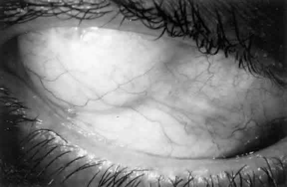
1. Katz LJ, Spaeth GL: Surgical management of the secondary glaucomas. Part 1. Ophthalmol Surg 18:826, 1987

2. Eid TE, Katz LJ, Spaeth GL et: Tube-shunt surgery versus neodymium:YAG cyclophotocoagulation in the management of neovascular glaucoma. Ophthalmology 104:1692, 1997

3. Rollet M: Le drainage au irin de la chambre anterieure contre l'hypertonie et al douleur. Rev Gen Ophthalmol 25:481, 1906

4. Zorab A: The reduction of tension in chronic glaucoma. Ophthalmolscope 10:258, 1912

5. Stefansson J: An operation for glaucoma. Am J Ophthalmol 8:681, 1925

6. Muldoon WE, Ripple PH, Wilder HC: Platinum implant in glaucoma surgery. Arch Ophthalmol 45:666, 1951

7. Troncoso MU: Use of tantalum implants for inducing a permanent hypotony in rabbit eyes. Am J Ophthalmol 32: 499, 1949

8. Bock RH: Subconjunctival draining of the anterior chamber by a glass seton. Am J Ophthalmol 33:929, 1950

9. Stewart RH, Kimbrough RL, Okercke PC: Trabeculectomy with implantation of the Mendez glaucoma section: Early results. Ophthalmol Surg 17:221, 1986

10. LaRocca V: Gonioplasty in glaucoma. Br J Ophthalmol 46: 404, 1962

11. Honrubia FM, Gomez ML, Hernandez A et al: Long term results of silicone tube in filtering surgery for eyes with neovascular glaucoma. Am J Ophthalmol 97:501, 1984

12. Igerer I: Silicone catheters used as setons in glaucoma surgery. Glaucoma 5:32, 1983

13. Kalijaca Z, Ljubojevic V, Manirov D: Draining implant for neovascular glaucoma. Am J Ophthalmol 96:372, 1983

14. Refojo MF: Current status of biomaterials in ophthalmology. Surv Ophthalmol 26:257, 1982

15. Epstein E: Fibrosis response in aqueous. Br J Ophthalmol 43:641, 1959

16. Richards RD, Van Bijsterveld OP: Artificial draining tube for glaucoma. Am J Ophthalmol 60:405, 1965

17. Krupin T, Kaufman P, Mandell A et al: Filtering valve implant surgery for eyes with neovascular glaucoma. Am J Ophthalmol 89:338, 1980

18. Sutton GE, Popp JC, Records RF: Krupin-Denver valve and neovascular glaucoma. Trans Ophthalmol Soc UK 102: 119, 1982

19. Folberg R, Hargett NA, Weaver JE et al: Filtering valve implant for neovascular glaucoma in proliferative diabetic retinopathy. Ophthalmology 89:286, 1982

20. Molteno ACB: New implant for draining in glaucoma. Br J Ophthalmol 53:609, 1969

21. Molteno ACB, Van Roogen MMB, Bartholomew RS: Implants for draining neovascular glaucoma. Br J Ophthalmol 61:120, 1977

22. Schocket SS, Nirankari VS, Lakhampal V et al: Anterior chamber tube shunt to an encircling band in the treatment of neovascular 
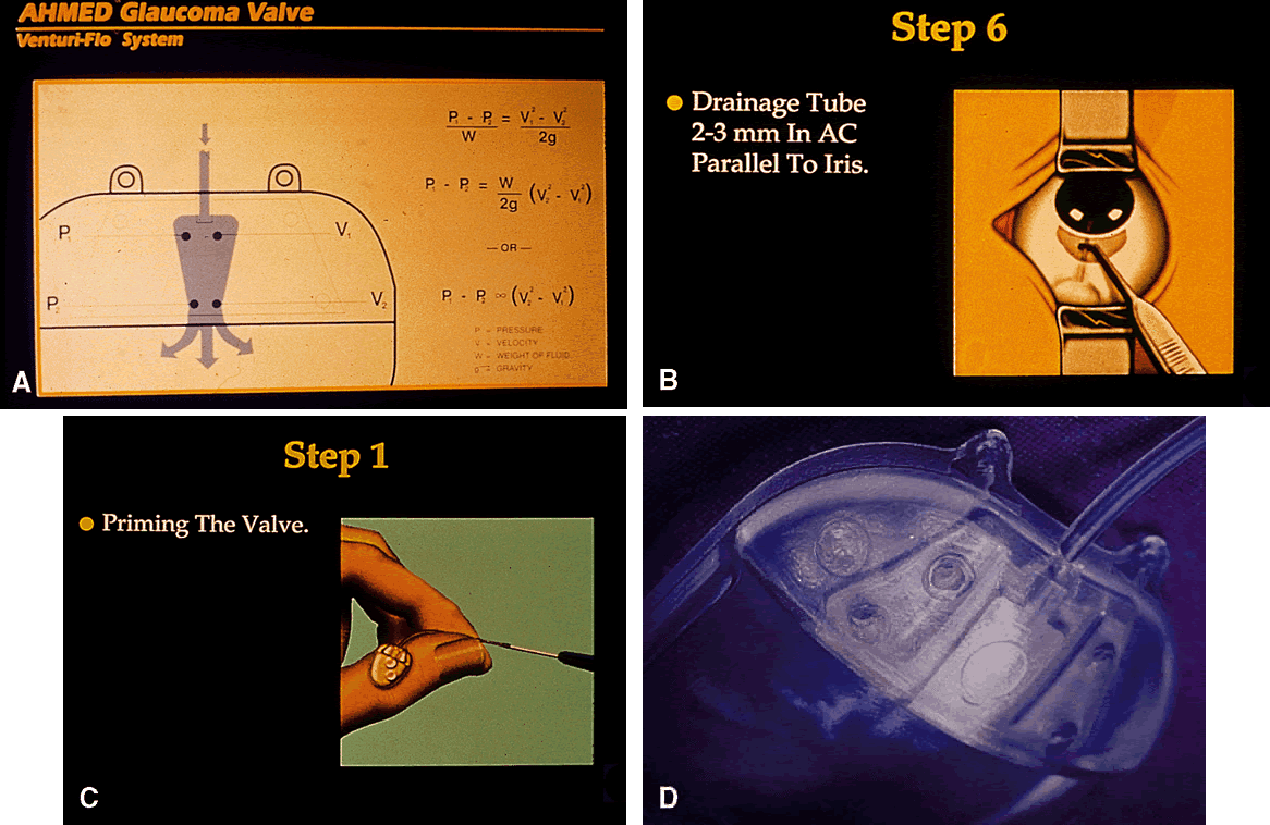
glaucoma and other refractory glaucomas. Ophthalmology 92:553, 1985

23. Krupin T, Ruderman JR, Rosenberg LE et al: Glaucoma valve to an external disc implant for filtration surgery. Invest Ophthalmol Vis Sci 33:948, 1992

23a. Coleman AL, Hill R, Wilson MR et al: Initial clinical experience with the Ahmed glaucoma valve implant. Am J Ophthalmol 120:23, 1995

24. Carmeron JD, White TC: Clinico-histopathologic correlation of a successful glaucoma pump shunt 
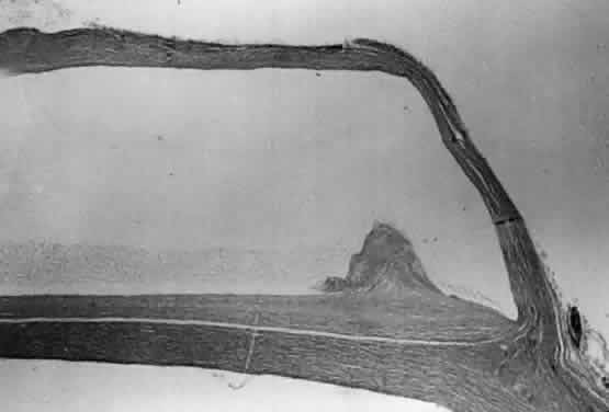
implant. Ophthalmology 95:1189, 1988

25. Minckler DS, Shammas A, Wilcox, M et al: Experimental studies of aqueous using the Molteno seton. Trans Am Ophthalmol Soc 85:385, 1987

26. Rubin B, Chan CC, Burnier M et al: Histopathologic study of the Molteno glaucoma implant in three patients. Am J Ophthalmol 110:371, 1990

27. Loeffler KU, Jay JL: Tissue response to aqueous drainage in a functioning Molteno implant. Br J Ophthalmol 72:29, 1988

28. Schocket SS: Investigations of the reasons for success and failure in the anterior shunt to the encircling band procedure in the treatment of refractory glaucoma. Trans Am Ophthalmol Soc 84:743, 1986

29. Lloyd MA, Baerveldt G, Quang HN et al: Long-Term histologic studies of the Baerveldt implant in a rabbit model. J Glaucoma 5:334, 1996

30. Folberg R, Hargett NA, Weaver JE et al: Filtering valve implants for neovascular glaucoma and proliferative diabetic retinopathy. Ophthalmology 89:286, 1982

31. Anker E, Molteno ACB: Molteno drainage implant for neovascular glaucoma. Trans Opthalmol Soc UK 102: 122, 1982

32. Brown RD, Cairns JE: Experience with the Molteno long tube implant. Trans Ophthalmol Soc UK 103:297, 1983

33. Freedman J: The use of single stage Molteno long tube seton in treating resistant cases of glaucoma. Ophthalmolic Surg 16:480, 1985

34. Hoare Nairne JEA, Sherwood D, Jacob JSH, Rich WJCC: Single stage insertion of the Molteno tube for glaucoma and methods to reduce postoperative hypotony. Br J Ophthalmol 72:846, 1988

35. Latina MA: Single stage Molteno implant with combination internal occlusion and external ligature. Ophthalmol Surgery 12(6):444, 1990

36. Minckler DS, Baerveldt G, Heuer DK: Clinical experience with the Molteno implant in complicated glaucoma cases. Ophthalmology 95:1181, 1988

37. Molteno ACB: The use of drainage implants in resistant cases of glaucoma. Late results of 110 operations. Trans Ophthalmol Soc NZ 32:101, 1983

38. Hill RA, Heuer DK, Baerveldt G et al: Molteno implantation for glaucoma in young patients. Ophthalmology 98: 1042, 1991

39. Fish L, Heuer DK, Baerveldt G: Molteno implantation for secondary glaucomas associated with advanced epithelial ingrowth. Ophthalmology 97:557, 1990

40. Freedman J, Rubin B: Molteno implants as a treatment for refractory glaucoma in black patients. Arch Ophthalmol 109:1417, 1991

41. Traverso CE, Tomey KF, Al-Kaff A: The long-tube single plate Molteno implant for the treatment recalcitrant glaucoma. Int Ophthalmol 13(1–2):159, 1989

42. Wilson RP, Cantor L, Katz, LJ: Aqueous shunts. Ophthalmology 99:672, 1992

43. Smith MF, Sherwood MB, McGorrary MB: Comparison of the double plate Molteno drainage implant with the Schocket procedure. Arch Ophthalmol 40:1246, 1992

44. Camras CB: Discussion of paper by Wilson RP et al: Aqueous shunts: Molteno vs. Schocket. Ophthalmology 99:676, 1992

45. Heuer DK, Lloyd MA, Abrams DA et al: Preliminary report of a randomized clinical trial of single plate versus double plate Molteno implantation for glaucoma in aphakia and pseudophakia. In Kriegelstein GK (ed): Glaucoma 
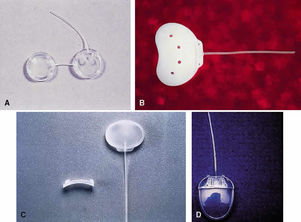
Update IV, pp 224–249. Berlin, Springer-Verlag, 1991

46. Wilson RP: The Schocket shunt. Ophthalmology 1:225, 1988

47. Omi CA, DeAlmeida GV, Cohen R et al: Modified Schocket implant for refractory glaucoma. Ophthalmology 98:211, 1991

48. Krupin T, Ritch R, Camras CB et al: A long Krupin-Denver valve implant attached to a 180° scleral explant for glaucoma surgery. Ophthalmology 95:1174, 1988

49. Saravitz EM, Toris CB, Camras CB et al: Opening/closing pressures and flow rates of Krupin-Denver valves in air and/or water. Invest Ophthalmol Vis Sci 33:948, 1992

50. Hitchings RA, Joseph NH, Sherwood MD et al: Use of one-piece valved tube and variable surface area explant for glaucoma drainage surgery. Ophthalmology 94:1079, 1987

51. Hitchings RA, Lavin MJ, Calthrope M: Glaucoma drainage tubes. Their role in glaucoma management. Int Ophthalmol 13:151, 1987

52. Lavin MJ, Franks WA, Wormaid RPL, Hitchings RA: Clinical risk factors for failure in glaucoma tube surgery. Arch Ophthalmol 110:480, 1992

53. Baerveldt G, Heuer DK, Martone JF et al: Clinical experience with the Baerveldt implant in complicated glaucomas. Personal communication, 1992

54. Smith SL, Starita RJ, Fellman RL et al: Extraocular muscle imbalance after anterior chamber tube shunt to the Baerveldt 350 mm2 Implant. Invest Ophthalmol Vis Sci 33: 949, 1992

55. Ohdara E, Kubota H, Takanashi T et al: Outcome of White pump shunt surgery for neovascular glaucoma in Asians. Ophthalmol Surg 23:666, 1992

56. Davidovski F, Stewart RH, Kimbrough RL: Long-term results with the White glaucoma pump shunt. Ophthalmol Surg 21:288, 1990

57. Ayyala RS, Zurakowski D, Smith JA et al: A clinical study of the Ahmed glaucoma valve implant in advanced glaucoma. Ophthalmology 105:1968, 1998

58. Prata JA Jr, Mermoud A, LaBree L et al: In vitro and in vivo flow characteristics of glaucoma drainage implants. Ophthalmology 102:894, 1995

59. Porter JM, Krawezyk CH, Carey RF: In vitro flow testing of glaucoma drainage devices. Ophthalmology 104:1701, 1997

60. Francis BA, Cortes A, Chen J et al: Characteristics of glaucoma drainage implants during dynamic and steady-state flow conditions. Ophthalmology 105:1708, 1998

61. Prata JA Jr, Minckler DS, Mermoud A et al: Effects of Intraoperative mitomycin-C on the function of Baerveldt glaucoma drainage implants in rabbits. J Glaucoma 5:29, 1996

62. Lee D, Shin DH, Birt CM et al: The effect of adjuctive mitomycin-C in Molteno implant surgery. Ophthalmology 104:2126, 1997

63. Lloyd MA, Heuer DK, Baerveldt G et al: Combined Molteno implantation and pars plana vitrectomy for neovascular glaucomas. Ophthalmology 98:1401, 1991

64. Gandham SB, Costa VP, Katz LJ et al: Aqueous tube-shunt implantation and pars plana vitrectomy in eyes with refractory glaucoma. Am J Ophthalmol 116:189, 1993

65. Fish LA, Heuer DK, Baerveldt G et al: Molteno implantation for secondary glaucomas associated with advanced epithelial ingrowth. Ophthalmology 97:557, 1990

66. Costa VP, Katz LJ, Cohen EJ, Raber IM: Glaucoma associated with epithelial downgrowth controlled with Molteno tube shunts. Ophthalmol Surg 23:797, 1992

67. McDonnell PJ, Rubin JB, Schanzlin DJ: Molteno implant for control of glaucoma in eyes after penetrating keratoplasty. Ophthalmology 93:364, 1988

68. Beebe W, Starita R, Fellman R et al: The use of Molteno implant and anterior chamber tube shunt to encircling band for the treatment of glaucoma in keratoplasty patients. Ophthalmology 97:1414, 1990

69. Billson F, Thomas R, Aylward W: The use of two stage Molteno implants in developmental glaucoma. J Pediatr Ophthalmol Strabismus 26:3, 1989

70. Munoz M, Tomey K, Traverso C et al: Clinical experience with the Molteno implant in advanced infantile glaucoma. J Pediatr Ophthalmol Strabismus 28:68, 1991

71. Hill RA, Heuer OK, Baerveldt G et al: Molteno implantation for glaucoma in young patients. Ophthalmology 98: 1042, 1991

72. Eid TE, Katz LJ, Spaeth GL et al: Long-term effects of tube shunt procedures on refractory childhood glaucoma. Ophthalmology 104:1011, 1997

73. Fellenbaum PS, Sidoti PA, Heuer DK et al: Experience with the Baerveldt implant in young patients with complicated glaucomas. J Glaucoma 4:91, 1995

74. Weiss HS: Postoperative manipulation of the Krupin valve. Ophthalmic Surg Lasers 27:151, 1996

75. Burchfield JC, Kass MA, Wax MB: Primary valve malfunction of the Krupin eye valve with disk. J Glaucoma 6: 152, 1997

76. Feldman RM, El-Harazi SM, Villanueva G: Valve membrane adhesion as a cause of Ahmed glaucoma valve failure. J Glaucoma 6:10, 1997

77. Azuara-Blanco A, Katz LJ, Gandham SB et al: Pars plana tube insertion of aqueous shunt with vitrectomy in malignant glaucoma. Arch Ophthalmol 116:808, 1998

78. Canning CR, Lavin M, McCartney AC: Delayed suprachoroidal hemorrhage after glaucoma operations. Eye 3:327, 1989

79. Franks WA, Hitchings RA: Injection of perfluoropropane gas to prevent hypotony in eyes undergoing 
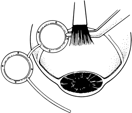
tube implant surgery. Ophthalmology 97:899, 1990

80. Kooher JS: Repair of Mottino implant during surgery. Am J Ophthalmol 117:673, 1994

81. Molteno ACB, Van Bijon G, Anker E: Two stage insertion of glaucoma drainage implants. Trans Ophthalmol Soc NZ 31:17, 1979

82. Molteno ACB, Polkinghorne PJ, Bowbyes JA: the Vieryl tie technique for inserting a draining implant in the treatment of secondary glaucoma. Aust NZ J Ophthalmol 14: 343, 1986

83. El Sayyad F, El-Maghraby A, Hetal M et al: The use of releasable sutures in Molteno glaucoma implant procedures to reduce postoperative hypotony. Ophthalmol Surg 22: 82, 1991

84. Price FW Jr, Whitsen WE: Polypropylene ligatures as a means of controlling intraocular pressure with Molteno implants. Ophthalmol Surg 20:781, 1989

85. Liebmann J, Ritch R: Intraocular suture ligature to reduce hypotony following Molteno seton implantation. Ophthalmol Surg 23:51, 1992

86. Traverso CF, Tomey KF, Al-Katita A: The long tube single plate Molteno implant for the treatment of recalcitrant glaucoma. Int Ophthalmol 13:159, 1989

87. Latina M: Single stage Molteno implant with combination internal occlusion and external ligature. Ophthalmol Surg 21:444, 1990

88. Kooner KS, Goode SM: Removable ligature during Molteno implant procedure. Am J Ophthalmol 114:102, 1992

89. Susanna R Jr: Modifications of the Molteno implant and implant procedure. Ophthalmol Surg 22:611, 1991

90. Egbert PR, Lieberman MP: Internal suture occlusion of the Molteno glaucoma implant for the prevention of postoperative hypotony. Ophthalmol Surg 20:53, 1989

91. Molteno ACB: The dual chamber implant: its use in neovascular glaucoma. Aust NZ J Ophthalmol 18:131, 1990

92. Freedman J: Clinical experience with the Molteno dual-chamber single-plate implant. Ophthalmol Surg 23:238, 1992

93. Chen PP, Palmberg PF: Needling revision of glaucoma drainage device filtering blebs. Ophthalmology 104:1004, 1997

94. Ayyala RS, Harman LE, Michelini-Norris B et al: Comparison of different biomaterials for glaucoma drainage devices. Arch Ophthalmol 118:233, 1999

95. Valimaki J, Tuulonen A, Airaksinen PJ: Capsule excision after failed Molteno surgery. Ophthalmic Surg Lasers 28: 382, 1997

96. Mills RP, Reynolds A, Emond M et al: Long-term survival of Molteno glaucoma drainage devices. Ophthalmology 103:229, 1996

97. Fiore PM, Melamed S: Use of neodymium:YAG laser to open an occluded Molteno tube. Ophthalmol Surg 20: 373, 1989

98. Singh K, Eid TE, Katz LJ et al: Evaluation of Nd:YAG laser membranectomy in blocked tubes after glaucoma tube-shunt surgery. Am J Ophthalmol 124:781, 1997

99. Zalloum JN, Ahuja RM, Shin D et al: Assessment of corneal decompensation in eyes having undergone Molteno shunt procedures compared to eyes having undergone trabeculectomy. CLAO J 25:57, 1999

100. Billson F, Thomas R, Grigg J: Resiting Molteno tubes. Ophthalmic Surg Lasers 27:801, 1996

101. Allinson RW: Paralimbal compression suture for Molteno implants. Ophthalmol Surg 22:750, 1991

102. Rapuano CJ, Schmidt CM, Cohen EJ et al: Results of alloplastic tube shunt procedures before, during, or after penetrating keratoplasty. Cornea 14:26, 1995

102a. Christmann LM, Wilson ME: Motility disturbances after Molteno implants. J Pediatr Ophthalmol Strabismus 29: 44, 1992

103. Ball S, Ellis GS, Herrington RG et al: Brown's superior oblique tendon syndrome after Baerveldt glaucoma implant. Arch Ophthalmol 110:1368, 1992

104. Ball SF, Lotfield K, Scharfenberg J: Molteno rip-cord suture hypopyon. Ophthalmol Surg 21:407, 1990

105. Perkins T: Endophthalmitis after placement of a Molteno implant. Ophthalmol Surg 21:733, 1990

106. Krebs DB, Liebmann JM, Ritch R et al: Late infectious endophthalmitis from exposed glaucoma setons. Arch Ophthalmol 110:174, 1992

107. Melamed S, Cahane M, Gutman I et al: Postoperative complications after Molteno implant surgery. Am J Ophthalmol 111:319, 1991

108. Lotufo DG: Postoperative complications and visual loss following Molteno implantation. Ophthalmol Surg 22: 650, 1991

109. Kramer T, Brown R, Lynch M et al: Molteno implants and operating microscope-induced retinal phototoxicity. Arch Ophthalmol 109:379, 1991

110. Huna R, Melamed S, Hirsh A et al: Retinal detachment adherent to posterior chamber IOL after Molteno implant surgery. Ophthalmol Surg 21:854, 1990

Back to Top