Chapter 7
Techniques of Phacoemulsification
MARK PACKER, I. HOWARD FINE, STEPHEN A. OBSTBAUM, RICHARD S. HOFFMAN, STEPHEN B. LICHTENSTEIN and HOWARD V. GIMBEL
Main Menu   Table Of Contents

Search

WOUND CONSTRUCTION AND ARCHITECTURE
CONTINUOUS CURVILINEAR CAPSULORHEXIS
HYDRODISSECTION AND HYDRODELINEATION
NUCLEOFRACTIS TECHNIQUES
CONCLUSION
REFERENCES

Phacoemulsification (phaco) means disassembly and removal of the crystalline lens. From its introduction in the late 1960s to the present, phaco has evolved into a highly effective method of cataract extraction. Incremental advances in surgical technique and the simultaneous redesign and modification of technology have permitted increasing safety and efficiency.

Among the advances that have shaped modern phaco are incision construction, continuous curvilinear capsulorhexis, cortical cleaving hydrodissection, hydrodelineation, and nucleofractis techniques. The refinement of cataract removal through a small incision has improved phaco and permitted rapid visual rehabilitation and excellent ocular structural stability. Perhaps the most outstanding characteristic of this era of phaco is the unrelenting quest for excellence that continues to challenge the innovative spirit of cataract surgeons.

Back to Top
WOUND CONSTRUCTION AND ARCHITECTURE
The availability of foldable intraocular lenses (IOLs) that can be inserted through small unsutured phacoemulsification incisions1 has created a trend away from scleral tunnel incisions to clear corneal incisions.2 Among the disadvantages of scleral tunnels are the need to perform conjunctival incisions and scleral dissections, and the need for cautery to prevent operating in the presence of blood. In addition, there is increased difficulty with oarlocking of the phaco tip and distortion of the cornea because of the length of scleral tunnels.

Kratz is generally credited as the first surgeon to move from the limbus posteriorly to the sclera in order to increase appositional surfaces, thus enhancing wound healing and reducing surgically induced astigmatism.3,4 Girard and Hoffman were the first to name the posterior incision a “scleral tunnel incision” and were, along with Kratz, the first to make a point of entering the anterior chamber through the cornea, creating a corneal shelf.5 This corneal shelf was designed to prevent iris prolapse. In 1989, McFarland used this incision architecture and recognized that these incisions allowed for the phacoemulsification and implantation of lenses without the need for suturing.6 Maloney, who was a fellow of Kratz, advocated a corneal shelf to his incisions which he described as strong and waterproof.7 Ernest recognized that McFarland's long scleral tunnel incision terminated in a decidedly corneal entrance. He hypothesized that the posterior “corneal lip” of the incision acted as a one-way valve, thus explaining the mechanism for self-sealability (Ernest PH. Presentation at the Department of Ophthalmology, Wayne State University School of Medicine, Detroit, MI, February 28, 1990). In April of 1992, Fine presented his self-sealing temporal clear corneal incision at the annual meeting of the American Society of Cataract and Refractive Surgery.8

There have been surgeons who have favored corneal incisions for cataract surgery prior to their recent popularization. In 1968, Charles Kelman stated that the best approach for performing cataract surgery was with phacoemulsification through a clear corneal incision utilizing a triangular tear capsulotomy and a grooving and cracking technique in the posterior chamber.9 Harms and Mackenson in Germany published an intracapsular technique using a corneal incision in 1967.10 Troutman was an early advocate of controlling surgically induced astigmatism at the time of cataract surgery by means of the corneal incision approach.11 Arnott in England utilized clear corneal incisions and a diamond keratome for phacoemulsification, although he had to enlarge the incision for introducing an IOL.12 Galand in Belgium utilized clear corneal incisions for extracapsular cataract extraction in his envelope technique13 and Stegman of South Africa has a long history of having utilized the cornea as the site for incisions for extracapsular cataract extraction (Stegmann R. Personal communication, December 3, 1992). Finally, perhaps the leading proponent of clear corneal incisions for modern era phacoemulsification was Kimiya Shimizu of Japan.14

In 1992, Fine began routinely utilizing clear corneal cataract incisions with closure by a tangential suture modeled after Shepherd's technique.15 Within a very short period, the suture was abandoned in favor of self-sealing corneal incisions.16 Through the demonstrated safety and increased utilization of these incisions by pioneers in the United States, including Williamson, Shepherd, Martin, and Grabow17 these incisions became increasingly popular and utilized on an international basis.

Rosen demonstrated by topographical analysis that clear corneal incisions 3 mm or less in width do not induce astigmatism.18 This finding led to increasing interest because of better predictability of T-cuts, arcuate cuts, and limbal relaxing incisions for managing pre-existing astigmatism at the time of cataract surgery. Surgeons recognized many other advantages of the temporal clear corneal incision, including better preservation of pre-existing filtering blebs19 and options for future filtering surgery, increased stability of refractive results because of decreased effects from lid blink and gravity, ease of approach, elimination of the bridle suture and iatrogenic ptosis, and improved drainage from the surgical field via the lateral canthal angle.

Single-plane incisions, as first described by Fine,20 utilized a 3.0-mm diamond knife. After pressurizing the eye with viscoelastic through a paracentesis, the surgeon placed the blade on the eye so that it completely applanated the eye with the point of the blade positioned at the leading edge of the anterior vascular arcade. The knife was advanced in the plane of the cornea until the shoulders, 2 mm posterior to the point of the knife, touched the external edge of the incision. Then the point of the blade was directed posteriorly to initiate the cut through Descemet's membrane in a maneuver known as the dimple-down technique. After the tip entered the anterior chamber, the initial plane of the incision was re-established to cut through Descemet's membrane in a straight-line configuration.

Williamson was the first to utilize a shallow 300- to 400-micron grooved clear corneal incision.21 He believed that the thicker external edge to the roof of the tunnel reduced the likelihood of tearing. Langerman later described the single hinge incision, in which the initial groove measured 90% of the depth of the cornea anterior to the edge of the conjunctiva.22 Initially, he utilized a depth of 600 microns and subsequently made the tunnel itself superficially in that groove, believing that this led to enhanced resistance of the incision to external deformation.

Surgeons employed adjunctive techniques to combine incisional keratorefractive surgery with clear corneal cataract incisions. Fine used the temporal location for the cataract incision and added one or two T-cuts made by the Feaster Knife with a 7-mm ocular zone for the management of pre-existing astigmatism. Others, including Lindstrom and Rosen, rotated the location of the incision to the steep axis. Kershner used the temporal incision by starting with a nearly full thickness T-cut through which he then made his corneal tunnel incision. For large amounts of astigmatism, he used a paired T-cut in the opposite side of the same meridian.23 Finally, the popularization of limbal-relaxing incisions by Gills24 and Nichamin,25 added an additional means of reducing pre existing astigmatism.

The 3-D Blade (Rhein Medical, Tampa, Florida) improved incision construction with differentially sloped bevels on its anterior and posterior surfaces (Fig. 1). This design allowed the surgeon to touch the eye at the site of the external incision location and advance the blade in the plane of the cornea without dimpling down. The differential slopes allowed the forces of tissue resistance to create an incision characterized by a linear external incision, a 2-mm tunnel, and a linear internal incision.26 The trapezoidal 3-D Blade also allowed enlargement of the incision up to 3.5-mm for IOL insertion without altering incision architecture.

Fig. 1. The differentially beveled diamond blade constructs a 2-mm tunnel incision that is 2.5-mm in width. Gentian violet has been used to mark the location of a limbal relaxing incision (LRI). The LRI may be constructed first and the clear corneal incision initiated at the mid-depth of the LRI.

Following phacoemulsification, lens implantation, and removal of residual viscoelastic, stromal hydration of the clear corneal incision can be performed in order to help seal the incision.16 Stromal hydration is performed by gently irrigating balanced salt solution into the stroma at both edges of the incision with a 26- or 27-gauge cannula. Once apposition takes place, the hydrostatic forces of the endothelial pump help seal the incision. In those rare instances of questionable wound integrity, a single 10-0 nylon or Vicryl suture is placed to ensure a tight seal.

Clear corneal incisions, by nature of their architecture and location, have some unique complications associated with them. If one incidentally incises the conjunctiva at the time of the clear corneal incision, ballooning of the conjunctiva can develop, which may compromise visualization of anterior structures. In this case, a suction catheter may be used to aid exposure. Early entry into the anterior chamber may result in an incision of insufficient length to be self-sealing. In addition, incisions that are too short or improperly constructed can result in an increased tendency for iris prolapse. A single suture may be required in order to assure a secure wound at the conclusion of the procedure. On the other hand, a late entry may result in a corneal tunnel so long that the phaco tip creates striae in the cornea and compromises the view of the anterior chamber.

Manipulation of the phacoemulsification handpiece intraoperatively may result in tearing of the roof of the tunnel, especially at the edges, resulting in compromise of the incision's self-sealability. Tearing of the internal lip can also occur, resulting in compromised self-sealability or, rarely, small detachments or scrolling of Descemet's membrane in the anterior edge of the incision.

Of greater concern has been the potential for incisional burns.27 When incisional burns develop in clear corneal incisions, there may be a loss of self-sealability. Closure of the wound may induce excessive amounts of astigmatism. In addition, manipulation of the incision can result in an epithelial abrasion, which can compromise self-sealability because of the lack of a fluid barrier by an intact epithelium. Without an intact epithelial layer, the corneal endothelium does not have the ability to help appose the roof and floor of the incision through hydrostatic forces.

In a large survey performed for the American Society of Cataract and Refractive Surgery by Masket,28 there was a slightly increased incidence of endophthalmitis in clear corneal cataract surgery compared to scleral tunnel surgery. However, the survey failed to note the incision sizes in those cases in which endophthalmitis in clear corneal incisions had occurred. Masket described several generally accepted techniques of prophylaxis, including preoperative topical antibiotics, 5% povidone-iodine prep, and draped eyelashes

Colleaux and Hamilton29 found no significant difference in the rate of endophthalmitis with respect to clear corneal versus scleral tunnel incisions in a retrospective review of 13,886 consecutive cataract operations. They reported a significant prophylactic effect of subconjunctival antibiotic injection but found no benefit to preoperative antibiotic drops. In an evidence-based update, Ciulla, Starr, and Masket found that current literature most strongly supports the use of preoperative povidone-iodine antisepsis.30 They found little change in the risk of endophthalmitis in the United States over time, from 0.12% in 1984 to 0.13% in 1994.

Clear corneal cataract incisions are becoming a more popular option for cataract extraction and IOL implantation throughout the world. With clear corneal incisions, we have achieved minimally invasive surgery with immediate visual rehabilitation. Clear corneal incisions have had a proven record of safety with relative astigmatic neutrality. In addition, clear corneal incisions result in an excellent cosmetic outcome. We expect that they will continue to increase in popularity, especially as newer modalities, such as bimanual microincision phaco, become the standard of care.

Back to Top
CONTINUOUS CURVILINEAR CAPSULORHEXIS
Implantation of the IOL in an intact capsular bag facilitates the permanent rehabilitative benefit of cataract surgery. For many years, surgeons considered a “can-opener” capsulotomy satisfactory for both planned extracapsular cataract extraction and phaco. Problems related to malposition and decentration of implanted posterior chamber IOLs were later recognized. In 1991, Wasserman and associates31 performed a postmortem study that showed that the extension of one or more V-shaped tears toward the equator of the capsule produced instability of the IOL and resulted in IOL malposition.

We are fortunate to have benefited from the work of Calvin Fercho, who developed continuous tear capsulotomy (“Continuous circular tear anterior capsulotomy,” Welsh Cataract Congress, 9 September 1986) and Gimbel and Neuhann, who popularized continuous curvilinear capsulorhexis (CCC).32–34

The technique of CCC is not difficult to learn if certain basic principles are observed:

  1. The continuous capsular tear should be performed in a deep, stable anterior chamber. We advocate using a viscoelastic substance that deepens the anterior chamber and stretches the anterior capsule. The use of a viscoelastic material accomplishes two important goals:
    1. It creates space for safe instrumentation in the anterior chamber.
    2. By making the anterior capsule taut and pushing the lens posteriorly it resists the action of posterior pressure, which tends to cause the capsular tear to move peripherally.

  2. The tear is started at the center of the capsule. This way the origin of the tear is included within the termination of the tear. Starting in the center of the capsule generates a flap with a peripheral edge that is smooth and continuous.
  3. Once the initial flap is mobilized, it is inverted to permit a smooth tearing action, such as would be achieved in tearing a piece of paper with one half held stable while the inverted half is torn to the desired configuration. This principle is the same whether a cystotome, bent needle, or forceps is used to create the capsulotomy.
  4. The continuous tear proceeds either clockwise or counterclockwise in a controlled and deliberate fashion, the surgeon regrasping with the forceps or repositioning the point of the cystotome or bent needle on the inverted flap to control the vector of the tear. On completion of the CCC, it is essential that the origin of the peripheral portion of the CCC be included within the circumference of the tear.

As we have indicated, it is essential to control the course of the capsular tear. A tear that begins moving peripherally or in a radial fashion is a signal that a condition exists that requires immediate attention. The first thing the surgeon must do is to recognize the situation. Further progress of the tear should be stopped and the depth of the anterior chamber assessed. Frequently, the cause of the peripheral course of the tear is shallowing of the anterior chamber and the effect of posterior pressure on the lens and anterior capsule. Adding more viscoelastic to deepen the anterior chamber opposes the posterior pressure, makes the lens capsule taut, widens the pupil, and permits inspection of the capsule to see whether zonular extension onto the anterior capsule is responsible for the misdirection of the tear. Generally, the tear can be redirected and continued.

If the tear has extended peripherally and cannot be safely redirected, one option is to create a small tangential incision at the origin of the CCC with Vannas' scissors and to direct the tear in the opposite direction to include the peripheral extension. If this maneuver cannot be accomplished and the discontinuity in the CCC remains, it is probably wisest to make several other small incisions in the capsular rim so that the peripheral force is distributed evenly, reducing the likelihood that a tear will extend around the lens equator.

A similar situation may occur on completion of the CCC. Again, at this point, it is essential that the origin of the peripheral portion of the CCC be included within the circumference of the tear. If this maneuver is performed correctly, it will result in a totally blended edge, or it will form a small centripetally peaked area (cardioid). If the end of the CCC results in a V-shaped centrifugally oriented peak, however, this peak acts as a discontinuity in the anterior capsular opening and may extend peripherally, with the attendant consequences mentioned earlier. The surgeon must convert this area to a smooth tear by either regrasping an edge to include the V-shaped tear or by making a small incision with Vannas' scissors to create a segmental secondary CCC.

The use of a vital dye to stain the anterior capsule in the absence of a good red reflex constitutes an important adjunctive technique for capsulorhexis construction. The surgeon makes the sideport incision and then fills the anterior chamber with air. The dye, either indocyanine green or trypan blue, is injected into the chamber. The air and residual dye is then exchanged for viscoelastic. Despite the absence of a red reflex, the capsule is now easy to see.

The technique of CCC has provided important advantages both for cataract surgery and IOL implantation. Because endolenticular or in situ phaco must be performed in the presence of an intact continuous capsulotomy opening, the capsulorhexis has also served as a stimulus for modification of phaco techniques.

Back to Top
HYDRODISSECTION AND HYDRODELINEATION
Hydrodissection of the nucleus in cataract surgery has traditionally been perceived as the injection of fluid into the cortical layer of the lens under the lens capsule to separate the lens nucleus from the cortex and capsule.35 With increased use of continuous curvilinear capsulorhexis and phacoemulsification in cataract surgery, hydrodissection became a very important step to mobilize the nucleus within the capsule for disassembly and removal.36–39 Following nuclear removal, cortical cleanup proceeded as a separate step, using an irrigation and aspiration handpiece.

Fine first described cortical cleaving hydrodissection, which is a hydrodissection technique designed to cleave the cortex from the lens capsule and thus leave the cortex attached to the epinucleus.40 Cortical cleaving hydrodissection usually eliminates the need for cortical cleanup as a separate step in cataract surgery, thereby eliminating the risk of capsular rupture during cortical cleanup.

CORTICAL CLEAVING HYDRODISSECTION

A small capsulorhexis, 5 to 5.5 mm, optimizes the procedure. The large anterior capsular flap makes this type of hydrodissection easier to perform. The anterior capsular flap is elevated away from the cortical material with a 26-gauge blunt cannula (e.g., Katena Instruments No. K7-5150) before hydrodissection. The cannula maintains the anterior capsule in a tented-up position at the injection site near the lens equator. Irrigation before elevation of the anterior capsule should be avoided because it will result in transmission of a fluid wave circumferentially within the cortical layer, hydrating the cortex and creating a path of least resistance that will disallow later cortical cleaving hydrodissection. Once the cannula is properly placed and the anterior capsule is elevated, gentle, continuous irrigation results in a fluid wave that passes circumferentially in the zone just under the capsule, cleaving the cortex from the posterior capsule in most locations. When the fluid wave has passed around the posterior aspect of the lens, the entire lens bulges forward because the fluid is trapped by the firm equatorial cortical-capsular connections. The procedure creates, in effect, a temporary intraoperative version of capsular block syndrome as seen by enlargement of the diameter of the capsulorhexis. At this point, if fluid injection is continued, a portion of the lens prolapses through the capsulorhexis. However, if before prolapse the capsule is decompressed by depressing the central portion of the lens with the side of the cannula in a way that forces fluid to come around the lens equator from behind, the cortical-capsular connections in the capsular fornix and under the anterior capsular flap are cleaved. The cleavage of cortex from the capsule equatorially and anteriorly allows fluid to exit from the capsular bag through the capsulorhexis, which constricts to its original size, and mobilizes the lens in such a way that it can spin freely within the capsular bag. Repeating the hydrodissection and capsular decompression starting in the opposite distal quadrant may be helpful. Adequate hydrodissection at this point is demonstrable by the ease with which the nuclear-cortical complex can be rotated by the cannula.

HYDRODELINEATION

Hydrodelineation is a term first used by Anis to describe the act of separating an outer epinuclear shell or multiple shells from the central compact mass of inner nuclear material, the endonucleus, by the forceful irrigation of fluid (balanced salt solution) into the mass of the nucleus.41

The 26-gauge cannula is placed in the nucleus, off center to either side, and directed at an angle downward and forward toward the central plane of the nucleus. When the nucleus starts to move, the endonucleus has been reached. It is not penetrated by the cannula. At this point, the cannula is directed tangentially to the endonucleus, and a to-and-fro movement of the cannula is used to create a tract within the nucleus. The cannula is backed out of the tract approximately halfway, and a gentle but steady pressure on the syringe allows fluid to enter the distal tract without resistance. Driven by the hydraulic force of the syringe, the fluid will find the path of least resistance, which is the junction between the endonucleus and the epinucleus, and flow circumferentially in this contour. Most frequently, a circumferential golden ring is seen outlining the cleavage between the epinucleus and the endonucleus (Fig. 2). Sometimes the ring will appear as a dark circle rather than a golden ring.

Fig. 2. The golden ring appears as a result of hydrodelineation, demonstrating that a cleavage plane has developed between the endonucleus and the epinucleus. However, the golden ring alone does not ensure free rotation of the endonucleus; the endonucleus should be rotated with the irrigation cannula prior to commencing phacoemulsification.

Occasionally, an arc will result and surround approximately one quadrant of the endonucleus. In this instance, creating another tract the same depth as the first but ending at one end of the arc, and injecting into the middle of the second tract, will extend that arc (usually another full quadrant). This procedure can be repeated until a golden or dark ring verifies circumferential division of the nucleus.

For very soft nuclei, the placement of the cannula allows for the creation of an epinuclear shell of any thickness. The cannula may pass through the entire nucleus if it is soft enough, so the placement of the tract and the location of the injection allow an epinuclear shell to be fashioned as desired. In very firm nuclei, one appears to be injecting into the cortex on the anterior surface of the nucleus, and the golden ring will not be seen. However, a thin, hard epinuclear shell is achieved even in the most brunescent nuclei. That shell will offer the same protection as a thicker epinucleus in a softer cataract.

Hydrodelineation circumferentially divides the nucleus and has many advantages. Circumferential division reduces the volume of the central portion of nucleus removed by phacoemulsification by up to 50%. This allows less deep and less peripheral grooving and smaller, more easily mobilized quadrants after cracking or chopping. The epinucleus acts as a protective cushion within which all of the chopping, cracking, and phacoemulsification forces can be confined. In addition, the epinucleus keeps the bag on stretch throughout the procedure, making it unlikely that a knuckle of capsule will come forward, occlude the phaco tip, and rupture.

Back to Top
NUCLEOFRACTIS TECHNIQUES
The evolution of phaco from the initial procedure as described by Kelman9 in the late 1960s to the techniques that we currently practice is nothing less than remarkable. The contributions of talented ophthalmic surgeons who persevered throughout these years should be commended, because they laid the groundwork for our present methods.

The major distinction between the phaco techniques practiced today and the earlier techniques is that modern methods have facilitated phaco of dense cataracts within the capsular bag, allowing the central endonucleus to be removed before the epinucleus is encountered. With previous techniques, we worked from the peripheral portion of the epinucleus/nucleus complex toward the center. This change was influenced by the recognition that the nuclear mass of firm and hard lenses could be divided into smaller pieces for controlled removal within the protective layer of the epinucleus and that a capsular opening produced by a CCC would withstand the forces involved in nuclear cracking. Retention of an intact CCC opening also required sequential microsurgical removal of the contents of the capsular bag, which is best achieved by performing phaco in the central and deepest portion of the anterior chamber.

DIVIDE AND CONQUER TECHNIQUE

Divide and conquer nucleofractis phaco, described by Gimbel,42 was the first nucleofractis (two-instrument) cracking technique developed. After adequate hydrodissection, a deep crater is sculpted into the center of the nucleus, leaving a dense peripheral rim that can later be fractured into multiple sections. It is important that the crater include the posterior plate of the nucleus; otherwise, fracturing of the rim is much more difficult. A shaving action is used to sculpt away the central nuclear material. When the central material is no longer accessible to the phaco probe, the lens should be rotated and additional central phaco performed to enlarge and deepen the crater. The size of the central crater should be expanded for progressively denser nuclei. Enough of the dense material must be left in place, however, to allow the phaco probe and second instrument to engage the rim and fracture the lens into sections.

The surgeon uses his experience as a guide to determine how deeply the central crater should be sculpted. The peripheral nuclear rim stretches the entire capsular bag and acts as a safety mechanism to prevent the posterior capsule from suddenly moving anteriorly and being cut by the phaco probe. For harder nuclei, small sections should be fractured from the rim. Rather than emulsify the sections as they are broken away, the sections should be left in place within the rim to maintain the circular rim and the tension on the capsule. Leaving the sections in place also facilitates rotation and the progressive fracturing of the remaining rim. It is sometimes advisable to initially remove one small section to allow space for fracturing of the other segments of the remaining rim. If only a small fragment is removed, the remaining segments can maintain capsular stretch and help to avoid rupture of the capsule. After the rim is fractured around the entirety of its circumference, each segment can then be brought to the center of the capsule for safe emulsification. One must be more cautious at this point because as more segments are removed, less lens material is available to expand the capsule and the capsule will have a greater tendency to be aspirated into the phaco tip, especially if high aspiration flow rates are used.

PHACO FRACTURE TECHNIQUE

In phaco fracture, a widely used nucleofractis technique described by Shepherd,43 the surgeon sculpts a groove from the 12- to 6-o'clock position after performing hydrodissection and hydrodelineation. The width of the groove should be one and a half to two times the diameter of the phaco tip. Using the phaco handpiece and a second instrument, the surgeon rotates the nucleus 90 degrees. A second groove is sculpted perpendicular to the first, in the form of a cross. Sculpting continues until the red reflex is seen at the bottom of the grooves. Additional rotations and removal of nuclear material is often necessary to accomplish adequate grooving. Care should be taken to avoid sculpting completely through to the cortex peripherally, because this procedure puts the equatorial and posterior capsule at increased risk of damage. A bimanual cracking technique is used to create a fracture through the nuclear rim in the plane of one of the grooves. The nucleus is then rotated 90 degrees, and additional fractures are made until four separate quadrants are isolated. The segments are then tumbled toward the center of the capsule for safe emulsification. A short burst of phaco power is used to embed the phaco tip into the bulk of the isolated quadrant, and then with the use of aspiration, the quadrant is gently pulled into the center for emulsification. Alternatively, the second instrument can be used to elevate the apex of the wedge to facilitate mobilization of the nuclear quadrant to the capsule's center.

CHIP AND FLIP TECHNIQUE

Introduced by Fine,44 and useful for softer grades of nuclei, this procedure relies on a nucleus that rotates freely within the capsular bag. Initially, a central bowl is sculpted in the nucleus until a thin central plate remains. The second instrument introduced through the side port incision engages the subincisional nuclear rim to move the inferior nuclear rim to toward the center of the capsule bag. Then clock-hour pieces of the rim are carefully emulsified as the nucleus is rotated. Once the entire rim is removed, the second instrument is used to elevate the remaining central thinned nuclear plate (the chip), which is then emulsified. The epinucleus is engaged at the 6-o'clock position with aspiration alone. As the phaco tip is moved superiorly, the second instrument pushes the epinucleus toward the 6-o'clock position, thereby tumbling the epinuclear bowl and permitting it to be aspirated (the flip).

CRACK AND FLIP TECHNIQUE

Fine and colleagues modified Shepherd's phaco fracture technique by adding hydrodelineation, resulting in the crack and flip technique.45 Sculpting two deep grooves at right angles to each other that extend to the golden ring permits bimanual nucleus cracking. Only the endonucleus cracks, because the epinucleus is separated from it by hydrodelineation. Each quadrant is then sequentially removed with the use of pulsed phaco and moderate aspiration. The second instrument elevates the apices of each quadrant so that the tip of the phaco needle can be totally occluded to aid in aspiration. Once the nucleus is removed, the epinucleus is aspirated as with the chip and flip technique.

There are several helpful tips in using the crack and flip technique or a modification of this method. The sculpting portion of the procedure is performed with minimal vacuum, relatively low aspiration flow, and low phaco power. The forward passes of the phaco needle only shave the nuclear material to ultimately sculpt a groove. The phaco tip is never totally occluded during this phase of the operation. Rather, only a portion of the phaco needle contacts the nucleus to remove controlled amounts of lens material. This process is continued until the grooves are deep. The appropriate depth can be assessed by a brightening of the red reflex, which suggests that the denser portion of the nucleus has been emulsified on the region of the grooves.

To achieve nuclear cracking, two instruments are placed deeply in the grooves and moved down and outward. The phaco needle and a second instrument introduced through the side port, paracentesis, or incision will crack the nucleus if the grooves are sufficiently deep and the instruments placed in the depth of the grooves. If cracking does not readily occur, additional deepening of the grooves is warranted. Phaco energy is not required during this step. The limit of the grooves is the golden ring, which represents the perimeter of the endonucleus. The loosened quadrants of the endonucleus remain within the cushion of the surrounding epinucleus.

Removing the nuclear fragments requires a change in the parameters for phaco. For this step, it is desirable to have lens material in contact with the phaco needle. Increasing the aspiration flow slightly directs lens material to the phaco tip, whereas increasing the vacuum encourages the nuclear fragments to be aspirated with application of only a minimum of phaco power. These parameters are influenced by the density of the nucleus, but in principle, these settings result in successful nucleus removal. A second instrument guides the control of nuclear fragments.

Removing the epinucleus is accomplished as described in the chip and flip section. The parameters can be modified such that (1) the aspiration flow is slightly reduced from the setting used in nuclear fragment removal, and (2) pulsed phaco power is used. If cortical cleaving hydrodissection is successful, the cortex is removed, along with the epinucleus during this step of the procedure.

PHACO CHOP

The phaco chop technique was initially introduced by Nagahara, who used the natural fault lines in the lens nucleus to create cracks without creating prior grooves (Nagahara K, American Society of Cataract and Refractive Surgery film festival, 1993). The phaco tip is embedded in the center of the nucleus after the superficial cortex is aspirated. A second instrument, the phaco chopper, is then passed to the equator of the nucleus, beneath the anterior capsule, and drawn to the phaco tip to fracture the nucleus. The two instruments are separated to widen the crack. This procedure is repeated until several small fragments are created, which are then emulsified.

Koch and Katzen46 modified this procedure because they encountered difficulty in mobilizing the nuclear fragments. They created a central groove or central crater, depending on the density of the nucleus. This modification permits ease of removing the nuclear fragments liberated by the phaco chop technique.

Advocates of these nucleus-dividing techniques have suggested that high levels of vacuum help remove the nuclear fragments and minimize the need for ultrasound energy. With some of the newer phaco instruments, higher vacuum power can be applied with minimal risk of anterior chamber collapse.

CHOO CHOO CHOP AND FLIP

Fine described the “choo-choo chop and flip” technique in 1998.47 Subsequently, Fine, Packer, and Hoffman correlated the reduction of ultrasound energy with this technique to improvement in uncorrected post operative day one visual acuity.48 A 30-degree standard bevel down tip is used throughout endonuclear removal. After aspirating the epinucleus uncovered by the capsulorhexis, a Fine/Nagahara chopper (Rhein Medical, Tampa, FL) is placed in the golden ring by touching the center of the nucleus with the tip and pushing it peripherally so that it reflects the capsulorhexis. The chopper is used to stabilize the nucleus by lifting and pulling toward the incision slightly (Fig. 3), after which the phaco tip lollipops the nucleus in either pulse mode at 2 pulses/second or 80 millisecond burst mode.

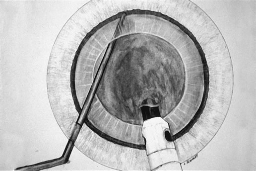
Fig. 3. The Fine-Nagahara chopper supports the endonucleus as the phaco tip is advanced in foot position three and achieves a firm purchase.

Burst mode is a power modulation that utilizes a fixed percent power (panel control), a programmable burst width (duration of power), and a linear interval between bursts. As one enters foot position 3, the interval between bursts is 2 seconds; with increasing depressions of the foot pedal in foot position 3 the interval shortens until at the bottom of foot position 3 there is continuous phaco. In pulse mode, there is linear power (%) but a fixed interval between pulses, resulting at 2 pulses/sec in a 250-millisecond pulse (linear power), followed by a 250-millisecond pause in power, followed by a 250-millisecond pulse, and so on. However, in both of these modulations with tip occlusion, vacuum is continuous throughout the pulse and pause intervals. With the energy delivered in this way, ultrasound energy into the eye is minimized and hold on the nucleus is maximized as vacuum builds between pulses or bursts. Because of the decrease in cavitational energy around the tip at this low pulse rate or in burst mode, the tunnel in the nucleus in which the tip is embedded fits the needle very tightly and gives us an excellent hold on the nucleus, thus maximizing control of the nucleus as it is scored and chopped (Fig. 4) in foot position 2.

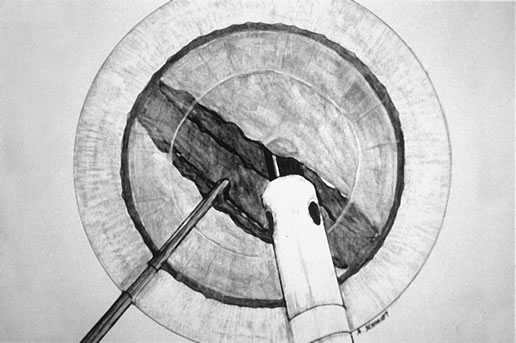
Fig. 4. Vacuum is maintained in foot position 2 as the chopper is brought to the side of the phaco tip and the two instruments are separated, hemisecting the endonucleus.

The Fine/Nagahara chop instrument is grooved on the horizontal arm close to the vertical “chop” element with the groove parallel to the direction of the sharp edge of the vertical element. In scoring the nucleus, the instrument is always moved in the direction the sharp edge of the wedge-shaped vertical element is facing (as indicated by the groove on the instrument), thus facilitating scoring. The nucleus is scored by bringing the chop instrument to the side of the phaco needle. It is chopped in half by pulling the chopper to the left and slightly down while moving the phaco needle, still in foot position 2, to the right and slightly up. Then the nuclear complex is rotated. The chop instrument is again brought into the golden ring (Fig. 5), the nucleus is again lollipopped, scored, and chopped with the resulting pie-shaped segment now lollipopped on the phaco tip (Fig. 6). The segment is then evacuated utilizing high vacuum and short bursts or pulse-mode phaco at 2 pulses/second. The nucleus is continually rotated so that pie-shaped segments can be scored, chopped, and removed essentially by the high vacuum assisted by short bursts or pulses of phaco. The short bursts or pulses of ultrasound energy continuously reshape the pie-shaped segments that are kept at the tip, allowing for occlusion and extraction by the vacuum. The size of the pie-shaped segments is customized to the density of the nucleus, with smaller segments for denser nuclei. Phaco in burst mode or at this low pulse rate sounds like “choo-choo-choo-choo”; therefore, the name of this technique. With burst mode or the low pulse rate, the nuclear material tends to stay at the tip rather than chatter as vacuum holds between pulses. The chop instrument is utilized to stuff the segment into the tip or keep it down in the epinuclear shell.

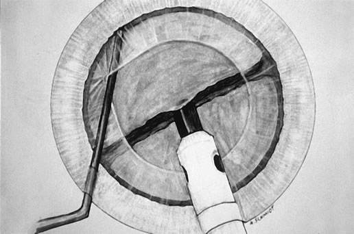
Fig. 5. Following rotation of the endonuclear complex, the chopper is repositioned in the golden ring and the phaco tip is buried in the distal heminucleus in preparation for the second chop.

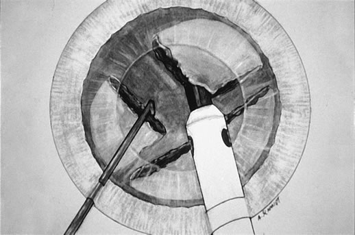
Fig. 6. The second chop is complete. The pie-shaped wedge of nucleus is positioned on the phaco tip, ready for aspiration by high vacuum and small pulses of ultrasound as required. The size of this wedge can be adjusted in inverse proportion to the density of the cataract.

After evacuation of the first heminucleus, the second heminucleus is rotated to the distal portion of the bag and the chop instrument stabilizes it while it is lollipopped. It is then scored (Fig. 7) and chopped. The pie-shaped segments can be chopped a second time to reduce their size (Fig. 8) if they appear too large to easily evacuate.

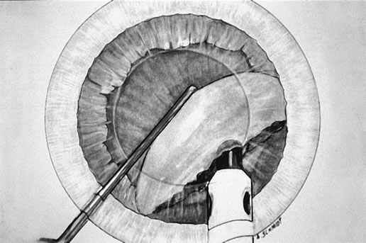
Fig. 7. After extraction of the first heminucleus, the remaining material is rotated distally and chopped.

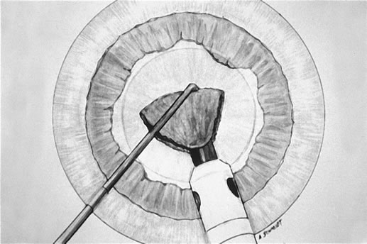
Fig. 8. The final pie-shaped wedge is chopped and extracted.

There is little tendency for nuclear material to come up into the anterior chamber with this technique. Usually, it stays down within the epinuclear shell, but the chop instrument can control the position of the endonuclear material. The 30-degree bevel-down tip facilitates occlusion, as the angle of approach of the phaco tip to the endonucleus through a clear corneal incision is approximately 30 degrees. This allows full vacuum to be quickly reached, which facilitates embedding the tip into the nucleus for chopping and allows mobilization of pie-shaped segments from above rather than necessitating going deeper into the endolenticular space as is necessary with a bevel-up tip. In addition, the cavitational energy is directed downward toward the nucleus rather than up toward the endothelium.

After evacuation of all endonuclear material, the epinuclear rim is trimmed in each of the three quadrants, mobilizing cortex as well in the following way. As each quadrant of the epinuclear rim is rotated to the distal position in the capsule and trimmed, the cortex in the adjacent capsular fornix flows over the floor of the epinucleus and into the phaco tip. Then the floor is pushed back to keep the bag on stretch until three of the four quadrants of the epinuclear rim and forniceal cortex have been evacuated. It is important not to allow the epinucleus to flip too early, thus avoiding a large amount of residual cortex remaining after evacuation of the epinucleus.

The epinuclear rim of the fourth quadrant is then used as a handle to flip the epinucleus. As the remaining portion of the epinuclear floor and rim is evacuated from the eye, most of the time the entire cortex is evacuated with it. Downsized phaco tips with their increased resistance to flow are less capable of mobilizing the cortex because of the decreased minisurge accompanying the clearance of the tip when going from foot position 2 to foot position 3 in trimming of the epinucleus.

After the IOL is inserted, these strands and any residual viscoelastic material are removed using the irrigation-aspiration tip, leaving a clean capsular bag.

If there is cortex remaining following removal of all the nucleus and epinucleus, there are three options. The phacoemulsification handpiece can be left high in the anterior chamber while the second handpiece strokes the cortex-filled capsular fornices. Frequently, this results in floating up of the cortical shell as a single piece and its exit through the phacoemulsification tip (in foot position two) because cortical cleaving hydrodissection has cleaved most of the cortical capsular adhesions.

Alternatively, if one wishes to complete cortical cleanup with the irrigation-aspiration handpiece before lens implantation, the residual cortex can almost always be mobilized as a separate and discrete shell (reminiscent of the epinucleus) and removed without ever turning the aspiration port down to face the posterior capsule.

The third option is to viscodissect the residual cortex by injecting the viscoelastic through the posterior cortex onto the posterior capsule. We prefer the dispersive viscoelastic device chondroitin sulfate-hyaluronate (Viscoat, Alcon Surgical, Fort Worth, Texas). The viscoelastic material spreads horizontally, elevating the posterior cortex and draping it over the anterior capsular flap. At the same time, the peripheral cortex is forced into the capsular fornix. The posterior capsule is then deepened with a cohesive viscoelastic device, and the IOL is implanted through the capsulorhexis, leaving the anterior extension of the residual cortex anterior to the IOL. Removal of residual viscoelastic material accompanies mobilization and aspiration of residual cortex anterior to the IOL, which protects the posterior capsule, leaving a clean capsular bag.

Chop techniques substitute mechanical forces (chopping) for ultrasound energy (grooving) to disassemble the nucleus. High vacuum is utilized as an extractive technique to remove nuclear material rather than utilizing ultrasound energy to convert the nucleus to an emulsate that is evacuated by aspiration. These techniques maximize safety, control and efficiency, allowing phaco of harder nuclei, even in the presence of a compromised endothelium. Chop techniques facilitate the achievement of two goals: minimally invasive cataract surgery and maximally rapid visual rehabilitation.

LASER PHACOEMULSIFICATION

Cataract extraction modalities employing laser energy currently include the Erbium:YAG Phacolase (Carl Zeiss Meditec, Jena, Germany), the Neodymium:YAG Photon Laser PhacoLysis System (Paradigm Medical, Salt Lake City), and the Dodick Q-switched Neodymium:YAG laser (ARC GmbH). Several potential advantages over ultrasound have maintained interest in laser, including relative reduction in the energy requirement for cataract extraction, the absence of any potential for thermal injury, and improved protection of corneal endothelial cells.

The erbium:YAG (2940 nm) laser energy is well absorbed by tissues with high water content and has a penetration depth of less than 1 micron. The laser energy is delivered though a fiber inside the aspiration port placed flush with the tip. Hoh and Fischer demonstrated that erbium laser is safe and effective for mild to moderate nuclear sclerosis.49

Surgeons may employ either a bimanual technique, separating irrigation from aspiration, or the more familiar coaxial set up. With the latter, Takayuki Akahoshi's counter prechop technique is used to effectively disassemble the lens nucleus into multiple wedge-shaped segments.50 A horizontal chopper such as the Fine-Nagahara chopper (Rhein Medical, Tampa, Florida) is inserted through the side port, touched against the anterior lens surface, and gently pushed under the distal anterior capsular flap, where it falls into the golden ring. The chopper supports the nucleus, while the Akahoshi Prechopper (ASICO, Westmont, Illinois) is passed through the 2.5 mm corneal incision directly into the core of the nucleus. The chopper in the golden ring is held in front of the prechopper to preclude rotational movement of the nucleus. Opening the prechopper then bisects the nucleus.

The nucleus is then rotated 90 degrees, and the first heminucleus is bisected in a similar fashion. The chopper supports the heminucleus from the golden ring, whereas the prechopper is inserted directly into the center of the heminucleus and opened. In this manner, the nucleus may be divided into four or more segments, each of which is a suitable size for laser phacoemulsification.

Nd: YAG photolysis represents a low energy modality for cataract extraction developed by Dodick.51 Kanellopoulos reported a mean intraocular energy use of 5.65 Joules per case.52 This level of energy compares favorably with values previously reported for ultrasound phacoemulsification, and approximates the level of energy reported for the chop and flip phacoemulsification technique using power modulations.48 Huetz and Eckhardt found mean total energy of 1.97 Joules for nuclear sclerosis up to grade 3, 3.37 Joules for Grade 3 and 7.7 Joules for Grade 4.53

Surgeons generally employ a groove and crack technique with the laser, sculpting in a bimanual fashion and cracking as soon as possible. Once superficial cortical material is aspirated, the laser tip is used to ablate and fragment the nucleus. The laser tip should only just touch the surface of the nucleus, and not be used to impale the cataract. Following central photofragmentation, the nucleus is handled much as it is with the classic divide and conquer technique. The total time that the tip is in the eye varies with the grade of nucleus, from 2.15 minutes for 1+ nuclear sclerosis to 9.8 minutes for 3+ nuclear sclerosis.52

Using the bimanual Dodick system, a cataract may be completely extracted through two 1.5-mm incisions. Now, IOL technology is becoming available to take advantage of this ultrasmall incision. Wehner and Ali have reported a series of cases implanted with a dehydrated acrylic IOL through a 1.5-mm incision.54

BIMANUAL ULTRASOUND PHACOEMULSIFICATION

The promise of bimanual, ultrasmall incision cataract surgery and companion IOL technology is becoming a reality through both laser and new ultrasound power modulations. New instrumentation is available for bimanual surgery, including forceps for construction of the capsulorhexis, irrigating choppers, and bimanual irrigation and aspiration sets (Fig. 9). Proponents of performing phaco through two paracentesis-type incisions claim reduction of surgically induced astigmatism, improved chamber stability in every step of the procedure, better flow characteristics due to the physical separation of infusion from ultrasound and vacuum, and greater ease of irrigation and aspiration with the elimination of one, hard-to-reach subincisional region. However, the risk of thermal injury to the cornea from a vibrating bare phaco needle has posed a challenge to the development of this technique.

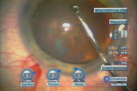
Fig. 9. Capsulorhexis forceps designed for bimanual phaco through two paracentesis incisions (ASICO, Westmont, Illinois).

In the 1970s, Girard attempted to separate infusion from ultrasound and aspiration, but abandoned the procedure because of thermal injury to the tissue.55, 56 Shearing and colleagues successfully performed ultrasound phaco through two 1.0-mm incisions using a modified anterior chamber maintainer and a phaco tip without the irrigation sleeve.57 They reported a series of 53 cases and found that phaco time, overall surgical time, total fluid use, and endothelial cell loss were comparable to those measured with their standard phaco techniques. Crozafon described the use of Teflon-coated phaco tips for bimanual high-frequency pulsed phaco, and suggested that these tips would reduce friction and therefore allow surgery with a sleeveless needle . 58 Tsuneoka, Shiba, and Takahashi determined the feasibility of using a 1.4-mm (19-gauge) incision and a 20-gauge sleeveless ultrasound tip to perform phaco.59 They found that outflow around the tip through the incision provided adequate cooling, and performed this procedure in 637 cases with no incidence of wound burn.60 Additionally, less surgically induced astigmatism developed in the eyes operated with the bimanual technique. Agarwal and colleagues developed a bimanual technique, “Phakonit,” using an irrigating chopper and a bare phaco needle passed through a 0.9 clear corneal incision.61 They achieved adequate temperature control through continuous infusion and use of “cooled balanced salt solution” poured over the phaco needle.

Soscia, Howard, and Olson have shown in cadaver eye studies that phacoemulsification with the Sovereign WhiteStar system (AMO, Santa Ana, California), using a bare 19-gauge aspiration needle, does not produce a wound burn at the highest energy settings unless all infusion and aspiration are occluded.62, 63 WhiteStar represents a power modulation of ultrasonic phacoemulsification that reduces the production of thermal energy by limiting the duration 
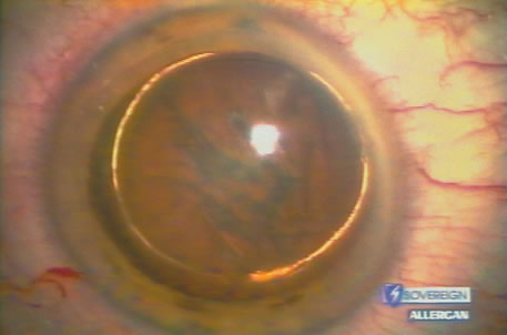
of energy pulses to the millisecond range (Fig. 10).

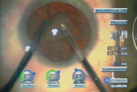
Fig. 10. Bimanual phaco through two paracentesis incisions with WhiteStar (AMO, Santa Ana, California) and an irrigating chopper (MicroSurgical Technology, Redmond, Washington). The final pie-shaped segment is ready for chopping, as shown in figure 8. High vacuum (307 mm Hg) and low levels of ultrasound (current 5%, total Effective Phaco Time to this point in the procedure, 0.05 second at 1.2% Average Power) create efficient nuclear removal.

Back to Top
CONCLUSION
Since the time of Charles Kelman's inspiration in the dentist's chair (while having his teeth ultrasonically cleaned), incremental advances in phacoemulsification technology have produced ever-increasing benefits for patients with cataract. The modern procedure simply was not possible even a few years ago, and until recently prolonged hospital stays were common after cataract surgery.

The competitive business environment and the wellspring of surgeons' ingenuity continue to demonstrate synergistic activity in the improvement of surgical technique and technology. Future advances in cataract surgery will continue to benefit our patients as we develop new phacoemulsification techniques and technology.

Back to Top
REFERENCES

1. Fine IH: Architecture and construction of a self-sealing incision for cataract surgery. J Cataract Refract Surg 17:672,1991

2. Leaming DV: Practice styles and preferences of ASCRS members—2001 survey. J Cataract Refract Surg 28:1681, 2002

3. Colvard DM, Kratz RP, Mazzocco TR, Davidson B: Clinical evaluation of the Terry surgical keratometer. Am Intraocular Implant Soc J 6:249,1980

4. Masket S:. Origin of scleral tunnel methods. [Letter to the Editor.] J Cataract Refract Surg 19:812,1993

5. Girard LJ, Hoffman RF: Scleral tunnel to prevent induced astigmatism. Am J Ophthalmol 97:450, 1984

6. McFarland MS: Surgeon undertakes phaco, foldable IOL series sans sutures. Ocular Surgery News 8, 1990

7. Maloney WF, Grindle L: Textbook of Phacoemulsification, p 31. Fallbrook, CA: Lasenda Publishers, 1988

8. Brown DC, Fine IH, Gills JP et al: The Future of Foldables. Panel discussion held at the 1992 annual meeting of the American Society of Cataract and Refractive Surgery. Ocular Surgery News August 15 (supplement) 1992

9. Kelman CD: Phacoemulsification and aspiration: a new technique of cataract removal: a preliminary report. Am J Ophthalmol 64:23, 1967

10. Harms H, Mackensen G: Intracapsular extraction with a corneal incision using the Graefe knife, p 144. In Blodi FL (ed): Ocular Surgery Under the Microscope. Stuttgart, Germany, Georg Thieme Verlag, 1967

11. Paton D, Troutman R, Ryan S: Present trends in incision and closure of the cataract wound. Highlights of Ophthalmology 14:3, 1973

12. Arnott EJ: Intraocular Implants. Transactions of the Ophthalmological Society of the United Kingdom 101:58, 1981

13. Galand A: La Technique de l'Enveloppe. Liege, Belgium, Pierre Mardaga publisher, 1988

14. Shimizu K: Pure corneal incision. Phaco & Foldables 5:5, 1992

15. Shepherd JR: Induced astigmatism in small incision cataract surgery. J Cataract Refract Surg 15:85, 1989

16. Fine IH: Corneal tunnel incision with a temporal approach. In Fine IH, Fichman RA, Grabow HB, (eds): Clear-Corneal Cataract Surgery & Topical Anesthesia, p 5. Thorofare, NJ, Slack, Inc., 1993

17. Fine IH, Fichman RA, Grabow HB: Clear-Corneal Cataract Surgery & Topical Anesthesia. Thorofare, NJ, Slack, Inc., 1993.

18. Rosen ES: Clear corneal incisions: a good option for cataract patients. A Roundtable Discussion. Ocular Surgery News , February 1, 1998

19. Park HJ, Kwon YH, Weitzman M, Caprioli J: Temporal corneal phacoemulsification in patients with filtered glaucoma. Arch Ophthalmol 115:1375, 1997

20. Fine IH: Self-sealing corneal tunnel incision for small-incision cataract surgery. Ocular Surgery News May 1, 1992

21. Williamson CH: Cataract Keratotomy Surgery. In Fine IH, Fichman RA, Grabow HB, (eds): Clear-Corneal Cataract Surgery & Topical Anesthesia, p 87. Thorofare, NJ, Slack, Inc., 1993

22. Langerman DW: Architectural design of a self-sealing corneal tunnel, single-hinge incision. J Cataract Refract Surg 20:84, 1994

23. Kershner RM: Clear corneal cataract surgery and the correction of myopia, hyperopia, and astigmatism. Ophthalmology 104:381, 1997

24. Gills JP, Gayton JL: Reducing pre-existing astigmatism. In Gills JP (ed): Cataract Surgery: The State of the Art, p 53 Thorofare, NJ, Slack, Inc., 1998

25. Nichamin L: Refining astigmatic keratotomy during cataract surgery. Ocular Surgery News April 15, 1993

26. Fine IH: New blade enhances cataract surgery, Techniques Spotlight. Ophthalmology Times September 1, 1996

27. Fine IH: Special Report to ASCRS Members: Phacoemulsification Incision Burns. Letter to American Society of Cataract and Refractive Surgery members, 1997

28. Taher B: Endophthalmitis: State of the prophylactic art. Eyeworld News August 42, 1997

29. Colleaux KM, Hamilton WK: Effect of prophylactic antibiotics and incision type on the incidence of endophthalmitis after cataract surgery. Can J Ophthalmol 35:373, 2000

30. Ciulla TA, Starr MB, Masket S: Bacterial endophthalmitis prophylaxis for cataract surgery. Ophthalmology 109:13, 2002

31. Wasserman D, Apple D, Castaneda V et al: Anterior capsular tears and loop fixation of posterior chamber intraocular lenses. Ophthalmology 98:425, 1991

32. Neuhann T: Theorie und operationstechnik der kapsulorhexis. Klin Monatsbl Augenheilkd 190:542, 1987

33. Gimbel HV, Neuhann T: Development, advantages and methods of the continuous circular capsulorhexis technique. J Cataract Refract Surg 16:31, 1990

34. Gimbel HV, Neuhann T: Letter to the editor: Continuous curvilinear capsulorhexis. J Cataract Refract Surg 17:110, 1991

35. Faust , KJ: Hydrodissection of soft nuclei. Am Intraocular Implant Soc J 10:75, 1984

36. Davison JA: Bimodal capsular bag phacoemulsification: A serial cutting and suction ultrasonic nuclear dissection technique. J Cataract Refract Surg 15:272, 1989

37. Sheperd JR: In situ fracture. J Cataract Refract Surg 16:436, 1990

38. Gimbel HV: Divide and conquer nucleofractis phacoemulsification: Development and variations. J Cataract Refract Surg 17:281, 1991

39. Fine IH: The chip and flip phacoemulsification technique. J Cataract Refract Surg 17:366,1991

40. Fine IH. Cortical cleaving hydrodissection. J Cataract Refract Surg 18:508, 1992

41. Anis A: Understanding hydrodelineation: The term and related procedures. Ocular Surg News 9:134, 1991

42. Gimbel HV: Divide and conquer nucleofractis phacoemulsification. J Cataract Refract Surg 17:281,1991

43. Shepherd JR: In situ fracture. J Cataract Refract Surg 16:436, 1990

44. Fine IH: The chip and flip phacoemulsification technique. J Cataract Refract Surg 17:306, 1991

45. Fine IH, Maloney WF, Dillman DM: Crack and flip phacoemulsification technique. J Cataract Refract Surg 19:797, 1993

46. Koch PS, Katzen LE: Stop and chop phacoemulsification. J Cataract Refract Surg 20:566, 1994

47. Fine IH: The choo-choo chop and flip phacoemulsification technique. Operative Tech Cataract Refract Surg 1:61, 1998

48. Fine IH, Packer M, Hoffman RS: Use of power modulations in phacoemulsification. J Cataract Refract Surg 27:188, 2001

49. Hoh H, Fischer E. Pilot study on erbium laser phacoemulsification. Ophthalmology 107:053, 2000

50. Akahoshi T: Phaco prechop: Manual nucleofracture prior to phacoemulsification. Operative Techniques in Cataract and Refractive Surgery 1:69,1998

51. Dodick JM: Laser phacolysis of the human cataractous lens. Dev Ophthalmol 22:58, 1991

52. Kanellopoulos AJ: Laser cataract surgery: A prospective clinical evaluation of 1000 consecutive laser cataract procedures using the Dodick photolysis Nd:YAG system. Ophthalmology 108:649, 2001.

53. Huetz WW, Eckhardt HB: Photolysis using the Dodick-ARC laser system for cataract surgery. J Cataract Refract Surg 27:208, 2001

54. Wehner W, Ali I: Implantation of a 1.5-mm IOL after dodick laser photolysis cataract extraction. Symposium of the American Society of Cataract and Refractive Surgery, Philadelphia, 2002.

55. Girard LJ: Ultrasonic fragmentation for cataract extraction and cataract complications. Adv Ophthalmol 37:127, 1978

56. Girard LJ: Pars plana lensectomy by ultrasonic fragmentation 1984, part II: operative and postoperative complications, avoidance or management. Ophthalmic Surg 15:217,1984

57. Shearing SP, Relyea RL, Loaiza A, Shearing RL: Routine phacoemulsification through a one-millimeter non-sutured incision. Cataract 2:6,1985

58. Crozafon P. “The use of minimal stress and the Teflon-Coated tip for bimanual high frequency pulsed phacoemulsification,” presented at the 14th Meeting of the Japanese Society of Cataract and Refractive Surgery, Kyoto, Japan, July 1999.

59. Tsuneoka H, Shiba T, Takahashi Y: Feasibility of ultrasound cataract surgery with a 1.4 mm incision. J Cataract Refract Surg 27:934, 2001

60. Tsuneoka H, Shiba T, Takahashi Y: Ultrasonic phacoemulsification using a 1.4 mm incision: Clinical results. J Cataract Refract Surg 28:81, 2002

61. Agarwal A, Agarwal A, Agarwal S et al: Phakonit: phacoemulsification through a 0.9 mm corneal incision. J Cataract Refract Surg 27:1548, 2001

62. Soscia W, Howard JG, Olson RJ: Bimanual phacoemulsification through 2 stab incision. A wound-temperature study. J Cataract Refract Surg 28:1039, 2002

63. Soscia W, Howard JG, Olson RJ: Microphacoemulsification with WhiteStar. A wound-temperature study. J Cataract Refract Surg 28:1044, 2002

Back to Top