
|
|
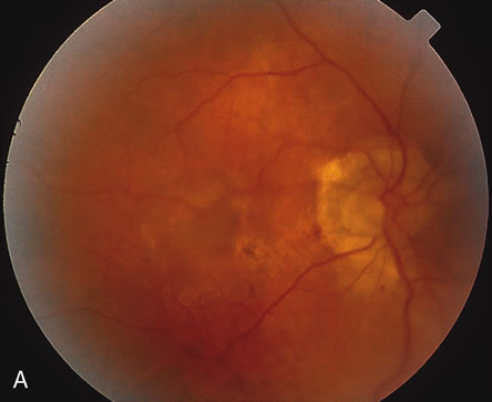
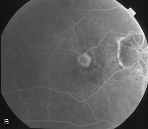
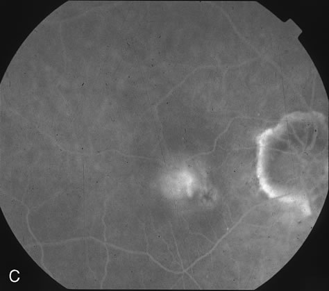
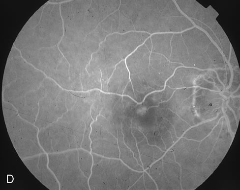
| Fig. 1. A. Color fundus photograph of subfoveal CNV in a patient with AMD as well as myopia. B. Early-phase FA (35 seconds) demonstrates predominantly classic subfoveal CNV (as defined by the TAP study).23 C. Late-phase FA (3:20) demonstrates hyperfluorescent leakage from subfoveal CNV. D. Early-phase FA (42 seconds) at day 7 status post verteporfin-PDT demonstrates the typical ring of hypofluorescence corresponding to the treated area. E. Late phase FA (3:01) at day 7 status post verteporfin-PDT demonstrates a small, central area of fluorescein staining. |