Chapter 88
Flaps, Grafts, and Alloplastic Materials in Lid Reconstruction and Repair
W. JACKSON ILIFF and ELBA M. PACHECO
Main Menu   Table Of Contents

Search

FLAPS
GRAFTS
ALLOPLASTIC MATERIALS IN EYELID RECONSTRUCTION
REFERENCES

This chapter was conceived to supplement the other excellent chapters in this text on lid reconstruction and orbital surgery by supplying some additional ideas and resources for the management of lid defects. Although it is not encyclopedic in scope, we hope it will supply both basic and more advanced techniques currently available that will improve our flexibility in difficult situations.

There are, of course, many ways to reconstruct a lid defect. Some can be repaired with flaps only, others with a combination of flaps and grafts, perhaps with the addition of alloplastic materials. Each individual reconstruction requires careful planning. Hopefully this chapter will supply alternatives. However, the goal is not to lead the surgeon step by step through each technique, although some will be illustrated. Basic suturing techniques and wound closure will not be covered. Instead, this material should lead, when necessary, to the appropriate resources in this or other texts or the original literature.

In association with planning the exact procedures necessary to reconstruct a given defect, a review of lid and adjacent anatomy and anesthesia considerations and applicable general surgical ones is appropriate. Other chapters in this text cover general and local anesthesia1,2 and provide a good review of the anatomy and basic techniques.3 Other texts on oculoplastic surgery also provide expanded treatments of these topics, including harvesting some of the grafts to be mentioned here.4

Back to Top
FLAPS
A flap is a tissue structure, usually skin or a combination of skin and muscle, that is relocated from its original site. The blood and possibly the neural supply remains intact and is not interrupted during the transfer. The recipient bed does not initially contribute to its nourishment. In the lids, the rich anastomosing network of vessels improves flap survival. Random flaps are dissected and moved perpendicular to their blood supply; thus, they are more poorly vascularized than axial flaps, which are elevated parallel with and incorporating the supplying vessels. It is generally said that a random flap length may not exceed four times its width. In the early days of tissue transfer without benefits such as microsurgical techniques and antibiotics, a one-to-one ratio was recommended, to be exceeded only if a vessel (thus an axial flap) was included in the dissection. In the lid, long flaps from the upper to lower lid, for instance, are truly part flap and part free graft, since these principles are routinely exceeded. So-called free flaps are not routinely used in lid reconstruction, but may be in more complex facial, orbital, and maxillofacial procedures. Free or microvascular flaps are, in essence, free grafts that incorporate both an artery and vein, which are reanastomosed microsurgically to corresponding vessels in the recipient area, thus becoming tissue transferred with a direct vascular supply at the close of the procedure.

Skin flaps and musculocutaneous flaps are most often used in lid reconstruction or for filling partial-thickness defects. These usually can supply tissue coverage for the entire anterior lamellae of the lid and can support grafts to the posterior portion of the lid. Alloplastic materials in reconstruction can also be supported. Mucosal flaps with or without tarsus may conversely rebuild the posterior lid and support a free skin graft anteriorly.

In addition to vascular supply, flaps are characterized by their shape. Four types are described: sliding, advancement, rotation, and transposition. A sliding flap is generally random and involves simply undermining the area surrounding a defect to allow primary closure. The skin and subcutaneous tissue is “slid” into position. An advancement flap is a more complicated sliding flap in that the tissue to be “advanced” is incised on three sides to allow greater motility. Closure is primary and generates no secondary defect. An example is the Cutler-Beard flap to reconstruct the upper lid.* The Hughes tarsoconjunctival flap* is another. The rotation flap is similar to an advancement flap in its basic construction in that a three-sided incision and elevation of tissue is performed. However, the defect to be filled is adjacent to the side of the flap rather than at its end, so that the flap is “rotated” into the defect. In doing so, a secondary defect is created opposite the primary. The secondary defect may be closed with a number of techniques, including primary closure if small enough, by developing sliding flaps (further undermining) perhaps with excision of small amounts of additional tissue to produce tension in a different direction, by additional smaller flaps or by a skin graft. Examples include the Tenzel semicircular flap and the Mustardé cheek rotation flap.* The transposition flap moves tissue to a nonadjacent site. Small transposition flaps, like small rotation and advancement flaps, may be random. Larger (i.e., longer) flaps, as in the midforehead,* more often need axial design for proper survival. Examples of transposition flaps are simple Z-plasties, rhomboid flaps, bilobed flaps,* and the tarsoconjunctival flaps described by Hewes and Leone (see Marginal Defects section).


See Figures later in the chapter

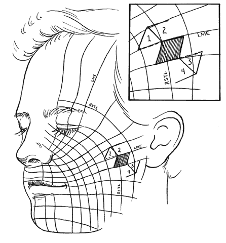
Specific incision placement for these procedures goes beyond the scope of this chapter, but some general comments and concepts are important to include. The concept of the relaxed skin tension lines (RSTL) (Fig. 1) or Langer's lines helps incision placement.5 Wrinkles and skin creases generally develop parallel to these lines as a result of the action of underlying musculature. Incisions ideally should be placed in preexisting creases because they are best hidden in these areas and avoid development of scars that may be more visible. If this is not possible, they should be parallel to the RSTL. The lines of maximum skin extensibility (LME) are perpendicular to the RSTL. The skin is most extensible (i.e., available for sliding, transposing) in the direction of the LME. Vectors of tension should be parallel to the LME. Once the possible donor sites are identified (there are usually several), the site with the most available skin is identified by pinching it up and pushing and pulling the surrounding structures to judge possible distortion caused by closure. For instance, significant lower-lid ectropion might be caused by one donor site and avoided by an alternate. This should be kept in mind when designing flaps and will be expanded further in the discussion of rhomboid flaps.

Fig. 1. Relaxed skin tension lines (RSTL) and lines of maximum extensibility (LME). Inset shows rhomboid flap superimposed in optimum orientation. Dotted lines are short diagonals of the flap and indicate preferred flap orientation. Numbers 1, 2, 3, and 4 indicate possible flaps.

DEFECTS THAT DO NOT INVOLVE THE LID MARGIN

Of course the most common method of closing a skin defect is simple direct closure, perhaps with just a little undermining. Undermining produces a sliding flap. However, the size of a defect or its proximity to another vital structure, such as lid margin, punctum, or canthus, may make primary closure difficult or may produce an unwanted side effect, such as ectropion or lagophthalmos. In this case, a flap closure may be appropriate. Additionally, skin grafts certainly can be used, especially with difficult-to-close areas, such as the medial canthus. Lid skin, being thin and elastic, often allows direct closure with sliding or simple rotation or advancement flaps. Cheek and brow skin is much thicker and less mobile, requiring more extensive undermining and more frequent transposition of skin.

Z-Plasty

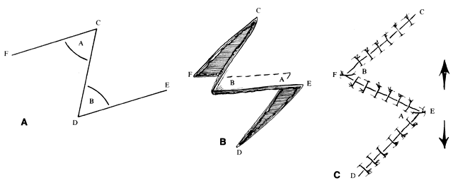
Z-plasty (as well as V-Y and Y-V) is one of the most basic techniques: it may be used in many simple as well as more complicated repairs. This technique (Fig. 2), which employs transposition of two flaps of skin, brings tissue from one direction and adds it to another perpendicular to the first. Z-plasty involves marking and dissecting two triangular flaps, which are transposed. The center of the Z (see Fig. 2, line CD) is aligned on the “short” axis of an area to be lengthened (such as a contracted scar). Each arm of the Z is equal in length and forms angles from 30° to 90°, with angles of up to 60° being the most useful. Larger angles provide the most lengthening but are technically more difficult and are rarely useful. The greater the angle, the greater the percentage gain in length along the central axis; the longer the central member, the greater the actual increase in length.6 A Z-plasty, in addition to being used in other reconstructions, can be used to lengthen a contracted scar, to realign a scar to fall in natural folds (along RSTL), or to make the scar less conspicuous by interrupting its continuity. Long scars can be revised by multiple Z-plasties along their length. Malpositioned structures, such as the brow or canthus, may be repositioned by including them in one of the flaps of a Z-plasty.

Fig. 2. Basic Z-plasty. A. Outline of flaps. All lines equal length. Angles A and B 30° to 90°, 60° maximum practical. B. Flaps undermined and transferring. C. Closure with lengthening parallel to line CD.

V-Y and Y-V Plasty

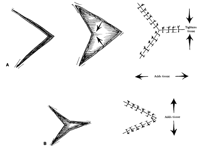
The Y-V and V-Y flaps (Fig. 3) are sliding flaps that also may be combined with other techniques, such as the five-flap procedure for epicanthal folds, which is discussed later. They may be used to close the donor site of another skin flap or to relieve tension on the lid margin, as from a tight scar. The flap is developed by making a V-shaped incision and recessing the central flap, closing to form a Y, (see Fig. 3A) or reversing the process by making a Y-shaped incision, advancing the flap to close as a V (see Fig. 3B). The Y-V flap is useful in the correction of epicanthal folds.

Fig. 3. A. V to Y procedure. B. Y to V procedure. These procedures tighten or add tissue in the direction indicated. They may be combined with other procedures such as Z-plasty.

Subcutaneous Pedicle Flaps

The subcutaneous pedicle flap is a flap of skin that is totally divided from surrounding skin but is left attached to the subcutaneous tissues for its blood supply. Adequate mobilization of the pedicle may allow greater mobility of the skin, and this flap usually combines the technique of V-Y closure. It may be used to close a number of defects in a fashion similar to the V-Y-S flap (Fig. 4A and B), described in the paragraph on medial canthal defects. The flap is elevated by incising all sides of a triangle and gently mobilizing the deep tissues. The skin may then be advanced to fill a defect, with closure of the secondary defect directly (Fig. 4C and D). Two opposing flaps may be elevated for larger defects. With adequate design, flaps such as the V-Y glabellar flap and variations of the median forehead flap (see V-Y Glabellar Flap section) can be developed with a subcutaneous pedicle and advanced to fill medial canthal, lower-lid, or cheek/nasolabial defects. This has the advantage of a one-stage procedure with a flap that can be shaped to lie flat without “dog-ears” of skin. The pedicle must be long enough to reach the recipient site without tension or kinking and to provide vascular elements sufficient to sustain the flap.7

Fig. 4. V-Y-S (A and B) and subcutaneous pedicle flap (C and D). Circular defect with ellipse outlined. A. Flaps incised and rotated. B. V-Y closure forming an S. C. Complete incision in the ellipse with advancement on subcutaneous pedicle. D. Closure using V-Y principle. This can be performed with one or two pedicles.

Rhomboid Flaps and Variations

The rhomboid flap5 is very useful for reconstructing cheek defects. It can also be used with some medial canthal or brow and temple sites. Because the flap has a precise geometric design, its principles help us to understand the principles of flap design and transfer and to execute other transposition flaps. Because of the rich vascularity of the cheek and relatively little concern of viability, randompattern flaps are the rule. They are raised at the level of the subdermal plexus, leaving a layer of subcutaneous tissue attached to the skin. Transposition skin flaps, such as the rhomboid flap, are designed adjacent to the defect and share all or part of one side of the defect.

The flap shapes may vary, for instance being oval rather than rectilinear, but can be thought of as having two sides, a distal end, and a base. If spacing and lesion shape allow, and excessive removal of normal skin is avoided, the rhombic shape is marked around the lesion with two sides parallel to the LME. These lines run perpendicular to the RSTL (see Fig. 1). As will be seen, the vector of traction will be roughly parallel to the LME if well constructed (see Fig. 1 inset; Fig. 5), producing the easiest closure. The flap is ideally drawn with all sides and the short diagonal equal (see Fig. 5, line D'F) in length. Essentially this represents two equilateral triangles base to base. The distal end of the flap (see Fig. 5, point E) is a continuation of the short diagonal of the defect (in effect, the base of the triangles). The sides of the flap, the edges of the defect, and the continuation of the short diagonal are all of equal length. There are four possible donor sites for any given rhomboid defect, two on each side of the defect on the extension of the short diagonal. These can be mentally drawn on the defect to decide which is best (see Fig. 1, inset). The vector of tension (VOT) is roughly parallel to the short diagonal of the flap (see Fig. 5A). If the defect is properly positioned with two sides parallel to the LME, the VOT of two of the possible four flaps will be parallel to the LME (see Fig. 1 inset—dotted lines, flaps 1 and 3). The other two possible flaps will have VOT roughly perpendicular or close to parallel to the RSTL, which is an undesirable condition (see Fig. 1 inset, flaps 2 and 4). Either of the preferred flaps can be used, but proximity to other structures will usually make one preferable over the other. The surrounding skin is pushed, pinched, or pulled to assess the possible distortion produced by each flap and the most advantageous one picked.

Fig. 5. Basic rhomboid flap construction. A. Dotted line D'F is short diagonal of flap. Vector of tension (VOT) is roughly parallel to D'F if flap is well constructed. B. Flap elevated. C. Sutured in place. VOT remains same (arrow). If F, pivot point, does not move adequately, additional VOT parallel to D'F is generated (see text).

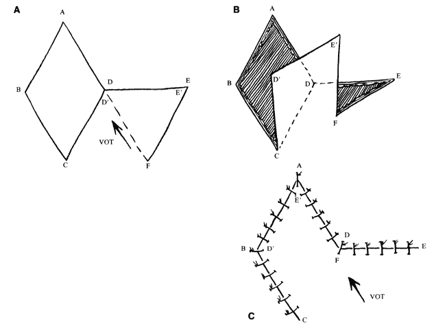
The near corner of the flap base is adjacent to the defect (see Fig. 5A, point C). The far corner of the flap base is called the pivot point of the flap (see Fig. 5A, point F). It is the width of the flap away from the near corner. The pivot point in a classic transposition flap does not move, but this requires a longer flap for closure. However, with this type of rhomboid flap, the pivot point moves toward the junction of the short diagonals of the defect and the flap (see Fig. 5A, point D). This facilitates closure of the donor defect and eliminates the need for a longer flap. If there is adequate tissue and the flap is well designed, it closes nicely with little tension and the VOT is parallel to the short diagonal of the flap (see Fig. 5A, D'F). However, as is sometimes the case, the pivot point is not mobile enough to close the defect easily. In that case, the junction point of the short diagonals (see Fig. 5A, point D) must move toward the pivot point. For this to occur, a near side of the defect Üol 0Ý(see Fig. 5A, line AD) must elongate, the end of the defect (see Fig. 5A, point A) must move toward the pivot point (which tends to widen the defect), or the far side of the defect (see Fig. 5A, line ABC) must move toward the flap. These movements change the shape of the defect and the flap and will induce a second VOT in the axis of the short diagonal of the flap (see Fig. 5C, D'F) as it is sutured in place. If this is excessive, flap viability is compromised.5 In addition, if the far side of the defect moves, it may distort adjacent structures, which is also undesirable; however, undermining the far side of the defect may allow closure with less tension if the pivot point is poorly mobile.

If the VOT is not ideal, it can be changed somewhat by extending or shortening the far side of the flap. By extending the far side of the flap (see Fig. 5A, line EF), the pivot point is moved more toward the long axis of the defect (see Fig. 5A, line AC). The more parallel the VOT is to the long axis of the defect, the less tendency there is for the defect to widen. However, this also moves the pivot point farther away from the junction of the short diagonals, making it more difficult to close the donor defect.

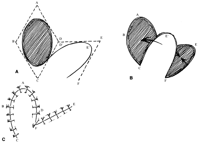
The concepts of the rhomboid flap may also be used with round, oval, and teardrop-shaped defects. For instance, with an oval defect, a rhombus may be drawn around the defect, the flap marked, and then an oval flap designed within the rhomboid to be transposed in a similar fashion (Fig. 6). The principles are the same. In this setting, however, there is somewhat more flexibility in the length of the far side of the flap and its angle with respect to the long axis of the defect. As noted, this allows some modification to the VOT. A longer far side of the flap (see Fig. 6B, line EF) and greater angle between the axis of the flap and the axis of the defect produces a VOT more parallel to the long axis of the defect. (Fig. 6A and B, angle between lines AC and EC, can be changed with oval defects, in contrast to true rhomboids.) As the long axis of the flap rotates, the VOT rotates in the same direction. A VOT more perpendicular to the axis of the defect produces a tendency for the defect to increase in width with closure. A VOT more parallel tends to pull downward on the proximal side of the defect without tending to widen it.5

Fig. 6. Oval defect closed by transposition flap using rhomboid principles. A. Defect with rhomboid lines drawn for design of flap. B. Flap elevated. C. Closure.

Bilobed Flaps

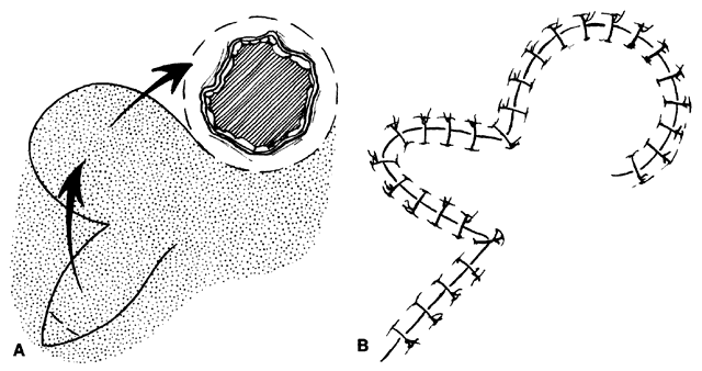
An additional variation on the rhomboid theme is the bilobed flap (Fig. 7). This flap can be used when it is believed that the flap necessary to close a given defect will leave a donor site too large to close primarily. An additional, smaller (about one-half) flap is elevated adjacent to the donor defect and rotated at the same time, leaving a smaller secondary donor defect, which can be closed primarily. This can be used multiply in some areas of thick, inelastic skin, such as around the nose.

Fig. 7. Bilobed transposition flap for closure of a circular defect. A. Outline of incisions. Stippled area is to be undermined. B. After closure.

V-T Closure

Harvey and Corin8 described a simple closure of skin defects not involving the lid margin or lashes. In the typical case of a tumor overlying the tarsus, the lesion is excised by producing a base-up triangle. An infralash incision is performed medially and laterally, extending the base of the triangle as necessary with undermining to allow rotation of skin flaps into the defect. Closure gives horizontal rather than vertical tightening and produces little vertical scarring forces. Lesions close to the lid margin are effectively removed in cases where a vertical ellipse, an alternative closure, may be difficult.

Other

Larger defects in the anterior lamellae, both superior and inferior, can be closed with larger advancement myocutaneous flaps, as described by Anderson and Edwards.9 In the upper lid, they used classic advancement flaps, which may include portions of the brow if necessary to fill upper nasal brow defects. The flap is dissected widely and extends far temporally to allow sufficient advancement. In the lower lid, the flap has similar features to the Mustardé flap in that dissection can be carried temporally as far as the ear if necessary. In this case, however, only the anterior skin muscle lamellae are moved. Flaps of this type can cover large defects if adequately developed.

MARGINAL DEFECTS

Marginal defects can be traumatic in origin, such as from severe motor vehicle accidents with loss of tissue or from human, dog, or other animal bites. More commonly, perhaps, they are the result of excision of malignant tumors.

Techniques vary somewhat for upper and lower lids and depending on whether they are nasal or temporal. A combination of two or more may allow closure of a defect in a more advantageous way than a single one, even though the final result may be similar. For instance, a large (greater than 50%) defect in the nasal lower lid too large to be closed by a Tenzel semicircular flap10,11 might be closed by a Hughes-type tarsoconjunctival flap with a skin graft. This requires closing the central visual axis for up to 6 to 8 weeks. This is undesirable, especially if it involves the only seeing eye. An alternative would be to use a rotation flap of the remaining lower lid moved nasally by performing a lateral canthotomy and cantholysis, thus creating a secondary defect laterally. The secondary defect may then be reconstructed with a rotation flap of tarsus and conjunctiva and a skin flap or graft from the upper lid. This produces a somewhat less objectionable lateral tarsorrhaphy, which will not obstruct vision as much and can be opened at a later time. As will be discussed further on in the chapter, Jordan and co-workers12 believe that nearly any defect bordered by remaining tarsus may be closed by their technique, which is a modification of the Tenzel flap. Yet a better approach might be a transposition flap of tarsus and conjunctiva from the upper lid, with a transposition skin flap or graft from the upper lid producing a reconstruction with no tarsorrhaphy.13,14 These techniques will be briefly illustrated, but there are many possible combinations to fit differing lids, locations, and sizes of defect.

In addition to studying the chapter in this text on general lid surgery3 to review the basic principles of techniques such as lid closure, suturing, and needles, we recommend becoming familiar with lateral and medial tarsal strip procedures and their modifications, as described by Anderson15,16 and others.17,18 These procedures are useful in a wide variety of oculoplastic operations, especially in some reconstructions. Direct closure of a lid defect has numerous advantages, including lack of obstruction of visual axis caused by sharing techniques, no need for tarsal substitutes (see grafts and alloplastic materials), no need for multistage procedures, a less complicated and quicker surgery, and good cosmesis. However, many times more complicated procedures are necessary.

Sliding/Rotation Flaps

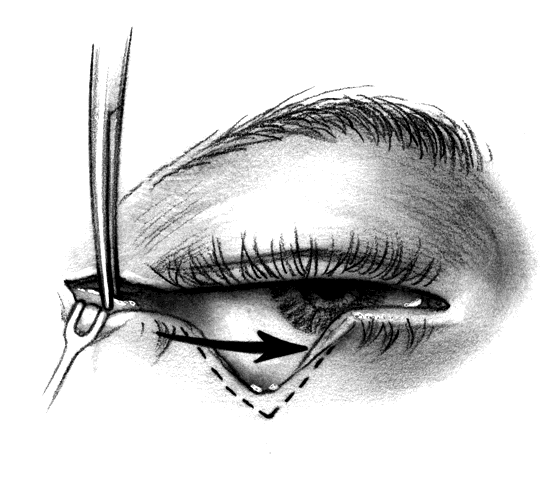
CANTHOTOMY AND CANTHOLYSIS.Lateral canthotomy and cantholysis, which can be applied to either the lower or upper lid, is probably the most useful technique in lid repair when simple primary closure is inadequate. This will allow closure of defects of 25% to 40% or slightly more if the lid is particularly lax. The procedure is simple and is performed with straight, fairly heavy scissors. They are placed horizontally at the lateral canthal angle, and the skin, tendon, and conjunctiva are divided. If fine enough, these same or a smaller pair of scissors are placed on the tendon between the skin and conjunctiva superiorly or inferiorly, and the tendon is divided down to the orbital rim (Fig. 8). The lid can be felt to release and slide nasally when the tendon is completely divided. There are often several strands that fan inferiorly that need to be cut for complete release. If conjunctiva is divided, it can be closed or the open area left to re-epithelialize. The skin is closed laterally at the canthus. Many defects can be closed with this simple technique, which gives excellent cosmetic results.

Fig. 8. Inferior cantholysis to release lid after lateral canthotomy.

If a canaliculus is nonfunctioning and can be sacrificed, a medial canthotomy and cantholysis can be performed in selected cases.19 This can also be combined with a similar lateral procedure to allow closure of central defects without sharing procedures or grafts.

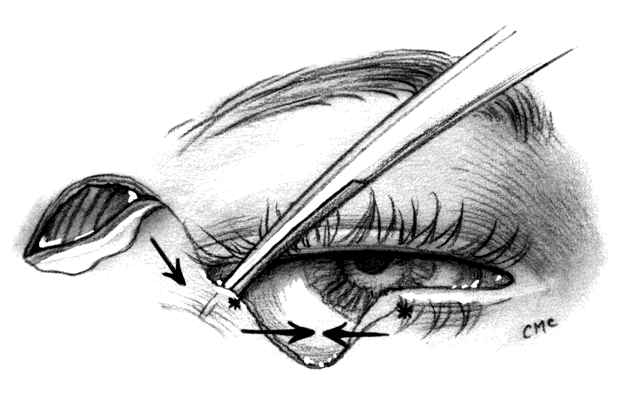
TENZEL ROTATION FLAP. In the mid to late 1970s, Tenzel10 and later Tenzel and Stewart11 described the semicircular flap to reconstruct the central one half of the lid. The results of earlier operations in this area were not as satisfactory as the results with Tenzel's technique, which has since become popular. Jordan and colleagues12 reported on some modifications of this technique in a medium-sized series. They found that they were able to close defects of 50% to 70% of the central and medial margin and defects of almost any size if medial and lateral stumps of tarsus remained. Their modifications involve making a more vertical incision at the canthus, more extensive undermining laterally, and if necessary, division of the conjunctiva and lower-lid retractors to allow further mobilization of lid remnants. Their extensive lateral dissection has the disadvantage of possible injury to the facial nerve (cranial nerve VII).

The semicircular flap technique as originally described10 involves making a skin and muscle incision at the lateral canthus, which curves superiorly and temporally in a semicircular fashion (Fig. 9). A lateral canthotomy and cantholysis are performed to slide the lateral portion of the lid nasally. Of course, this assumes there is a remnant of tarsus available laterally. If there is not, other procedures may be used, such as a sharing procedure from the upper lid with advancement or transposition flaps or a graft. Closing the primary defect with standard techniques may require removing a piece of conjunctiva inferiorly under the primary defect. Tenzel suggested using this tissue to reline the flap laterally. The semicircular skin flap stretches and moves into the area vacated by the tarsal remnant and forms the anterior lamellae of the lid in that area. With slight undermining and sliding, the semicircular flap temporally can be closed primarily. Small dog-ears (Burows' triangles) of skin may need to be removed to achieve a flat closure.

Fig. 9. Tenzel semicircular flap for closure of lower lid defect. Inferior cantholysis to slide lid nasally.

Especially with larger defects, the modification by Jordan and associates,12 wherein they suggested a more vertical incision and inclusion of additional deep muscle by making a beveled incision, would seem to provide some advantage and more lateral support to the new lid. However, the more extensive lateral dissection has the disadvantage of risking injury to the facial nerve. Indeed, as they pointed out, with larger procedures this becomes very similar to the well-known Mustardé rotation flap20 (see next section), although the dissection is more superior (temporal area rather than cheek).

As with the Tenzel10,11 flap, the modification of Jordan and colleagues12 can be used with upper-lid reconstructions as well. The semicircular flap or vertical incision is made inferiorly instead of superiorly, the superior limb of the canthal tendon is divided, the lid remnant is moved nasally, and the skin muscle flap is used to reconstruct the lateral portion of the lid. Careful closure laterally to reattach the new portion of lid to the remaining tendon of the lid is important for support and redevelopment of the canthal angle.

Jordan and co-workers12 also pointed out that their technique ends with apparent excess of tissue at the lid margin in the reconstructed area. This tissue will contract and smooth with time, which may help prevent a lid notch. With large defects, other procedures may be combined with the Tenzel-type flap. McCord and Nunery21 suggested the addition of a periosteal flap elevated from the lateral orbital rim to support the lateral part of the lid. Careful placement of this flap gives good upward support to the lateral lid. A strip about 5 mm in width, with a length appropriate for the defect size, is elevated from the rim and hinged at the orbital rim. It is sutured to the lid remnant nasally and effectively produces a new canthal tendon. The anterior lamella is reformed by the semicircular flap. The inner aspect is lined with elevated conjunctiva. If a large lateral defect is present, an ear cartilage graft (or, for that matter, a nasal chondromucosal, hard palate mucosa, or tarsoconjunctival graft) can be used to reform the inner lamella laterally, with the semicircular flap to reconstruct the anterior lid. Both these modifications can be used with lateral defects that extend to the canthus, where there is no tarsal stump to advance.

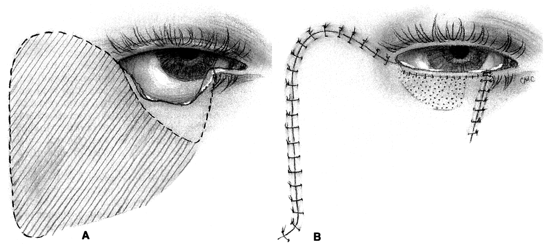
MUSTARDÉ CHEEK ROTATION FLAP. The Mustardé flap20 has the reputation of being a major operation. If necessary, it can be used to close big defects that are difficult to manage by other techniques. Although we perhaps see very large primary tumors less frequently now than in the past, recurrent lesions (especially morphea-type basal cell carcinoma) can be widespread by the time they are detected, resulting in large defects at the time of removal. Large skin grafts require the use of less than optimum donor sites (see Grafts section) or two or more sites. If possible, split-thickness grafts should not be used on the lids or face to avoid contraction. A large flap, with grafts for the posterior lamella, may therefore be the best option and can give satisfactory results when well performed. However, the cheek rotation flap has the inherent disadvantage of producing an inferior vector of traction during healing, which is aggravated by gravitational forces; this makes ectropion or lid retraction with scleral show more likely. Other disadvantages include a large scar and the possible necessity of excising considerable amounts of normal tissue, since in addition to the lid defect from primary tumor excision, a large superiorly based triangle of skin with the nasal edge at the nasolabial fold will also need to be excised. Possible facial nerve damage from the dissection of the full-thickness musculocutaneous flap may also occur.

The flap (Fig. 10) is developed by outlining the area from the lateral canthus extending superiorly and temporally to or above the brow.3,21 This helps to provide superior support to the lateral lid. Many illustrations of this procedure do not emphasize this point. The incision is extended laterally to just in front of the ear and then inferiorly over the jaw. The flap is extensively undermined prior to rotation. The posterior lamella of the lid is reformed by a variety of grafts, again including ear or nasal cartilage, tarsus, or possibly hard palate mucosa. The flap is rotated into position with trimming of the large redundant triangle nasally and of the flap laterally to avoid puckering. If the canthal tendons were removed, careful suturing of the graft and flap to remnants is necessary to provide good support to help prevent ectropion and lid displacement from the globe.

Fig. 10. Mustarde's cheek rotation flap. A. Defect. Dotted line beneath lid is additional tissue to be removed. Striped area is to be undermined. B. Closure. Stippled area is chondromucosal or tarsal graft.

Tarsoconjunctival Flaps

Although many lid defects, even large ones, can be repaired by the relatively easy techniques of canthotomy with cantholysis or semicircular flaps, some defects may be best fixed with a sharing procedure from the opposite lid. Many have been described, including the classic Hughes22 and Cutler-Beard23 operations. Both have been modified, and we will present several examples.

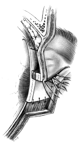
HUGHES PROCEDURE AND MODIFICATIONS. In 1937, Hughes22 presented his first description of the now classic procedure to reconstruct the lower lid and emphasized in a later article the following concept: “When it is necessary to provide new lid structures nothing replaces lid tissue so well as lid tissue itself, both functionally and cosmetically.”24 The original technique involved splitting the upper lid into anterior (skin-muscle) and posterior (tarsoconjunctival) flaps. Because of possible instability or distortion of the lid margin or loss of lashes, this technique has been modified. The generally accepted technique places the incision in the tarsus about 4 mm above the margin, thus leaving the margin intact.

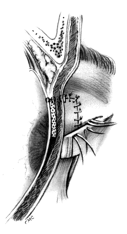
The basic technique for the Hughes procedure has been nicely described by Shore and associates3 and will be outlined only briefly here (Fig. 11). After excision of the lower-lid lesion, the remaining lid segments are drawn toward each other to judge the minimal amount that must be borrowed from the opposite lid. The lid is everted on a retractor or over a plate. In the upper lid, a horizontal incision about 4 mm from the margin is made through tarsus. Vertical incisions are made to the superior tarsal border. The tarsoconjunctival flap is elevated, and the levator muscle of the upper eyelid and Müller's muscle are dissected free. The conjunctiva is left attached and advanced with the tarsus to the lower lid, where the tarsus is sutured to the conjunctiva and lower-lid retractors inferiorly, and medially and laterally to the cut edges of the tarsus remaining in the defect. After thus repairing the posterior lamellae, the anterior can be reconstructed with a skin graft, a transposition flap from the upper lid, an advancement flap from the lower lid inferiorly (increased risk of producing ectropion), or a laterally based advancement flap, depending on the location of the defect and the laxity and availability of skin. The tarsorrhaphy thus created is left in place for 4 to 8 weeks (Hughes suggested 3 months).

Fig. 11. Hughes tarsoconjunctival flap. Tarsus is incised 4 mm above margin and advanced inferiorly to close to the conjunctiva and retractors inferiorly. The defect anteriorly is closed with skin flap or graft.

After opening the lid, margins and donor site may require some trimming, but will re-epithelialize rapidly. Some redness is expected and will fade in 6 months to a year. Care must be taken not to advance the levator tendon, or else lid retraction will result. This modification has an advantage over the original technique in that it does not violate the upper-lid margin or disturb the lashes. Hughes describes grafting lashes from the brow to the lower lid midway through healing. In many cases, a lower lid without lashes gives an acceptable appearance and avoids the possibility of misdirection with trichiasis, but grafting can be considered if cosmesis requires.

Although this basic technique was described and is best used for lower-lid reconstruction, a “reverse” Hughes procedure has been described for upper-lid reconstruction. Not surprisingly, Hughes reported this himself in his 1945 article24 on lid reconstruction. Several authors, including Leone25 and Mauriello and Antonacci,26 have presented their experience with this modification, which technically is similar to the upper-lid procedure. Jordan and colleagues27 also reported using features of the classic Hughes operation to rebuild the central upper lid in patients in whom complete tarsal excision was not necessary. They found that if at least 3 mm of tarsus remained superiorly, it could be advanced to reform the lid margin with appropriate closure to the remaining lid fragments and could be covered with a graft or advancement flap.

Leone25 used virtually the same procedure, supplemented with a reverse Hughes' flap from the lower lid, the combination filling a large upper defect. As with the standard Hughes procedure and other lid-sharing techniques, the reverse Hughes' flap employs tarsorrhaphy for eye closure, which remains for up to several months (also a disadvantage).

A further variation of the Hughes-type tarsoconjunctival flap to close a lateral upper-lid defect was presented by McCord and Wesley.28(pp80–82) Again, this is a modification of a procedure Hughes previously described in which he used aspects of his procedure to help rebuild the upper lid with smaller (one half or less) medial or lateral defects.24 McCord and Wesley elevated a tarsoconjunctival flap centrally, which in effect is a partial-width Hughes' flap. The flap is used to fill the lateral upper-lid defect by rotating it into the lateral defect where it is sutured to the canthal tendon remnants and the lid margin at the edge of the tarsal defect. The flap is then covered by a skin flap or graft.

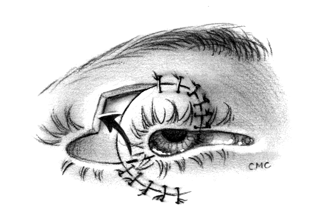
We have also used a modification of the Hughes' flap in what amounts to a large Elschnig's tarsorrhaphy laterally to fill lower-lid defects at the canthus produced by directly excising tumors there or by sliding the lid nasally to fill a defect. The upper lid is split, as Hughes originally described, and a triangular flap based at the lateral canthus is rotated inferiorly to the lower-lid defect with appropriate closure of lid margins. The tarsoconjunctival flap is covered with sliding or advancement flaps inferiorly by a transposition from the upper lid, or by a skin graft. This has given very satisfactory results. The tarsorrhaphy is opened in 6 weeks.

An interesting modification of the classic Hughes' flap was presented by Hargiss,29 in which he avoided the occlusion of the visual axis by bringing the flap down on two tubed vascular pedicles constructed of conjunctiva and Müller's muscle. The tarsoconjunctival flap is elevated in the usual fashion without dissecting Müller's muscle from conjunctiva. A horizontal incision is made through conjunctiva about 1.5 mm above the tarsal border centrally and measuring 6 to 8 mm less than the defect width. The extra conjunctiva above the tarsal border folds anteriorly to form a mucosalined margin. Vertical incisions centrally are then made to form two strips of conjunctiva and Müller's muscle, which are sutured to form tubes. The remainder of the operation is completed by standard techniques. After several weeks, the pedicles can be gently pinched to judge the adequacy of neovascularization to the new lower lid before division. Although this has several additional steps, the procedure has the distinct advantage of not occluding the visual axis.

OTHER TARSOCONJUNCTIVAL FLAPS. In addition to the several techniques already described for rebuilding the lateral lower lid, some of which would produce a tarsorrhaphy, the tarsal transposition flaps described by Hewes and co-workers13 and modified by Leone and Van Gemert14 can be useful in producing a nice reconstruction with a one-stage procedure. A tarsoconjunctival flap, based laterally to include the tendon, is elevated from the upper portion of the lateral tarsus. This avoids the lid margin to avoid distortion or loss of lashes, but care should be taken to avoid the fornix as well so as not to damage the lacrimal secretory system. A flap 4-mm wide is adequate and is fashioned so that it just closes the lower defect with slight tension. Leone and Van Gemert14 suggest taking the flap from the center of the tarsus rather than from the upper border, as suggested by Hewes and associates.13 This obviates the need for closure and avoids possible ductule damage. However, Stephenson and Brown30 found that midtarsal donor sites were associated with greater donor lid distortion and did not recommend them. As with other techniques, the anterior surface can be covered with grafts from a variety of sources or the skin and muscle advancement or transposition flaps as suggested by the above authors.

Cutler-Beard and Other Full-Thickness Procedures

In 1955, Cutler and Beard23 reported a method for repair of partial and total upper-lid defects. As expected, their technique has been modified in an attempt to improve it by avoiding some of its problems. However, the operation is relatively straightforward and gives quite satisfactory results. Basically it involves a full-thickness advancement flap from the lower lid to the upper lid, excluding the margin (Fig. 12). Because the lower tarsus is narrow, it is not included in the flap. The skin, muscle, retractors, and conjunctiva are tunneled under the bridging lid margin and advanced superiorly. The tarsorrhaphy is opened after 2 months. The inferior edge of the margin is freshened and closed to the remains of the flap. Although there is often enough scar tissue or thickening in the flap to give reasonable support to the upper margin and prevent entropion, some believe this lack of tarsal replacement to be inadequate and have modified the procedure by adding sclera,31 cartilage,32 and aorta.33 If completely covered by conjunctiva, an alloplastic material such as polytetrafluoroethylene (Gore-tex, see appendix) might be used.34

Fig. 12. Cutler-Beard procedure after closure. Note bridge of lower lid margin. Stippled area represents sclera graft.

Disadvantages of the Cutler-Beard procedure include possible necrosis of the bridge flap, a serious problem possibly caused by pressure patching after the procedure, and irregularity of the lid margin, loss of lower-lid lashes, and lid retraction or ectropion. Beyer-Machule35 suggested performing a lid-tightening procedure on the lower lid at the time of reconnection of the bridge and the advancement flap. In addition, there are no lashes in the reconstructed upper lid, a failing of most similar operations. As in other tarsoconjunctival flaps, the need to occlude the visual axis for an extended period is a major drawback.

Anderson and colleagues have presented techniques for full-thickness unipedicle36 and bipedicle37 flaps for lower-lid reconstruction. These techniques present alternatives to the above-mentioned procedures and may be suitable where others have failed or are inappropriate. The authors have reported several advantages: (1) an excellent tissue match in a single-stage reconstruction; (2) lack of occlusion of the visual axis; (3) use of tarsus rather than substitutes in the posterior lamella; (4) functional orbicularis anteriorly; (5) pedicle support to the lower lid; and (6) simultaneous repair of canthal defects. Careful study of their report is recommended. These procedures do, however, have disadvantages: (1) their technical aspects require careful attention to the microvascular anatomy of the lid; and (2) the procedures seem somewhat more difficult than the lower-lid procedures already described.

In cases where lashes in the reconstructed upper lid are desirable, a full-thickness pedicle flap from the lower-lid margin to the upper lid (Fig. 13) can be performed. The procedure that Mustardé described38,39 can be used for near-total upper-lid reconstruction when combined with other techniques to repair the donor lower lid. The operation works best for defects of up to one half of the upper lid and uses up to one quarter of the lower lid. The marginal vessels supply the flap until new vessels develop, supporting the lash follicles. The technique has the disadvantages of being a two-staged procedure and partially occluding the visual axis. Normal lid laxity allows closure of the remaining lower lid directly in most cases, and the 6- to 7-mm flap provides enough length to close the upper lid. In larger defects, a larger lower-lid flap may require lower-lid reconstruction by canthotomy and cantholysis or by a Tenzel flap. The pedicle should be not less than 5 mm to avoid the marginal vessels, although the skin may be incised to within 3 mm of the margin. The underlying orbicularis is left intact. Likewise, the conjunctival surface and partial-thickness tarsus may be incised to 3 mm from the margin. The incisions should not be closed under tension. The hinge of the flap should be centered under the center of the upper-lid defect. Care should be taken to avoid the punctum. With larger defects, the levator muscle of the upper eyelid should be sutured to the flap to avoid ptosis. In 2 to 3 weeks,39 the pedicle can be divided; however, some surgeons recommend up to 4 to 6 weeks.28 Minor revision of the lid margin is performed by excision of small triangles vertically from the pedicle edges to allow the margin to close smoothly.

Fig. 13. Full-thickness lower- to upper-lid pedicle flap. Illustrated partially closed. After revascularization, divide at dotted line and revise lid margins.

Sutcliffe40 recently presented the use of a total full-thickness advancement flap of the lower to the upper lid. A relaxing incision is made in the fornix to divide the retractors of the lower lid, and the lash follicles and mucosa at the lid margin are excised. The lid is then advanced to attach superiorly to the levator muscle. The entire tarsus is moved to the upper lid, and when the flap is divided after 3 weeks, the lower tarsal border becomes the lid margin and the incision line the upper-eyelid crease. He reported a good functional and cosmetic result, although both eyelids have no lashes. Purported advantages included simplicity of technique and maximum tissue transfer of all lower-eyelid tissues to the upper eyelid.

MEDIAL CANTHAL DEFECTS

Medial canthal defects are usually the result of excision of malignant lesions. If the lesion requires only skin excision, and if the canthal tendons, canaliculi, and deeper structures are not sacrificed, a skin graft will suffice to cover the defect. It heals quickly, has good color and texture match when obtained from eyelid, has little tendency to retract, and conforms nicely to the concavity of the medial canthus. Leaving such defects to heal by secondary intention has also been reported to produce very satisfactory results, although several weeks of healing may be necessary.41 However, larger or deeper defects—especially if sutures, wires, screws, or plastics are necessary to reform the medial lids, tendons, or lacrimal system—require flap coverage with thicker musculocutaneous flaps. The glabellar and midforehead flaps can supply these tissues. Large anterior lamellar defects in the upper lid can also be covered by the midforehead or temporal forehead flap, although these produce a thicker lid skin than a graft or myocutaneous advancement flap from the eyelid itself.9 However, circumstances may dictate such a flap. In addition to these flaps, smaller ones are also described in the following sections. The V-Y-S and rhomboid flaps can also be useful with smaller defects. We will also briefly discuss some methods for repair of epicanthal folds in blepharophimosis.

V-Y-S-Plasty

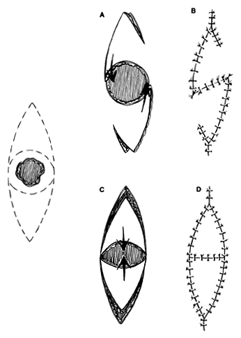
Small, round defects in the medial canthal region can be closed in many ways. Direct closure is possible with small defects, often with undermining. Small dog-ears may need to be excised. However, a straight line closure that crosses the valley of the medial canthal area may bowstring and produce a web. As already noted, skin grafting and healing by secondary intention, or the glabellar flap described later, are options. The V-Y-S-plasty42,43 can be an effective method of closure when direct suturing might distort adjacent structures or produce a web. No undermining in surrounding areas is required or desired. Tissue conservation is maximized. The procedure can be used in other areas of the face as well.

The flaps are designed (see Fig. 4) by outlining the lesion to be excised, with a margin. This is then enclosed by an additional mark, elliptical in shape, that effectively outlines the skin that would be excised and discarded if the defect were to be closed primarily. The long axis is parallel to a natural wrinkle or fold (RSTL) if possible. After excision of the lesion, two marked triangles remain with the defect between. One side of a triangle and the opposite side on the opposing triangle is incised with a short back cut to give an S-shaped configuration. Minimal undermining of the flaps, not the surrounding area, allows them to rotate into the defect for closure. The secondary defects can be closed readily, producing a small V-Y configuration.

V-Y Glabellar Flap

Deep defects in the medial canthus require coverage. Care must be taken in cases of tumor excision to ensure complete removal with frozen-section control. Residual tumor could otherwise be buried, allowing orbital extension. The glabellar V-Y flap (Fig. 14) is taken from the bridge of the nose and glabellar region, occasionally with extension somewhat superiorly to include some skin of the forehead. Color and texture match is reasonably good, and if the defect is deep, the thickness of the skin is not a great disadvantage. Blood supply is good, so a relatively small pedicle will safely support a long flap. Some narrowing of the interbrow distance may be noted but is more a problem with the larger median forehead flap. The flap is raised and rotated/advanced using the V-Y advancement principle (see Fig. 3). The flap can be used to cover defects of up to 15 mm in the medial canthus or larger if combined with other flaps. The height of the flap is about three times its width at its widest inferiorly and is based on the opposite side of the nose. After sliding the flap down, the forehead is closed in an inverted Y. The skin is undermined and usually closes, leaving scars that tend to hide well. The operation has the advantage of being one-staged.

Fig. 14. V-Y glabellar flap. A. Medial canthal defect. Dotted line indicates incision and area to be rotated. B. After closure.

Median Forehead Flap

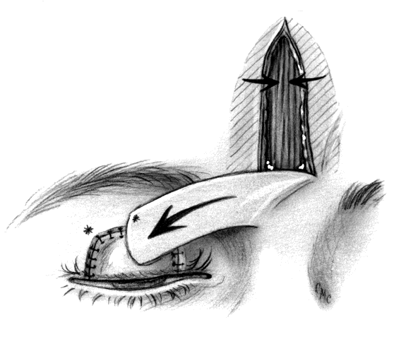
The median forehead flap is used infrequently because other techniques for upper- or lower-lid reconstruction often give better results. Occasionally, however, very large defects of the upper or lower lid and cheek may arise. Circumstances such as loss of both the upper and lower lids due to trauma, or large tumor removal that renders sharing tissue impossible, are rare but not unheard of, and may necessitate its use.

The flap28,44,44a is elevated from the central forehead and is based at the root of the nose (Fig. 15). There is a good blood supply in this area, which makes it possible to create a long, relatively narrow flap. The flap can extend to the hair line if necessary and up to about 2.5 cm in width, slightly more if the forehead is especially loose. The flap is raised in the connective tissue overlying the galea aponeurotica. An estimate of the width of the flap available can be made by making one of the vertical incisions, undermining widely, and overlapping the forehead skin to judge how large a defect can be closed. The superior end of the flap should be tapered to allow smooth closure at the top. Rotating this type of flap will produce a significant dog-ear, which will need to be revised during a second procedure 2 to 3 weeks later. Longer delay may result in the formation of a significant amount of granulation tissue or scarring, or may even result in the pedicle's becoming tubed. However, if this flap is used for total reconstruction of a lid and is lined with a mucous membrane or mucosal-chondral graft, more time must be given for ingrowth of vessels. McCord and Wesley28 recommend 8 weeks. Excising the granulation or scar tissue must be done carefully to avoid damage to the vessels of the flap. The unused portion of the flap may be returned to the forehead after appropriate trimming, thus lessening the narrowing of the interbrow distance.

Fig. 15. Midforehead flap to reconstruct anterior lamella over a tarsal or cartilage graft already sewn in place.

Epicanthal Folds

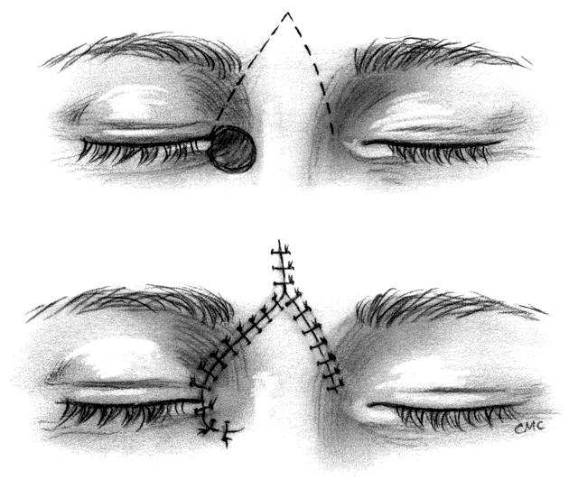
Epicanthal folds occur frequently at birth, but most are minor, produce no significant cosmetic blemish, and diminish with time. Occasionally they are significant and require surgical correction, especially when associated with ptosis in the blepharophimosis-ptosis syndrome. Descriptions of the varieties of folds45 and of the blepharophimosis-ptosis syndrome46 are available elsewhere. Many procedures for the repair of epicanthal folds have been described, including varieties of direct excision, Y-V-plasties, excision of skin from the nose, and a wide variety of flap techniques. All have their disadvantages, including lack of long-lasting effect, scarring, technical difficulty, and suboptimal cosmetic result. Mustardé47 developed a flap technique that improved upon prior procedures and reduced some of the above-mentioned problems. We have used Mustardé's procedure with success but agree with Anderson and Nowinski48 that the measurement of incision lines and angles makes it somewhat difficult. For that reason, we suggest their five-flap technique for this repair in instances where simpler procedures are unsuccessful. The operation combines fairly straightforward double Z-plasty with a Y-V-plasty, thus producing a five-flap operation. It superficially appears similar to Mustardé's procedure, but does not require measurement of lines or angles. Their description also suggests that, after excising excess deep muscle, fat, and fibrous tissue and shortening or repositioning the medial canthal tendon, the flaps fit together more neatly and with less trimming than with Mustardé's procedure.

The basic procedure involves marking the canthus at the medial extent and at a point of the intended new canthal angle. This is approximately one half the distance from the pupil to the center of the nasal bridge. Paramarginal lines superiorly and inferiorly from the canthal angle form the Y. A vertical line through the angle of the Y on the apex of the fold is drawn, and finally back-cut lines equal in length to the paramarginal lines and roughly parallel to them are drawn, thus completing the Z. The flaps are elevated, excess tissue excised, and the Y-V flap advanced with a heavy suture from the canthus to the posterior insertion of the tendon or periosteum. Once the canthus is in position, the flaps are transposed, the apices are closed, and the skin is closed as desired.

Back to Top
GRAFTS
A graft is a tissue fragment moved from one location to another without maintaining vascular or neurologic continuity (compare with free flaps discussed in the Flaps section). Skin is the most common tissue grafted (as well as flapped), but in the lids several other common tissues are used for reconstruction of the posterior lamellae. We will also discuss some uncommon donor sites, which under special circumstances may provide reconstruction resources when other sites are precluded. Autologous grafts are derived from the patient, whereas homologous grafts are harvested, generally from cadavers, and stored and preserved in a variety of ways (e.g., fresh, frozen, tissue culture media [as with corneas], dried). Of course, homologous grafts have the risk of transmitting infectious diseases, which is a great concern. Although some tissues can be sterilized (e.g., gamma irradiation used on fascia lata in tissue banks), this is not always the case, and even the most careful screening cannot guarantee lack of infectious agents. The banking of tissues is time consuming and likely to be expensive, considering the relatively low demand. Not all are commercially available, although fascia, dura, and cartilage are (see appendix). At this time, there are no appropriate heterologous (nonhuman) sources of donor tissue. Tissue culturing, cloning, and other new techniques may in the future give us a safe supply of tissues to replace skin, tarsus, and mucosa.

SKIN

Replacement of skin lost as a result of trauma, tumor removal, or overzealous cosmetic surgery can be carried out by skin grafting or by flaps, as described in the preceding section. The major difficulty is in obtaining skin that is thin enough and pliable enough to be used in the lid, especially the more mobile upper lid. Texture and color match are also important, and these are affected by the type of graft and by the donor site.

Skin grafts are either full or split thickness. Split-thickness grafts tend to darken more with time and sun exposure. Although more pliable initially, they contract more during healing than full-thickness grafts. Because of the loss of sweat and sebaceous glands (see next section, paragraph 1), the texture may also be affected. However, split-thickness grafting allows coverage of large areas, which is difficult with full-thickness grafts because of donor site limitations. Thin split-thickness grafts have a greater viability, especially on poorly vascularized beds, such as irradiated areas or orbital bone after exenteration. Full-thickness grafts give the best texture and color match and exhibit the least contraction. For these reasons, unless there is no other choice, we do not recommend split-thickness grafts on the lid.

Split-Thickness Grafts

Split-thickness skin grafts involve harvesting skin that includes the epidermis and partial thickness of the dermis. They are generally classified as thin, medium, and thick. Thin and medium splitthickness grafts have few or no sweat or sebaceous glands and no hair follicles and relatively little dermis. Thick split-thickness grafts have more dermis, and some of these skin elements may be present. The skin appendages are usually present in full-thickness grafts. Thin split-thickness grafts include only a small amount of superficial dermis, whereas the thicker ones include deeper layers. More dermis produces poorer graft pliability and more contraction. The relative thickness of a graft varies with the donor site. A 0.015-inch split-thickness graft from a donor site of thin skin would be considered a thick graft, but if taken from a thicker donor site would be a medium or thin graft. For instance, a split-thickness graft designed for orbital lining of 0.010 to 0.015 inches6 or lid and facial reconstruction of 0.016 to 0.017 inches49 may be considered a thick graft if the lids or retroauricular area (about 0.017 inches thick) were the donor area, but only a thin split if taken from another area where the skin may be up to 0.150 inches thick. The major advantage of split-thickness grafts is the potential availability of large amounts to cover large defects. Small49,50 advocated the use of split-thickness grafting to cover large defects after Mohs' surgery. His reports show good coverage of large defects, but also a significant incidence of lid malposition secondary to contraction of the grafts, which frequently needed secondary repair. Perhaps a combination of full- and split-thickness grafting might improve this.

Full-Thickness Grafts

Full-thickness grafts contain the full thickness of dermis as well as epidermis and include the dermal glandular structures and hair follicles. Some capillaries may also be included and contribute to later revascularization. Donor sites (see next section) are usually limited to the lids and retroauricular areas because of the thickness of the skin in other sites. However we have used other sites occasionally with satisfactory results in the periorbital area. The best other sites are relatively hairless, but not totally so, and some hairs may be transplanted. The major advantages of full-thickness grafts are better texture, color match, and less contraction.

DONOR SITES. Many possible donor sites are available. At any given site, a split-thickness graft will be more pliable, but eventual texture is sacrificed because of lack of dermal glands. A full-thickness graft from the same site may give better color and texture match than a split-thickness graft, but may be excessively thick or insufficiently pliable or may have poor viability on poorly vascularized sites. The following donor sites are listed in the order of our preference. The first three supply only full-thickness grafts, whereas the rest can be split- or full-thickness. It is always preferable to use full-thickness lid skin when available.

Lid. Upper-lid skin is by far the best for resurfacing the lids. It is very thin, with minimal dermis and virtually no hair. In the elderly or those with early-onset dermatochalasis, fairly large grafts can be harvested. If more is needed, the other lid can serve as a second donor site. Lower-lid skin is similar in quality, but because resection of significant amounts may lead to ectropion, it is less useful. Repair of a postblepharoplasty lower-lid ectropion is often challenging because the skin of the upper lid usually has been excised and discarded. Because this procedure is usually done for cosmetic reasons, it is especially unfortunate that a less-than-ideal graft may need to be placed, compromising the cosmetic result even further. Retroauricular skin is the next best option. Thermal and chemical burns, prior surgery, young age, and other factors may also reduce the availability of skin from the upper lid.

Retroauricular Skin. Skin from behind the ear is a close second to lid skin in thinness, color, and texture, although the dermis is slightly thicker and requires thinning during preparation of the graft. Dermal thinning is performed by stretching the skin on a peg board or by holding it on a gauze square over your finger while gently excising any subcutaneous tissue and thinning the dermis with fine-tipped sharp scissors. The skin is virtually hairless, and sizable grafts can be obtained. An ellipse of skin is taken from the crease at the juncture of the ear and skull and should overlap evenly on either side.3 Any potential donor site should be carefully inspected before use. We have seen a case of rather diffuse basal cell carcinoma involving the back of an ear that had previously been used as a skin donor site to reconstruct a lid with a similar tumor. In contrast to other sites (supraclavicular excepted), the ear can receive a considerable amount of sun exposure.

Genital Area. The foreskin can provide a source of rather large amounts of pliable donor skin. It is quite thin, nearly matching the lids. However, it is often unavailable because of prior circumcision, and the common genital hyperpigmentation can produce a poor color match. Iliff and colleagues6(pp131–132) illustrated a case in which foreskin was used to repair severe cicatricial ectropion of the upper lid secondary to herpes zoster ophthalmicus. The eventual color as well as functional result was quite good, but we have seen another similar case in which the eventual color match was very poor. The assistance of a urologist in harvesting the graft may be helpful.

Supraclavicular Area. The supraclavicular area also can provide reasonably thin, relatively hairless skin, though not in some men. This skin must be thinned somewhat. The graft is taken as an ellipse parallel to the clavicle. In female patients, this may not be a desirable location for a scar, as it is often exposed. This should be considered in preoperative discussions. A split-thickness graft can be harvested by inflation of the subcutaneous tissue with 100 to 200 mL of saline to provide a tense, convex surface on which to apply a dermatome.

Upper Inner Arm and Thigh. The upper inner arm and thigh often can be considered a relatively thin donor site. The amount of hair is variable and must be individually assessed. Again, scar placement in a female patient must be considered, especially if a split-thickness graft is to be harvested, since a large area may be denuded.

Abdomen. Abdominal skin is probably too thick for a full-thickness graft, but we have used it as a split-thickness donor site with success.

TARSUS

Tarsus is clearly the best material for reconstructing the posterior lamella of a lid defect. Other substitutes, such as cartilage (see Cartilage section), tend to be thick and stiff. Sclera often absorbs or is too flexible, resulting in instability. Unfortunately, donor sources are severely limited. We have previously discussed the use of tarsus in flap reconstructions. Free grafting of tarsus may also give a very satisfactory result, but it requires a flap instead of skin grafting for proper vascularization.

Either technique means sacrificing the donor lids' architecture and thereby possibly its stability or the integrity of the lid margin, which may have cosmetic implications.30 Autologous tarsus also has the distinct advantage, lacking in some other substitutes, of having a mucosal lining. This allows faster healing and protection of the eye, especially when used in the upper lid. This is such a valuable consideration in the upper lid that use of lower-lid tarsus to reconstruct an upper-lid defect followed by a graft from another site to repair the lower lid may be preferable to primary reconstruction of the upper with some other substitute. This is more commonly accomplished with flaps, however.

Tarsus as a Free Graft

Because the lower-lid tarsus is so narrow, in order to obtain a reasonable piece the entire tarsus would need to be harvested, sacrificing the lid margin. This is generally not done, but the upper portion of the upper-lid tarsus can be used. The technique is similar in design to the Hughes' flap initially in that a strip of about 4 mm is left at the margin, and the remainder of the tarsus may be harvested after carefully dissecting the levator muscle of the upper eyelid and Müller's muscle from the anterior surface and upper margin. The graft is kept moist until used and, as with any graft, must be in apposition to a vascularized surface to survive.

Brown51 described the use of preserved homologous tarsus with good results for lid reconstruction and repair. She used fresh cadaver tarsus processed by preservation in alcohol or glycerin, similar to bank sclera. The development of a somewhat leathery texture with alcohol preservation was noted, which is not seen when tarsus is preserved in glycerin. Shrinkage was minimal, and oversizing the grafted tissue by about 1 mm was suggested. Donor screening is recommended, but this report predates recognition of the AIDS epidemic, and the use of nonsterilized homologous tissue is now neither warranted nor wise unless no alternatives exist and the patient gives explicit consent.

Jordan and associates52,53 reported on the use of banked, irradiated tarsus in both experimental and clinical reconstructive settings. They detailed their technique of tissue harvesting, storage (similar to Brown's51), and sterilization and described the use of the stored tissue. It would appear that the techniques are simple enough that arrangements with a local eye bank probably could be made to provide a ready source of tissue at a reasonable cost. They noted all the advantages that might be expected from ready availability of the best replacement tissue, despite slight texture changes produced by the storage. The only significant disadvantage seemed to be slight resorption at the exposed lid margin, which is the least nourished area. The tissue must, of course, be covered with a vascularized flap. As already mentioned, preserved homologous tissue must be screened for potential infectious agents, although their sterilization techniques were 10 times the necessary dose for HIV inactivation.

In a large series, Stephenson and Brown30 reported their results of 121 cases involving autologous tarsus used for reconstruction as well as for repair of other problems, such as cicatricial entropion, symblepharon, and distichiasis. They had few problems but did note that when the tarsal graft was taken from midtarsus rather than the upper border, distortion of the donor lid occurred and therefore did not recommend midtarsal grafts.

Shorr and colleagues53 described several techniques for using both contralateral and ipsilateral free tarsal grafts in the management of cicatricial entropion of the upper lid. Portions of tarsus from the superior tarsal border of the same or opposite lid were grafted just superior to the lid margin (2 mm) by making an incision through tarsus and conjunctiva extending across the lid at this point. The lid margin and distal tarsus is rotated anteriorly and inferiorly, making a bed in which the approximately 4-mm graft from either the same or opposite lid is sutured in place. The graft extends slightly beyond the margin, giving the initial appearance of overcorrection. They also described advancing the tarsus in a similar fashion as a bipedicle flap to achieve the same rotation of the lid margin. The margin is incised and rotated, and then the remainder of the tarsus is dissected free from Müller's muscle and the levator muscle of the upper eyelid except medially and laterally, forming a bipedicle flap. The whole tarsus is then advanced inferiorly while hinged medially and laterally. It is sutured in a slightly overcorrected position, as with the grafts.

Composite Grafts

The use of a full-thickness composite lid graft including skin, lashes, lid margin, and tarsus with conjunctiva was introduced by Callahan in55 Although in many cases the lashes fall out, this technique has the potential advantage of replacing missing lashes, a significant cosmetic defect, and leaving a nearly normal lid margin. Callahan initially transplanted skin as well as tarsus and lid margin, requiring revascularization of the entire segment to grow in from the edges, risking sloughing of some segments, especially central margin and lashes.

Putterman56 and subsequently others57–59 have described a modification that greatly enhances viability. After harvesting the graft, the skin and orbicularis 2 to 3 mm superior to the lid margin is excised, leaving tarsus and lid margin. The graft is then covered with a myocutaneous flap to provide a good vascular bed in close approximation to the vital grafted tissues. The small piece of excised skin can be used to help cover secondary defects produced by the flap procedure. Werner and co-workers59 reported a sizable series of such cases in 1993 and showed the clear benefits of composite grafting in selected cases. Lash survival was not high, but the lid margin maintained its configuration well. Obviously, as with other grafting procedures, there is a risk of damage to the donor sites, but this was not a problem in their series and should not be a problem with careful surgical technique. Although this technique cannot reconstruct a total lid as with a Hughes or Cutler-Beard procedure, the one-stage reconstruction with no visual axis occlusion is a benefit. In addition, the repair of the donor site can leave a near-normal appearance of the donor lid, something that cannot always be said of the Hughes, Cutler-Beard, or Sutcliff's procedures (see Flaps section).

What about the larger defects that one composite graft cannot close well, even with the addition of canthotomy and cantholysis or a Tenzel flap? Beyer-Machule and colleagues57 described the double composite flap in 1985 and have reported the histology in two cases.60 This innovation uses two modified composite grafts side by side and covered by a myocutaneous flap. The grafts are taken from any of the remaining lids. Putterman and Migliori58 described repairing large upper- and lower-lid defects, including those involving the canthus and portions of both lids, by using a combination of techniques. Some portions were repaired with standard Hughes or Cutler-Beard flaps, with the addition of composite grafts to fill additional segments. Myocutaneous flaps were used to cover the anterior lamellar defects as necessary.

LASHES

Eyelash transplantation is not technically difficult, but the results are rarely satisfactory and therefore seldom performed. Grafted lashes tend to be unsightly due to an abnormal growth orientation of the new lashes. The lashes of the lower lid are more sparse and tapered and are shorter than those in the upper lid. The lower lid is often cosmetically acceptable without lashes, especially if the upper lid is normal.20 Considering the risk of scar distorting the grafted lashes causing trichiasis, lower-lid grafting is not recommended.

If grafting of lashes to the upper lid is deemed essential, it can be performed. Hughes22 recommended doing so while the lids were still joined after reconstruction. This delays opening the tarsorrhaphy and does not guarantee that distortion and trichiasis will not occur with opening. The graft can be obtained from the brow (ipsilateral or contralateral), the temporal area, or the occipital area behind the ear. Hughes suggested obtaining a strip from the nasal contralateral brow and inverting it prior to suturing in place to give best lash orientation in the lower lid. A narrow strip of skin with two or three rows is chosen from a site where the hairs are vertical. Although survival is not ensured, transplanting more than three rows may give an excessively thick and abnormal appearance if all survive. Too thin is probably better than too thick. Care should be taken to incise the skin so as to not cut through the follicles. The lid margin is incised on the outer edge anteriorly and the graft sutured into the bed. Many lashes will initially fall out but will regrow in 6 to 8 weeks. Hughes22 recommended training the lashes with colloidion, but Mustardé20 rather emphatically stated that in the presence of contracting scar this method does not work. Lashes from nonbrow sites will need to be trimmed occasionally.

CARTILAGE

After tarsus, cartilage is probably the most favored material for reconstructing the posterior part of the lid. It is readily available either autologously61,62 or in the form of irradiated preserved tissue,63,64 although the latter is less commonly seen in ophthalmology operating rooms. It may be obtained either with (nasal or nasal septum) or without (ear or costal) a mucosal lining. The tissue does not shrink appreciably, especially when fresh and when covered with mucosa. Both fairly flat and slightly curved pieces may be obtained, which may be helpful at certain reconstruction sites. The cartilage may readily be trimmed to shape and thinned as necessary, but it is rather friable and it is easy to pull sutures through it. Cartilage has been used interchangeably with tarsus, sclera, periosteum, hard palate mucosa, and other tissues in reconstructions. It does produce a somewhat stiffer lid that may be less mobile for that reason, and we have seen it give the appearance of a free-floating foreign body, albeit accomplishing its designed task: providing a spacer for reducing lower-lid retraction.

MUCOSAL GRAFTS

Other than when used as composite grafts, mucosal grafts are more commonly used for relining contracted sockets and repairing symblepharon and cicatricial entropion than for reconstruction. Conjunctiva is more often utilized as a flap in this setting, and donor tissue is in short supply due to potential donor-site damage. Buccal mucosa, either from the cheek or lower lip, can provide a ready source of lining material in these settings. Both can be harvested as split- or full-thickness grafts, although we prefer the full-thickness type. They are dissected freehand, and the donor site is not closed. Vaginal mucosa has also been occasionally used6(pp219–220) when large areas require coverage, as in a severely contracted socket. Generally this tissue is too thick for routine ocular use.

For reconstruction as well as relining purposes, hard palate mucosa has recently become popular because it provides not only a mucosal lining but is thick enough to replace tarsal function as well.65–69 The articles by Cohen and Shorr65 and Beatty and associates69 give a very thorough review of donor site anatomy and surgical technique as well as a presentation of several procedures using this material. Until becoming well familiar with the surgical anatomy of this area, the potential for significant intraoperative problems (e.g., uncontrollable hemorrhage) suggests that the assistance of an oral surgeon is essential. Donor site problems were reported to be minor, but included pain lasting 7 to 10 days, postoperative bleeding, and 3 to 4 weeks for complete healing.65 Beatty and co-workers69 recommended an intraoral appliance to aid postoperative comfort and hemostasis. Foster and colleagues,70 in a series of 35 patients, reported postoperative bleeding (n = 4), graft necrosis (n = 5), oronasal fistula (n = 1), prolonged pain (n = 2), significant thickening of the lower lid (n = 2), corneal epithelial irregularities (n = 2), and persistent keratinization of the graft (n = 1).

SCLERA

Although sclera, fresh or preserved, has been used for years for various reinforcing procedures on the globe and for wrapping implants used with enucleation, it was first used in lid repair by Quickert and Dryden for lower-lid retraction in71 Its use expanded rapidly72 as a fairly readily available tissue, and it has worked well as a spacer in settings such as cicatricial entropion and lid retraction.6(pp150–157,211),73–75 It has been used to line the posterior surface and will eventually epithelialize but has some tendency to absorb and contract and may be too soft to provide good replacement for tarsus when used alone. Wesley and McCord31 described good results using sclera between the conjunctiva and skin muscle flap of a Cutler-Beard procedure to provide additional stiffness to the reconstructed upper lid.

OTHER

Several additional donor materials have been occasionally used in lid reconstruction and repair. Autologous sites include periosteum,76 which also is used as a flap to reconstruct canthal tendons,21 epiglottis,77 and fascia. Epiglottis is certainly a novel material and not one to jump at you as a likely source. However, Adams and co-workers77 make a good case for its use as a composite graft in lid reconstruction, and it has been used for laryngeal and tracheal repairs. Concern about donor site effects seem to be minimal, as the structure seems to be expendable. Adams and associates quote Pressman and Keleman's Physiology of the Larynx: “The epiglottis is primarily an organ of olfaction. Its fundamental purpose is not to seal over the larynx and protect it during swallowing, and it does not play any important role in phonation. As a matter of fact, the epiglottis in man can be removed in toto with no harmful effects to either swallowing or phonation.” The tissue is lined by mucosa with a thin cartilage lamella, a potentially ideal posterior lid replacement. The major disadvantage is, not surprisingly, the need for expertise in harvesting the tissue, not something in the average ophthalmologist's armamentarium. Bleeding and the need for general anesthesia are also negative factors.

FASCIA LATA

Fascia has been used for years in plastic and reconstructive surgery. There are occasional instances where fascia lata may be used in reconstruction of the lids, such as replacement of a canthal tendon or bridging a gap in a retractor.78 Periosteal flaps are also used in similar situations. Fascia has also been used as a supportive strip or slings in lid and facial malpositions caused by facial nerve (cranial nerve VII) paralysis. However, the most common use in ophthalmology still remains frontalis suspension for severe congenital or acquired ptosis, where we believe it is still the best tissue to use, although many other materials have been suggested (see Alloplastic Materials section).46 Autologous fascia is best, but preserved irradiated fascia works equally well in most cases and is readily commercially available as well (see appendix). Lyophilized fascia has also been used.79

Irradiated homologous aorta has been used by Jordan and colleagues and presented in two reports.33,80 These thorough experimental and clinical reports are similar to their studies of homologous tarsus.52,53 In addition to using this tissue as a layer between conjunctiva and a myocutaneous flap for lid reconstruction, an aortic graft was also used as a sandwich in a Cutler-Beard procedure, as described by Wesley and McCord31 using sclera. The major disadvantage noted was significant resorption when not covered on both sides by vascularized tissue, but this was minimal when placed between vascular layers. They found the working characteristics to be similar to tarsus, and the tissue is plentiful. Although recognizing the inherent difficulties of such a material (harvesting, sterilization, storage, etc.), they offered it as a reasonable addition to our variety of substitutes for tarsus.

Back to Top
ALLOPLASTIC MATERIALS IN EYELID RECONSTRUCTION
Alloplastic materials provide options to the surgical armamentarium in eyelid reconstruction, although they are generally reserved for cases where autologous tissue is unavailable. This section will present those materials currently in use in eyelid reconstruction, but will not delve into those used for orbital surgery or anophthalmic socket problems, although some sources are listed in the appendix. Orbital defects and their reconstruction have been covered elsewhere in this text.

POLYTETRAFLUOROETHYLENE (GORE-TEX AND ePTFE)

Polytetrafluoroethylene (PTFE) is a synthetic, woven, nonabsorbable, pliable, inert, Teflon-related (Du Pont, Wilmington, DE) material available in sheets and strips (see appendix) with several applications in oculoplastic surgery. The original 1-mm thick Gore-Tex patch was made using a random fibrillar structure, which afforded it properties of relative solidity and a pore size of 22 μm, making it difficult for fibrovascular ingrowth to occur.81,82 Any graft not completely covered by conjunctiva or other tissue would extrude. More recently, an expanded PTFE (ePTFE) has been developed, which contains pores of 50 to 70 μm in size, and channels that extend completely through the thickness of the 1-mm sheet. Fibrovascular ingrowth has been demonstrated in these sheets within 4 to 6 weeks.83 Although this material may offer great promise as a spacer graft in the eyelid, the studies are not yet available to support its presumed advantages. ePTFE strips have been used successfully in ptosis surgery84 and to wrap orbital implants following enucleation.83

In the eyelid, PTFE has been used as an interpositional graft in place of cartilage, tarsus, or sclera in the treatment of upper and lower eyelid retraction.82 It has also been advocated as a good substitute to fascia lata used in frontalis suspensions for cases of congenital, myogenic, or traumatic ptosis, where levator function is poor.84,85 PTFE may also be used as a temporary graft to obviate the need for mucous membrane grafting in patients with cicatricial conjunctival disease.86 In these cases, an appropriately shaped PTFE graft is sutured to the edges of the remaining conjunctival epithelium, overlapping the same by about 1 mm. The graft covers the surgical bed created by dissection, as is done in preparation for mucosal grafting. After approximately 3 weeks, or when the underlying bed has epithelialized, the PTFE graft or grafts are removed.

PTOSIS REPAIR BY SUPRAMID SUTURE, SILICONE RODS, AND BANDS

Correcting severe ptosis in the presence of poor levator function is generally accomplished by means of a frontalis suspension. Efforts to simplify this procedure and to allow for postoperative adjustment in eyelid height at any point following the operation have been pursued for decades. In 1948, Friedenwald and Guyton87 described the first suture ptosis repair utilizing a silk suture and a single rhomboid sling. This procedure has been substituted by better materials, including 4-0 nylon polyfilament cable-type suture (Supramid, see appendix) and silicone rods or No. 40 bands, such as those used in scleral buckling surgery (Storz Instruments, see appendix).

Supramid has been widely used for the treatment of all types of ptosis cases with poor levator function, perhaps in part because of the ease of handling and its ready commercial availability, despite a significant failure and complication rate. In their comparative study of Supramid and homologous fascia lata, Wagner and co-workers88 found that 40% of Supramid eyelids failed either because of recurrence of ptosis or granuloma formation compared with an 8% failure rate in the fascia group. Ptosis recurrence is thought to be due to cheese-wiring. As a result, Supramid is reserved for temporary ptosis repair, as in complete congenital ptosis, where a later permanent procedure is planned, and for some cases of myogenic ptosis (e.g., chronic progressive external opthalmoplegia [CPEO], oculopharyngeal syndrome), where reversibility of the sling may be a high priority and where the risk of cheese-wiring is minimized by the presence of poor orbicularis function in those patients.

Silicone rods and bands have been reported89,90 to be successful sling materials and to be better tolerated by the eyelids with a greater success rate. They have demonstrated an additional advantage of causing little, if any, tissue reaction, making reoperations for height adjustments simple long after the initial surgery.89 Their elasticity was touted as an advantage early on, anticipating that it would improve eyelid closure during blinking without compromising height, but enthusiasm for this and other potential benefits has been dampened by late failures.6 Silicone slings are best reserved for cases of myogenic ptosis, where the orbicularis function is affected along with the function of the levator extraocular muscles, reducing the forces that may lead to cheese-wiring and thus improving their long-term success.

GOLD WEIGHTS

Improving the dynamic function of the eyelid in patients with cranial nerve VII paralysis has become the goal in the visual rehabilitation of these patients in preference to previously favored, visually obstructive, and more disfiguring procedures such as tarsorrhaphy. In patients with permanent paralysis, the use of a gold weight is particularly attractive because it may offer a long-term benefit. In many instances, placement of the gold weight is coupled with a horizontal tightening of the lower eyelid in the presence of significant paralytic ectropion.91,92

Gold weights are available in 0.6-, 0.8-, 1.0-, 1.2-, 1.4-, and 1.6-g sizes. Early investigators have tested other materials, such as lead93 and tantalum gauze,94 but gold gained wider acceptance because of its inert nature and higher density (permitting the use of less bulky implants). Recent modifications have been made in gold implants to make them thinner and to provide smoother, more tapered edges. Both the original and modified gold weights may be obtained from MEDDEV (see appendix). Proper placement of the weight in a pocket dissected under the levator aponeurosis through a small eyelid crease incision and fixation of the weight to the tarsal plate maximize the implant's stability and have also been reported to reduce the risk of extrusion.95,96 Centration and the weight of the implant to be placed should be determined preoperatively with the patient in the sitting position. A “silver-weight sizing” kit is available from the manufacturer at a moderate cost, which allows for ordering of the individual weights instead of requiring the maintenance of a full stock. A weight that maximizes closure of the eyelid during a passive blink without producing unacceptable ptosis in the primary position should be selected.

Back to Top
REFERENCES

1. Libonati MM: General anesthesia. In Tasman W, Jaeger EA (eds): Clinical Ophthalmology, Vol 6, Chap 1. Philadelphia, JB Lippincott, 1992

2. Wilson RP: Local anesthesia in ophthalmology. In Tasman W, Jaeger EA (eds): Clinical Ophthalmology, Vol 6, Chap 2. Philadelphia, JB Lippincott, 1992

3. Shore JW, McCord CD, Popham JK: Surgery of the eyelids. In Tasman W, Jaeger EA (eds): Clinical Ophthalmology, Vol 6, Chap 123. Philadelphia, JB Lippincott, 1992

4. Wilkins RB, Kulwin DR, McCord CD Jr, Tanenbaum M: Skin and tissue techniques. In McCord CD, Tanenbaum M (eds): Oculoplastic Surgery, Ch 1, 2nd ed. New York, Raven Press, 1987

5. Gunter JP, Sheffield RW: Transposition cheek skin flaps: The rhombic flap and variations. In Strauch B, Vasconez LO, Hall-Findley EJ (eds): Encyclopedia of Flaps, Vol 1, Chap 83. Boston, Little, Brown & Co, 1990

6. Iliff CE, Iliff WJ, Iliff NT: Oculoplastic Surgery. Philadelphia, WB Saunders, 1979

7. Barron JN, Saad MN: Subcutaneous pedicle skin flaps. In Strauch B, Vasconez LO, Hall-Findley EJ (eds): Encyclopedia of Flaps, Vol 1, Chap 38. Boston, Little, Brown & Co, 1990

8. Harvey J, Corin S: V to T reconstruction of anterior lamellar eyelid defects. Ophthalmic Surg 20:497, 1989

9. Anderson RL, Edwards JJ: Reconstruction by myocutaneous eyelid flaps. Arch Ophthalmol 97:2358, 1979

10. Tenzel RR: Reconstruction of the central one half of an eyelid. Arch Ophthalmol 93:125, 1975

11. Tenzel RR, Stewart WB: Eyelid reconstruction by semicircular flap technique. Trans Am Soc Ophthalmol Otolaryngol 85:1164, 1978

12. Jordan DR, Anderson RL, Holds JB: Modifications to the semicircular flap technique in eyelid reconstruction. Can J Ophthalmol 27:130, 1992

13. Hewes EH, Sullivan JH, Beard C: Lower eyelid reconstruction by tarsal transposition. Am J Ophthalmol 81:512, 1976

14. Leone CR, Van Gemert JV: Lower eyelid reconstruction with upper eyelid transposition grafts. Ophthalmic Surg 11:315, 1980

15. Anderson RL, Gordy DP: The tarsal strip procedure. Arch Ophthalmol 97:2192, 1979

16. Anderson RL: Tarsal strip procedure for correction of eyelid laxity and canthal malposition in the anophthalmic socket. Ophthalmology 88:895, 1981

17. Jordan DR, Anderson RL, Thiese SM: The medial tarsal strip. Arch Ophthalmol 108:120, 1990

18. Tenzel RR: Treatment of lagophthalmos of the lower lid. Arch Ophthalmol 81:336, 1969

19. Holds TB, Anderson RL: Medial canthotomy and cantholysis. Am J Ophthalmol 116:218, 1993

20. Mustardé JC: Reconstruction of eyelids. In: Repair and Reconstruction in the Orbital Region: A Practical Guide, Ch 7, pp 116–162. Baltimore, Williams & Wilkins, 1966

21. McCord CD Jr, Nunery WR: Reconstruction of the lower eyelid and outer canthus. In McCord CD Jr, Tanenbaum M (eds): Oculoplastic Surgery, 2nd ed, Ch 4, pp 105–110. New York, Raven Press, 1987

22. Hughes WL: A new method for rebuilding a lower lid. Arch Ophthalmol 17:1008, 1937

23. Cutler NL, Beard C: A method for partial and total upper lid reconstruction. Am J Ophthalmol 39:1, 1955

24. Hughes WL: Reconstruction of the lids. Am J Ophthalmol 28:1203, 1945

25. Leone CR: Tarsalconjunctival advancement flaps for upper eyelid reconstruction. Arch Ophthalmol 101:945, 1983

26. Mauriello JA, Antonacci R: Single tarsoconjunctival flap (lower eyelid) for upper eyelid reconstruction (“reverse” modified Hughes procedure). Ophthalmic Surg 25(6):374, 1994

27. Jordan DR, Anderson RL, Nowinski TS: Tarsoconjunctival flap for upper eyelid reconstruction. Arch Ophthalmol 107:599, 1989

28. McCord CD Jr, Wesley RE: Reconstruction of the upper eyelid and medial canthus. In McCord CD Jr, Tanenbaum M (eds): Oculoplastic Surgery, 2nd ed, Ch 3. New York, Raven Press, 1987

29. Hargiss JL: Bipedicle tarsoconjunctival flap. Ophthalmic Plast Reconstr Surg 5(2):99, 1989

30. Stephenson CM, Brown BZ: The use of tarsus as a free autologous graft in eyelid surgery. Ophthalmic Plast Reconstr Surg 1:43, 1985

31. Wesley RE, McCord CD Jr: Transplantation of eyebank sclera in the Cutler-Beard method of upper eyelid reconstruction. Ophthalmology 87:1022, 1980

32. Callahan A: Lid reconstruction. In Tessier P, Callahan A, Mustardé JC, Salyer KE (eds): Symposium on Plastic Surgery in the Orbital Region. St. Louis, CV Mosby, 1976

33. Jordan DR, McDonald H, Anderson RL: Irradiated homologous aorta in eyelid reconstruction: Part II. Human data. Ophthalmic Plast Reconstr Surg 10:227, 1994

34. Karesh JW, Fabrega MA, Rodrigues MM: Interpositional polytetrafluoroethylene grafts. Ophthalmic Plast Reconstr Surg 7(4):278, 1991

35. Beyer-Machule CK: Upper and lower lid reconstruction. In Beyer-Machule CK, von Noorden GK (eds): Atlas of Ophthalmic Surgery. New York, Thieme-Stratton, 1985

36. Anderson RL, Jordon DR, Beard C: Full thickness unipedicle flap for lower eyelid reconstruction. Arch Ophthalmol 106:122, 1988

37. Anderson RL, Weinstein GS: Full thickness bipedicle flap for total lower eyelid reconstruction. Arch Ophthalmol 105:570, 1987

38. Mustardé JC: Surgery of the medial canthus. In: Repair and Reconstruction in the Orbital Region: A Practical Guide, Ch 11, pp 195–211. Baltimore, Williams & Wilkins, 1966

39. Mustardé JC: Skin-muscle-tarsoconjunctival (Esser) flap from the lower to the upper eyelid. In Strauch B, Vasconez LO, Hall-Findley EJ (eds): Encyclopedia of Flaps, Vol 1, Chap 20. Boston, Little, Brown & Co, 1990

40. Sutcliffe RT: Upper eyelid reconstruction by full thickness lower eyelid sliding flap. Poster presentation, American Society of Ophthalmic Plastic and Reconstructive Surgery, Atlanta, 1995

41. Fox SA, Beard C: Spontaneous lid repair. Am J Ophthalmol 58:947, 1964

42. Argamoso RV: V-Y-S plasty for closure of a circular defect of the medial canthal area. In Strauch B, Vasconez LO, Hall-Findley EJ (eds): Encyclopedia of Flaps, Vol 1, Chap 25. Boston, Little, Brown & Co, 1990

43. Argamoso RV: V-Y-S plasty for closure of a round defect. Plast Reconstr Surg 53:99, 1974

44. Mustardé JC: Tissue loss—deep. In: Repair and Reconstruction in the Orbital Region: A Practical Guide, Ch 4, pp 57–85. Baltimore, Williams & Wilkins, 1966

44a. Mustardé JC: Techniques for transfer of resurfacing materials. In: Repair and Reconstruction in the Orbital Region: A Practical Guide, Ch 19, pp 353–375. Baltimore, Williams & Wilkins, 1966

45. Johnson CC: Epicanthus. Am J Ophthalmol 66:939, 1968

46. Iliff WJ, Pacheco EM: Ptosis surgery. In Tasman W, Jaeger AE (eds): Clinical Ophthalmology, Chap 117. Philadelphia, JB Lippincott, 1992

47. Mustardé JC: Ptosis: epicanthus and telecanthus. In: Repair and Reconstruction in the Orbital Region: A Practical Guide, Ch 18, pp 338–349. Baltimore, Williams & Wilkins, 1966

48. Anderson RL, Nowinski TS: The five flap technique for blepharophimosis. Arch Ophthalmol 107:448, 1989

49. Small RG, Sahl WJ: The use of split-thickness skin grafts for eyelid and facial reconstruction after Mohs' fresh-tissue surgery. Ophthalmic Plast Reconstr Surg 5(4):266, 1989

50. Small RG: The use of split-thickness skin grafts in ophthalmic plastic surgery. Ophthalmic Surg 3:154, 1972

51. Brown BZ: The use of homologous tarsus as a donor graft in lid surgery. Ophthalmic Plast Reconstr Surg 1:91, 1985

52. Jordan DR, Tse DT, Anderson RL, Hanson SO: Irradiated homologous tarsal plate banking: A new alternative in eyelid reconstruction, Part I. Ophthalmic Plast Reconstr Surg 6(3):158, 1990

53. Jordan DR, Tse DT, Anderson RL, Hanson SO: Irradiated homologous tarsal plate banking: A new alternative in eyelid reconstruction: Part II. Ophthalmic Plast Reconstr Surg 6(3):168, 1990

54. Shorr N, Christenbury JD, Goldberg RA: Tarsoconjunctival grafts for upper eyelid cicatricial entropion. Ophthalmic Surg 19:316, 1988

55. Callahan A: The free composite lid graft. Arch Ophthalmol 45:539, 1954

56. Putterman AM: Viable composite grafting in eyelid reconstruction. Am J Ophthalmol 85:237, 1978

57. Beyer-Machule CK, Shapiro A, Smith B: Double composite lid reconstruction: a new method of upper and lower lid reconstruction. Ophthalmic Plast Reconstr Surg 1:97, 1985

58. Putterman AM, Migliori ME: Combined viable composite grafting and eyelid sharing techniques to prevent blepharoptosis after extensive tumor excision. Am J Ophthalmol 106:53, 1988

59. Werner MS, Olson JJ, Putterman AM: Composite grafting for eyelid reconstruction. Am J Ophthalmol 116:11, 1993

60. Budenz DL, Beyer-Machule CK, Albert DM: Histology of partial thickness double composite eyelid graft. Ophthalmic Surg 20:362, 1989

61. Baylis HI, Rosen N, Newhaus RW: Obtaining auricular cartilage for reconstructive surgery. Am J Ophthalmol 93: 709, 1982

62. Baylis HI, Perman KI, Felt DR, Sutcliffe RT: Autologous auricular cartilage grafting for lower eyelid reconstruction. Ophthalmic Plast Reconstr Surg 1:23, 1985

63. Linberg JV: Preserved irradiated homologous cartilage for orbital reconstruction. Ophthalmic Surg 11:457, 1980

64. Schenk W, Linberg JV, McCormick S: Preserved irradiated homologous cartilage implant in canine eyelids. Ophthalmic Plast Reconstr Surg 1:267, 1985

65. Cohen MS, Shorr N: Eyelid reconstruction with hard palate mucosa grafts. Ophthalmic Plast Reconstr Surg 8:183, 1992

66. Silver B: The use of mucous membrane from the hard palate in the treatment of trichiasis and cicatricial entropion. Ophthalmic Plast Reconstr Surg 2(3):129, 1986

67. Kersten RC, Kulwin DR, Levartovsky S et al: Management of lower lid retraction with hard palate mucosal grafting. Arch Ophthalmol 108:1339, 1990

68. Hollsten DA, Ross J: Considerations on the use of hard palate grafts in eyelid reconstruction. Ophthalmology 96: 117, 1991

69. Beatty RL, Harris G, Bauman GR: Intraoral palatal mucosal harvest. Ophthalmic Plast Reconstr Surg 9:120, 1993

70. Foster JA, Holck DE, Dutton JJ et al: Complications of hard palate mucosal grafts. Symposium Presentation, American Society of Ophthalmic Plastic and Reconstructive Surgery, Atlanta, 1995

71. Quickert ME, Dryden RM: Lower eyelid advancement. Symposium Presentation, American Society of Ophthalmic Plastic and Reconstructive Surgery, Las Vegas, 1971

72. Soll DB: Scleral transplantation in ophthalmic plastic surgery. Trans Am Ophthalmol Otolaryngol 83:679, 1977

73. Doxanas MT, Dryden RM: The use of sclera in the treatment of dysthyroid ophthalmopathy. Ophthalmology 88: 887, 1981

74. Flanagan JC: Eyebank sclera in oculoplastic surgery. Ophthalmic Surg 5(3):45, 1980

75. Dryden RM, Soll DB: The use of scleral transplantation in cicatricial entropion and eyelid retraction. Trans Am Soc Ophthalmol Otolaryngol 83:669, 1977

76. Hurwitz JJ, Corin SM, Tucker SM: The use of free periosteal grafts in extensive lower lid reconstruction. Ophthalmic Surg 29:415, 1989

77. Adams JJ, Olson NR, Siders DB: The use of the epiglottis as an autologous composite graft in eyelid reconstruction. Ophthalmic Plast Reconstr Surg 9:206, 1993

78. Beyer CK, Albert DM: The use and fate of fascia lata and sclera in ophthalmic plastic and reconstructive surgery. Ophthalmology 88:869, 1981

79. Broughton WL, Matthews JG, Harris DJ: Congenital ptosis: results of treatment using lyophilized fascia lata for frontalis suspension. Ophthalmology 89:1261, 1982

80. Jordan DR, McDonald H, Addison DJ et al: Irradiated homologous aorta in eyelid reconstruction, Part I. Ophthalmic Plast Reconstr Surg 10:75, 1994

81. Karesh JW: Polytetrafluoroethylene as a graft material in ophthalmic plastic and reconstructive surgery. Ophthalmic Plast Reconstr Surg 3:179, 1987

82. Karesh JW, Fabrega MA, Rodrigues MM et al: Polytetrafluoroethylene as an interpositional graft material for the correction of lower eyelid retraction. Ophthalmology 96:419, 1989

83. Durette JF, Dube I, Winter MH et al: Orbital implants covered with expanded polytetrafluoroethylene (ePTFE): practical considerations and early clinical results. J Am Soc Ocularists 25:10, 1994

84. Adenis JP, Lebraud P, Mathon M: Utilisation du PFTE (Goretex) dans la suspension palpebro-frontale pur le ptosis. J Fr Ophtalmol 10:604, 1987

85. Arnaud B, Dupeyron G, Malrieu C: Utilisation de Gore-tex dans la chirurgie du ptosis par suspension au muscle frontal. Bull Soc Ophtalmol Fr 3:483, 1989

86. Levin PS, Dutton JJ: Polytef (Polytetrafluoroethylene) alloplastic grafting as a substitute for mucous membrane. Arch Ophthalmol 108:282, 1990

87. Friedenwald JS, Guyton JS: A simple ptosis operation: utilization of the frontalis by means of a single rhomboid-shaped suture. Am J Ophthalmol 31:411, 1948

88. Wagner RS, Mauriello JA, Nelson LB et al: Treatment of congenital ptosis with frontalis suspension: a comparison of suspensory materials. Ophthalmology 91:245, 1984

89. Tillett CW, Tillett GM: Silicone sling in the correction of ptosis. Am J Ophthalmol 63:521, 1966

90. Leone CR, Rylander G: A modified silicone frontalis sling for the correction of blepharoptosis. Am J Ophthalmol 85: 802, 1978

91. Seiff SR, Chang J: Management of ophthalmic complications of facial nerve palsy. Acoustic Neuroma II 25:669, 1992

92. Soll DB: New surgical approaches to the management of ocular exposure secondary to facial paralysis. Ophthalmic Plast Reconstr Surg 4:215, 1988

93. Smellie GD: Restoration of the blink reflex in facial palsy by a simple lid-load operation. Br J Plast Surg 19:279, 1966

94. Sheehan E: Progress in correction of facial palsy with tantalum wire and mesh. Surgery 27:122, 1950

95. Jobe RP: A technique for lid loading in the management of lagophthalmos of facial palsy. Plast Reconstr Surg 53:29, 1974

96. Seiff SR, Sullivan JH, Freeman LN et al: Pretarsal fixation of gold weights in facial nerve palsy. Ophthalmic Plast Reconstr Surg 5:104, 1989

Back to Top
APPENDIX

We have listed here some sources for tissue and alloplastic materials used in lid surgery and some orbital reconstruction as well, although the latter is not discussed in the text.


Tissue/MaterialManufacturer/Distributor
Fascia lata, dura, demineralized bone, bone (several varieties), pericardium, temporalis fasciaBiodynamics International (813) 979-0042 Distributes through: IOP,3100 Airway Ave., Costa Mesa, CA 92626;(800) 535-3545
 and
 OR Specialties, Inc., PO Box 466, Lutherville, MD 21093; (800) 673-8598,FAX(410) 560-1397
Cornea, sclera, skin, cartilage, Biodynamics productsTissue Banks International, 815 Park Avenue, Baltimore, MD 21201; (410)752-2020
 Distributes through:
  OR Specialties, Inc., PO Box 466, Lutherville, MD, 21093; (800) 673-8598,FAX(410) 560-1397
Fascia lataUniversity of Toronto, Fascia-Lata Bank, 1 Spadian Crescent, Toronto,Canada M55 2J5CAN;(416)978-7355
SupramidS. Jackson Inc., 4815 Rugby Ave., Washington, DC 20014; (800) 368-5225
Hydroxyapatite implantsIntegrated Orbital Implants, 12526 High Bluff Drive, Suite 300, San Diego,CA 92130; (619) 792-3565
PTFE-Gore-Tex: Mycro Mesh, Soft tissue patch, facial implant, cardiovascular patch, surgical membrane, sutureW. L. Gore & Assoc., 3750 West Kiltie Lane, Flagstaff AZ 86002-0900; (602) 779-2771, FAX (602) 779-1456; order info (800) 528-8763, tech info (800) 437-8181
ePTFE: ePTFE-covered spheres and implants, covers, orbital floor implants, conformersOculo-Plastik, 1170 Henri-Bourassa E., Montreal, Quebec, Canada H2C IG4; (514) 381-1849, (800) 363-7004, FAX (514) 381-1164
ePTFEIMPRA, PO Box 1740, Tempe, AZ 85280-1740; (602) 894-9515
SiliconeStorz Instruments, 3365 Tree Court Indust. Blvd., St. Louis, MO 63122; (800) 325-9500
Gold Weights testing kit (silver)MEDDEV, PO Box 1352, Los Altos, CA 94023-1352;(800) 543-2789
MEDPORE-Porous Polyethylene Spheres, plates, blocksPorex Surgical, 4715 Roosevelt Hgwy., College Park, GA 30349; (404)969-8145, (800) 521-7321, FAX (404) 969-8045


Back to Top