Chapter 82
Enucleation, Evisceration, and Exenteration Techniques
MARK ALFORD*
Main Menu   Table Of Contents

Search

*The author has no financial interest in any of the products discussed in this manuscript.

HISTORICAL PERSPECTIVE
ALTERNATIVES TO REMOVAL OF THE EYE
ENUCLEATION AND EVISCERATION
MANAGEMENT OF THE ANOPHTHALMIC SOCKET
EXENTERATION
CONCLUSION
REFERENCES

The removal of an eye or the orbital contents is one of the most serious and difficult decisions a patient and physician must consider. The patient facing the loss of an eye has often recently been through multiple ophthalmic surgeries, experienced severe ocular trauma, or been diagnosed with a potentially life-threatening disease. Patients facing the removal of an eye are often feeling depressed and overwhelmed.

Enucleation is removal of the entire globe, including the cornea, sclera, and a portion of the optic nerve (Fig. 1). Evisceration surgery involves removal of the contents of the globe while leaving the sclera and optic nerve in place. The cornea can be retained in some evisceration cases. Exenteration is the removal of the globe, as well as the complete or partial removal of the soft tissues of the orbit. The ultimate goals of these surgeries are to safely and effectively remove the diseased eye or orbital contents using advanced surgical techniques, eliminate the severe underlying ocular pathology, and provide excellent long-term cosmesis. In addition to the surgical procedure, the ophthalmologist often must coordinate the patient's ancillary care such as radiation therapy, systemic work-up, and prosthetic evaluations. Although every surgeon who performs enucleations, eviscerations, or exenterations must provide emotional support to the patients; certain patients will need psychologic referrals and contact with support groups.

Fig. 1. The globe is removed from the socket using silk traction sutures. The optic nerve is clamped with a large hemostat.

The objective of this chapter is to provide the reader with a general understanding of underlying principles and surgical techniques to manage this challenging group of patients. The indications and contraindication for enucleation, evisceration, and exenteration are discussed. Surgical techniques and an explanation of the various implant materials are reviewed. The chapter also provides guidance in managing the anophthalmic patient postoperatively.

Back to Top
HISTORICAL PERSPECTIVE
Dating back thousands of years, there is evidence that the Egyptians and Sumerians were making artificial eyes and performing enucleations. It was not until the 1500s that enucleations were reported in the medical literature. Noyes1 first reported evisceration of the ocular contents in 1874. In 1885, Mules was the first physician to report the use of an orbital implant, a glass sphere, after an evisceration.2 Advances in surgical techniques, anesthesia, implant materials, and prosthetics over the past decade have greatly improved surgical outcomes and patient satisfaction. In addition, the ability to deal with the long-term complications of the anophthalmic socket such as enophthalmos, ptosis, contraction, exposure, extrusion, and lower lid laxity have improved significantly. Today, most patients can confidently return to their daily activities with good cosmetic results following the removal of an eye.
Back to Top
ALTERNATIVES TO REMOVAL OF THE EYE
Not all blind, painful disfigured globes require removal. In many cases, patients are relieved to try an alternative treatment to surgery while adjusting to the loss of vision. The use of topical steroids, cycloplegics, antiglaucoma medications, and oral analgesics can provide relief for varying periods. The use of retrobulbar alcohol injections has been used in the past in selected patients. It has been my experience that alcohol injections are painful, provide temporary relief, and makes future enucleation surgery more difficult because of the postinjection scarring of the orbit. In a debilitated patient that is a very poor surgical risk, it is a reasonable choice for temporary pain control. The only permanent solution is removal of the eye, which is often safer and definitely more effective than alternative treatments.

Patients with blind, disfigured eyes without pain can be managed effectively with a custom made, shell conformer (Figs. 2 and 3). Conformers provide excellent cosmesis and good motility for many patients. A custom fit by a skilled ocularist is essential for a good appearance and comfort. Corneal sensitivity and globe phthisis may limit the patient's ability to tolerate the shell or for the ocularist to create a cosmetically acceptable appearance. Although the appearance is improved with a shell conformer, the eye must continue to be followed for problems. Because the risk of uveal melanomas in phthisical eye is reported to be between 4% and 15%, routine ocular ultrasound should be performed in atrophic eyes.3

Fig. 2. Note the small phthisical left eye.

Fig. 3. The same patient with a custom shell over the left eye.

The likelihood of sympathetic ophthalmia should also be discussed with any patient deciding against removal and electing to keep a traumatized or phthisical eye. Sympathetic ophthalmia is a rare, bilateral granulomatous panuveitis that occurs after injury to one eye. The exact incidence is unknown but reports have shown it to be as common as 0.28% to 1.9% following penetrating trauma.4 The incidence is less following intraocular surgery. The cause of sympathetic ophthalmia is unknown, but theories include hypersensitivity to retinal or uveal pigments, infectious agents, or sensitivity to retinal S antigen.5,6 It is believed that enucleation within 2 weeks of the initial injury prevents the onset of sympathetic ophthalmia. Some reports suggested that the removal of the exciting, traumatized eye within 2 weeks of the identification of sympathetic ophthalmia might benefit the other eye with a more benign course and better visual outcome.7,8

Enucleation during childhood has been reported to cause orbital growth retardation.9 Promoting bony growth remains a challenge to the oculoplastic surgeon. Traditional treatment includes progressive conformers, orbital implant exchange, orbital expanders, dermis fat grafts, and craniofacial surgery. However, Fountain and coworkers10 have shown that children who undergo enucleation leading to acquired anophthalmia at an early age maintain normal orbital and periorbital soft tissue growth when large static spherical implants are placed at the time of surgery. Implant exchange and enlarging conformers were not found to be necessary.

Back to Top
ENUCLEATION AND EVISCERATION

INDICATIONS FOR ENUCLEATION

Recommending enucleation is one of the most difficult therapeutic decisions in ophthalmic surgery. The most common indications for enucleation are treatment of intraocular malignancies, relief of pain in blind eyes; removal of severely traumatized, deformed, or phthisical eyes without visual potential; and prevention of sympathetic ophthalmia.

Choroidal melanoma is the most common primary intraocular malignancy in adults. Its treatment remains controversial and must be individualized to each patient based on tumor size, tumor location, metastatic spread, and patient preference. The mainstay of treatment has been enucleation.11 The Collaborative Ocular Melanoma Study (COMS) has investigated the role of enucleation versus I-125 radioactive plaque therapy in medium-sized tumors. The COMS also studied enucleation alone versus pre-enucleation external beam radiation in eyes with large melanomas. The COMS found that survival rates were similar in the large melanoma group. In 2003, the COMS will have completed at least 5 years of follow-up for all patients in the medium-sized tumor trial, and initial results regarding survival and quality of life are anticipated at that time.12

The Zimmerman theory postulated that enucleation might induce metastatic spread of the disease because of an increase in the intraocular pressure during surgery. This theory was based on the findings in 1146 patients with uveal melanomas that metastatic disease was found in only 1% of patients preoperatively but increased to 8% during the second year of follow-up.13 From information obtained in the COMS, there is evidence that enucleation does not induce metastasis. Although this topic remains controversial, most surgeons advocate gentle handling of the tissues, as well as avoidance of opening the globe during enucleation surgery to decrease the theoretical increased risk of tumor spread during enucleation surgery.

Retinoblastoma is the most common intraocular malignancy of children. Although other means of treatment exist such as chemotherapy, cryotherapy, radiation, and photocoagulation, most eyes affected by retinoblastoma are enucleated. Enucleation of the affected eye has resulted in survival rates as high as 90%.14

In a review of 24,444 enucleation cases over a 55-year period, Spraul and Grossniklaus15 found trauma to account for 40.9% of cases, whereas tumors were the cause of enucleation in 24.2% of cases. Brackup and colleagues16 found that 34% of patients with open globe injuries eventually required enucleation for pain control in a university referral practice. Custer17 reviewed 58 cases of enucleation for blind, painful eyes and found that 45% of patients had sustained prior trauma. In the same study, 202 cases of enucleation during a 15-year period were reviewed; intraocular malignancy accounted for 54% of cases, blind painful eyes accounted for 33%, and disfigured phthisical eyes for 6% of cases.17 Margo18 found that blind, painful eyes accounted for 43% of cases and was the most common indication for enucleation in a study of enucleations in a community hospital practice.

The issue of primary enucleation must sometimes be addressed by any ophthalmologist who cares for ocular trauma patients. In most cases, surgeons initially try to salvage even severely traumatized eyes. Assessment of visual acuity and obtaining proper informed consent may be impossible. There is also some advantage in allowing the patient and the patient's family to realize that the eye is no longer functional and to come to terms emotionally with the loss of vision. Primary enucleation is appropriate in certain circumstances. In patients who have experienced explosive, thermal, or gunshot injuries to the eye, there is typically no appreciable ocular tissue left to repair and primary enucleation may be indicated. In other patients large corneoscleral lacerations extending posterior to the ora serrata where no light perception (NLP) vision is documented and prolapsed uveal and retinal tissue is verified by frozen section, primary enucleation is a viable option. Of course, complete examination to evaluate the status of the other eye should be completed before surgery and detailed informed consent by the patient or patient's guardian must be obtained. They must be made well aware of the condition and realize that no chance of vision will be possible in the future and that the eye will be removed.

Typically associated with penetrating ocular trauma or surgery, sympathetic ophthalmia is a rare, granulomatous panuveitis affecting both the injured and uninjured eye. Characteristically, there is a diffuse lymphocytic infiltration of the uveal tract with nonnecrotizing granulomas. The precise cause and incidence is unknown. Most studies quote incidence rates between 0.001% and 1.9% in traumatized eyes.19 Lubin and coworkers8 found that 65% of cases occurred between 2 weeks and 3 months following the initial injury. Although the condition is extremely rare, enucleation is the only known prophylaxis for sympathetic ophthalmia.19 Based on the previous information, enucleation has been recommended within 2 weeks of the injury to prevent sympathetic ophthalmia. However, current thoughts on the subject are under debate.

Levin and coworkers4 undertook a chart review and survey of the members of the American Society of Ophthalmic Plastic and Reconstructive Surgery, Uveitis Society, and Eastern Ophthalmic Pathology Society in an effort to evaluate the relationship between evisceration and sympathetic ophthalmia. First, of the fifty-one patients who underwent evisceration at their institution, none were found to have clinical evidence of sympathetic ophthalmia. The survey portion of the study found no documented cases of sympathetic ophthalmia among the respondents following evisceration. Less that five cases of sympathetic ophthalmia were recalled but not documented by the surveyed physicians. From this information, the authors concluded that sympathetic ophthalmia is a rare disease and is infrequently associated with evisceration. Levin and coworkers concluded that evisceration is safe and effective procedure. It should be considered in cases in which direct examination or ultrasound excludes intraocular tumor, when there is adequate scleral volume, and when the pathologic specimen is not important.4

THE ENUCLEATION PROCEDURE

The most devastating surgical complication in ophthalmology would be the removal of the incorrect eye. It is absolutely essential that the eye to be removed is appropriately identified before surgery. Reviewing the patient's chart and obtaining informed consent personally are the first steps on the day of surgery. After the patient is asleep in the operating room, verifying the correct eye by another chart review, as well as direct examination of the patient, is essential. In patients with intraocular tumors in which the eye typically appears normal externally, a dilated examination is extremely helpful. I like to dilate only the operative eye and identify the tumor just before draping the patient. Marking the cornea of the operative eye with a surgical marking pen also provides another safety check. Finally, a shield should be placed over the nonoperative eye, and the patient should be prepped and draped by the operating surgeon. These measures, although time consuming, are important in preventing a disastrous situation.

Enucleation is best performed under general anesthesia. After the patient is asleep and has been prepped and draped by the surgeon, a retrobulbar injection of 3 cc of a 50/50 mixture of 1% lidocaine and 0.5% bupivacaine 
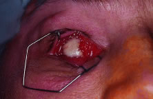
with 1:100,000 units of epinephrine is performed. An eyelid speculum is placed, and a 360-degree limbal peritomy is performed with blunt-tipped Westcott scissors and small toothed forceps. Using blunt-tipped Steven's tenotomy scissors, the four quadrants between the recti muscles are cleared (Fig. 4). This is performed by grasping the edge of conjunctiva and Tenon's capsule, advancing the scissors posteriorly along the sclera to just past the equator, and spreading the tissue by opening the scissors. Next, all four recti muscles are identified, tagged with a double-armed 5-0 Vicryl suture in a locking fashion, and cut free from the globe. Each rectus muscle is identified and isolated on a muscle hook and excess facial attachments are bluntly removed from the muscle's surface with a cotton-tipped swab. One end of a double-armed 5-0 Vicryl is then passed through the midportion of the muscle belly approximately 1 mm from its insertion towards the edge of the muscle (Fig. 5). The suture is then passed through the underside of the muscle approximately 1 mm from the muscle edge and pulled through so as to lock the suture. The other end of the suture is passed similarly by starting the pass where the first end started and exiting at the opposite muscle edge. The sutures are then clamped or taped to the drapes. After all four recti muscles have been removed from the globe, the superior oblique 
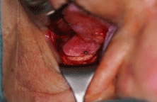
muscle tendon is cut with Westcott scissors. Next, the inferior oblique muscle is isolated with a muscle hook in the inferior medial quadrant, cauterized 
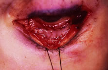
across its belly, and cut (Fig. 6). A 4-0 silk suture is then placed in a running fashion through the remaining muscle tissue of the medial and lateral recti muscles to be used as traction sutures. While applying gentle upward traction, a large hemostat is advanced towards the optic nerve, posterior to the globe, starting at the lateral canthus. With the hemostat closed, the optic nerve is identified. With the clamp superior to the nerve, gentle inferior movement of the clamp will cause the eye to rotate superiorly. The opposite will be observed with the hemostat inferior to the nerve. With the position of the optic nerve accurately identified, the clamp is opened, retracted slightly, advanced over the nerve, displaced a few millimeters posteriorly, and closed. Again, gentle movement of the clamp 
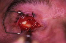
should verify that the nerve is securely clamped. With the nerve still clamped, enucleation scissors are then advanced into the orbit, anterior to the clamp, and the optic nerve is transected (Fig. 7). The globe is elevated with the traction sutures while any remaining tissue adherent to the globe is cut with scissors. The socket is immediately packed with two gauze sponges. During this time the orbital implant can be wrapped with an appropriate wrapping material and prepared for implantation. The globe is grossly examined and sent to the pathology laboratory for examination. The packing is removed, the cut edge of the optic nerve is isolated, and bipolar cautery is applied. The clamp is carefully released under direct visualization to ensure that no significant hemorrhage is present. The socket is then examined for any sign of pathologic process. A small rent in posterior Tenon's capsule corresponding to the opening for the optic nerve can be repaired at this time using a single 5-0 Vicryl suture. Repair of this defect in Tenon's capsule prevents unintentional posterior migration of the orbital implant.

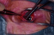
Fig. 4. Blunt-tipped scissors are placed in the inferior-nasal quadrant and opened carefully while the conjunctiva and Tenon's capsule is retracted superiorly. This action clears the quadrant of Tenon's tissue and allows for isolation of the recti muscles. All four quadrants are cleared in a similar fashion.

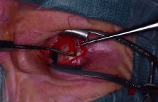
Fig. 5. A double-armed suture is place in the lateral rectus muscle.

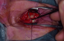
Fig. 6. The inferior oblique muscle is isolated on a muscle hook before cauterization and transection.

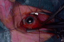
Fig. 7. After the optic nerve is clamped, curved enucleation scissors are used to cut the optic nerve just anterior to the clamp.

In certain instances, it is appropriate to place the implant posterior to Tenon's capsule, directly into the orbital fat. In these cases, placing blunt-tipped scissors into the defect and spreading easily enlarges the rent in posterior Tenon's capsule allowing for placement of the implant directly into the muscle cone.

To provide for the best possible prosthetic appearance and motility, an implant with attached extraocular muscles should be placed within Tenon's capsule. The largest implant that comfortably fits into the socket should be used to decrease the risk of a superior sulcus deformity. Most adults can accommodate a 20- or 22-mm implant without difficulty. Wrapping an implant provides approximately an additional 2 mm of diameter. (The average ocular diameter is 24 mm.) In general, the recti muscles are attached to the implant directly or to the various materials used to wrap orbital implants in a location corresponding to their normal anatomic position. A complete discussion on the wide variety of orbital implants and wrapping materials is found in the section on implant and wrapping materials. A careful layered closure is mandatory to decrease the risk of implant exposure. Tenon's capsule is closed first with multiple interrupted 5-0 Vicryl sutures. Care should be taken not to incorporate any conjunctiva into the deep closure. This may allow for cyst formation, implant exposure, or extrusion. The conjunctiva is then closed with a running suture of 7-0 Vicryl (Fig. 8). Antibiotic ointment and a conformer are then placed between the eyelids, and the socket is pressure patched for 4 to 7 days. Some surgeons place patients on prophylactic oral antibiotics for the first few days following surgery. After the patch is removed, the patient is asked to apply antibiotic ointment to the socket twice a day for the next 2 to 4 weeks. Continued wear of the conformer is used to prevent shortening of the conjunctival fornices. The patient is ready to see the ocularist 6 to 8 weeks after surgery for the prosthesis fitting. Patients should be encouraged to wear polycarbonate glasses to protect the remaining eye.

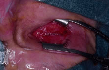
Fig. 8. The conjunctiva is closed with a running absorbable suture.

INDICATIONS FOR EVISCERATION

Evisceration is the removal of the ocular contents while leaving the sclera and optic nerve and, in some cases, the cornea intact. Evisceration offers several advantages over enucleation. Operating time is shorter, the operation is less technically challenging, and the procedure can be performed under retrobulbar anesthesia. There is less disruption to the orbital tissues, better motility, and a more cosmetically acceptable orbit when compared with enucleation. It is the preferred treatment for endophthalmitis because it allows for extirpation and drainage of the ocular contents without orbital invasion. In cases of endophthalmitis, orbital implantation has typically taken place as a secondary procedure. However, a recent study has found that primary implant placement is a viable technique.20 Disadvantages include the theoretical risk of sympathetic ophthalmia and a less complete specimen for pathologic examination to detect intraocular malignancy or spread. Although controversy exists as to the exact indications for evisceration and enucleation, evisceration should never be performed in cases of suspected intraocular tumor.

THE EVISCERATION PROCEDURE

The appropriate eye for removal must be established as described previously. Unlike enucleation, evisceration can be performed using a retrobulbar injection and intravenous sedation. Whether monitored anesthesia or general anesthesia is used, a retrobulbar injection of 3 cc of a 50/50 mixture of 1% lidocaine and 0.5% bupivacaine with 1:100,000 units of epinephrine is injected to control oozing and provide postoperative pain control. The patient is then prepped and draped by the surgeon. An eyelid speculum is placed and a 360-degree limbal peritomy is performed with blunt-tipped Westcott scissors and small toothed forceps. Using Steven's scissors, the four quadrants between the recti muscles are cleared. This is performed by grasping the edge of conjunctiva and Tenon's capsule, advancing the scissors posteriorly along the sclera to just past the equator and spreading the tissue by opening the scissors.

Approximately 1 to 2 mm posterior to the limbus, a small full-thickness scleral incision is made. Westcott scissors are then used to make a circumferential incision around the globe to remove the cornea. If the cornea is to be left in place, the incision is stopped just short of completion leaving a small scleral hinge. The intraocular contents are then separated from the sclera using an evisceration spoon or Freer periosteal elevator. Bleeding from the optic nerve or penetrating vessels can be controlled with gentle bipolar cautery. The pigment is meticulously removed using absolute alcohol on a cotton-tipped applicator. The scleral cavity is then copiously irrigated with antibiotic solution. Windows oriented in an anterior to posterior direction are cut in the sclera in the four quadrants between the recti muscles using scissors. The sclera can also be opened around the optic nerve.21 These scleral windows allow for vascular ingrowth if a porous implant is placed. Scissors are then used to make two cuts at the anterior opening of the sclera in an inferior-medial and superior-lateral direction 
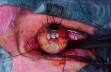
to facilitate implant placement into the sclera. A sphere implant measuring from 14 to 18 mm is placed into the scleral cavity (Fig. 9). Redundant sclera is trimmed and the sclera is closed with multiple interrupted 5-0 Mersiline sutures. Tenon's capsule is closed first with multiple interrupted 5-0 Vicryl sutures. The conjunctiva is then closed with a running suture of 7-0 Vicryl (Fig. 10). Antibiotic ointment and a conformer are then placed between the eyelids, and the socket is pressure patched for 4 to 7 days. Some surgeons place patients on prophylactic oral antibiotics for the first few days following surgery. After the patch is removed, the patient is asked to apply antibiotic ointment to the socket twice a day for the next 2 to 4 weeks. Continued wear of the conformer is essential to prevent shortening of the conjunctival fornices. The patient is ready to see the ocularist 6 to 8 weeks after surgery for the prosthesis fitting. As with any monocular patient, polycarbonate glasses should be worn routinely to protect the remaining eye.

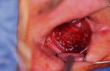
Fig. 9. A polyethylene spherical implant rests inside the scleral cavity before closure.

Fig. 10. The sclera, Tenon's capsule, and conjunctiva are closed in layers.

IMPLANT AND WRAPPING MATERIALS

Much of the interest in enucleation and evisceration surgery over the past few years has concentrated on new types of orbital implant materials. Ideally, the purpose of an orbital implant is to provide adequate orbital volume to compensate for the absent globe, promote prosthesis motility, and be responsible for minimal complications following surgery. Common complications that have arisen include exposure, extrusion, infection, inflammation, and migration of the implant within the anophthalmic socket.

Since 1885 when Mules placed the first orbital implant consisting of a blown glass sphere, various types of materials have been placed in the orbit following removal of an eye.2 Glass, rubber, steel, gold, silver, silicone, acrylic, titanium mesh, and polymethylmethacrylate (PMMA) spheres have been used (Fig. 11). Many of these materials are well tolerated by the host and provide adequate orbital volume. However, direct extraocular muscle attachment is impossible and motility is limited. Further refinement occurred with the use of quasi-integrated implants such as the Allen, Iowa, and Universal models. In these implants, the extraocular muscles were attached the implants directly and implant movement was transferred to the prosthesis via matching irregularly shaped surfaces on the implant and prosthesis. Quasi-integrated implants are now rarely used because of their difficulty of implantation and higher risk of migration and extrusion.

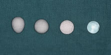
Fig. 11. From left to right: conical polyethylene, spherical polyethylene, hydroxyapatite, and polymethylmethacrylate (PMMA) orbital implants.

In 1985, Perry introduced the use of a hydroxyapatite (HA) orbital implant formed from a salt of calcium phosphate that is found in the mineralized portion of human bone.22 The material is biocompatible, nontoxic, and nonallergenic. Its extensive system of channels and pores facilitates fibrovascular ingrowth that allows the implant to become vascularized and integrated into the orbital tissues. The most common type of HA spheres (Bioeye; Integrated Orbital Implants, Inc., San Diego, CA) currently used in the United States are derived from sea coral. A synthetic HA is manufactured in France (FCI, Issy-Les-Moulineaux, France). A HA derivative derived from the mineral portion of calf bone is available under the brand name M-sphere (IOP Inc., Costa Mesa, CA). Although it has a long history of use in other fields of surgery, the M-sphere is extremely fragile and requires great care when using. A new bioceramic implant made of the material Alumina (aluminum oxide) was approved for use in the United States in early 2000. It represents a new type of porous integrated orbital implant that is manufactured without disruption to marine ecosystems. Studies are underway regarding its long-term success as an orbital implant.

Because of the rigid nature of the material, HA implants must be wrapped to facilitate attachment of the extraocular muscles. Donor sclera is the most commonly used wrapping material. The main concern with the use of donor sclera is the extremely low risk of disease transmission. Autologous fascia or dermis can be used but harvesting the tissue can greatly increase surgical time and leave undesirable scars. Other effective wrapping materials producing similar results are now available on the market. Processed human sclera (Tutoplast), bovine pericardium (Ocu-guard), and acellular dermis are all appropriate wrapping materials. Some surgeons have used polyglactin 910 (Vicryl) mesh as a wrapping material. It has the advantage of being easy to use, extremely inexpensive, and readily available with no risk of disease transmission.23 The use of Vicryl mesh is associated with an increased risk of exposure unless the extraocular muscles are sutured anterior to their normal anatomic position. This results in the implant being placed deeper into the socket. Although polyglactin mesh facilitates implant insertion and extraocular muscle attachment, it does not provide a permanent barrier to exposure.17

Wrapping HA implants adds to the cost and complexity of the surgery. For these and other reasons, the use of porous polyethylene implants (Medpor) were investigated.24 Porous polyethylene has been used successfully in reconstructive surgery for many years. It is biocompatible with channels that allow for vascularization and integration similar to HA. However, porous polyethylene implants have several advantages over HA. The material is less expensive at approximately $400 compared with $650 for HA. It is soft enough that the muscles can be directly attached to the material. It can also be shaped and carved with a scalpel if needed. Some authors have advocated truncation of the anterior surface of the implant to facilitate prosthesis movement. Thus far, there is no indication that this improves motility and may allow for implant deviation to become a problem.17 I prefer to use polyethylene (Medpor) as my implant of choice. I recommend attaching an approximately 10- to 15-mm piece of sclera, fascia, periosteum, or acellular dermis to the anterior surface of the porous polyethylene implant with 5-0 Merseline suture (Fig. 12). However, some surgeons use polyethylene orbital implants without any wrapping. I think it is easier to suture directly to the implant if has been warmed for a few minutes in sterile saline. The implant is then placed into Tenon's capsule, and the extraocular muscles are sewn to the implant directly a few millimeters anterior to their normal anatomic position. This corresponds to a position slightly anterior to the edge of the piece of barrier tissue. I have also had excellent success wrapping the anterior half of the implant with processed whole sclera (Tutoplast) or donor sclera (Fig. 13). I make sure that the sclera is securely attached to the implant by using multiple nonabsorbable 5-0 Merseline sutures. With this method, I cut four windows in the sclera measuring 2 mm by 5 mm corresponding to the positions of the extraocular muscle attachments. Each extraocular muscle is then advanced through its respective window and sutured to the sclera and implant. As with any implant material, Tenon's capsule and conjunctiva are then meticulously closed in layers.

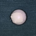
Fig. 12. A polyethylene sphere with a piece of sclera attached to its surface using nonabsorbable suture.

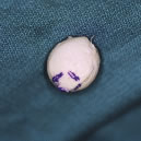
Fig. 13. A scleral-wrapped porous orbital implant. Windows to allow for attachment of the extraocular muscles are visible.

Unlike alloplastic implants, extrusion and migration of HA integrated implants is uncommon. The fibrovascular ingrowth prevents most exposed 
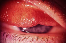
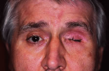
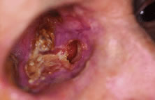
integrated implants from extruding.25 However, tissue breakdown and exposure of the anterior surface of the implant is reported to occur in 0% to 11.1% of cases25–27 (Fig. 14). Shields and coworkers28 reviewed 249 enucleation cases in which scleral-wrapped HA spheres were placed and found an acceptable exposure rate of 1.6%. The risk of exposure is reduced after vascularization of the anterior portion of the implant. Exposure can be reduced with meticulous layered surgical closure, proper implant size, drilling holes into the sphere before implantation, delayed fitting of the prosthesis, and vaulting of the posterior surface of the initial prosthesis to reduce pressure on the tissues covering the anterior surface of the implant.

Fig. 14. An exposed polyethylene orbital implant.

Care must also be taken to avoid exposure problems when using porous polyethylene implants as well. Karesh and Dresner24 first proposed the use of unwrapped porous polyethylene implants following enucleation, evisceration, and secondary orbital implant surgery with no signs of exposure in 21 patients. However, other investigators have found exposure rates to be high ranging from 9% to 13%.29,30 Although some debate exists regarding the need to wrap porous polyethylene implants, uncovered porous polyethylene implants appear to have a complication rate higher that wrapped HA implants.17 Soparkar and coworkers31 studied the vascularization of porous polyethylene cubes placed in rabbits. From the results of the study, they suggest an alteration in surgical technique to improve and accelerate implant vascularization. They advocate the use of a tissue barrier (sclera, fascia, and dermis) attached to the anterior surface of the implant to prevent exposure but believe that wrapping the implant is unnecessary. They also recommend increasing the amount of muscle tissue in direct contact with the implant by attaching the muscles anterior to their normal anatomic position, placing posterior fixation sutures, and removing the muscle capsule from the anterior 5 to 6 mm of each extraocular muscle. Currently, investigation is also underway to determine if growth factors or angiogenesis-promoting agents may enhance implant vascularization.32

Improved implant motility has always been a goal in implant design and advancement. The motility of wrapped alloplastic and HA has been compared in two studies.33,34 Both concluded that there is no motility benefit of nonpegged HA over spherical alloplastic implants. Motility is due to the attachment of the extraocular muscles to the wrapping and not to the implant material. Custer and coworkers also determined that larger implants showed more movement and that motility declines with advancing age. Other investigators have shown that oval implants show no advantage over spherical implants.30 Integrated implants might be unnecessary in patients who do not wish to consider peg placement.

In motivated patients desiring maximal motility, coupling pegs made of titanium are available for use with both the HA and porous polyethylene types of implants. The pegging systems are placed into the buried, vascularized implant allowing direct coupling with the prosthesis. Before peg placement, implant vascularization is typically confirmed 6 to 12 months following surgery. This has been done in the past using bone scan technology. Recent evidence has shown contrast-enhanced magnetic resonance imaging (MRI) best suited for assessing the vascularization of the porous polyethylene and HA implants.35 However, some investigators believe that titanium motility pegging systems can be placed safely and effectively at the time of enucleation in Medpor or HA implants.36,37

The current pegging system for HA implants supplied by Integrated Orbital Implants, Inc. (San Diego, CA) consists of a threaded titanium sleeve with a hollow core and various shapes and sizes of coupling pegs. The pegging procedure is typically done under retrobulbar anesthesia and sterile conditions. With the implant stabilized, a hole is first created in the HA implant with progressively larger hypodermic needles. No power drill is required. The threaded titanium screw is then advanced into the implant with the aid of a sleeve driver until the top of the sleeve is flush with the implant surface. A flat peg is placed into the hollow core of the sleeve. The patient is treated with topical antibiotic for a month during the healing process. The patient is then sent to the ocularist to have the implant coupled to the prosthesis. The ocularist will determine the best technique for coupling and will select the appropriate titanium attachment. Complications such as peg extrusion, peg loosening, chronic discharge, pyogenic granuloma, infection, and conjunctival breakdown and exposure do arise from pegging. Jordan and coworkers found 37.5% of patients who underwent the pegging procedure in HA implants experienced problems. However, most cases used the older polycarbonate pegging system. It is thought that the newer titanium systems will have fewer complications.

A pegging system is also available for use with Medpor implants. The motility coupling post (Porex Surgical, Inc., College Park, GA) consists of a threaded titanium peg that protrudes 4 mm above the implant surface (Figs. 15 and 16). Although the manufacturer recommends local anesthesia, some patients may require a retrobulbar injection before the pegging procedure. The conjunctiva is marked, and a small opening is made in the conjunctiva using a disposable cautery. The implant is then stabilized with a provided clamp. A hole is then carefully placed into the implant using a manual drill bit attached to a screwdriver. The equipment is packaged in a kit from the manufacturer. It is important to maintain the appropriate orientation of the drill, which is perpendicular to the implant, while making the initial hole. The peg is then twisted into placed using a special screwdriver until the threads are no longer visible. A conformer and antibiotic ointment is then placed. At approximately 1 month after pegging, the patient is ready for coupling with the prosthesis by the ocularist. Reported complications associated with the motility coupling post are uncommon but include pyogenic granuloma and conjunctival overgrowth.

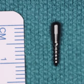
Fig. 15. The threaded peg used with Medpor implants is shown next to a surgical ruler.

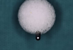
Fig. 16. Although the peg is typically placed after orbital implant vascularization within the socket, this photo shows how the peg extends from the implant.

When pegging either the HA or polyethylene implants, preoperative planning with the ocularist is very helpful in determining the best position of the peg.

Back to Top
MANAGEMENT OF THE ANOPHTHALMIC SOCKET
The goals of enucleation and evisceration surgery are removal of a diseased eye and the production of a healthy, stable anophthalmic socket. The long-term goals in the management of the anophthalmic socket patient are to achieve and maintain a comfortable socket and a natural-appearing ocular prosthesis.

GENERAL CARE

All patients who elect to wear an ocular prosthesis following removal of an eye should have a custom-made, impression-fitted ocular prosthesis constructed by a certified ocularist. “Off the shelf” or stock prostheses should be discouraged. The ocularist typically follows the patient along with the ophthalmologist and assures that the prosthesis is clean and fitting appropriately. The prosthesis is generally cleaned and polished yearly and replaced every 7 to 10 years. Patients are encouraged to handle and remove the artificial eye as little as possible. Monthly removal and cleaning is reasonable, but daily removal and cleaning should be discouraged. A dry socket is the most common cause of problems in patients wearing a prosthetic eye. Although most patients do not require lubricants, in patients with dryness and good eyelid closure, the application of artificial tears will often suffice. In cases of more significant dryness, silicone, aloe vera, or mineral oil can be used. Patients with partial eyelid closure often develop a dry residue on the surface of the prosthesis. In these cases, the application of an ophthalmic lubricating ointment following cleaning improves the problem.

DISCHARGE FROM THE SOCKET

Socket problems, prostheses problems, eyelid problems, or lacrimal problems can all be responsible for discomfort, irritation, and discharge from the anophthalmic socket.

The most common socket problem causing discharge is dryness and decreased tear production. As discussed previously, the use of artificial tears and ointments often improves or eliminates the problem. Bacterial or viral conjunctivitis may cause discharge and are treated with antibiotic drops when appropriate. Discharge from the socket may be the first indication of orbital implant extrusion or dehiscence of the conjunctiva. Special attention must be paid to patients enucleated for ocular neoplasia who complain of irritation, discomfort, discharge, or a change in the fit of the prosthesis. A careful search for tumor recurrence should be performed in these patients.

A poorly fitting prosthetic eye or an old prosthetic eye often causes discharge. The poorly fitting eye can cause chronic irritation of the socket tissues and the production of increased amounts of mucus. Patients will often complain of an increase in drainage during movement of the artificial eye. This is likely due to pooling of secretions behind the poorly fitting prosthesis that is expelled with ocular movement. The pooling of mucus and debris in a dead space behind the prosthesis also makes the chance of a bacterial conjunctivitis more common. Older prosthetic eyes may also cause mechanical irritation in the socket because of scratches or the buildup of debris on its surface. Modification of the existing prosthesis, polishing, or the construction of a new prosthesis may be indicated. Consultation with the ocularist is essential in these cases.

Giant papillary conjunctivitis (GPC) is the most common eyelid problem leading to socket discharge (Fig. 17). GPC causes a mucoid discharge, irritation, and itching. Large papillae are seen on the tarsal conjunctiva. The exact cause of GPC is unknown, but it likely represents a hypersensitivity reaction to environmental allergens or materials in or on the prosthesis. Preservatives in artificial tears, lipoproteins in the tear film, microbes, and the acrylic materials used to make the prostheses have been implicated. Treatment of GPC involves cleaning and polishing of the prosthesis by the ocularist. Replacement of the prosthesis is sometimes required. Topical steroid preparation such as prednisolone acetate 1% three times per day for 2 weeks is effective in relieving the symptom quickly. The long-term use of 4% cromolyn sodium or a topical nonsteroidal anti-inflammatory drop may help prevent recurrences and make the socket more comfortable.

Fig. 17. Note the typical large papillae associated with giant papillary conjunctivitis.

Lid laxity, ectropion, and entropion may also lead to a poor prosthesis fit, inability to retain the prosthesis, socket discharge, and discomfort. With time, the added weight of an ocular prosthesis can cause lengthening of the lower eyelid. To a large extent, the lower eyelid supports the weight of the prosthesis. Lid laxity leads to a poor prosthesis fit and often exacerbates a superior sulcus deformity. Lid laxity and ectropion are treated with a horizontal lid shortening procedure such as the lateral tarsal strip. Cases of entropion are typically due to conjunctival cicatricial changes leading to shortening of the upper or lower fornices. Procedures such as the tarsal fracture or marginal rotation can be used to reposition the eyelid margin. In cases of more extensive fornix shortening, mucous membrane grafting to lengthen the fornix is indicated.

PTOSIS

Ptosis can often be corrected by modification of the prosthesis by the ocularist to elevate the eyelid and change contour without surgical intervention. If further elevation of the eyelid cannot be provided by the prosthesis, ptosis correction surgery can be performed (Fig. 18). A lengthening and rarefication of the levator aponeurosis is the most common etiology of the ptosis. An external levator advancement procedure is effective in correcting the ptosis in most cases. I do not recommend a posterior tarsoconjunctival muscle resection because of the possibility of shortening or disrupting the superior conjunctival fornix. In cases with poor or absent levator function, a conservative silicone or fascia lata sling procedure can be performed. Coordination with the ocularist is important. Before the ptosis procedure, an optimal prosthesis should be constructed and in place to assure proper lid height, contour, and movement.

Fig. 18. Ptosis of the right upper eyelid.

SUPERIOR SULCUS DEFORMITY

The superior sulcus deformity is extremely common and is most evident by the appearance of a deepened superior sulcus (Fig. 19). The characteristics of the anophthalmic socket that produce the superior sulcus deformity are ptosis of the ocular prosthesis, decreased orbital volume, and lower lid laxity. The decrease in orbital volume is a result of atrophy of the orbital fat, as well as the relative loss of volume produced by the orbital implant compared with the globe. In the anophthalmic socket, 6 cc of tissue is removed at enucleation. The orbital implant replaces 2 to 4 cc and the prosthetic eye provides another 1 to 2 cc. This still leaves 1 to 3 cc of volume deficit. Orbital implant migration and shifting in the socket may exacerbate the condition. Treatment of the superior sulcus defect is best addressed by orbital volume augmentation. Through a subciliary lower eyelid incision, the inferior orbital rim is identified. Again, I try to avoid a conjunctival incision, if possible. The periosteum is opened, and the periorbita from the orbital floor is elevated. Various implant materials can then be placed in the subperiorbital space to provided added orbital volume. The implant should also push the orbital implant upward and outward to improve the superior sulcus defect. Many different floor implant materials have been used in the past including calvarian bone, harvested or donor rib cartilage, silicone, or polyethylene blocks. I prefer and frequently use the specially shaped Medpor enophthalmos wedge implant. (Porex Surgical, Inc., College Park, GA) After the implant is placed, it is fixated to the bone with one or two titanium microscrews and the periosteum is closed with absorbable sutures (Fig. 20). The skin incision is then closed and a lateral tarsal strip procedure is performed.

Fig. 19. Superior sulcus deformity on the right side in a young anophthalmic patient.

Fig. 20. A Medpor enophthalmos wedge implant in excellent position in the subperiosteal space along the right orbital floor. The implant is fixated to the inferior orbital rim with a single titanium screw while the orbital contents are retracted superiorly.

In very mild cases with minimal enophthalmos and no lower eyelid laxity, a conservative blepharoplasty on the opposite eye can improve superior eyelid symmetry. Some authors have suggested implantation of autologous fat or a dermis fat graft into the upper eyelid for correction.

SOCKET CONTRACTION

Socket contraction is a condition typically noted months to years after enucleation or evisceration surgery. The shrinkage of the conjunctiva leads to shallow fornices, entropion, and the inability to wear a prosthesis (Figs. 21 and 22). This complication is incompletely understood but various contributing factors have been suggested: traumatized tissues before enucleation surgery, poor surgical technique, chronic inflammation or infection, poor prosthesis fit, a result of not wearing a prosthesis or conformer, or a result of the absence of an intraorbital implant.

Fig. 21. A patient with a severely contracted socket on the left side. The patient has entropion of the lower eyelid and is unable to wear a prosthesis comfortably.

Fig. 22. A mucosal graft is sutured into the lower fornix.

In eyes removed because of trauma or following multiple ocular surgeries, there may be a loss of conjunctiva or Tenon's capsule. In an attempt to close the tissues over an alloplastic implant, the conjunctiva and Tenon's capsule may be advanced excessively leading to a shallow fornix. In these cases, a dermis fat graft may be required at the time of enucleation to prevent socket contraction.38 A dermis fat graft provides volume to the socket and allows for the preservation of the fornices in sockets that have undergone significant conjunctival loss or scarring.

With the patient under general anesthesia, the patient is positioned to provide access to the buttocks. An 18- to 22-mm circle of skin is marked on the upper, outer quadrant of the buttocks. The area is infiltrated with local anesthetic with epinephrine to control bleeding. A scalpel is used to make an incision through skin and into the underlying fat. The epidermis is then removed with a scalpel or dermatome. The dermis and underlying plug of fatty tissue, approximately the size of the globe, is removed. Hemostasis is achieved, and the incision is closed in layers. Following enucleation, the dermis fat graft is then placed within Tenon's capsule and its size is assessed. The graft should fill the defect but should not be overly tight. If needed, careful sculpting of the graft can be performed to assure a proper fit. Excessively large grafts cause fat necrosis, whereas small grafts lead to postoperative enophthalmos. After the proper size graft is placed in the defect, the four recti muscles are sutured, using absorbable suture, to the edges of the graft corresponding to their normal anatomic positions. The edges of Tenon's capsule and conjunctiva are sewn to the edges of the dermis using multiple interrupted absorbable sutures. Antibiotic ointment and a large conformer are placed over the graft followed by a pressure patch for 5 to 7 days. The conjunctival epithelium grows over the dermal graft in the first few weeks after surgery. A conformer is continued for 8 weeks at which time the prosthesis can be fit. Some graft shrinkage, up to 20% to 30% of the original volume, can be expected. Excessive shrinkage or discharge from the graft may indicate graft necrosis. Graft necrosis is managed with systemic antibiotics to prevent infection and with careful follow-up.

The surgeon should take great care to maintain the inferior fornix during enucleation surgery and postoperatively. I recommend the placement of an orbital implant in all patients. The implant prevents migration of orbital tissue inferiorly and reduces the chance of a shortened inferior fornix. Fornix shortening may also result from excessive advancement of Tenon's capsule anteriorly during closure. Undermining Tenon's capsule from the conjunctiva before the final layered closure over the orbital implant best prevents this complication. The edges of Tenon's capsule are then closed over an appropriately sized implant followed by the conjunctival closure under little or no tension. Conformers should be worn continuously following surgery. A temporary tarsorrhaphy may be required in the early postoperative period to assure that the conformer stays in place. This is particularly effective in sockets with significant postoperative chemosis and swelling.

Significant conjunctival contraction producing shortened conjunctival fornices, entropion, and the inability to wear a prosthesis requires extensive socket reconstruction using full-thickness mucous membrane grafts harvested from the buccal mucosa. It should first be determined if the lower fornix alone or both the upper and lower fornices should be deepened. In most cases, it is the lower fornix that is severely shortened because of contraction.

With the patient under general anesthesia, a 4-0 silk traction suture is placed through the inferior tarsal plate. The lid is everted and a no. 15 blade is used to make a conjunctival incision along the full length of the eyelid just inferior to the tarsus. Using sharp dissection with Westcott scissors, any scar tissue that exists between the conjunctiva and underlying Tenon's capsule is lysed along the entire length of the incision. A conjunctival defect now exists. A template is then made from the surgical drapes and placed in the defect. The size of the graft must be large enough to reform the fornix so that it is typically twice as large as the existing defect. Attention is next directed towards the buccal mucosa along the lateral vestibule of the mouth. The parotid (Stensen's) duct must be located and marked. The duct is located opposite the neck of the superior second molar. The mucosa is then infiltrated with local anesthetic with epinephrine. The graft is marked with a surgical marking pen using the template. The tissue is incised with a scalpel and elevated with forceps and Steven's scissors. The underling medial pterygoid muscle should not be disturbed. The oral incision is closed with a running locking suture of 4-0 chromic. The graft is thinned to remove any fatty tissue and placed in the conjunctival defect. The graft is sutured into position with multiple interrupted sutures of 7-0 Vicryl (see Fig. 22). The same procedure is used to repair the upper or lower fornix. Antibiotic ointment and a conformer are then placed in the socket with or without fornix-forming sutures. I believe that good result can be obtained without the use of fornix-forming sutures as long as the conformer stays in place. Other options include preplaced holes in the conformer allowing for fornix sutures to stabilize the conformer or a complete tarsorrhaphy to prevent conformer loss. The patients are asked to keep the mouth clean by swishing with a dilute mouthwash solution three times per day for 1 week. The graft is stable 4 to 6 weeks following surgery at which time a new prosthesis can be made.

IMPLANT EXPOSURE AND EXTRUSION

With the use of acrylic or PMMA sphere implants, implant migration within the socket and extrusion of the implant were a concern. Impending implant extrusion was often heralded by a conjunctival or Tenon's capsule dehiscence followed by loss of the implant (Fig. 23). In some cases, evidence of a thin layer of conjunctiva was found surrounding the orbital implant that would not allow the conjunctival incision to heal. Treatment involved excision of the conjunctival capsule and replacement of the implant posterior to or within Tenon's capsule.

Fig. 23. Impending extrusion of a silicone implant placed 17 years earlier. Laterally the implant is completely exposed, whereas centrally it is covered with only a thin layer of conjunctiva.

With the increasing use of vascularized, porous, integrated, orbital implants, the risk of implant extrusion is decreasing. However, implant exposure resulting from conjunctival defects overlying the newer porous implants is more common than extrusion. These complications are often seen in the early postoperative period or more uncommonly years after enucleation or evisceration surgery. Typically, conjunctival defects with small areas of exposed polyethylene or HA are noted. The patients may complain of increased socket discharge or pain.

Conjunctival defects may be related to poor prosthesis fit, excessive socket manipulation, poor surgical technique, the rough surface of the implant, poor vascularization of the implant, or unknown causes. Patients with a conjunctival defect are at increased risk of orbital infection. Correction of the problem can sometimes be accomplished by elevating conjunctiva and Tenon's capsule from the implant, freshening the edges, and closing in a layered fashion. More commonly, the conjunctival edges must be elevated from the implant and sutured to a piece of graft material such as sclera, dermis, or fascia. With time, the conjunctival epithelium grows over the graft material and seals the defect. I have had good success in repairing conjunctival defects over vascularized implants using a patch graft of buccal mucous membrane (Fig. 24). An appropriately sized graft is harvested from the mouth, and the conjunctival edges are sutured to the graft with interrupted absorbable sutures. Vascularized flaps from the eyelid can also be used to repair larger defects.39 Significant defects and conjunctival shortening may necessitates implant removal and replacement with a dermis fat graft. After surgical treatment, consultation with the ocularist is essential to ensure a proper prosthesis fit.

Fig. 24. A buccal mucosal graft is used to cover an exposed porous orbital implant.

Back to Top
EXENTERATION
Orbital exenteration involves the removal of the eye and orbital soft tissues. Exenterations vary in the amount of tissue removed. In a subtotal exenteration, the globe and adjacent tissues are removed, but the periorbita and some or all of the eyelids are preserved. In a total exenteration, all of the orbital tissues, including the periorbita are removed. An extended, or radical, exenteration involves the removal of all of the orbital tissues, as well as adjacent structures such as bone, sinuses, muscular tissue, and skin.

INDICATIONS

Most commonly, exenteration is performed in cases of life-threatening malignancy in an attempt to prolong life and decrease morbidity.5,40 Tumors extending secondarily from the adjacent sinuses, cranium, face, globe, conjunctiva, and eyelids account for approximately 75% of the cases requiring exenteration in an extensive review at Duke University.25 Squamous cell carcinoma, basal cell carcinoma, and melanoma are the most common orbital tumors requiring exenteration. Primary orbital tumors such as vascular malformations, primary lacrimal gland tumors, and meningiomas are less common causes of exenteration. Orbital exenteration is often required in cases of extensive rhino-orbital phycomycosis in conjunction with systemic antifungal therapy and hyperbaric oxygen. Wide surgical debridement is required in the treatment of mucormycosis; however, some reports have shown positive results without extensive exenteration.41 Less commonly, orbital exenteration is performed to in cases of severe orbital pain or deformity.

SURGICAL PROCEDURE

Patients with life-threatening malignancies or infections are typically under the care of a team of physicians at the time of exenteration. A complete systemic evaluation is required before surgery to establish the extent of the disease process and to determine the patient's prognosis following any surgical and/or medical treatment. Preoperative imaging should also be performed to help identify the extent of the orbital involvement and to aid in surgical planning. Obvious extension outside the orbit mandates the need for surgical consultation with a Mohs' surgeon, head and neck surgeon, or neurosurgeon. Often in cases of secondary orbital tumors, an interdisciplinary surgical approach is required. Consultation with a tumor board often proves valuable in formulating an overall treatment plan for the patient. Some tumors benefit from preoperative chemotherapy, whereas others require postoperative radiation therapy. In cases in which extensive systemic disease is found, a major disfiguring surgery might be contraindicated and palliative care might be undertaken instead. A definitive tissue diagnosis using permanent histopathology material is always required before exenteration.

After general anesthesia is induced, an initial incision line is marked just inside the orbital rim. Intravenous broad-spectrum antibiotics are given. The area is infiltrated with local anesthesia with 1:100,000 unit of epinephrine injected subcutaneously. A 4-0 silk suture placed through the eyelid margin is used to close the eyelids and provide a means of traction during the procedure. An incision is then made using a no. 15 blade through skin and into superficial orbicularis muscle fibers along the previously marked incision line (Fig. 25). The dissection is then continued in the suborbicularis plane using scissors or unipolar cautery to the bony orbital rim. If possible, the septum should be left intact. The periosteum is then cut just outside the orbital rim around the entire orbit. The periosteum is dissected from the orbital rim and elevated along the orbital walls towards the apex using a Freer periosteal elevator. Adequate suction and a headlight are essential for proper visualization. The zygomaticofacial and zygomaticotemporal neurovascular bundles along the lateral orbital wall are cauterized and cut. The periorbita elevates easily from most surfaces of the orbit. Firm attachments are seen at the lateral orbital tubercle, the trochlea, and in the inferomedial orbit near the nasolacrimal duct and inferior oblique muscle attachment. When the inferior and superior orbital fissures are encountered, incremental cautery followed by cutting with large scissors facilitates the dissection and decreases bleeding. Care must be taken when dissecting the periorbita from the medial orbital wall to prevent breaking the bone. Penetration into the sinuses may result in the formation of sino-orbital fistulas. After the periorbita has been elevated to the apex, the apical stump is transected using enucleation or Metzenbaum scissors. The specimen is removed; cottonoids soaked in dilute epinephrine, Afrin, or thrombin are placed over the remaining orbital tissue; and the socket is firmly packed with 4- × 4-in. gauze sponges. After 5 minutes, the packing is removed and hemostasis is achieved with bipolar cautery to the stump. Inspection of the orbit is then performed for any evidence of abnormal tissue. Frozen section monitoring should be used to ensure complete resection in areas where the tumor approaches the surgical margins. Extension into the cranial vault or sinuses requires the expertise of a neurosurgeon and head and neck surgeon to ensure adequate removal of tumor.

Fig. 25. The dashed line marks indicate the placement of the initial exenteration incision.

Immediate postoperative complications following standard exenteration surgery are uncommon and are best avoided with meticulous surgical technique. Complications include hemorrhage, cerebrospinal fluid leak, osteomyelitis, and intracranial infection. Any postexenteration patient with altered mental status must be evaluated for an intracranial infection.

ORBITAL RECONSTRUCTION

After the resection is complete, the options for reconstruction of the orbit must be assessed and individualized for each patient. Following craniofacial resection, the use of distant microsurgically attached, free myocutaneous flaps are typically required for closure and protection of vital structures. Primary flap reconstruction also allows for early postoperative radiation therapy. Following a standard exenteration in which the orbital bones are left intact, immediate reconstructive options include granulation, split-thickness skin grafts, or regional flaps.

Granulation of the socket is the simplest reconstructive technique. It is complete approximately 2 months following surgery. Granulation produces a thin skin covering of the orbit, which allows easy inspection and identification of tumor recurrence. Following resection, the orbit is irrigated and packed with iodine-dampened gauze and patched for 1 week. After removal of the patch, the gauze is removed daily and the socket is cleaned with hydrogen peroxide or soapy water. After granulation is complete, the patient may elect to wear a patch or silicone orbital prosthesis.

A split-thickness skin graft can also be placed in the exenterated socket. The skin graft is commonly harvested from the thigh with a dermatome with a thickness of 0.015 to 0.017 inches. The graft is then placed in the socket and sewn to the skin edges with absorbable sutures. The skin graft is then covered with an antibiotic ointment and the socket is packed with iodine-soaked gauze for 1 to 2 weeks. Advantages of the split-thickness skin graft method include rapid healing and a thin orbital covering. Disadvantages include pain at the graft donor site and hyperkeratosis of the socket requiring occasional debridement. A drawback of both granulation and split-thickness skin grafting is the relatively high occurrence of fistula formation between the orbit and adjacent sinus or nasal cavities (Fig. 26). The incidence of fistula formation following granulation ranges from 35% to 68%.40,42

Fig. 26. A fistula between the exenterated socket and sinus cavity. Also note the build-up of keratin within the socket.

Some surgeons prefer primary closure of the orbital defect at the time of resection with local, regional, or microvascular free flaps. Frontotemporal, parietotemporal, and free flaps are the most commonly used flaps for orbital reconstruction. Mohr and Esser42 argue that intra-orbital tumor recurrence is rare and that if it occurs, the prognosis is poor. Furthermore, they state that primary closure has many advantage over granulation, which include decreasing fistula formation, protecting against intracranial infection, rendering early postoperative radiation therapy possible, and improving functional and aesthetic outcome. Bartley and coworkers40 at the Mayo Clinic consider healing by secondary intention as their preferred choice of socket reconstruction. They state that early recurrences are not hidden, operating time is reduced, socket color matches the surrounding skin, and the socket is shallow compared with a split-thickness skin graft. Although fistula formation was seen in 35% of their cases, they believe that careful attention to preserving the orbital bones makes fistula formation less likely. Repair of sino-orbital fistulas often requires the placement of a vascularized flap. However, repair is not mandatory because fistulas are typically considered more of a nuisance than a problem.

After the socket has healed, silicone prosthetic devices can be constructed to provide an excellent cosmetic result. Prosthetic devices are attached to the skin with various types of adhesives or are affixed to spectacles (Figs. 27 and 28).The use of new osseointegration techniques, which involve the permanent placement of bone-anchored titanium implants, can also be used to successfully support maxillofacial prosthetic devices.43

Fig. 27. An exenterated socket with osseointegration hardware. (Courtesy of Jeffrey Nerad, MD.)

Fig. 28. The same patient wearing an orbital prosthesis. (Courtesy of Jeffrey Nerad, MD.)

Back to Top
CONCLUSION
Enucleation and evisceration procedures have been performed in medical practice for hundreds of years. Recently, there has been renewed interest in these procedures. Much of the interest has been stimulated by the introduction of new implant and wrapping materials. Pegging systems that directly couple the implant to the prosthesis allow for better motility in selected patients. Information from the COMS has shown that enucleation continues to be a mainstay of treatment for choroidal melanomas.

As discussed throughout the chapter, the decision to remove an eye must be individualized to each patient. Various advantages and disadvantages exist among the many choices of surgical techniques, implant materials, and wrapping materials. Whether enucleation, evisceration, or exenteration is performed, the goals of the procedures are the safe removal of the diseased eye, placement of an appropriately sized orbital implant, and the ability to wear a comfortable and cosmetically acceptable ocular prosthesis.

Back to Top
REFERENCES

1. Noyes W. Treatise on Diseases of the Eye. New York: Wood, 1881:189

2. Mules PH. Evisceration of the eye and its relation to the bacterial theory of origin of sympathetic disease. Trans Ophthalmol Soc U K 1885;5:200

3. Nunery WR, Hetzler K. Enucleation. In Hornblass A (ed). Oculoplastic, Orbital and Reconstructive Surgery. Baltimore: Williams & Wilkins, 1990:1200–1220

4. Levine MR, Pou CR, Lash RH. Evisceration: Is sympathetic ophthalmia a concern in the new millennium? Ophthalmic Plast Reconstr Surg 1999;15:4

5. Levin PS, Dutton JA. A 20-year series of orbital exenteration. Ophthalmology 1991;112:496

6. Rao NA, Robin J, Hartmann D. The role of the penetrating wound and the development of sympathetic ophthalmia. Arch Ophthalmol 1983;101:102

7. Raynard M, Riffenburgh RS, Maes EF. Effect of corticosteroid treatment and enucleation on the visual prognosis of sympathetic ophthalmia. Am J Ophthalmol 1983;96:290

8. Lubin JR, Albert DM, Weinstein M. Sixty-five years of sympathetic ophthalmia: A clinicopathologic review of 105 cases (1913-1978). Ophthalmology 1980;87:109

9. Osborne D, Hadden B, Deeming LW. Orbital growth after childhood enucleation. Am J Ophthalmol 1974;77:756

10. Fountain TR, Goldberger S, Murphree AL. Orbital development after enucleation in early childhood. Ophthalmic Plast Reconstr Surg 1999;15:32

11. Diener-West M, Hawkins BS, Markowitz JA et al. A review of mortality from choroidal melanoma, II: A meta-analysis of five year mortality rates following enucleation, 1966-1988. Arch Ophthalmol 1992;110:245

12. Collaborative Ocular Melanoma Study (COMS) randomized trial of I-125 brachytherapy for medium choroidal melanoma. Ophthalmology 2001;108:348

13. Zimmerman LE, McLean IW, Foster WD. Does enucleation of the eye containing a malignant melanoma prevent or accelerate the dissemination of tumour cells? Br J Ophthalmol 1978;62:420

14. Abramson DH, Niksarli K, Ellsworth RM et al. Changing trends in the management of retinoblastoma 1951-1965 versus 1966-1980. J Pediatr Ophthalmol Strabismus 1994;31:32

15. Spraul C, Grossniklaus H. Analysis of 24,444 surgical specimens accessioned over 55 years in an ophthalmic pathology laboratory. Int Ophthalmol 1998;283

16. Brackup AB, Carter KD, Nerad JA et. al. Long-term follow-up of severely injured eyes following globe rupture. Ophthalmic Plast Reconstr Surg 1991;7:194

17. Custer PL. Enucleation: Past, present, and future. Ophthalmic Plast Reconstr Surg 2000;16:316

18. Margo CE. Surgical enucleation in community hospitals. Am J Ophthalmol 1989;108:452

19. Albert DM, Diaz-Rohena R. A historical review of sympathetic ophthalmia and its epidemiology. Surv Ophthalmol 1989;34:1

20. Dresner SC, Karesh JW. Primary implant placement with evisceration in patients with endophthalmitis. Ophthalmology 2000;107:1661

21. Kostick DA, Linberg JV. Evisceration with hydroxyapatite implant surgical technique and review of 31 case reports. Ophthalmology 1995;102:1542

22. Perry AC. Integrated orbital implants. Adv Ophthalmic Plast Reconstr Surg 1990;8:75

23. Jordan DR, Allen LH, Ells A et al. The use of Vicryl mesh (Polyglactin 910) for implantation of hydroxyapatite orbital implants. Ophthalmic Plast Reconstr Surg 1995;11:95

24. Karesh JW, Dresner SC. High-density porous polyethylene (Medpor) as a successful anophthalmic socket implant. Ophthalmology 1994;101:1688

25. Dutton JA. Coralline hydroxyapatite as an ocular implant. Ophthalmology 1991;98:370

26. Nunery WR, Heinz GW, Bonnin JM. Exposure rate of hydroxyapatite spheres in the anophthalmic socket: Histopathologic correlation and comparison with silicone sphere implants. Ophthalmic Plast Reconstr Surg 1993;9:96

27. Christmas MJ, Gordon CD, Murray TG et al. Intraorbital implants after enucleation and their complications: A ten year review. Arch Ophthalmol 1998;116:1199

28. Shields CL, Shields JA, Depotter P et al. Problems with the hydroxyapatite orbital implant: Experience with 250 consecutive cases. Br J Ophthalmol 1994;78:702

29. Remulla HD, Rubin PA, Shore JW et al. Complications of porous spherical orbital implants. Ophthalmology 1995;102:586

30. Cook T, Lucarelli MJ, Lemke BN. Results of unwrapped conical and spherical porous polyethylene enucleation implants. Presented at American Society of Ophthalmic Plastic and Reconstructive Surgery, Nov 1999, Orlando, FL.

31. Soparkar CNS, Wong JF, Patrinely JR et al. Porous polyethylene implant fibrovascularization rate is affected by tissue wrapping, agarose coating, and insertion site. Ophthalmic Plast Reconstr Surg 2000;16:330

32. Bigham WJ, Stanley P, Cahill JM et al. Fibrovascular ingrowth in porous ocular implants: The effect of material composition, porosity, growth factors, and coatings. Ophthalmic Plast Reconstr Surg 1999;15:317

33. Custer PL, Trinkaus KM, Fornoff J. Comparative motility of hydroxyapatite and alloplastic enucleation implants. Ophthalmology 1999;106:513

34. Colen TP, Paridaens DA, Lemij HG et al. Comparison of artificial eye amplitudes with acrylic and hydroxyapatite spherical enucleation implants. Ophthalmology 2000;107:1889

35. De Potter P, Duprez T, Cosnard G. Postcontrast magnetic resonance imaging assessment of porous polyethylene orbital implant (Medpor). Ophthalmology 2000;107:1656

36. Rubin PAD, Fay AM, Remulla HD. Primary placement of a motility coupling post in porous polyethylene orbital implants. Arch Ophthalmol 2000;118:826

37. Perry AC. New surgical techniques in orbital implant surgery-placement of the motility peg. Presented at 1999 fall symposium, American Society of Ophthalmic Plastic and Reconstructive Surgery, Orlando, FL

38. Nunery WR. Dermal-fat graft as a primary enucleation technique. Ophthalmology 1985;92:1256

39. Soparkar CN, Patrinely JR. Tarsal patch-flap for orbital implant exposure. Ophthalmic Plast Reconstr Surg 1998;14:391

40. Bartley GB, Garrity JA, Waller RR et al. Orbital Exenteration at the Mayo Clinic 1967-1986. Ophthalmology 1989;96:468

41. Kohn R, Hepler R. Management of limited rhino-orbital mucormycosis without exenteration. Ophthalmology 1985;92:1440

42. Mohr C, Esser J. Orbital exenteration: Surgical and reconstructive strategies. Graefes Arch Clin Exp Ophthalmol 1997;235:288

43. Nerad JA, Carter KD, LaVelle WE et al. The osseointegration technique for the rehabilitation of the exenterated orbit. Arch Ophthalmol 1991;109:1032

Back to Top