Chapter 63
Ocular Abnormalities in Leprosy
NAUSHAD HUSSEIN and IVAN R. SCHWAB
Main Menu   Table Of Contents

Search

PATHOGENESIS
EPIDEMIOLOGY
OCULAR MANIFESTATIONS OF LEPROSY
TREATMENT AND PROGNOSIS
PREVENTION
REFERENCES

Leprosy, also known as Hansen's disease, is a chronic disease of the skin and peripheral nerves that produces skin lesions, peripheral and facial neuropathy, absorption of digits, limb loss, and facial deformities. In the Western Hemisphere, patients and many medical professionals prefer the term Hansen's disease because of the opprobrium attached to the word leprosy in some regions. In the technically oriented medical literature, however, the word leprosy is the more widely used term and is also used in World Health Organization (WHO) documents on the disease. In patients with leprosy, superimposed eye involvement frequently leads to blindness that can be prevented by surveillance and specific, timely intervention during the life of the individual. Because the disease produces loss of sensation in the limbs or digits, the simple tasks of daily living present a heavy burden for patients with leprosy who are blind or have partial visual loss.
Back to Top
PATHOGENESIS
Leprosy is caused by Mycobacterium leprae, a gram-positive and strongly acid-fast bacillus. The organism was first recognized by Hansen in 1874 but has never been cultured on artificial media. It is an obligate intracellular parasite, primarily of macrophages and Schwann cells and is the only mycobacterium that infects peripheral nerves. It was first shown to replicate in the foot pads of mice in 1960 by Shepard.1 Recently, three species of monkeys have been infected, and one of these species was probably infected naturally.2,3 The organism grows slowly, requiring about 12.5 days' generation time (doubling time)4 compared to 14 to 15 hours for Mycobacterium tuberculosis5 and 20 minutes for Escherichia coli.6 One result of this extremely slow growth is the long incubation period for leprosy of 1 to 10 years.7 It may be 20 or more years after exposure before the diagnosis is made.

Leprosy is a chronic, granulomatous infection of the skin, mucous membranes, nerves, eyes, and adnexal tissues of the eyes. The major organs involved are the skin and peripheral nerves, although any organ can be affected, especially later in the disease. Although the bacillus prefers tissues that are cooler than 37°C, bacilli have been found in almost every organ.8

The immunologic defect in leprosy is caused by the selective anergy of T cells to M. leprae and its antigens. This immunodeficiency is either hereditary or acquired through an infection that leads to tolerance from gradual exposure of the patient to the organism. Consequently, cell-mediated immunity is inadequate, and the host is unable to mount an effective response.

Using the Madrid classification scheme, there are basically three forms of Hansen's disease: tuberculoid, borderline, and lepromatous, depending on the immune response of the host.9 Diagnosis of these three forms of the disease is made on clinical and histologic findings. The Ridley and Jopling classification includes two polar forms (tuberculoid TT and lepromatous LL), the intermediate form (borderline BB), and a continuum between the two polar forms, giving five disease states (polar lepromatous LL, borderline lepromatous BL, intermediate II, borderline tuberculoid, BT and polar tuberculoid TT).10,11 The disease in any one patient may evolve from one form to another, depending on duration of treatment and immunity.

In tuberculoid leprosy, the granulomatous lesions resemble those of tuberculosis, with epithelioid cells, lymphocytes, and giant cells. Acid-fast staining reveals few bacilli in the affected tissues. Clinically, tuberculoid leprosy exhibits only a few discrete skin lesions because of localization of the bacilli. The early skin lesions are sharply demarcated, hypopigmented, and hypoesthetic. The dermal nerves and skin are affected, often symmetrically, with few bacilli being present. Although sensory deficits are a prominent feature, there is little, if any, internal involvement.

In lepromatous leprosy, the patient has multiple diffuse lesions. The skin and, to a somewhat lesser degree, the peripheral nerves of the cooler portions of the body are affected early in this form, but sensory changes appear late in the disease process. The skin lesions are less clearly outlined and may be elevated. Later, the skin of the face becomes thickened, producing leonine facies. The internal organs and the eye are more frequently affected than in the tuberculoid form. There appears to be a defect in cell-mediated immunity,12 and the replication of the bacillus is poorly constrained. Histologically, numerous acid-fast bacilli may be seen intracellularly and extracellularly. The lesions typically have many lipid-laden macrophages and many histiocytes. The acid-fast bacilli surround and are within vessel walls. There may also be a mild inflammatory perivascular infiltrate.

Borderline leprosy has some characteristics of both the tuberculoid and lepromatous forms of leprosy. The patient may evolve toward either of the polar forms or may remain borderline throughout the course of the disease. The lesions of intermediate leprosy may have variable presentations of skin and peripheral nerve involvement, as well as internal organ and ocular manifestations. The skin lesions are often multiple with either discrete or irregular margins. The histology has characteristics of both polar forms of the disease, with a mixture of epithelioid cells, histiocytes, and some lymphocytes. Acid-fast staining reveals several to many acid-fast bacilli, often found in the nerves. Although the immune status is less well defined, it probably lies between the two polar forms, with some abnormality in cell-mediated immune function but not the profound defect found in lepromatous leprosy.12

Occasionally, one sees reactional states during the course of the disease. These episodes may cause deformities from nerve and soft tissue inflammation and destruction. The reactional states are reversal (type I lepra reactions) and erythema nodosum leprosum (ENL) (type II lepra reactions).

In a type I reaction, the reversal reaction represents a delayed hypersensitivity reaction and is directed toward bacillary antigens. The surrounding tissue is damaged as a result of the reaction. This reaction usually occurs in the borderline and unstable states of leprosy and usually after initiation of treatment; it rarely occurs without treatment. A reversal reaction usually arises from an abrupt increase in cell-mediated immunity, and there is a shift of the disease toward the tuberculoid end (called upgrading reaction). It is clinically characterized as fever, edema, hyperemia, infiltration of skin lesions, and severe peripheral neuritis. In borderline disease, it is responsible for loss of nerve function and deformity. A downgrading reaction indicates development of decreased immunity. In this form of reversal reaction, borderline leprosy drifts toward the lepromatous pole. It generally occurs in the absence of treatment.

ENL, a type II reaction, is an immune-complex reaction. It occurs in lepromatous or borderline lepromatous patients as a result of immune complex deposition in skin. ENL is characterized by painful erythematous nodules or plaques on the face, arms, and thighs. Fever, malaise, arthritis, orchitis, iridocyclitis, lymphadenopathy, and proteinuria may all occur. The reaction is triggered by a transient imbalance in the patient's immunoregulatory mechanisms. There is an increase in the CD4:CD8 (T helper/suppressor) ratio resulting in release of bacillary antigens from macrophages, which sets the stage for antibody and antigen to combine, fix complement, and attract neutrophils.

The Lucio phenomenon, which has occurred in a few lepromatous patients from Mexico and Central America, is probably a variant of ENL. It represents an acute allergic vasculitis from immune-complex formation and deposition within the vascular walls caused by release of antigens from infected endothelial cells. The Lucio phenomenon results in infarction of the overlying skin.

Humans are the only known natural host of M. leprae. Patients with untreated lepromatous leprosy are most likely the source of this infection with organisms being shed in nasal secretions and discharge from skin ulcers. The port of entry is probably the mucous membrane of the upper respiratory system and the skin. The disease process begins with bacillary infiltration, during a bacillemia, of predisposed cool and dopa-rich nerve and pigmented tissues13 (e.g., skin, nasal mucosa, the eye, and ocular adnexa), followed by an inflammatory response leading to functional loss with eventual atrophy and deformity of the involved structures.

Back to Top
EPIDEMIOLOGY
Leprosy continues to remain a public health problem in some 14 countries in Africa, Asia, and Latin America, both in the tropical and subtropical regions. For some 20 years, WHO has focused on a global strategy to eliminate leprosy as a public health problem as defined by a prevalence rate, on the global level, of less than 1 case per 10,000 persons. During this period, more than 12 million patients have been diagnosed with leprosy, treated, bacteriologically cured, and discharged from leprosy control programs. The prevalence rate of the disease has decreased by 90% and active reportable leprosy has been eliminated from 108 of the 122 countries where leprosy was considered a public health problem in 1985. Approximately 755,000 new cases were detected in 2001. Intensive efforts are still needed to reach the leprosy elimination targets in Brazil, India, Madagascar, Mozambique, Myanmar, and Nepal; these countries accounted for 90% of the prevalence of the disease in the world in early 2002.14,15 However, complications and disabilities associated with this disease continue to be a major challenge facing leprosy control efforts all over the world.

The worldwide distribution of leprosy does not parallel patterns of ocular complications seen in the disease, which is influenced by type of leprosy, age distribution, implementation of multidrug therapy (MDT) in the area, socioeconomic status, and eye care services available to patients.16 The available data on ocular leprosy and its complications rely almost exclusively on institutionalized patients. These data, therefore, include mainly the more advanced cases with serious debilities but rarely include earlier and milder cases living in their own communities. A worldwide study on ocular complications of leprosy has revealed blindness caused by leprosy in 3.2% and grade 2 visual disability in 7.1% of the sample analyzed.17 There are approximately 1.5 million patients with leprosy undergoing MDT treatment and another 10 million have completed and been released from MDT treatment. It is estimated that 1.5% to 2% are blind from leprosy-related causes and another 2% are blind from nonleprosy causes, mostly age related cataracts (defined as vision less than 20/400 [3/60] in the better eye; the metric equivalent is given after Snellen notation).18,19

Major causes of blindness in this group include corneal opacity from exposure, iridocyclitis and its sequelae, and cataract. Because these causes may be considered in other categories of blindness (e.g., cataract is recognized as a major worldwide blinding condition), leprosy is largely ignored in blindness prevention efforts. Yet, this group of patients is clearly at high risk to have an avoidable blindness whatever the cause.

The apparent susceptibility of persons to infection and the expression it takes varies according to the gender, race, and geographic distribution of the involved population. The disease occurs in clusters and in families, probably because of shared environment, genetics, and contact. A history of prolonged exposure to the disease is common in new cases. Within families, the disease is spread by untreated lepromatous cases where the attack rate among the spouses is 5% to 10%.20 Among young children exposed to untreated lepromatous cases, 30% to 50% develop a mild episode (usually a single lesion) that heals spontaneously.21 Documented cases in previously healthy adults (e.g., exposed medical personnel) are uncommon.

In most prevalence studies, males outnumber females by 2:1 or 3:1,22–26 but this may represent a reporting bias instead of an increased male susceptibility. Race or climate also appears to be important in determining the disease pattern. Prevalence studies suggest that lepromatous leprosy is more common in the temperate climates of Asia, South America, Northern India, and Nepal,27 while tuberculoid leprosy is more common in tropical central Africa.28,29 However, racial differences may play some role in this differing distribution. Some workers believe that the Mongolian races are more susceptible to lepromatous leprosy and that climate plays a less important role.30,31 The diffuse form of lepromatous leprosy with skin ulcers (Lucio reaction),32 which occurs principally in Mexico, has been cited as an example of genetic influence.

Ocular involvement and blindness differ according to the type of leprosy. In lepromatous leprosy, there is a higher rate of intraocular complications, particularly in the various forms of uveitis.33,34 Patients with tuberculoid leprosy, on the other hand, have more problems of the outer eye such as lagophthalmos, exposure keratitis, and corneal opacities.21,28 A study in Egypt, however, indicated a higher rate of intraocular disease (uveitis) than expected in patients with long-term tuberculoid leprosy.35

Although a great deal of data has been collected on leprosy and to a lesser extent on ocular leprosy, caution should be used in comparing the data because of the vast differences in reporting, examination techniques, equipment used, and background and training of the observers. Ocular involvement has been reported in 6% to 90% of patients, and some workers believe that all leprosy patients eventually develop eye diseases.28,36–39 Estimates of blindness among patients with leprosy also vary. For example, a study of 466 patients with leprosy in Nepal revealed that the prevalence of blindness, defined as a visual acuity of less than 20/200 (6/60), depended on the duration and type of disease. Of patients with leprosy of less than 10 years' duration, 8.3% were blind, but of those who had had leprosy more than 20 years, 30% were blind.40 Data on blindness in patients with Hansen's disease are notoriously incomplete and often unreliable. There simply are many problems in obtaining representative population-based estimates.

Back to Top
OCULAR MANIFESTATIONS OF LEPROSY

EXTERNAL ADNEXAL TISSUES

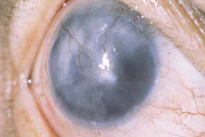
Brow loss is one of the most common manifestations of leprosy41–43 and occurs more frequently in the tuberculoid form (Fig. 1). Brow loss begins temporally and progresses nasally, perhaps because the temporal brow is somewhat cooler. Madarosis, or eyelash loss, is also common, affecting the lower lid before the upper lid. The severity of both brow loss and madarosis is related to the duration of disease.44

Fig. 1 Patient with borderline leprosy with classic leonine facies of leprosy from accentuated facial skin folds. Note the brow loss, lash loss and madarosis, nasal septum involvement, and ectropion secondary to facial nerve paralysis.

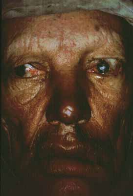
Seventh cranial nerve paralysis is common and results in lagophthalmos and ectropion of the lower eyelid (see Fig. 1). When fifth-nerve involvement also occurs, a marked form of anesthetic corneal exposure may result. It has been believed that inflammation caused by leprosy may cause upper lid entropion,43,45 but trachoma is often endemic in these populations so the upper lid deformity may often be caused by trachoma. Unfortunately, multibacillary patients develop paresis later in their disease when they often have corneal sensory loss. These patients have little urge to blink, nor can they effectively blink when they try. Such patients are at high risk for corneal ulceration, and surgical intervention should be considered early.

Eyelid nodules, ENL, and erysipeloid reactions of the lid are recognized less frequently, probably because they occur less frequently, or perhaps because they are transient. Loss of skin elasticity, infiltration of the marginal and pretarsal fibers of the orbicularis oculi muscle by M. leprae, and loss of muscle tone contributes to dermatochalasis and heavy, drooping upper lids (see Fig. 1). Further atrophy at the canthal tendons and tarsal plates create heavy, floppy lids allowing ectropion or perhaps entropion, and trichiasis.

Meibomian gland infiltration leads to atrophy and an inadequate lipid production with associated tear dysfunction. Leprosy involves the lacrimal gland by causing denervation of the lacrimal gland that helps to contribute to a dry-eye syndrome. Leprotic dacryocystitis secondary to nasal disease is a common complication, and may contribute to subsequent corneal ulceration and microbial keratitis.

CORNEAL AND CONJUNCTIVAL INVOLVEMENT

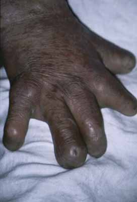
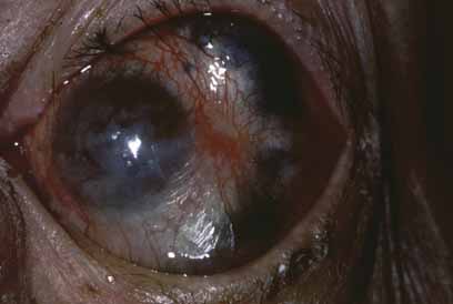
Exposure keratitis occurs frequently in patients with leprosy. Seventh cranial nerve involvement with lagophthalmos results in exposure keratitis inferiorly. Fifth cranial nerve involvement with an anesthetic cornea and decreased blinking also contributes to the problem. Patients may inadvertently traumatize themselves by rubbing their eyes with calloused, insensitive, and often infected fingers (Fig. 2). This constant exposure is often combined with lid deformities (trichiasis) so that corneal ulceration and secondary bacterial infection are frequent events.46 These ulcers may perforate, leading to phthisis and blindness. Even when the corneal ulcer heals, there is stromal scarring and thinning. Corneal hyposensitivity or anesthesia is seen more commonly in lepromatous leprosy, and is coincident with more severe complications.47

Fig. 2 Left hand of a patient with borderline leprosy. Note the loss of digits and severe deformities. The remaining fingers are insensitive and clumsy.

Enlarged, edematous corneal nerves are found in lepromatous leprosy. The nerve involvement has the appearance of focal swelling resembling beads on a string and consists of masses of M. leprae and a surrounding granulomatous response within the corneal nerve. These nerve swellings are pathognomonic of leprosy and may be the first ocular or systemic finding.48

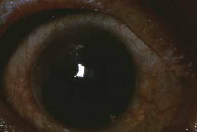
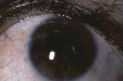
There is also avascular or punctate keratitis consisting of chalky, white, punctate subepithelial opacities that first form in the superior temporal quadrant of the cornea. These are miliary lepromas with macrophages, lymphocytes, and M. leprae.49 The individual lesions gradually become less demarcated with a surrounding haze and eventually becoming confluent. Histologically within these nebulae, discrete calciumlike deposits may be seen, often associated with destruction of Bowman's layer. Superficial neovascularization occurs late in the disease to produce the classic leprous pannus (Fig. 3).49 The bacilli, which produces avascular keratitis, probably reach the cornea by way of the nerves, but deposits also occur adjacent to trachomatous pannus vessels, so that a vascular route is also possible.

Fig. 3 Avascular keratitis may be seen initially in the superior temporal quadrant, beginning as subepithelial opacities, later coalescing with vascularization, as seen here.

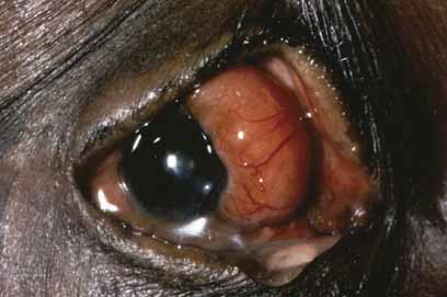
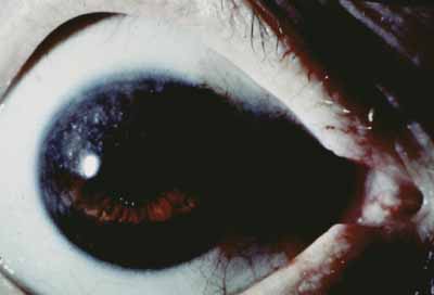
Corneal lepromas (Fig. 4) occur more commonly in South America50 and Japan51 but are relatively infrequent. These lesions are large granulomas with epithelioid cells, lymphocytes, and bacteria. They may become large enough to involve the visual axis and compromise vision. Lepromas usually arise at the limbus (most commonly the lateral limbus) or on the sclera and then encroach onto the cornea.

Fig. 4 Ocular lepromas occasionally occur in the palpebral aperture. These may occur at the limbus or in the episcleral tissues where bacilli are localized with a granulomatous response. It may progress to involve the visual axis.

Interstitial keratitis begins in the superior temporal quadrant or in the superior quadrants. It is probably a more severe form of the avascular keratitis, with necrosis and then vascular invasion. Interstitial keratitis may begin without a preceding avascular keratitis in the midstroma of the cornea (superior nasal quadrant) or as a discoid lesion often located superiorly. The interstitial keratitis may progress to involve the visual axis (Fig. 5), with a decrease in visual acuity. Ghost vessels are occasionally seen in mid- to deep stroma in interstitial keratitis, indicating previous inflammatory activity.

Fig. 5 Interstitial keratitis may occur by two possible mechanisms. The interstitial keratitis may be secondary to a deeper extension of the avascular keratitis with necrosis, as probably occurred here. However, interstitial keratitis may also be the result of an immunologic response without a preceding area of avascular keratitis or necrosis.

SCLERAL AND UVEAL LESIONS

Episcleritis, scleritis, and uveitis are the initial presenting complaints in up to 16% of leprosy patients.52 The nodular episcleritis that occurs consists of focal leproma with surrounding inflammatory response. There are also more diffuse forms of episcleritis. Episcleritis commonly occurs as part of the ENL reaction, and may be associated with iridocyclitis. Lepromas may occur in the interpalpebral fissure more commonly because this area is cooler. The episcleritis and scleritis may be the result of direct bacillary invasion, but there is strong evidence that some cases may be mediated by immune responses, such as the deposition of immune complexes. Chronic or recurrent scleritis in leprosy may result in scleromalacia and staphyloma (Fig. 6) and, in some cases, disorganization of the globe.

Fig. 6 Intercalary staphylomas are a result of long-standing episcleritis and scleritis secondary to leprosy. The frequent attacks may result in scleromalacia, scleral thinning, and staphylomas, as seen here. This patient also exhibits upper lid entropion from concomitant trachoma. The keratitis sicca, xerosis, and keratinization of the conjunctiva have also contributed to this patient's corneal blindness.

Uveitis in leprosy may be the result of several different mechanisms. Uveitis with the erythema nodosum leprosum reaction probably results from a hypersensitivity reaction (probably antigen-antibody mediated)25 and usually occurs with therapy or occasionally after termination of therapy. This iridocyclitis, which also occurs spontaneously, has an abrupt onset and fulminating course with hypopyon, posterior synechiae, and elevated intraocular pressure. Occasionally, there is a spontaneous hyphema presumably because of fragile vessels from previous attacks. This granulomatous uveitis is usually associated with greasy, keratic precipitates.

There is also a chronic, low-grade uveitis seen commonly in lepromatous leprosy, which is the principal cause of blindness in ocular leprosy.33,34 This insidious anterior uveitis causes gradual iris atrophy as manifested by loss of iris stroma with a moth-eaten appearance, frank iris holes (Fig. 7), and small, nonreactive pupils. In this condition, iris stromal nerve involvement and disintegration of iris stroma associated with leprosy bacilli can be demonstrated histologically.53 In this “white” iritis, patients have few anterior chamber cells and mild to moderate anterior chamber flare with minimal ciliary flush or conjunctival injection. There are a few scattered, fine, white keratic precipitates. Posterior synechiae are not common and, when present, may be the result of a previous attack of acute iritis. The dilator muscle is affected earlier and to a greater extent than the sphincter muscle, resulting in a small, nonreactive pupil.54 The pinpoint pupil may lead to marked visual loss if there is a corneal or lens opacity. This chronic, low-grade uveitis is more common in patients with lepromatous leprosy, although it may be present in those with tuberculoid leprosy.35

Fig. 7 The chronic iritis often associated with lepromatous leprosy may result in marked iris atrophy with stromal loss, iris pigment epithelium loss, and frank iris holes. This severe iris atrophy is symptomatic of long-standing disease. Although the cause is unknown, Mycobacterium leprae bacilli seem to affect preferentially the nonmyelinated iris nerves with a “neurotrophic” iritis and iris atrophy.

Iris pearls arise from the surface connective tissue of the iris and can be recognized on the iris surface even early in the disease (Fig. 8). These creamy white particles consist of bacilli and monocytes and are rarely associated with an inflammatory response.49 Iris pearls are pathognomonic of ocular leprosy and look like grains of sand on the iris surface, often occurring near the pupillary border. After several years, they may coalesce and drop into the inferior angle, where they can be observed by gonioscopy. Pearls have been reported on the retina55 but probably represent the migration of such spheres to the posterior segment rather than formation there.

Fig. 8 Iris pearls can be seen on the pupillary border (see text for discussion). There is also typical avascular keratitis in the superior temporal quadrant.

The ciliary body may represent the site of bacillary activity in early leprosy. Several clinical signs suggest ciliary body dysfunction early in the disease. Premature presbyopia in lepromatous leprosy is common and is probably caused by a parasympathetic neuropathy affecting accommodation.56,57 Low intraocular pressures has been observed in some studies with a large fluctuation in postural intraocular pressure change early in the disease without other evidence of ocular involvement.58–60 Decreased intraocular pressure can also be found in household contacts of leprosy patients compared to endemic controls.61

Rarely, there may be a uveal effusion associated with an overlying scleritis and episcleritis. Choroiditis has been reported,62,63 but it is doubtful if leprosy was the cause.

Glaucoma is a result of seclusion of the pupil by posterior synechiae and resulting iris bombé. This condition is readily corrected by peripheral iridectomy or pupil transfixion. There is also a higher rate of elevated intraocular pressure in leprosy patients with a deep anterior chamber. It is not yet known if this is caused by peripheral anterior synechiae or by other forms of involvement of the outflow channels.

Back to Top
TREATMENT AND PROGNOSIS
Early diagnosis and prompt treatment with antimicrobials (dapsone, rifampin, and clofazimine given orally) may prevent late complications such as ectropion, lagophthalmos, exposure, and other forms of keratitis as well as some forms of uveitis. The introduction of MDT by WHO in 1981 has rendered this disease curable within 6 to 12 months of treatment in paucibacillary leprosy (PB, patients with one to five skin patches) and multibacillary leprosy (MB, patients with more than five skin patches) patients, respectively. The transmission of this mildly contagious infection is interrupted after the first dose of MDT.15 Early treatment of keratitis and uveitis with topical corticosteroids combined with appropriate antimycobial therapy may reduce the binding complications of these problems. The risk of the ENL reaction is present with treatment, and this reaction may affect the seventh cranial nerve leading to partial or complete paralysis. Prompt intervention by use of corticosteroids may prevent or reduce these complications from the reaction.

Lid deformities caused by leprosy or concomitant diseases such as trachoma, including ectropion, entropion, and lagophthalmos, should be corrected by appropriate surgical procedures to provide adequate corneal protection. These lid procedures are relatively simple to perform. Appropriately trained nonspecialist health personnel can carry them out in developing countries where ophthalmologists may be scarce. Sector iridectomy may restore vision in patients with nonreactive pupils whose visual pathways are blocked by small corneal or lens opacities. Angle-closure glaucoma resulting from seclusion of the pupil by posterior synechiae can be treated with peripheral iridectomy. Finally, cataract is the most common cause of visual loss and blindness in developing countries63 and may occur more often and at an earlier age in patients with leprosy; simple cataract extraction can restore useful vision in many cases. Fortunately, intraocular surgery is well tolerated by patients with leprosy.64 Intraocular lens (IOL) implantation after cataract extraction could be well tolerated in select patients if careful attention is paid to preoperative selection of patients, aggressive management of intraocular inflammation with corticosteroid, minimum intraocular manipulation of the iris, in-the-bag IOL implantation, and careful postoperative management and follow-up.65

Most developing countries have special leprosy hospitals for advanced cases with severe disability and deformities. Although the practice of hospitalizing patients with leprosy for long-term care is controversial, those with ocular complications can be identified and treated with only a modest deployment of resources. The patients themselves or the regular hospital staff can provide primary eye care by promptly treating all minor eye infections with topical antibiotics and by protecting an exposed cornea with a topical ointment. An ophthalmologist can review patients periodically, because these patients rarely require emergency ocular surgery, except of course for acute angle-closure glaucoma. Much of the blindness caused by leprosy can therefore be prevented or easily cured at relatively little cost. Patients with leprosy who are blind have a higher risk of death compared to sighted patients.66 It is important to not neglect patients with or without ocular complications during and after treatment programs, because they do need follow-up for life and treatment of blinding condition as and if they develop.

Back to Top
PREVENTION
Blindness can largely be prevented through meticulous attention to local factors, as discussed previously, throughout the life of the patients even after they have been discharged from MDT treatment programs. Preventing the disease itself is more problematic. A specific vaccine made from bacille Calmette-Guérin (BCG) plus heat-killed M. leprae bacilli has been undergoing field trials but will require several years to evaluate effectiveness. Early results have been promising.67 Because the supply of leprosy bacilli from animals is limited, other workers are using DNA transfer techniques to obtain large quantities of leprosy antigens from other sources. Recent strategic research has focused on sequencing the genome of the M. leprae to develop a diagnostic skin test to understand the transmission of the disease, its epidemiology for early treatment and a step toward disease eradication.

Chemotherapy remains the primary avenue for dealing with existing infection. All patients should be evaluated and managed by a leprologist. Several MDT regimens have been suggested. Current recommendations are dapsone and rifampin for all cases and the addition of clofazimine for multibacillary cases. Patients with paucibacillary disease should receive dapsone (50 mg) daily (unsupervised) rifampin (10 mg/kg or as much as 600 mg) monthly (supervised) for a minimum period of 6 months. These patients should then be observed for a minimum of 3 years. Patients with multibacillary disease should receive clofazimine (1 to 4 mg/kg) daily (supervised at least once monthly) in addition to the above until slit-skin smears are negative or for a minimum period of 12 months. Drugs that are useful for reactional states are analgesics (e.g., aspirin), corticosteroids, thalidomide, and clofazimine. Leprosy patients should also be encouraged to avoid stress and have proper rest because physical and emotional stress can precipitate a reactive episode.

Dapsone effectively reduces infectivity, but a few viable bacilli (“persisters”) may remain for many years, so that secondary sulfone resistance may appear after 5 to 20 years of favorable response to the drug.20 A small but increasing number of previously untreated patients also appear to be resistant to dapsone (primary dapsone resistance).68 Fortunately, this resistance is usually only partial, and does not appear to pose major clinical problems yet.69 Rifampin is effective against the leprosy bacillus and is bactericidal, but unfortunately relapses have occurred in patients treated only with this drug. Clofazimine is also effective alone and, as discussed above, is recommended as a supplement to dapsone therapy. Unfortunately, it is expensive and produces hyperpigmentation of the skin in light-skinned persons. Other drugs that are not suitable for use alone but occasionally supplement other agents include prothionamide, ethionamide, ofloxacin,70 minocycline,71 thiambutosine, and thiacetazone.17 Other sulfones are used much less frequently and include sulfoxone and acadapsone.

The global strategy for the elimination of leprosy by WHO, based on the implementation of MDT with case finding, is working well in reducing the prevalence of leprosy. It is recommended that research in leprosy be continued especially to improve patient care and in addressing posttreatment program issues of progressive complications and disabilities of the disease.72–75

Back to Top
REFERENCES

1. Shepard CC: The experimental disease that follows the injection of human leprosy bacilli into footpads of mice. J Exp Med 112:445, 1960

2. Baskin GB, Wolf RH, Gormus BJ, et al: Experimental leprosy in the Mangabey (Cercocebus atys): necrospy findings. Int J Lepr Other Mycobact Dis 53:269, 1985

3. Baskin GB, Gormus BJ, Martin LN, et al: Experimental leprosy in African green monkeys (Cercopithecus aethiops): a model for polyneuritic leprosy. Am J Trop Med Hyg 37:385, 1987

4. Shepard CC: Rodents, Edentulates, and Other Animal Models for Leprosy: Cultivation of the Etiologic Agent, Immunology, Animal Models. Washington, D.C.: Pan American Health Organization, 1977:57–62

5. Runyon EH, Wayne LG, Kubica GP: Mycobacteriaceae. In Buchanen RE, Gibbons NE, eds: Bergey's Manual of Determinative Bacteriology. Baltimore: Williams & Wilkins, 1974:681–701

6. Thimann KV: The Life of Bacteria. New York: Macmillan, 1963:631

7. Feldman IRA, Sturdivant M: Leprosy in the United States, 1950–1969. South Med J 69:970, 1976

8. Fasal P: A primer of leprosy. Cutis 7:525, 1971

9. Memoria del VI Congreso Internacional de Leprologia, Madrid, October 1953

10. Ridley DS, Jopling WH: A classification of leprosy for research purposes. Lepr Rev 33:119, 1962

11. Ridley DS, Jopling WH: A classification of leprosy according to immunity. A five-group system. Int J Lepr 34:255, 1966

12. Van Voorhis WC, Kaplan G, Sarno EN, et al: The cutaneous infiltrates of leprosy. N Engl J Med 307:1593, 1982

13. Prabhakaran K: Oxidation of 3,4-diphydroxyphenylalanine (DOPA) by Mycobacterium leprae. Int J Lepr 35:42–51, 1967

14. Major Conclusions and Recommendations, 7th WHO Expert Committee on Leprosy Report, June 1997

15. WHO. Leprosy, Fact Sheet No: 101, Revised January 2003

16. Courtright P: The epidemiology of ocular complications of leprosy—symposium paper. Indian J Lepr 70:33, 1998

17. Ffytche TJ: The prevalence of disabling ocular complication of leprosy: a global study. Indian J Lepr 70:49, 1998

18. World Health Organization: Methods of Assessment of Avoidable Blindness. WHO offset publication No. 54. Geneva: World Health Organization, 1980

19. Lewallen S, Tungpakorn NC, Kim SH, et al: Progression of eye disease in “cured” leprosy patients: implications for understanding the pathophysiology of ocular disease and for addressing eye care needs. Br J Ophthalmol 84:817, 2000

20. Shepard CC: Leprosy. In Harrison TR, ed: Principles of Internal Medicine. New York: McGraw-Hill, 1980:711–714

21. Duke-Elder S: System of Ophthalmology, Vol VIII. St. Louis: CV Mosby, 1965:802, 844

22. Hobbs HE: Leprotic iritis and blindness. Int J Lepr 40:366, 1972

23. McLaren DS, Shaw MJ, Dulley KR: Eye disease in leprosy patients: A study in central Tanganyika. Int J Lepr 29:20, 1961

24. Wangspa S, Limpaphayom P: Ocular leprosy in Thailand. Trans Asia Pac Acad Ophthalmol 5:153, 1976

25. Gibson JB: Eye complications of leprosy. Med J Aust 1:8, 1950

26. Wasty IA, Abdel-Rehim DE: Ocular leprosy in Assiut. Bull Ophthalmol Soc Egypt 64:299, 1971

27. Ffytche TJ: The eye and leprosy. Lepr Rev 52:111, 1981

28. Ticho U, Sira IB: Ocular leprosy in Malawi: clinical and therapeutic survey of 8,325 leprosy patients. Br J Ophthalmol 54:107, 1970

29. Chatterjee S, Chaudhury DS: Pattern of eye diseases in leprosy patients of northern Ghana. Int J Lepr 32:53, 1964

30. Cameron AN: Leprosy and its ocular manifestations. Trans Ophthalmol Soc UK 81:637, 1961

31. Cochrane RG: Leprosy in Korea. Lepr Rev 27:19, 1956

32. Latapi F, Zamora AC: The ‘spotted’ leprosy of Lucio—an introduction to its clinical and histological study. Int J Lepr 16:421–429, 1948

33. Choyce DP: Diagnosis and management of ocular leprosy. Br J Ophthalmol 53:217, 1969

34. Weerekoon L: Ocular leprosy in Ceylon. Br J Ophthalmol 53:457, 1969

35. Schwab IR, Nassar E, Malaty R, et al: Leprosy in a trachomatous population. Arch Ophthalmol 102:240, 1984

36. Alien JH, Brand M: Ocular leprosy. In Locatcher-Khorazo D, Seegal BC, eds: Microbiology of the Eye. St. Louis: CV Mosby, 1972

37. Elliot DC: An interpretation of the ocular manifestations of leprosy. Ann NY Acad Sci 54:84, 1951

38. Choyce DP: Discussion on ocular leprosy. Trans R Soc Trop Med Hyg 64:43, 1970

39. Hobbs HE, Choyce DP: The binding lesions of leprosy. Lepr Rev 42:131, 1971

40. Malla OK, Brandt F, Anten JGF: Ocular findings in leprosy patients in an institution in Nepal (Khokana). Br J Ophthalmol 65:226, 1981

41. Choyce DP: Diagnosis and management of ocular leprosy. Br J Ophthalmol 53:217, 1969

42. Harrell JD: Ocular leprosy in the Canal Zone. Int J Lepr 45:56, 1977

43. Richards WW: Ocular leprosy. Trans Pacif Coast Oto-oph-thalmol Soc 52:161, 1971

44. Cakiner T, Karacorlu MA: Ophthalmic findings of newly diagnosed leprosy patients in Istanbul Leprosy Hospital, Turkey. Acta Ophthalmol Scand 76:100, 1998

45. Holmes WJ: Leprosy of the eye. Trans Am Ophthalmol Soc 55:145, 1957

46. John D, Daniel E: Infectious keratitis in leprosy. Br J Ophthalmol 83:173, 1999

47. Karacorlu MA, Cakiner T, Saylan T: Corneal sensitivity and correlations between decreased sensitivity and anterior segment pathology in ocular leprosy. Br J Ophthalmol 75:117, 1991

48. Daniel E, Koshy S, Rao GS, et al: Ocular complications in newly diagnosed borderline lepromatous and lepromatous leprosy patients: baseline profile of the Indian cohort. Br J Ophthalmol. 86:1336, 2002

49. Allen JH, Byers JL: The pathology of ocular leprosy. Arch Ophthalmol 64:216, 1960

50. Aparisi T: Lepra ocular. Arch Oftalmol Hispano-Am 10:107, 1950

51. Holmes WJ: Leprosy of the eye. Lepr Rev 28:108, 1957

52. Brand ME: Early Ocular Changes in Leprosy. Presented at the IX International Leprosy Conference, London, 1968

53. Hobbs HE, Harman DJ, Rees JW, et al: Ocular histopathology in animals experimentally infected with Mycobacterium leprae and M. lepraemurium. Br J Ophthalmol 62:516, 1978

54. Ffytche TJ: Role of iris changes as a cause of blindness in lepromatous leprosy. Br J Ophthalmol 65:231, 1981

55. Somerset EJ, Sen NR: Leprotic lesions of the fundus oculi. Br J Ophthalmol 40:167, 1956

56. Slem G: Clinical studies of ocular leprosy. Am J Ophthalmol (Suppl 77):431, 1971

57. Lamba PA Rohatgi J, Sehgal VN: Evidence of sub-clinical uveal involvement in leprosy. Indian J Ophthalmol 34:319 1986

58. Hussein N, Courtright P, Ostler HB: Low intraocular pressure and postural change in intraocular pressure in Hansen's disease. Am J Ophthalmol 108:80, 1989

59. Lewallen S, Courtright P, Lee HS: Ocular autonomic dysfunction and intraocular pressure in leprosy. Br J Ophthalmol 73:946, 1989

60. Daniel AE, Arunthathi S, Bhat L, et al: Intraocular pressure in leprosy patients without clinically apparent anterior segment pathology. Indian J Lepr 66:165, 1994

61. Hussein N, Chiang T, Ehsan Q, et al: Intraocular pressure decrease in household contacts of patients with Hansen's disease and endemic control subjects. Am J Ophthalmol 114:479, 1992

62. Prendergast JJ: Ocular leprosy in the United States. Arch Ophthalmol 23:112, 1940

63. Tarizzo ML: Cataract: a major cause of unnecessary blindness. Doc Ophthalmol Proc Series 21:5, 1979

64. Ffytche TJ: Cataract surgery in the management of the late complications of lepromatous leprosy in South Korea. Br J Ophthalmol 65:243, 19981

65. Daniel E, Koshy S: Intraocular lens implantation in leprosy. Int J Lepr Other Mycobact Dis 70:9, 2002

66. Courtright P, Kim SH, Lee HS, et al: Excess mortality associated with blindness in leprosy patients in Korea. Lepr Rev 68:326, 1997

67. Convit J, Ulrich M, Aranzazu N, et al: The development of a vaccination model using two microorganisms and its applications in leprosy and leishmaniasis. Lep Rev 57:263, 1986

68. Pearson JMH, Ross WF, Rees RJW: DOS resistance in Ethiopia—a progress report. Int J Lepr 44:140, 1976

69. Gelber RH, Rea TH, Murray LP, et al: Primary dapsoneresistant Hansen's disease in California. Arch Dermatol 126:1584, 1990

70. Grosset JH, Guelpa-Lauras C-C, Perani EG, et al: Activity of ofloxacin against Mycobacterium Leprae in mice. Int J Lepr 56:259, 1988

71. Gelber RH: Activity of minocycline in Mycobacterium leprae–infected mice. J Infect Dis 156:236, 1987

72. Fytche TJ: Residual sight-threatening lesion in leprosy patients completing multidrug theraphy and sulphone monotheraphy, Lepr Rev 62:35, 1991

73. Courtright P, Lewallen S, Tungpakorn N, et al: Cataract in leprosy patient: cataract surgical coverage, barriers to acceptance of surgery and outcomes of surgery in a population based in Korea. Br J Ophthalmol 85:643, 2001

74. Ostler HB: Hansen's disease. Int Ophthalmol Clin 30:42 1990

75. Ostler HB: Bacterial Infections—Diseases of the External Eye and Adnexa. A Text and Atlas. Baltimore: Williams & Wilkins, 1993:458–469

Back to Top