Chapter 61
Corneal Opacification in the Developing World
GULLAPALLI N. RAO
Main Menu   Table Of Contents

Search

ETIOLOGIC FACTORS
TRAUMA
XEROPHTHALMIA
CONGENITAL/HEREDITARY DISEASES
IATROGENIC DISEASES
HARMFUL EYE PRACTICES
PREVENTIVE STRATEGIES
MANAGEMENT AT THE PRIMARY HEALTH CARE LEVEL
TREATMENT
WORLDWIDE SITUATION
EYE BANKING IN INDIA
RECIPIENT CORNEAL PATHOLOGY
CARE OF THE TRANSPLANT
REFERENCES

Blindness is a major public health problem in most developing countries. Corneal opacification, as a cause of blindness, is second only to cataract in magnitude.1 One major difference between the two entities is the age of onset, as corneal opacification is more often seen at a younger age. In a series of 2111 corneal transplants performed over an 8-year period in a single center in India, 53% of the patients were younger than age 45.2

It is estimated that about 30% of all blindness in some developing countries is due to corneal opacification,3 while the corresponding rate in most developed countries is very low. These data, however, are based on information from only a few countries.

According to 1990 figures from the World Health Organization (WHO), there are 37.9 million bilaterally blind (i.e., visual acuity less than 3/60 in the better eye) people in the world.1 These estimates indicate that 5.5 million are bilaterally blind from trachoma and 0.5 million from xerophthalmia. It is quite possible that these figures will increase by 1995 to at least 40 million blind people, of which at least 7 million are expected to be bilaterally blind from corneal disease. This constitutes approximately 17% to 20% of all blindness in the world.4 The number of people with severe bilateral visual impairment (i.e., less than 6/60 but equal to or better than 3/30) or unilateral blindness from corneal disease is not known. It is likely that there are at least 3 million people with severe bilateral visual impairment and probably another 30 million people with unilateral blindness from corneal disease.4

Lack of public education about prevention, poor personal hygiene, poor standards of sanitation, malnutrition, inadequate safety regulations in industry, consanguineous marriages, and poor standards of medical and ophthalmic practice are factors contributing to corneal blindness. A multitude of disease mechanisms may lead to corneal involvement and opacification. However, accurate information is not available on their relative importance. Corneal opacification often leads to unilateral blindness, as exemplified by trauma, which is the leading cause of unilateral blindness in the world with a prevalence of 2%.3

Back to Top
ETIOLOGIC FACTORS
Infections, trauma, xerophthalmia, congenital and hereditary diseases, iatrogenic problems, and harmful eye practices lead to corneal blindness.

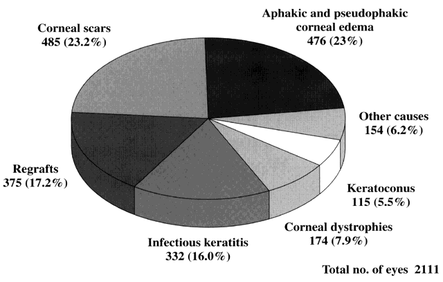
In a series of 2111 eyes that underwent penetrating keratoplasty at L.V. Prasad Eye Institute in Hyderabad, India, corneal opacification was due to corneal scars (23.2%), aphakic and pseudophakic corneal edema (23.0%), regrafts (17.2%), active infectious keratitis (16.0%), corneal dystrophies (7.9%), keratoconus (5.5%), and various other causes (6.2%)2 (Fig. 1).

Fig. 1. Graph showing the indications for penetrating keratoplasty in a series of 2111 eyes done over an 8-year period at L. V. Prasad Eye Institute, Hyderabad, India.

INFECTIONS

A variety of microbial agents cause corneal stromal degradation with consequent scarring. Of the infectious diseases, trachoma continues to be the leading cause of corneal blindness in the world. According to the WHO, almost 6 to 9 million people are blind from trachoma and more than 500 million people inhabit areas endemic to blinding trachoma. The cornea is affected secondarily to chronic progressive conjunctival pathology; blindness usually ensues after 50 years of age.

Corneal infections caused by bacteria, fungi, viruses, and parasites are a frequent occurrence in most developing countries. Poverty, illiteracy, ignorance, inaccessibility to medical care, delayed or incorrect diagnosis, nonavailability of appropriate drugs, and exposure to harmful treatment practices are impediments to effective prevention of corneal blindness. Medical attention is often sought at an advanced stage. It is not uncommon to find a large segment of eye beds occupied by patients with infectious keratitis.

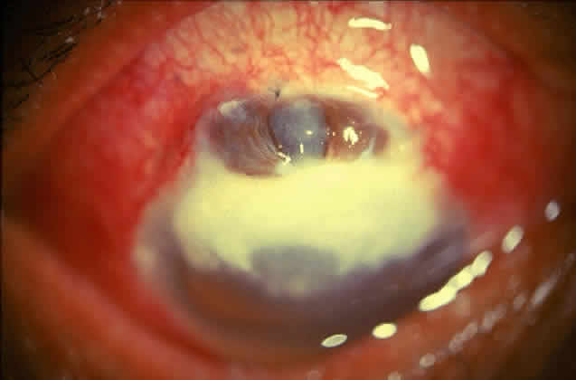
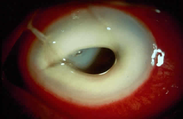
Bacterial corneal infections are caused by a wide variety of organisms. Pneumococcus seems to be the most common causative organism (Fig. 2); however, staphylococcal, pseudomonal, and gonococcal infections frequently are encountered.3 This may be related to chronic dacryocystitis.5

Fig. 2. Photograph demonstrating a pneumococcal corneal ulcer.

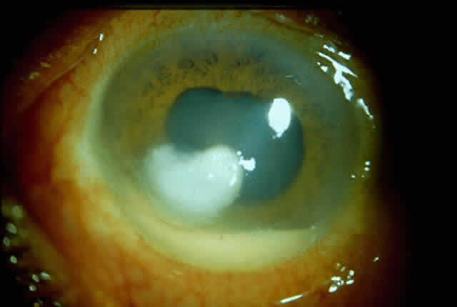
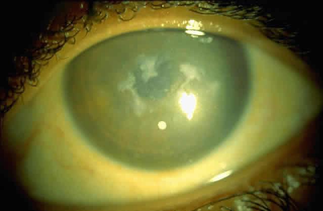
Fungal keratitis is as frequently seen as bacterial keratitis. Aspergillus and Fusarium are the two most commonly isolated organisms. Predisposing factors include trauma, foreign body, and injudicious usage of topical steroids or other indigenous medications.

Delay in diagnosis, the relative ineffectiveness of antifungal agents in most parts of the world, and the nonavailability of these drugs hinder prevention and management (Fig. 3).

Fig. 3. Advanced stage of corneal ulcer caused by Fusarium solani.

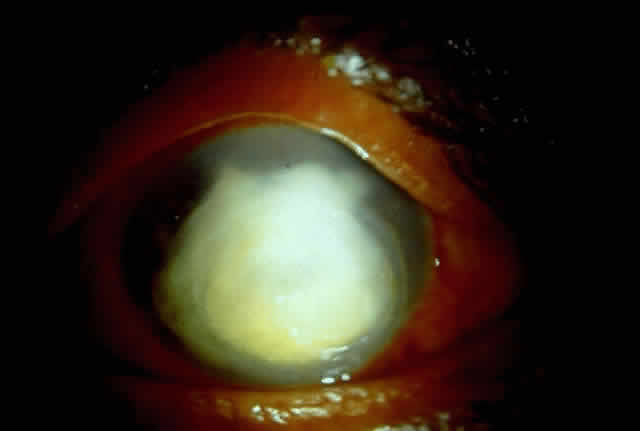
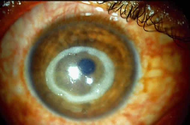
Acanthamoeba infection of the cornea is being reported with increasing frequency. While this is associated with contact lens wear in industrialized countries, in developing countries it is almost exclusively seen in non-contact lens wearers (Fig. 4).6 Misdiagnosis and inappropriate treatment are common.

Fig. 4. Typical ring infiltrate in a case of Acanthamoeba keratitis in a non-contact lens wearer.

Herpes simplex keratitis has the potential for corneal devastation, particularly when it presents as stromal disease. Failure to make the clinical diagnosis, lack of laboratory facilities, and inappropriate treatment often lead to recalcitrant stromal disease and consequent corneal opacification.

Ophthalmia neonatorum is being reported with increasing frequency.7 Absence of proper prenatal screening for venereal disease and low utilization of Crede's prophylaxis are responsible. The emergence of penicillin-producing strains of gonorrhea is yet another problematic factor.

Ocular Leprosy

As many as 10 to 12 million people worldwide suffer from leprosy, and about 250,000 million are blind from ocular leprosy.8 Involvement of the eye in leprosy has been reported from a variety of corneal manifestations, some of which may lead to blindness.9 Lagophthalmos with exposure keratitis and consequent infection resulting in scarring is an important mechanism. Hypoesthesia of cornea can also can lead to corneal ulceration and ultimate opacity.10

Onchocerciasis

Microfilariae, dead or alive, in the corneal stroma, produce “snowflake” or onchocercal punctate keratitis. Onchocerciasis can be a mild and reversible phenomenon, but severe longstanding invasion of numerous microfilariae into the cornea may lead to sclerosing keratitis, especially along the lower limbus, gradually spreading to the central optical zone.11 Sclerosing keratitis is usually seen in severe cases and is claimed to be the most common pathway to blindness in the savannas.

According to a WHO report on corneal blindness,3 nine main “ecological and behavioral niches” promote corneal ulceration through a consequence of risk factors, one of which is often infectious in origin. Adequate prevention and treatment measures include the following, which are listed according to patient or environmental characteristics:

  1. Newborn babies: Prevention of gonococcal ophthalmia in newborn babies by ocular prophylaxis at birth with tetracycline ointment, improved prenatal screening, and control of sexually transmitted diseases
  2. Children: In children 6 months to 6 years, especially those 1 to 3 years of age, immunization against measles, sound weaning practices to improve sanitation, and oral vitamin A supplements to prevent serophthamla (particularly in cases of malnutrition), malabsorption (prolonged diarrhea), measles, or corneal ulcers of unknown cause
  3. Measles epidemic: During an epidemic of measles, the prevention of overcrowding of children and removal of contact cases, vitamin A supplementation, eye ointment, and immediate treatment for corneal ulcers is essential
  4. Famine and disasters: In famine and disasters, displaced persons should receive immunization against measles, vitamin A supplementation, and immediate treatment of corneal ulcers
  5. Arid, crowded, or tropical rural areas: In these areas, where trachoma and communicable ophthalmia are predominant, prevention and management include prophylaxis against injuries and control of trachoma.
  6. Lush, humid, tropical areas and industrial settings: In these areas, prevention of and first aid for injuries is essential, particularly corneal trauma from agricultural products that frequently cause secondary (often fungal) infections

In East Africa between 1% and 4% of children with measles develop corneal ulcers, and almost half (46%) of unilateral and more than three quarters (79%) of bilateral corneal ulcers occur within 3 months of onset of rash. Vitamin A deficiency was the cause of 50% of these cases and 55% of cases of bilateral corneal ulcers.12 Use of local remedies and herbs were the other major etiologic causes of potentially preventable blindness.

The causes of bilateral blindness in children in Tanzania were reported to be as follows13:

  Vitamin A deficiency: 45%
  Harmful eye practices: 15%
  Herpes simplex virus: 5%
  Ophthalmia neonatorum: 5%
  Cataract, uveitis, and others: 30%

Again, 40% to 50% of all cases of childhood blindness were associated with measles.

Back to Top
TRAUMA
Ocular trauma is responsible for 1% to 10% of corneal blindness. The geographic location, pattern of injury, causative agent, and age are some of the factors that determine the degree of damage (Table 1).


TABLE 1. Pathways to Corneal Blindness from Trauma


Corneal foreign bodySecondary infectionCorneal ulcer and opacity
Chemical injuriesKeratitisCorneal opacification
Blunt injuryLacerationCorneal opacification
Penetrating injury (including intraocular foreign body)Wound & secondary infectionCorneal opacification
(Adapted from World Health Organization: Report of the International Meeting on Control of Corneal Blindness Within Primary Health Care Systems pp 1–22. Geneva, WHO, 1988.)


In urban areas and in the industrialized world, chemical injuries, accidents at the workplace, and automobile injuries are common. In rural areas of developing countries, minor trauma due to hazardous practices in agriculture, cottage industries, and other work places, as well as sports accidents, are responsible for a large proportion of corneal blindness. In addition, use of hazardous objects such as bows and arrows and lack of implementation of industrial safety regulations increase corneal morbidity from trauma.

Back to Top
XEROPHTHALMIA
Most developing countries continue to have malnutrition-related ocular problems due to vitamin A deficiency. This condition remains the single largest cause of childhood blindness in the world.3 It is estimated that there are about 10 million children with ocular signs of vitamin A deficiency and probably about 250,000 to 500,000 children who become blind from this problem every year, mainly in Asia and Africa. Vitamin A deficiency is a predisposing factor of respiratory tract infections, diarrhea, corneal ulceration, blindness, and increased child mortality. Vitamin A deficiency occurs from decreased intake of the vitamin or its precursors or from an increased bodily requirement of the vitamin during infections, particularly measles. Predisposing factors for acute corneal ulceration from vitamin A deficiency are protein-energy malnutrition, malabsorption (chronic diarrhea), and measles.

Corneal involvement manifests as bilateral, deep, punched-out ulcers with lack of luster in the remaining cornea, leading to rapid perforation (keratomalacia).14

Back to Top
CONGENITAL/HEREDITARY DISEASES
Corneal blindness due to congenital/hereditary problems occur frequently in developing countries, particularly in areas such as the Middle East, which has high rates of consanguinity. Manifestations range from sclerocornea in the newborn to corneal dystrophies in various age groups (Fig. 5).

Fig. 5. Cornea showing macular corneal dystrophy.

The exact prevalence of corneal blindness due to congenital/hereditary problems is not clearly established. However, in a tertiary-care setting in India, corneal blindness due to congenital/ hereditary problems constitutes 7.9% of the total number of keratoplasties performed over an 8-year period.2

Back to Top
IATROGENIC DISEASES
With advances in ophthalmology, a number of new surgical techniques are being employed. Adequate infrastructural support, such as modern equipment and adequate training of ophthalmologists, are prerequisites for the success of these new techniques.

Intracapsular cataract extraction continues to be the most common method of cataract surgery in most developing countries. The infrastructure available in such countries is not appropriate for modern extracapsular cataract extraction with intraocular lens implantation. The cost of infrastructure, inadequate manpower, lack of training facilities, and inaccessibility to standardized and safe drugs and other supplies are major obstacles.

The prevalence of aphakic and pseudophakic corneal edema is high and now constitutes the second most common indication for penetrating keratoplasty (Fig. 6).2 Poor surgical technique, use of noxious pharmalogic agents, poor quality of intraocular lenses, and delay in the diagnosis and treatment of complications aggravates the problem.

Fig. 6. Corneal edema in an eye that underwent intraocular lens implantation with an iris-claw lens.

Back to Top
HARMFUL EYE PRACTICES
Certain home remedies and traditional medicines are harmful to the eye and may cause corneal opacification, sometimes even severe scarring of the conjunctiva and eyelids. Home remedies are often contaminated and may thus lead to superimposed infections of the eye. Some remedies may cause a chemical burn in the eye.

The extent of these harmful eye practices is not well known, but data from African countries indicate that they may be responsible for a significant proportion of childhood blindness (26% in Malawi, 20% in the United Republic of Tanzania).3 Usually such remedies are applied to both eyes, and their effects are often seen in the form of superimposed infections of the cornea. Children often show signs of having been subjected to harmful eye practices, and these cases are often clustered, especially in association with epidemics of measles or in relation to a particular traditional healer. The diagnosis is often difficult, as the patient is often reluctant to reveal the procedure applied.

The use of corticosteroids in eye drops and ointments is a dangerous situation, particularly where these are freely available without a prescription. Injudicious usage of topical steroids aggravates infectious diseases such as fungal keratitis and herpes simplex keratitis.

Back to Top
PREVENTIVE STRATEGIES
The WHO advisory committee on corneal blindness recommends three levels of prevention3:

  Primary: Aims at preventing the recurrence of disease.
  Secondary: Aims at preventing the more severe complications.
  Tertiary: Focuses on reducing the existing disability from disease complications.

New models of comprehensive eye care are being developed with prevention programs as a major component.

The main preventive strategies at the primary health care level can be considered as follows3:

  1. Eye health education for the promotion and protection of eye health, both for the individual and in a community setting. This education may be focused on specific groups, such as school children, mothers, community health workers, and the general public.
  2. Early detection/diagnosis of eye diseases, ranging from simple recognition of the need for eye care for a member of a family, or more elaborate diagnosis of eye disease by a health worker. In most countries, the primary health care network is extensive, and incorporation of primary eye care in public health programs will have a salutary effect.
  3. Easy access to primary treatment. This implies the need for a functioning primary eye care scheme by a trained health worker at the community level. The regular availability of basic ophthalmic drugs at this level is of critical importance.
  4. Easy and prompt referral of patients in need of further, definitive eye care at a higher level in the health care system. This is an invaluable preventive strategy.
  5. Surveillance of major, common diseases causing blindness to allow for the proper planning, maintenance, and evaluation of intervention. Such surveillance may be population-based, in the form of repeated surveys on selected population groups or sentinel clinics, or it may focus on specific groups, such as school children, through school eye health services.
  6. Intersectoral cooperation for improved living standards or sustained behavioral changes. This may reduce or eliminate eye disease and visual loss.
Back to Top
MANAGEMENT AT THE PRIMARY HEALTH CARE LEVEL
Both general and specific measures for the primary prevention of diseases leading to corneal blindness must be considered.

The general measures should include the following main areas:

  1. Good ocular and personal hygiene
  2. A balanced and nutritious diet
  3. Proper water supply and sanitation
  4. Awareness of potential risk factors, including harmful eye practices
  5. Genetic counseling
  6. Community participation and intersectoral cooperation for promotion of eye health

For example, specific primary preventive measures against corneal ulcers include personal hygiene as well as general awareness of the potential danger of neglecting a red and painful eye. Early treatment of minor ocular trauma, particularly that caused by a foreign body, is another important preventive measure that can be easily applied at the family or community level by means of tetracycline eye ointment.

Prevention at the secondary level (i.e., once a corneal ulcer has already appeared) must focus on early recognition and treatment. Health workers should be trained to recognize patients' complaints (e.g., a creamy spot on the cornea in a red and painful eye) and to deal with such cases promptly. The capacity for treating corneal ulcers at the primary health care level is usually limited, but should at least include tetracycline eye ointment. Children should be given vitamin A supplementation in addition to the ointment. The patient should then be referred for definitive treatment at a higher level.

The tertiary prevention of complications of severe infections includes the appropriate identification of causative organisms and prompt and appropriate medical treatment. In the case of corneal opacification, a sequela of corneal ulcer, appropriate surgical treatment should be considered, particularly penetrating keratoplasty.

More than 95% of bacterial ulcers in developing countries are sensitive to a single ointment containing tetracycline, chloramphenicol, and gentamycin.3 A very high proportion of fungi are sensitive to econazole and even more so to the new imidazole saperconazole. Drugs such as natamycin are now manufactured in countries such as India at low cost.

It is expected that optimal combination preparations will be developed and listed for prophylactic prevention of infection of minor injuries and for immediate therapy of “infectious ulcers” through primary health care centers.

MICROBIOLOGIC SUPPORT

Identification of the causative organism is extremely valuable in selecting appropriate antimicrobial drugs. While sophisticated microbiology laboratories cannot be established in most health care facilities, low-cost models are now available. Ophthalmologists and paramedical personnel can receive training in techniques required for microbial identification. A simple laboratory can be established for less than $2000 (US). Such steps will have a positive impact on the prevention of blindness from infectious keratitis.

Back to Top
TREATMENT
Once corneal opacification occurs, visual rehabilitation becomes possible only by corneal transplantation. Advances in biology, pharmacology and microsurgery have made corneal transplantation a very successful procedure. The outcome of a corneal transplantation is dependent on a combination of factors, which include (1) quality of the donor cornea, (2) recipient corneal pathology, and (3) care of the transplant.

Developing countries are not yet conducive to high success rates for corneal transplants.

DONOR CORNEA: EYE BANKS

The quality of the donor cornea is of paramount importance for the success of penetrating keratoplasty. This requires rigorous quality-control measures in eye banking.

The concepts of modern eye banking are not familiar to most eye care organizations in developing countries. Those who are involved in eye banking in these countries should be made aware of the need to apply modern methods.

Structure of Eye Banks

An eye bank is an autonomous organization and should be run by a board of directors representing a cross-section of the community, having overall responsibility to all the activities of the eye bank. Functionally, each eye bank has an administrative section and a medical/technical section. The administrative section promotes public programs, liaison with government and other local agencies, fund raising, and day-to-day management. The medical/technical section is headed by a medical director, who should be a qualified corneal surgeon, and is assisted by trained technicians.

Function of Eye Banks

Eye banks have specific functions that include (1) promotion of public awareness about eye donation, (2) tissue harvesting, (3) tissue evaluation, (4) tissue presentation, and (5) tissue distribution.

Lack of knowledge among both professionals and the general public, orthodox religious practices, and prevailing myths about eye donation are impediments to progress in eye banking. Sources of large numbers of donor corneas are needed. In India alone, about 150,000 corneal transplants need to be performed annually to make any significant impact on prevailing corneal blindness. At the present rate, this will be achieved only if approximately 400,000 corneas are collected annually worldwide. In 1994, however, about 15,000 eyes were collected and about 5,000 corneal transplantation procedures were performed in India.15 These figures are a clear index of the gulf between need and supply.

A number of strategies are being developed in several developing countries to improve the situation. Public awareness programs using methods of communication are becoming more common. Appropriate legislative measures should be enacted. “Required request laws” and “pressured consent laws” have boosted the procurement of donor corneas in several countries. Replication of these models in other countries is crucial to the progress of eye banking. Most developing countries already have legislation facilitating eye donation; however, prospective donors are often discouraged by family members.

Government

Legislative measures that promote eye donation need to be enacted, and the creation of an appropriate infrastructure for eye banks and corneal transplantation centers should be encouraged. This may require only duty exemption for import of equipment, instruments, and supplies, as well as exemptions from income tax for contributions. The largest providers of surgical corneas are task eye banks utilizing these programs. In the Tissue Bank International network, 85% of corneas obtained through this route are surgically suitable, compared with 35% of all cadaver procurement programs.16

VOLUNTARY ORGANIZATIONS. In addition to improving public awareness, these agencies can help to generate financial support. Eye banking is dependent upon funding from philanthropists as minimal income-generating opportunities exist.

MEDICAL PROFESSION. In most developing countries, the medical curriculum does not adequately expose students to the concept of organ donation and organ transplantation. Consequently, it is often not recognized that procedures such as corneal transplantation can be highly successful, routine surgical procedures. Eye banks such as Arpan Eye Bank in Bombay have demonstrated the success of eye procurement programs wherein general practitioners are active participants in the campaign. In many developing countries, where the population is mostly rural, this concept should be encouraged, since only general practitioners work in these areas. All eye banking organizations should involve the medical profession in expanding the base of support.

The medical profession and hospitals can help by instituting grief counseling programs and also by facilitating required request laws. This has been shown to increase eye donations substantially.17

Back to Top
WORLDWIDE SITUATION
With the active participation of organizations such as the International Federation of Eye Banks (IFEB), Eye Bank Association of India (EBAI), and Orbis International, much progress has been made. Their planning and recommendations have helped many countries develop successful networks of eye banks.

The medical standards established by the Eye Bank Association of America18 are followed in a large number of developed countries. A similar situation is evolving in developing countries.

TISSUE HARVESTING

All eye banks should have the necessary equipment and staff to mount a prompt response to a call from the donor's family. Each eye bank should have around-the-clock coverage with a dedicated telephone line and transplantation facilities.

After a prompt response, the person involved in enucleation should be familiar with the required documentation.

The next step is the retrieval of corneal tissue, either by enucleation of the eyes or by in situ excision of the corneoscleral button. As of now, enucleation of the whole globe continues to be the most prevalent practice in developing countries. Aseptic measures using good quality surgical instruments can prevent problems during enucleation as well as postoperative complications in the recipient (e.g., infection). These procedures must be performed by trained personnel.19 However, meticulous attention to these details is not currently being followed by most eye banks in developing countries, resulting in avoidable disasters.

TISSUE EVALUATION

Tissue evaluation begins with collection of data on the donor in accordance with the recommendations of the Eye Bank Association of America.

Gross examination and slit-lamp biomicroscopy are mandatory before utilization of corneas for corneal transplantation. Grading of the cornea based on these observations will help the surgeon judge the utility of a donor cornea. While specular microscopy provides additional information, in the presence of financial constraints, it is not obligatory. Where conditions permit, this technique enhances the ability to identify corneas with a compromized endothelium.

In most developing countries, standards for eye banks and ophthalmic training are low, leading to an unacceptable rate of primary graft failure and other complications. Microbiologic investigations of the corneoscleral rim gives valuable information to prevent disastrous postoperative infections (particularly postkeratoplasty endophthalmitis, which is linked to positive cultures20).

TISSUE PRESERVATION

While the amount of corneal tissue is far below the requirements for corneal transplantation, wasted tissue is not uncommon. This is due to inherent limitations in the eye banking system, particularly in regard to the preservation and distribution systems.

Corneal preservation continues to be done through short-term techniques, such as a moist chamber system. While this has the advantage of simplicity, it wastes corneal tissue because it does not preserve it long enough. This is a clear indication for employment of other systems, such as McCarey-Karfman (M-K) medium.21 A number of newly emerging eye banks in many parts of the world are switching to this medium. IFEB has helped many centers to establish facilities for the manufacture of M-K medium. During its first year of operation, the L.V. Prasad Eye Institute provided M-K medium to all eye banks in South Asia and some East European countries.

Before modern preservation systems can be employed, the eye bank technicians and corneal surgeons have to be trained in the proper excision of corneoscleral buttons and their preservation. These surgeons must perform these procedures either in a sterile environment using a laminar flow hood or in an operating room. While this adds to the cost of eye banking, it provides a greater supply of viable corneas.

TISSUE DISTRIBUTION

A proper and equitable system of tissue distribution is important for the success of eye banking. All trained corneal surgeons and all persons requiring corneal transplantation should have access to corneal tissue. Most eye banks in developing countries are associated with single hospitals or ophthalmologists, limiting access. In India, this pattern resulted in the enormous proliferation of eye banks, generally with poor standards. This problem is currently under evaluation, and a proper system of licensing of eye banks implementing uniform medical standards is under consideration.

Back to Top
EYE BANKING IN INDIA
India has more than 140 eye banks, barely 10 of which are implementing the necessary systems required for proper eye banking. To improve the situation, both in terms of quality and quantity, the EBAI was formed about 6 years ago. Efforts are currently underway:
  1. To improve the public awareness about eye donation and corneal transplantation
  2. To work with the government to get appropriate legislative measures enacted
  3. To develop uniform medical standards for all eye banks across the country
  4. To provide manpower to train all segments of the eye banking community
  5. To assist existing eye banks in making necessary modifications and to help in the creation of new eye banks where none exist
  6. To develop a nationwide network of eye banks, thus increasing the utilization of available corneal tissue

MODEL FOR EYE BANKING

Since most people in developing countries live in rural areas with limitations in communication and transportation, eye banking in these countries needs modification. One of the concepts being developed is presented here (Table 2).


This model envisages one state-of-the-art central eye bank at each of several “eye donation centers” in rural areas. These eye donation centers will be part of a hospital or health care facility.

Staffing consists of paramedical personnel, who will be assisted in public education programs by community eye care field staff and volunteers in each village. This plan envisages one volunteer for every 5000 population, with 10 volunteers working with one full-time field worker (50,000 population) and 10 field workers reporting to the rural eye or medical center.

The transportation of a serum sample from the donor along with the eyes ensures implementation of world-class standards of serologic investigation without incurring the expense of multiple serology laboratories.

As this systems evolves, some of the eye donation centers can be transmitted into eye banks.

Role of International Agencies

IFEB has made a significant commitment to the promotion of eye banking around the world and has helped in the establishment of a number of eye banks in developing countries (Fig. 7). EBAA and Orbis are primarily involved in providing technical and advisory support. In addition, other international nongovernmental organizations, such as Sight Savers International, Christoffel Blinden Mission, Lions International, and Rotary International have provided support to several eyebanking activities around the world.

Fig. 7. World map showing the distributions of eye banks in the network of International Federation of Eye Banks.

Back to Top
RECIPIENT CORNEAL PATHOLOGY
The state of the recipient cornea helps determine the prognosis of the corneal transplant. Conditions such as corneal dystrophies and keratoconus carry an excellent prognosis all over the world. While the visual results of penetrating keratoplasty for aphakic and pseudophakic corneal edema from developed countries are encouraging,22 a similar rate of success is not seen in developing countries,23 even though the success in terms of graft clarity is almost the same. A greater degree of anterior segment distortion and delay in seeking treatment seem to be relevant factors.

Active corneal infections in advanced stages are encountered more often by corneal surgeons in developing countries compared with their counterparts in developed countries (Fig. 8). The extent of corneal involvement associated with other intraocular damage often compromises the success of corneal transplantation. In a series from our institution, the success rate for graft clarity was poorer24 compared with other series.25 Ophthalmologists involved in corneal surgery should be familiar with this information and be prepared for the necessary intraoperative manipulations (Fig. 9).

Fig. 8. Corneal ulcer with extensive necrosis and perforation in the superior part.

Fig. 9. Large central perforation in a case of infectious keratitis.

Back to Top
CARE OF THE TRANSPLANT
Proper utilization of donor corneas, proper case selection, meticulous surgical technique, close follow-up, and appropriate postoperative care are essential for long-term transplant success.

PROGRAMS FOR TRAINING CORNEAL SURGEONS

As is obvious from the previous discussion, a program for training corneal surgeons, exposing them to all aspects of corneal disease and transplantation, is essential. As of now, only a few such programs exist in developing countries.

A multipronged approach can improve these opportunities:

  A 1- to 2-year program to train full-fledged corneal specialists
  A 3-month program to enhance the skills of ophthalmologists who are already involved in a corneal speciality
  A 2-week “observer” program whose intention is to expose a large number of practicing ophthalmologists to the care of corneal diseases and transplants

The impact of such programs over a 5-year period has been felt all across India and in other developing countries. It is essential that such programs be modeled in other developing countries. Initiatives are also required to modify the curriculum of residency training programs to provide residents greater exposure to corneal problems.

INFRASTRUCTURE

Corneal transplantation is a quality-intensive surgical procedure and consequently demands proper equipment, instrumentation, and supplies. Diagnostic equipment, operating microscopes, surgical instruments such as trephines and punches, and all the necessary drugs should be available. In developing countries, however, these supplies usually are in short supply.

FOLLOW-UP CARE

Close postoperative follow-up is mandatory for all corneal transplant patients. This revolves around three major areas:

  1. Access to knowledgable ophthalmologists
  2. Access to appropriate drugs
  3. Access to visual rehabilitation

Access to Ophthalmologists

Quite often, the corneal transplant recipient may live far from the corneal surgeon. Access to an ophthalmologist who is knowledgeable in the care of corneal transplants is critical. Early recognition and prompt treatment of complications such as graft rejection is essential. However, few ophthalmologists in developing countries even use slit-lamp biomicroscopy routinely, making early diagnosis of graft-related complications problematic.

Access to Drugs

Long-term dependence on drugs such as topical corticosteroids is frequent. The scarcity of some steroidal preparations raises additional problems. In some countries, all the drugs are imported, and cost becomes a major problem.

Access to Visual Rehabilitation

One of the most frequently encountered postoperative problems after a successful corneal transplant is high degrees of postoperative astigmatism preventing good visual recovery. Rigid gas-permeable contact lenses or refractive surgical procedures are needed to achieve visual potential. Such expertise is available in only a few centers in the developing countries.

ALTERNATE STRATEGIES

In addition to following this traditional path, alternatives may have to be looked at as a supplement to these efforts. Experimental work on “artificial corneas” is in progress, using a wide array of materials and employing current knowledge of biomaterials with better compatibility.26 There are initial indications of success in some animal models, but this needs careful verification before these artificial corneas can be used in humans. It is not clear whether the expertise and needed follow-up will be any less demanding.

FUTURE DIRECTIONS

The problems of corneal blindness and the requirements to combat the problems are clear. As of now, the situation in developing countries is not conducive to meet the challenge. Much effort needs to be made. This is possible only through a cooperative effort of all agencies involved in eye care and the concerned governments.

As the 20th century comes to a close, the problem of corneal blindness in developing countries is beginning to get greater attention. Several models of what needs to be done are in place. The successful implementation of the new programs as well as expansion of such programs in all these countries will be required. The current goal is to establish a greater number of high-quality eye banks supplying excellent donor corneal tissue to well-trained corneal surgeons.

Back to Top
REFERENCES

1. Thylefors B, Negrel AD, Pararajasegaram R, Dadzie KY: Global data on blindness. Bull WHO 72:115, 1995

2. Gangopadhyay N, Readdy MK, Bansal AK et al: Unpublished data, 1996

3. World Health Organization: Report of the International Meeting on Control of Corneal Blindness Within Primary Health Care Systems, pp 1–22. Geneva, WHO, 1988

4. Foster A: Data on global corneal blindness. (personal communication) 1995

5. Reddy MK, Krishnamachary M, Rao GN: Qualification of risk factors in pneumococcal keratitis. Invest Ophthalmol Vis Sci 36:5156, 1995

6. Sharma S, Srinivasan M, George C: Acanthamoeba keratitis in non-contact lens wearers. Arch Ophthalmol 108: 676, 1990

7. World Health Organization: Conjunctivitis of the newborn: Prevention and treatment at the primary health care level, p 31. Geneva, WHO, 1986

8. Courtwright P, Johnson GJ: Prevention of blindness in leprosy, pp 1–39. London, International Centre for Eye Health, 1988

9. Hobbs HE, Choyce DP: The blinding lesion of leprosy. Lepr Rev 42(3):131, 1971

10. Schwab IR, Ostler HB, Dawson CR: Hausen's disease of the eye. In Tasman W, Jaeger E (eds): Duane's Clinical Ophthalmology, Chap 63, pp 1–9. Philadelphia, JB Lippincott, 1994

11. Newland HS, White AT, Green BM et al: Ocular manifestations of onchocerciasis in rain forest area of West Africa. Br J Ophthalmol 75:163, 1991

12. Foster A, Sommer A: Corneal ulceration, measles, and childhood blindness in Tanzania. Br J Ophthalmol 71: 331, 1987

13. Foster A, Sommer A: Childhood blindness from corneal ulceration in Africa: causes, prevention and treatment. Bull WHO 64:619, 1986

14. Sommer A, Sugana T: Corneal xerophthalmia and keratomalacia. Arch Ophthalmol 100:404, 1982

15. Eye Bank Association of India: Report: Statistics on eye donations and corneal transplantations. Eye Bank Association of India, 1994

16. Tissue Banks International: Statistical report, 1990. Baltimore, TBI, February 1991

17. Verble MS, Worth JR: Family counseling: Tissue procurement. In Brightbill F (ed): Corneal Surgery: Theory, Technique and Tissue, pp 694–699. St. Louis, CV Mosby, 1993

18. Mannis MJ, Reinhart WJ: Medical standards for eye banks. In Brightbill F (ed): Corneal Surgery: Theory, Technique and Tissue, pp 531–548. St. Louis, CV Mosby, 1993

19. Lisitza MA: Tissue removal. In Brightbill F (ed): Corneal Surgery: Theory, Technique and Tissue, pp 563–569. St. Louis, CV Mosby, 1993

20. Cameran JA, Antonios SR, Cotter JB, Habash NR: Endophthalmitis from contaminated donor corneas following penetrating keratoplasty. Arch Ophthalmol 109: 54, 1991

21. McCarey BE, Kaufman HE: Improved corneal storage. Invest Ophthalmol 13:165, 1974

22. Waring GO III: Management of pseudophakic corneal edema with reconstruction of the anterior ocular segment. Arch Ophthalmol 105:709, 1987

23. Agarwal V, Vagh MM, Sangwan V, Rao GN: Penetrating keratoplasty for bullous keratopathy. Indian J Ophthalmol 42:75, 1994

24. Reddy MK: Results of penetrating keratoplasty for active infectious keratitis. Personal communication, 1995

25. Killingsworth DW, Stern GA, Driebe WT et al: Results of therapeutic penetrating keratoplasty. Ophthalmology 100: 534, 1993

26. Legeais JM, Renand G, Thevenin D, Pouliquen Y: Advances in artificial corneas. Invest Ophthalmol 36:1466, 1995

Back to Top