Chapter 55
Ocular Protection From Solar Radiation
JONATHAN C. JAVITT and HUGH R. TAYLOR
Main Menu   Table Of Contents

Search

DESCRIPTION OF ULTRAVIOLET RADIATION
EFFECT OF ULTRAVIOLET RADIATION ON OCULAR TISSUES
UNDERSTANDING SUNGLASSES
PATIENTS AT HIGHEST RISK OF SOLAR DAMAGE
SUMMARY
REFERENCES

Each of us is exposed to some amount of sunlight. The extent of exposure can vary greatly depending on a person's occupation and recreational activities. As a visual organ, the eye is very much affected by light. Visual pigments in photoreceptor outer segments are constantly bleached, shed, and rapidly regenerated. With ordinary exposure, these light-induced changes. are short-lived and rapidly reversed. However, intense exposure to either the broad band of visible light or to narrower specific bands in the visible spectrum, such as those produced by a laser, can cause permanent ocular damage. For example, the occurrence of retinal burns in eclipse blindness is well known,1 and initial experiments in retinal photocoagulation were performed with highly focused solar energy.2

Not all bands of electromagnetic radiation emanating from the sun are in the visible spectrum, and many of the nonvisible bands can have a serious impact on biologic function. Although most harmful solar radiation is filtered out by the atmosphere, the sunlight that does reach the earth's surface contains sufficient amounts of ultraviolet radiation (UVR) to cause sunburn3 and various skin cancers.4,5 The changing life-style of persons in developed countries is causing a rapid increase in exposure to sunlight and consequently to UVR. The dermatologic and epidemiologic literature, as well as the lay press, documents a sudden rise in the rate of skin cancers.5 Just as topical sun-blocking agents are prudent measures in protecting the skin from harmful solar radiation, simple practical measures such as wearing appropriate spectacles or a hat can effectively protect the eyes from exposure to UVR type B (UV-B).

Back to Top
DESCRIPTION OF ULTRAVIOLET RADIATION

PHYSICAL DEFINITION OF UVR

The spectrum of nonionizing radiation ranges from short-wavelength UVR (wavelength 100 nm) to far-infrared radiation (1 mm or 100,000 nm) (Fig. 1). The visible spectrum lies between 400 nm (indigo) to 760 nm (red). Beyond the visible spectrum is infrared radiation, and below the visible spectrum are the shorter wavelengths of nonionizing radiation called ultraviolet radiation (UVR). Although UVR is only 5% of the sun's energy, it is the most hazardous portion encountered by humans. The physical spectrum of UVR ranges from 100 to 400 nm. However, much of the nonionizing radiation is absorbed by the earth's atmosphere. Wavelengths less than 290 nm are totally absorbed by the ozone layer in the stratosphere, and longer wavelengths are absorbed to a lesser extent. Thus, in nature, UVR less than 290 nm does not reach the earth's surface and is not encountered.6

Fig. 1. The spectrum of ultraviolet and visible light.

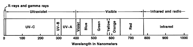
Based on the biologic effects of the different wavelengths or bands, UVR has been subdivided into three bands: UV-A (400 to 320 nm), UV-B (320 to 290 nm), and UV-C (290 to 100 nm).7 UV-A, or near UVR, produces tanning (the browning of the skin due to an increase in the skin content of melanin) and photosensitivity reactions. UV-A is emitted by so-called black lights, which are often used to make objects fluoresce and are also used in tanning salons. UV-B causes sunburn (painful erythema and blistering), and increased exposure to UV-B is associated with an increased rate of skin cancer.6–9 UV-C, which is germicidal and may also cause skin cancer, is not normally encountered on the earth's surface and comes entirely from artificial sources such as germicidal ultraviolet lamps or arc welding.

ENVIRONMENTAL, TEMPORAL, AND GEOGRAPHIC VARIATIONS IN ULTRAVIOLET RADIATION

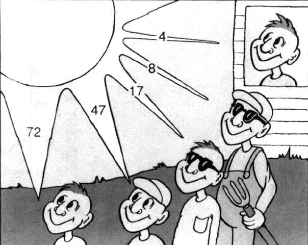
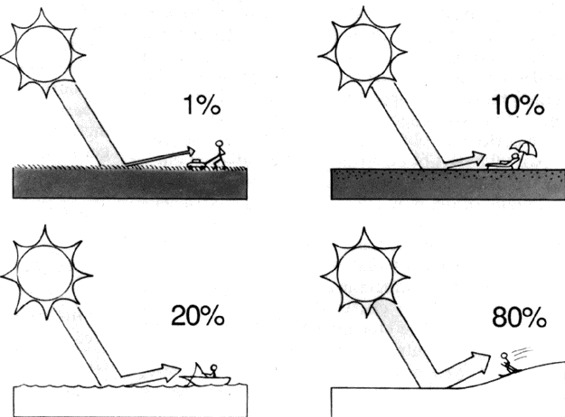
UVR is scattered across the whole sky by the Rayleigh effect, just as blue light is scattered.8 Light or broken clouds do not significantly reduce the level of UVR, although levels are reduced by heavy cloud cover.8 A sky with a clear horizon for 360° provides for maximal exposure; when hills, trees, or buildings obstruct part or all of the horizon, the UVR exposure is reduced proportionally.10 UVR can also be reflected by the ground, the amount dependent greatly on the type of surface (Fig. 2). Grass and soil reflect only 1% to 5% of UV-B, water 3% to 13%, sand and concrete about 7% to 18%, and fresh snow up to 88%.10

Fig. 2. There is substantial variation in the amount of UVR reflected by different surfaces. The extremely high reflectivity of snow and ice accounts for the risk of snow blindness (photokeratitis) encountered during spring skiing at high altitudes. (Javitt JC, Taylor HR: Focal Points: Clinical Modules for Ophthalmologists 9(3). Reprinted with permission from the American Academy of Ophthalmology, 1991)

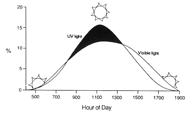
As the sun makes its daily transit, the spectral content of sunlight changes substantially (Fig. 3). At low incident angles, nearly all visible and ultraviolet energy is reflected or absorbed by the atmosphere, giving the familiar reddish hue to early morning and late evening sunlight. The UV-A and UV-B content of sunlight increases as the sun reaches its zenith and progressively decreases during the afternoon. Similarly, the farther from the equator, the more oblique is the angle of sunlight incidence and the lower the UVR level on the ground. Many people have learned the hard way that an hour's exposure to tropical sunlight imparts a far greater dose of UV-B than does an hour on the beach in New England or the Pacific Northwest. As the Earth tilts to produce changing seasons, so too does the angle of sunlight incidence and the resulting UVR content of sunlight, with summer sunlight imparting far greater doses of UV-B than sunlight of other seasons.

Fig. 3. Percent of total energy by hour of day. The relative UVR content of sunlight varies during the day and reaches its peak at noon. (Javitt JC, Taylor HR: Focal Points: Clinical Modules for Ophthalmologists 9(3). Reprinted with permission from the American Academy of Ophthalmology, 1991)

The ambient dose of UVR also increases somewhat at higher altitudes, because there is less atmosphere to filter the sun's energy. UV-B exposure increases approximately 20% per 10,000 feet. Climbers have frequently discovered that they are vulnerable to sunburn even when the sun seems to exert little warming effect. Persons climbing and skiing on snow-covered peaks are at particular risk because of the extremely high ultraviolet reflectivity of snow and ice.

The eyes are normally protected and shielded from UVR by a number of factors11,12aand only receive a small fraction of ambient UV-B under normal circumstances. The normal horizontal alignment of the eyes and the orbits significantly reduces ocular exposure to whole-sky irradiation. Further anatomical protection is provided by the brows, the nose, and the cheeks.13 The eyelids provide protection that is further enhanced by squinting, a common reflex in bright sunlight. The eyes are relatively unprotected laterally, although the transmission of UVR by internal reflection in the cornea may lead to a concentration of ultraviolet irradiation at the nasal limbus.14 Other personal factors that can decrease ocular UVR exposure in a given environment include wearing a hat and protective eyeglasses.11,12 Taken together, these different factors result in an ocular exposure that is between 7% and 17% less than the ambient UVR level.12

OCULAR TRANSMISSION OF ULTRAVIOLET RADIATION

Only a fraction of the UVR entering the eyes reaches the retina-a fact with important photobiologic implications (Fig. 4). The amount of radiation that is absorbed determines the potential for damage to the absorbing tissue. Energy from the dissipation of the absorbed radiation results in tissue damage. Radiation that is not absorbed by a superficial tissue will be transmitted and can affect deeper tissues.

Fig. 4. Absorption of light by ocular structures varies with its wavelength. (Javitt JC, Taylor HR: Focal Points: Clinical Modules for Ophthalmologists 9(3). Reprinted with permission from the American Academy of Ophthalmology, 1991)

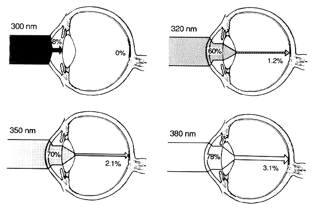
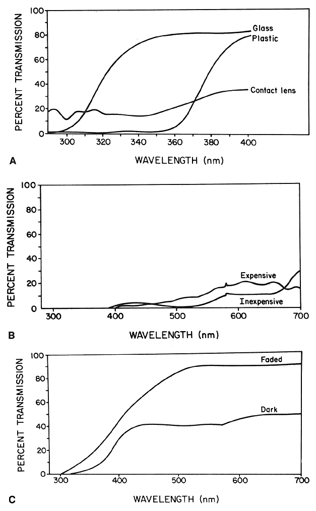
The cornea absorbs almost 100% of UV-C radiation (below 290 nm), but transmission rapidly increases for longer wavelengths, so that, for instance, 60% of radiation at 320 nm is transmitted by the cornea.15–18 A normal young human lens absorbs most UVR below 370 nm. As the human lens yellows with age, it absorbs even more UV-A and also shorter blue wavelengths.18,19 In adults, less than 1% of radiation between 320 and 340 nm and only 2% of radiation of 360 nm reaches the retina.20 The lens is exposed to and absorbs most of the UV-B entering the eye.

MECHANISMS OF PHOTOTOXICITY

The energy carried by a photon is directly proportional to its frequency; thus, the shorter the wavelength, the higher the energy. A photon's energy is absorbed by the atom or molecule with which it collides. Low-energy infrared photons carry enough energy to affect the rotational or vibrational state of an atom or a molecule and produce warming. Higher-energy UVR photons can alter the energy state of the electrons, making the atom or molecule relatively unstable, leading to oxidative reactions including cross-linking and denaturation of proteins and free-radical formation. Higher-energy photons such as gamma rays cause an electron to be removed entirely from the molecule, thereby causing ionization.

The capacity of a given atom or molecule to absorb radiant energy is dependent on its physicochemical properties, and the characteristics of a tissue are in turn dependent on the properties of its constituents. The lens proteins are rich in the amino acid residues of tryptophan, tyrosine, and phenylalanine, which absorb most of the radiant energy below 300 nm. Cross-linking of these proteins is implicated in the pathogenesis of cataract. Other chromophores and pigments in the lens appear to absorb most of the energy in the 300 to 400 nm range.21

Back to Top
EFFECT OF ULTRAVIOLET RADIATION ON OCULAR TISSUES

CORNEA

Photokeratitis from Acute Exposure

Exposure to UV-B and UV-C may cause a superficial punctate keratopathy that appears up to 6 hours after exposure. This is the ocular analogue of acute blistering sunburn. The condition is most frequently encountered as “welder's flash,” caused by even momentary exposure to UV-C during arc welding. Clusters of photokeratitis have been reported in association with defective glass envelopes surrounding mercury vapor lamps, most frequently in school gymnasiums. Naturally occurring UV-B in sufficient doses also causes photokeratitis. This condition, often termed snow blindness, is usually encountered when the UVR reflectivity of the environment is extremely high, such as during spring skiing. Snow blindness is of historical interest and importance in the annals of arctic and Antarctic explorers. Like welder's flash, snow blindness is a self-healing epithelial injury that resolves in 8 to 12 hours. Photokeratitis can occur in more hospitable environments as well. Near-threshold exposures of UV-B can be received during the summer on beaches in the southern United States.

The corneal epithelium can tolerate only a certain dose of UV-B before breakdown occurs. Although this threshold may be nearly reached during a full day of winter skiing, skiers may notice only irritation during the evening, and epithelial repair will occur during sleep. With the increased UV-B content in spring sunlight, this threshold can be exceeded in 1.5 to 2 hours of noontime skiing, especially at higher altitudes. Thus, by evening, spring skiers may have painful photokeratitis and a matching sunburn.

Photokeratitis accounts for 59% of all injuries associated with tanning booths.22 It can occur if the protective goggles are defective or removed even briefly or if the glass envelope of the lamp is defective and allows UV-C to escape.

Pinguecula and Pterygium from Chronic Exposure

Pingueculae are localized yellowish-gray fleshy lesions that appear close to the limbus on the nasal or temporal interpalpebral bulbar conjunctiva. Pterygia are somewhat similar in appearance but involve the peripheral cornea as well. Histologically, both lesions contain deposits of degenerating collagen fibers, elastoid fibers, and an increased population of metabolically active stromal fibrocytes. Similar histologic changes are found in the dermis of sun-exposed skin.

Pterygium occurs more commonly in tropical or sunny areas than in more temperate regions and has been clearly associated with exposure to UVR.23,24 Although geographic and ecological differences in pterygium rates have been postulated to result from non-UVR factors, such as corneal drying or microtrauma caused by smoke, sand, and dust particles, studies have confirmed the importance of UVR exposure.

A study of 838 men who work on Chesapeake Bay showed that the risk of pinguecula and pterygium was significantly associated with increased levels of UV-A and UV-B.25 For those in the highest quartile of annual UV-B or UV-A exposure, the odds ratio for the development of pterygium was 3.06.

Climatic Droplet Keratopathy

Climatic droplet keratopathy (CDK), which has also been called Labrador keratopathy, chronic actinic keratopathy, proteinaceous degeneration, and keratinoid degeneration, is a spheroidal degeneration of the superficial corneal stroma. The condition is characterized by yellow, oily-appearing subepithelial droplets replacing Bowman's layer or lying in the superficial stroma. In general, their distribution is similar to that of band keratopathy, but with a clear zone of separation from the limbus and corneal vessels. The changes are thought to be caused by degeneration of serum proteins that normally diffuse through the cornea.

In the study of Chesapeake Bay watermen, CDK was even more strongly associated with UV-B exposure than was pterygium.25The odds ratio for average annual UV-B exposure in the upper quartile was 6.36 for CDK, as compared with the lower quartile. Similar ratios were shown for exposure to UV-A.

ASSOCIATION BETWEEN ULTRAVIOLET RADIATION AND CATARACT

Experimental Basis for an Association Between UVR and Cataract

A number of experiments have shown that UVR exposure causes cataracts in animals, particularly cortical and posterior subcapsular Cataracts. Both guinea pigs and rabbits acutely exposed to UVR (293 to 303 nm) develop clouding of the anterior cortex.26 Subcapsular and punctate conical opacities develop in albino mice chronically exposed to broadband UVR from 40-watt blacklight lamps (300 to 400 nm).26 Posterior migration of undifferentiated lens epithelium (posterior subcapsular cataract) in response to UVR has also been noted.27–28

The Epidemiologic Basis for an Association Between UVR and Cataract

Epidemiologic and clinical observations also suggest a link between sunlight exposure and cataract formation. Cataracts occur more commonly in tropical areas with higher sunlight exposures than in more temperate regions.29–32 People undergoing cataract surgery are more likely to have brunescent cataracts if they live closer to the equator or work outdoors.33 People in the United States who live more than half their lives in areas with high sunlight or UVR levels have a greater risk of cortical cataract.34–36 This association does not hold for nuclear cataract.36

Studies of Australian aborigines show an association between cataract and local levels of UV-B radiation.37,38 The association was specific and consistent and showed a dose-response relationship. Studies in China, Nepal, Tibet, and India all support such an association.39–41

An analysis of cataract surgery rates across the United States for the years 1986 and 1987 shows a strong, statistically significant relationship between the age-adjusted rate of cataract surgery in a community and latitude, elevation, and July sunlight hours in that community.42 All three geographic factors are indirect measures of ambient UVR. Ultraviolet levels have been directly measured in nine U.S. cities and are found to correlate even more strongly with cataract surgery rates than the geographic variables.42

These studies all assume that all individuals in a region are exposed to the outdoor ambient level of UVR. Case-control studies that measure personal levels of UVR exposure also confirm the association between UVR and cataract.43,44

In order to quantify the dose-response relationship between UV-B exposure and cortical cataract, an epidemiologic survey was conducted of 838 watermen.45 The annual ocular exposure for each participant was calculated from age 16 by combining a detailed occupational history with laboratory and field measurements of sun exposure. Cataracts were graded by ophthalmologic examination for type and severity. Some degree of cortical cataract was found in 111 (13%) of the watermen, and some degree of nuclear opacity in 229 (27%). A doubling of cumulative exposure to UV-B increased the risk of cortical cataract by 60%. Assessed another way, those whose annual exposure was in the upper quartile had a 3.3-fold higher risk of cortical cataract compared with those in the lowest quartile. No association was found between nuclear cataracts and UV-B exposure, nor was any association detected between cataract and UV-A exposure. Bochow and coworkers performed a case-control study in the same region and assessed the association between UVR and posterior subcapsular cataract (PSC).46

One hundred sixty-eight patients who had undergone cataract extraction for PSC and 168 phakic controls were interviewed regarding sunlight exposure, drug use, occupational history, and history of diabetes, hypertension, and other diseases. A history of high UV-B exposure was strongly associated with increased risk of PSC, as was a history of diabetes.

ASSOCIATION BETWEEN ULTRAVIOLET RADIATION AND MACULAR DEGENERATION

Much less is known about the role of chronic UVR exposure and macular damage. Although the lens filters out most of the radiation below 370 nm, the retina has been shown to be particularly sensitive to any UVR that does reach it.47–49Light can damage the retina by thermoacoustic means (as with Nd:YAG laser treatment), by thermal effects or burn (as with xenon photocoagulation), or by photochemical mechanisms.1,48 The processes involved are essentially similar to those outlined for damage to the lens, although the retina has a more active metabolism with much higher oxygen consumption and higher levels of protection. Furthermore, in the retina, damaged proteins can be replaced by normal cellular repair mechanisms.

Short, intense exposure to sunlight leads to retinal burns known as eclipse blindness or solar retinopathy--probably the result of photochemical damage to the photoreceptors.48 Similar damage may be caused inadvertently by ocular instruments.49,50 Experimentally, the histologic changes induced by cumulative photic injury in rhesus monkeys resemble those seen in age related macular degeneration (AMD) in humans.51

Although much attention has been given to the retinal effects of short, intense light exposures, the effect of long-term sunlight or UVR on the retina is unclear. Animal studies by Ham and others have demonstrated that longer wavelength UVR and blue light can cause retinal damage48,52–54 at light levels below those that cause photocoagulation. Damage from repetitive exposures may be additive.55,56

Some investigators have found AMD to be associated with light iris and hair color.56 It has been suggested that light pigmentation may reflect a lower level of melanin and thus less protection against photo-oxidative damage because of the direct absorption of light.57 A small case-control study reported that the degree of dermal elastotic degeneration in sun-protected skin was predictive of exudative maculopathy.58 Such information suggests that individuals whose elastic fibers are more susceptible to photic or other degenerative stimuli may also have an increased risk of developing AMD.

As discussed earlier, the aging lens becomes increasingly brown and acts as a blue-light filter to protect the retina from shorter wavelengths. Eyes with nuclear sclerosis have been reported to have less AMD than eyes without nuclear sclerosis.59 However, another study suggested that the apparent increased risk of macular degeneration in aphakia may be due to obscuration of existing, subtle macular changes by nuclear opacities.60

Because removal of the crystalline lens allows a higher percentage of UVR to reach the macula, there is some concern that aphakic patients may be at increased risk for macular degeneration. A study of eight patients who underwent implantation of a non-UVR-blocking intraocular lens (IOL) in one eye and a UVR-blocking IOL in the other showed a loss of short-wave cone sensitivity in the non UVR-protected eye.61

In the study of Chesapeake Bay watermen, exposure to UVR was not associated with increased risk of AMD in phakic individuals, even with high levels of sunlight exposure.62 Photographs were graded in a standardized fashion for the presence of exudative disease, geographic atrophy, focal hyperpigmentation of the retinal pigment epithelium, and drusen that were large or confluent. The lack of an association between UVR exposure and AMD is not surprising, because the lens absorbs almost all UV-B, and only very small amounts of this waveband can reach the retina. However, further analysis of the waterman data suggests that longer wavelengths of blue light (400 to 500 nm) or visible light (500 to 700 nm) may exert a cumulative damaging effect on the retina. Persons with severe, vision-impairing macular degeneration had a statistically greater recent exposure to blue or visible light over the preceding 20 years.63

The most intense bleaching and shedding of cone outer segments is caused by blue light. The night vision of lifeguards who wore blue filtering sunglasses was shown to be superior to those who wore sunglasses with no blue attenuation, implying that intensely bleached and shed photoreceptor outer segments may not be instantly repaired.64 Macular degeneration is often thought to be related to a diminished ability of the retinal pigment epithelium to phagocytize and degrade those outer segments, leading to deposition of drusen. Thus the retina may have a repair mechanism for light-induced damage that can protect healthy young eyes, but this effect may diminish with age. Consequently, in older people, even low-grade damage caused by visible or blue light exposure may be additive, and ocular protection in wavelengths below 430 to 450 nm should be considered for those with high levels of occupational exposure. Additional studies are needed to quantify the association between light exposure and adult macular degeneration.

Back to Top
UNDERSTANDING SUNGLASSES
Given these findings, what are the implications for the general population? Certainly, it seems prudent to protect the eyes from unnecessary exposure to UVR. The amount of ambient UVR varies markedly during the day, reaching its peak between 10 A.M. and 2 P.M. (see Fig. 3). The periods of high levels of UV-B are usually well recognized, because this is the time when the skin is most likely to become sunburned. As a public health recommendation, therefore, people should be advised to use ocular protection at those times. However, patients should be reminded that overcast skies may create a false sense of security.

Protecting the eyes from UVR has become a multimillion-dollar-a-year industry with the development of UVR-absorbing spectacles, sunglasses, intraocular lenses, and, most recently, contact lenses. Furthermore, there are strong reasons to believe that we may be facing significantly higher levels of UVR exposure because of progressive changes in the Earth's atmosphere.65 Recent data indicate that chlorofluorocarbon compounds are causing a significant reduction in the stratospheric ozone layer, which is the main atmospheric filter of UVR.

In addition to sunglasses, there are two easy ways to reduce UV-B exposure short of staying indoors. A brimmed hat will reduce ocular exposure by half, and ordinary, close-fitting plastic spectacles can reduce it by about 95%.12 The effects of hats and glasses are additive (Fig. 5). Although special UVR-absorbing lenses can stop all UV-B transmission, a sample of 40 clear spectacle lenses showed that they all significantly reduced ocular UV-B exposure, although plastic lenses were much more effective than glass.12

Fig. 5. Protection from ultraviolet light afforded by hats and sunglasses. Ocular exposure expressed as percent of ambient UV radiation under normal conditions.

LENS MATERIAL AND COLOR

Ideally, sunglasses should decrease visible light to a comfortable level while entirely eliminating invisible but harmful UVR. Some authors have expressed concern that sufficient blocking of visible light without concurrent blocking of UVR might cause pupillary dilatation and a higher dose of UVR to the internal structures of the eye.13 Although that is a theoretical possibility, especially for UV-A, even “poor” sunglasses absorb most UV-B. However, the chief danger of poor UVR-absorbing characteristics is to lull patients into the mistaken notion that they are providing themselves with adequate protection. The ability of a lens to filter UVR is largely a function of chromophores (UVR-absorbing molecules) embedded in the plastic material. These chromophores can have little or no effect on the color or darkness of the lens. Therefore, the color or darkness of the lens gives no indication of the UVR-absorbing characteristics. Even clear spectacle lenses have significant UVR-absorbing properties (Fig. 6A). Color should be chosen on the basis of personal preference and the visual needs of the individual (Table 1). Gray lenses do not alter the perception of natural color, but as the gray becomes darker, contrast is diminished. For many people, amber and brown lenses seem to provide a more pleasant environment without significantly altering natural colors. Green introduces the greatest degree of color distortion and may occasionally diminish the ability to recognize traffic signals. Yellow or pink lenses absorb little visible light but may still have good UV-B absorption if manufactured of the proper material. Blue light is refracted and scattered more than other visible wavelengths within the eye. Some hunters and other sportsmen report that yellow lenses that block all blue light improve. distance acuity out-of-doors. Although yellow sunglasses are marketed with these claims, scientific validation is lacking. Polarizing lenses reduce glare substantially and are favored by many individuals, especially fishermen and boaters. Although it may provide significant advantages in terms of comfort, polarization has little effect on the UVR-absorbing properties of lenses. Similarly, mirror finishes by themselves have little effect on UVR transmission.

Fig. 6. Spectral transmission curves for different lenses. A. Prescription lenses, clear glass; prescription lenses, clear plastic; gray hard contact lens. B. UVR-blocking sunglasses, inexpensive ($10 price range) and expensive ($150 price range). C. Photochromic lens, unexposed (faded) and exposed (darkened) at 25°C. (Javitt JC, Taylor HR: Focal Points: Clinical Modules for Ophthalmologists 9(3). Reprinted with permission from the American Academy of Ophthalmology, 1991)


TABLE 1. Optical Characteristics Imparted by Lens Color


GrayDoes not alter perception of natural colors Reduced contrast with darker tints
GreenDistortion of natural colors
Brown, amberRelatively undistorted natural colors
 Good contrast
YellowEliminates most visible blue light
 Said to improve distance acuity


LABELING REQUIREMENTS

With increased public awareness of potential UVR hazards, manufacturers of sunglasses have started to market their products based upon UVR-absorbing properties. In recent years there has been a movement toward improving the labeling of commercially available sunglasses. The American National Standards Institute (ANSI) classifies sunglasses in three categories by their ability to screen UVR, as shown in Table 2. It is important to note that the ANSI standards allow for up to 40% UV-A transmission.


TABLE 2. Current Sunglass Labeling Standards—Absorptive Requirements


SunglassVisible  
DesignationLightUV-AUV-B
Cosmetic60%20%70%
General purpose92%60%95%
Special purpose97%60%99%


Given the evidence linking both UV-A and UV-B to ocular disease, prudence suggests seeking maximum UVR protection from spectacles, as long as the cost is reasonable. In fact, the addition of UVR-absorbing chromophores during manufacture of lenses introduces negligible cost. Many sunglasses manufacturers voluntarily exceed those standards and label their products as being 100% UVR absorbing. Some even specify “100% absorbing below 400 nm,” which is the labeling patients should seek whenever possible.

A number of studies have examined the UVR-absorbing properties of nonprescription sunglasses (Table 3; Fig. 6B).66,67 The UV-B incident on the eyes of specially constructed mannequins equipped with sunglasses has also been measured.68 Thirty-two pairs of “discount” (i.e., less than $7.00) sunglasses were purchased at random in local pharmacies. None of the lenses transmitted more than 1.8% of total UV-B, suggesting that manufacturing processes have recognized the importance of UVR-absorbing chromophores in recent years.


TABLE 3. Comparison of Sunglasses and Prescription Eye Wear with Respect to UVR Attenuation(% Transmission or Exposure)


 SunglassesPlastic LensesGlass Lenses
Transmission UV-B0.20.615.6
Transmission UV-B and UV-A1.51. 147.2
Percent ocular exposure with proper positioning2.97.846.4
Percent ocular exposure with 6 mm displacement from brow25.035.546.6


Consumer Reports tested more than 200 brands of randomly purchased sunglasses, ranging in price from $7 to $195.69 All were marketed to meet the ANSI “general purpose” standard. However, the vast majority met the stricter “special purpose” standard, even though they were not marketed as such. Every plastic lens tested, and most glass lenses, absorbed at least 90% of UV-A, far exceeding the ANSI standard. Many of those tested absorbed 98% to 99% of UV-A. The editors' conclusion was that adequate UVR protection was to be found in all price ranges of sunglasses.

Photochromic lenses that darken when exposed to ultraviolet light constitute a special situation (Fig. 6C). When faded, they attenuate more than 95% of UV-B but allow up to 40% transmission of wavelengths at the upper end of UV-A (370 to 380 nm). As such, they just meet the ANSI general standard. In their totally darkened state at 25°C, they reduce UV-A transmission (again up to 380 nm) to about 6%.

It is important to note that lens transmission alone is not the only factor in determining ocular UVR exposure, as explained later.

LENS SHAPE AND CONTOUR

The shape and position of the glasses are as important in reducing UV-B exposure as the actual lens material. Wraparound glasses give almost complete protection, whereas regular flames still let 4% to 5% of ambient UV-B reach the eyes by way of backscatter around the frames. The wearing position of the spectacles is also important. When glasses are displaced 6 mm from the forehead, ocular exposure to UV-B increases tenfold (Table 3). Proper wearing position of sunglasses is critical for full protection. For complete protection, patients should be advised to wear wraparound glasses or to install side shields on current glasses. An elastic strap or similar device may help maintain proper lens position and thus ensure adequate protection.

Ideal lens shape is determined by the facial contours of the individual. The chief rule of thumb is to minimize light leaks around the edges of the lenses. Because spectacles and sunglasses are available in such a wide array of shapes and sizes, there should be no difficulty in fitting any specific patient.

QUALITY AND CONSTRUCTION OF SUNGLASSES

Higher cost does not necessarily guarantee superior UVR protection, and other features of sunglasses should also be considered. Although sunglasses of adequate quality can be found among even the least expensive, patients should be cautioned to inspect lenses for surface scratches and optical quality. The cheapest lens manufacturing technique is “draping,” in which a clear sheet of plastic is tinted before being heated and bent into shape. This method may lead to poor optical quality and distorted images. The lenses of well-manufactured sunglasses are ground and polished just like those of prescription spectacles. Furthermore, the pigment of cheap lenses may scratch off entirely, and cheap frames may splinter on impact or break at the hinges.

When inspecting lenses for surface scratching and optical quality, the simplest approach is to examine a fixed linear object through the lens and observe any distortion as the lens is moved up and down and from side to side. By flexing the frames in the store, patients can select frames that will not splinter if hit. Durable frames should flex but readily regain their shape.

Of course, sunglasses do not provide satisfactory protection from ocular trauma. In recent years, several types of protective spectacles have been designed and marketed for athletic and other high-risk activities. They have been shown to prevent severe ocular trauma when fitted and worn properly. Patients participating in activities in which they are liable to be struck by flying objects or body parts should be strongly advised to purchase and wear protective eye wear.

Back to Top
PATIENTS AT HIGHEST RISK OF SOLAR DAMAGE

APHAKIC AND PSEUDOPHAKIC PATIENTS

Aphakic patients receive an even greater retinal dose of UVR than do phakic individuals. A report by the National Health and Nutrition Examination Survey indicated that aphakic patients may have more macular degeneration than phakic individuals of the same age, race, and gender.70 This may not necessarily be a causal relationship, because the physiological factors that caused the lens opacity and led to cataract surgery may also play a role in macular degeneration. However, the implication is that increased retinal exposure to UVR is harmful.

UVR-blocking intraocular lenses are commonly available and widely used. However, they have not been universally implemented, partly because of concerns about long-term toxicity should the UVR-absorbing chromophores come into contact with ocular tissues, although no such evidence has emerged. All aphakic and pseudophakic patients should be cautioned to use proper sunglasses, possibly with side shields, as well as a broad-brimmed hat when out-of-doors. Because these patients represent an age-group that is particularly prone to basal cell carcinoma, wearing a hat serves several useful functions.8,9

PATIENTS WITH CONGENITAL OR PHARMACOLOGICALLY INDUCED PHOTOSENSITIVITY

Patients with xeroderma pigmentosa lack an essential cellular pathway for repairing UVR-induced damage and are therefore at permanently high risk. Although these patients are likely to have been counseled by a dermatologist to stay out of the sun and use the highest levels of sun block, ophthalmologists should ensure that they wear adequate eye protection as well.

Although there is no proof yet, it has been suggested that patients with retinitis pigmentosa are at increased risk of photic injury. Certainly, wearing inexpensive UVR-blocking sunglasses is a simple and prudent intervention that is wise in any case. There is no current basis to justify the cost of the more expensive blue-blocking spectacles that eliminate all blue light up to 500 nm. However, patients who desire this possible degree of added protection or find that their vision is improved by the optical properties should not be discouraged, because little other therapy can be offered. Psoralen therapy, used to treat several dermatologic disorders including psoriasis and vitiligo, puts patients at increased risk from UVR exposure because of its photosensitizing properties. Psoralen is only one of several medications that can cause increased photosensitivity and may predispose users to severe sunburn. UVR-induced ocular damage has not been demonstrated in these patients but should be protected against.

Table 4 presents a list of medications that may cause severe photosensitivity in some patients. Of particular note is tetracycline, in that it is frequently prescribed for the prevention and treatment of traveler's diarrhea. Therefore, it may be used by individuals who are traveling to areas of high UVR exposure and who are unfamiliar with the practices (such as staying out of midday sun and wearing hats) that are common to natives of those regions.


TABLE 4. Pharmacologic Agents Reported to Cause Phototoxicity

  Adriamycin

  Aldose reductase inhibitors
  Allopurinol
  Griseofulvin
  Phenothiazines: chlorpromazine, thioridazine
  Porphyrin derivatives
  Sulfonamides
  Oral hypoglycemic agents
  Chloroquine derivatives (antimalarials and antiarthritics)
  Oral contraceptives (not all)
  Psoralens
  Retinoids (vitamin A derivatives)
  Tetracycline, minocycline, doxycycline


Back to Top
SUMMARY
Ocular exposure to ultraviolet radiation (UVR) is associated with damage to the cornea and lens. There is early evidence that it may be associated with retinal damage to aphakic patients and others at particular risk. Prescription and nonprescription sunglasses that provide at least 95% attenuation of UV-B are readily available at low cost. They are only effective when worn firmly against the brow. Patients should be strongly encouraged to purchase them and wear them properly.

Ophthalmologists should take the lead in encouraging patients to select properly labeled sunglasses. With changes in manufacturing trends, there is no difficulty in finding correctly designed and labeled sunglasses in any pharmacy or convenience store. Additional work is needed to determine more precisely whether blue light in general causes retinal damage.63,64,70,71 Early experimental evidence has suggested that this is a cause for concern and that aphakic patients and those on photosensitizing medications should be especially encouraged to wear proper absorptive lenses. Similarly, UVR-absorbing intraocular lenses have been shown to filter light at different wavelengths.70 Additional research is needed to determine the appropriate spectral cutoff for use in IOLs and other protective lenses.

The current epidemic of skin cancer is often attributed to a change in life-style, with far greater sun exposure than in previous generations. If so, we should be particularly concerned about a higher incidence of cataracts as the current generation of sunbathers ages. If current atmospheric trends continue, depletion of the ozone layer will result in a substantially higher UVR component in ambient sunlight, also resulting in increased cataract risk.

Back to Top
REFERENCES

1. Guerry RK, Ham WT, Mueller HA: Light toxicity in the posterior segment. In Duane TD, Jaeger EA (eds): Duane's Clinical Ophthalmology, Vol 3, pp 1–17. Philadelphia, Harper & Row, 1986

2. American Academy of Ophthalmology: Eye and visual system abnormalities. In Eye Care for the American People, Supplement to Ophthalmology. San Francisco, American Academy of Ophthalmology, 1987

3. Hausser KW, Vahle W: In Urbach F (trans): Sunburn and suntanning. In Urbach F (ed): Biological Effects of Ultraviolet Radiation, pp 3–21. Oxford, Pergamon Press, 1969

4. Jones RR: Ozone depletion and cancer risk. Radiat Res 2: 443, 1987

5. Weinstock MA: The epidemic of squamous cell carcinoma. JAMA 262:2138, 1989

6. Scotto J, Fears TR, Fraumeni JF: Solar Radiation. In Schottenfeld D, Fraumeni JF (eds): Cancer Epidemiology and Prevention, pp 254–276. Philadelphia, WB Saunders, 1982

7. Scotto J, Fears TR: The association of solar ultraviolet and skin melanoma incidence among Caucasians in the United States. Cancer Invest 5:275, 1987

8. Robertson DF: Solar ultraviolet radiation in relation to sunburn and skin cancer. Med J Aust 2:1123, 1968

9. Glass AG, Hoover RN: The emerging epidemic of melanoma and squamous cell skin cancer. JAMA 262:0297, 1989

10. Sliney DH: Physical factors in cataractogenesis: Ambient ultraviolet radiation and temperature. Invest Ophthalmol Vis Sci 27:781, 1986

11. Rosenthal RS, Safran M, Taylor HR: The ocular dose of ultraviolet radiation from sunlight exposure. Photochem Photobiol 42: 163, 1985

12. Rosenthal FS, Phoon C, Bakalian AE et al: The ocular dose of ultraviolet radiation in outdoor workers. Invest Ophthalmol Vis Sci 29:649, 1988

12a. Rosenthal FS, Bakalian AE, Taylor HR: The effect of prescription eyewear on ocular exposure to ultraviolet radiation. Am J Public Health 76:1216, 1986

13. Sliney DH: Eye protective techniques for bright light. Ophthalmology 90:937, 1983

14. Coroneo MT: Albedo Concentration in the Anterior Eye and the Location of Some Solar Diseases. Master's thesis, University of Sydney, Sydney, Australia, 1988

15. Kinsey VE: Spectral transmission of the eye to ultraviolet radiations. Arch Ophthalmol 39:508, 1948

16. Bachem A: Ophthalmic ultraviolet action spectrum. Am J Ophthalmol 41:969, 1956

17. Boettner EA, Wolter JR: Transmission of the ocular media. Invest Ophthalmol Vis Sci 1:776, 1962

18. Cooper G, Robson J: The yellow colour of the lens of man and other primates. J Physiol (Lond) 203:411, 1969

19. Lerman S: Human ultraviolet radiation cataracts. Ophthalmic Res 12:303, 1980

20. Rosen ES: Filtration of non-ionizing radiation by the ocular media. In Cronly-Dillon J, Rosen ES, Marshall J (eds): Hazards of Light: Myths and Realities of Eye and Skin, pp 145–152. Oxford, Pergamon Press, 1986

21. Zigman S: Photobiology of the lens. In Maisel H (ed): The Ocular Lens: Structure, Function, and Pathobiology, pp 301–347. New York, Marcel Dekker, 1985

22. Injuries associated with ultraviolet tanning devices-Wisconsin. MMWR 38:333, 1989

23. Anderson JR: A pterygium map. Acta Ophthalmol 3: 1631, 1954

24. Moran D J, Hollows FC: Pterygium and ultraviolet radiation: A positive correlation. Br J Ophthalmol 68:343, 1984

25. Taylor HR, West SK, Rosenthal FS et al: Corneal changes associated with chronic ultraviolet radiation. Arch Ophthalmol 107:1481, 1989

26. Zigman S, Yulo T, Schultz J: Cataract induction in mice exposed to near UV light. Ophthalmic Res 6:259, 1974

27. Zigman S, Schultz J, Yulo T: Possible roles of near UV light in the cataractous process. Exp Eye Res 13:462, 1974

28. Spector A: The search for a solution to senile cataracts (Proctor Lecture). Invest Ophthalmol Vis Sci 25: 130, 1984

29. Leske MC, Sperduto RD: The epidemiology of senile cataracts: A review. Am J Epidemiol 118:152, 1983

30. Mohan M, Sperduto RD, Angra SK et al: India-US case-control study of age-related cataracts. Arch Ophthalmol 107:670, 1989

31. Pacurariu I, Marin C: Changes in the incidence of ocular disease in children and old people. Ophthalmologica 17: 289, 1973

32. van Heyningen R: The human lens. I. A comparison of cataracts extracted in Oxford (England) and Shikarpur (W. Pakistan). Exp Eye Res 13:136, 1972

33. Zigman S, Datiles M, Torczynski E: Sunlight and human cataracts. Invest Ophthalmol Vis Sci 18:462, 1979

34. Hiller R, Giacometti L, Yuen K: Sunlight and cataract: An epidemiologic investigation. Am J Epidemiol 105:450, 1977

35. Hiller R, Sperduto RD, Ederer F: Epidemiologic associations with cataract in 1971-72 National Health and Nutrition Examination Survey, Am J Epidemiol 118:239, 1983

36. Hiller R, Sperduto RD, Ederer F: Epidemiologic associations with nuclear, cortical, and posterior subcapsular cataracts. Am J Epidemiol 124:916, 1986

37. Taylor HR: The environment and the lens. Br J Ophthalmol 64:303, 1980

38. Hollows F, Moran D: Cataract-the ultraviolet risk factor. Lancet 2: 1249, 1981

39. Mao WS, Hu TS: An epidemiologic survey of senile cataract in China. Chin Med J [Engl] 95:813, 1982

40. Xu J, Mao W, Zhu S et al: A case-control study of senile cataract in Lhasa, Tibet, China. Invest Ophthalmol Vis Sci (suppl) 28:396, 1987

41. Brilliant LB, Grasset NC, Pokhrel RP et al: Associations among cataract prevalence, sunlight hours, and altitude in the Himalayas. Am J Epidemiol 118:250, 1983

42. Javitt JC, Canner JK, Kolb ME et al: Association between rates of cataract surgery in Medicare beneficiaries and ambient sunlight. (unpublished data) 1990

43. Cameron LL, Auer CL, McCormick PA et al: Association of sunlight with senile macular and lens changes. Invest Ophthalmol Vis Sci (suppl) 24:202, 1983

44. Cameron LL, Emmett EA, Abbey H et al: Association of senile lens opacities with UV and other factors. Invest Ophthalmol Vis Sci (suppl) 27:45, 1986

45. Taylor HR, West SK, Rosenthal FS et al: Effect of ultraviolet radiation on cataract formation, N Engl J Med 319: 1429, 1988

46. Bochow TW, West SK, Axar A et al: Ultraviolet light exposure and risk of posterior subcapsular cataracts. Arch Ophthalmol 107:369, 1989

47. Ham WT Jr, Ruffolo JJ Jr, Mueller HA et al: Histologic analysis of photochemical lesions produced in rhesus retina by short wavelength light. Invest Ophthalmol Vis Sci 17:1029, 1978

48. Tso MOM, LaPiana FG: The human fovea after sungazing. Trans Am Acad Ophthalmol Otolaryngol 79:788, 1975

49. Calkins JL, Hochheimer BF: Retinal light exposure from operation microscopes. Arch Ophthalmol 97:2363, 1979

50. Hochheimer BF, D'Anna SA, Calkins JL: Retinal damage from light. Am J Ophthalmol 88:1039, 1979

51. Borges JM, Li ZY, Tso MOM: The cumulative effects of repeated photic exposures on the monkey macula. Invest Ophthalmol Vis Sci (suppl) 28:47, 1987

52. Ham WT Jr, Mueller HA, Ruffolo JJ Jr et al: Action spectrum for retinal injury from near-ultraviolet radiation in aphakic monkey. Am J Ophthalmol 93:299, 1982

53. Noell WK, Walker VS, Kang BS et al: Retinal damage by light in rats. Invest Opthalmol 5:450, 1966

54. Kuwabara T, Gom RA: Retinal damage by visible light; an electron microscopic study. Am J Ophthalmol 79:69, 1968

55. Lawwill T, Crockett S, Currier G: Retinal damage secondary to chronic light exposure. Soc Ophthalmol 44:379, 1977

56. Mainster MA: Light and macular degeneration: A biophysical and clinical perspective. Eye 1:304, 1987

57. Weiter J J, Delori FC, Wing GL et al: Relationship of senile macular degeneration to ocular pigmentation. Am J Ophthalmol 99: 158, 1985

58. Blumenkranz MS, Russell SR, Robey MG et al: Risk factors in age-related maculopathy complicated by choroidal neovascularization. Ophthalmology 93:552, 1986-

59. Sperduto RD, Hiller R, Seigel D: Lens opacities and senile maculopathy. Arch Ophthalmol 99: 1004, 1981

60. Bressler NM, West SK, Bressler SB et al: Relationship of cataract and macular degeneration in a population-based study. Invest Ophthalmol (suppl) 30:108, 1989

61. Werner JS, Steele VG, Pfoff DS: Loss of human photoreceptor sensitivity associated with chronic exposure to ultraviolet radiation. Ophthalmology 96: 1552, 1989

62. West SK, Rosenthal FS, Bressler NM et al: Exposure to sunlight and other risk factors for age-related macular degeneration. Arch Ophthalmol 107:875, 1989

63. Munoz B, West S, Bressler N et al: Blue light and risk of age-related macular degeneration. Invest Ophthalmol (suppl) 32:49, 1990

64. Mainster MA: Light and macular degeneration: A biophysical and clinical perspective. Eye 1:304, 1987

65. Farmer CB, Toon GC, Schaper PW et al: Stratospheric trace gases in the spring 1986 Antarctic atmosphere. Nature 329:126, 1987

66. Anderson WJ, Gebel RKH: Ultraviolet windows in commercial sunglasses. Appl Optics 16:515, 1977

67. Borgwardt B, Fishman GA, Vander Meulen D: Spectral transmission characteristics of tinted lenses. Arch Ophthalmol 99:293, 1981

68. Rosenthal FS, Bakalian AE, Lou C et al: The effect of sunglasses on ocular exposure to ultraviolet radiation. Am J Public Health 78:72, 1988

69. Sunglasses. Consumer Reports 53:504, 1988

70. Liu IY, White L, LaCroix AZ: The association of age-related macular degeneration and lens opacities in the aged. Am J Public Health 79:765, 1989

71. Mainster MA: The spectra, classification, and rationale of ultraviolet-protective intraocular lenses. Am J Ophthalmol 102:727, 1986

Back to Top