Chapter 44
Ophthalmic and Systemic Manifestations of Child Abuse
MARY LOUISE Z. COLLINS and ALEX V. LEVIN
Main Menu   Table Of Contents

Search

PHYSICAL ABUSE
SEXUAL ABUSE
NEGLECT AND NONCOMPLIANCE
EMOTIONAL ABUSE
ROLE OF THE OPHTHALMOLOGIST
REFERENCES

Child abuse and neglect may be defined as maltreatment of children by their parents or other caretakers that results in harm to their physical, psychological, or developmental state. Child abuse can be characterized as physical abuse, sexual abuse, neglect, or emotional abuse. In the United States, there are more than 3 million cases reported to child protection agencies each year, of which 30% are substantiated. Over half of the reported cases have had prior child protection or abuse issues. It is estimated that abusive violence against children results in 2000 deaths per year in the United States alone: more than 5 deaths each day. Child abuse is the leading cause of trauma death in infants and toddlers, with most victims younger than 4 years old. More than 18,000 children are left disabled annually at a cost for lost productivity estimated to be 658 million to 1.3 billion dollars. Yet despite this pandemic, over one fifth of legally mandated reporters do not do so.1

Although child abuse involves all socioeconomic classes, ethnic groups, races, genders, sexual preferences, professions, and religions, in affected families there is a higher incidence of social dysfunction, substance abuse, depression, and isolation. Overreporting of visible minorities living in adverse social situations, and underreporting of white patients of higher socioeconomic status is well recognized. Other factors related to poverty, unemployment, and parental as well as parent–child discord, may increase the likelihood of abuse in a family.2 The cycle of violence hypothesis proposes that children who are abused become abusers. A study by Widom3 suggests that abuse or neglect in childhood increases the potential for violent or criminal behavior in adulthood. Certain qualities of the child may predispose him or her to abuse by a parent or caretaker who is ill equipped to handle the child's needs. A physical or mental handicap or illness is a risk factor for abuse. Behavioral problems in a child, including being tempermental or difficult behavior, as well as hyperactivity, may cause increased stress in a family and predispose the child to abuse.2 The most common inciting factor for abuse in infancy is parental frustration over the child's crying or feeding. Often, however, there are no identifiable predisposing factors for abuse other than the proximity of the child to the abuser.4

In general, with the rare exception perhaps of psychotic perpetrators, perpetrators of abuse do not truly wish to harm their victims and often regret their actions. Yet this does not excuse those responsible from culpability. Abuse of a child is a crime with all physicians bound by law to report their suspicion. It can be particularly difficult for the ophthalmologist to maintain a professional distance from this emotional issue while trying to provide care for the child and parents, particularly when the parent is a suspected perpetrator. But physicians should not allow themselves to slip into the role of police, prosecuting attorney, judge, or jury. Our duty is to recognize and report abuse rather than identify or punish the perpetrator.

Although child abuse has been present since ancient times, its recognition as a major cause of morbidity and mortality in the pediatric age group is a recent phenomenon.5 Earlier papers reported clinical findings that would not be recognized for decades as child abuse.2, 5 It was not until 1962 that Kempe and co-workers first described the battered child syndrome and stated, “A physician needs to have a high initial level of suspicion in instances of subdural hematoma, multiple unexplained fractures at different stages of healing, failure to thrive, when soft tissue swellings or skin bruising are present, or in any other situation when the degree and type of injury is at variance with the history given regarding its occurrence or in any child who dies suddenly.”6 In 1972, Caffey defined what has come to be known as the Shaken Baby syndrome (SBS).7 Over the past 30 years, there has been a plethora of research into almost every aspect of child abuse including the ophthalmic manifestations.

Perhaps the first report of ophthalmic manifestations of child abuse was published in 1928: a child with retinal and subdural hemorrhage.8 The father was convicted after the child's sibling died with a skull fracture. Approximately 40% of physically abused children manifest signs of ocular trauma, occasionally presenting first to the ophthalmologist.9,10 Any ocular injury can be the result of abuse, and any form of child abuse can have ocular manifestations. Ophthalmologists are mandated reporters of suspected abuse. Failure to report this suspicion could potentially be perceived as an act of abuse in itself, and there is legal precedent for such prosecution. Therefore, it is incumbent on every medical professional to include child abuse in the differential diagnosis of eye injury, sexually transmitted diseases to the eye, neglect, and other potential ophthalmic indicators.

Back to Top
PHYSICAL ABUSE

THE BATTERED CHILD: SYSTEMIC MANIFESTATIONS

The battered child syndrome refers to bruises, lacerations, fractures, burns, intra-abdominal trauma, blunt head injury, and other forms of physical abuse. The possibility of child abuse should be considered whenever there is wide discrepancy between the explanation for the child's condition, and the physical findings (Table 1). For example, an accidental fall from a couch or bed is a common household occurrence, but serious injury is extremely unlikely. A parent may state that the child fell against a table. On examination, however, the child is found to be covered with multiple bruises. Another clue is inconsistency of the degree of injury with the developmental skills of a child. For example, falling off a tricycle at age 1 year when a child is unable to ride a tricycle is a doubtful explanation for a child's injury. Bruising of almost any kind in the first few months of life is worrisome. Histories that change repeatedly on inquiry from different members of the health-care team or new explanations that seem to arise only after a mechanism for injury is suggested should also be considered suspect. A careful history or subsequent investigation may reveal that the child has been treated at different hospitals for a series of injuries of varying severity.


Table 1. Diagnostic Features of the Battered Child Syndrome

  1. Fractures and soft tissue injury in different stages of healing with no organic cause
  2. Disproportionate degree of injury as compared to history
  3. Reported mechanism of injury incompatible with physical findings
  4. Multiple admissions to different hospitals for injuries
  5. No fresh lesions during hospitalization
  6. Unexplained delay in seeking care

Modified from Harcourt B: The role of the ophthalmologist in thediagnosis and management of child abuse. Ophthal Surg 4:37, 1973.


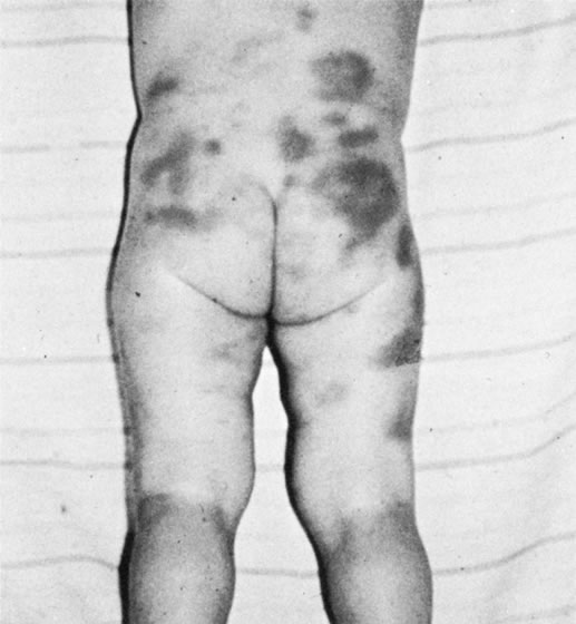
Physical examination may provide additional clinical evidence of child abuse (Table 2). Of course, accidental bruising and abrasion is part of normal childhood but these injuries are usually confined to the knees, anterior surface of the lower legs, the face, and other exposed areas. Accidental injuries do not follow artificial nonbiologic patterns, usually have a viable clear explanation, and are appropriate for the child's developmental level. Bruises that are found on certain parts of the body should raise the possibility of child abuse (Figs. 1 and 2). Bruises that predominate on the buttocks and lower back are often related to punishment (Fig. 3). Likewise, the abdomen and the back of the arms and legs are unusual sites for accidental injuries. Bruises can be morphologically similar to the implement used to inflict the trauma (Fig. 4).11,12 For example, the imprint of a belt buckle or hand may be seen clearly on a child's skin. Genital or innerthigh bruises may be the result of punishment for toileting mishaps. Bruises and scars at multiple stages of healing imply repeated abuse.


Table 2. Physical Manifestations of the Battered Child Syndrome

Brain injury or intracranial hemorrhage from blunt trauma
Bruises in different stages of healing with patterns and distribution inconsistent with accidental causation
Pattern skin injuries
Lacerations or abrasions without adequate explanation
Unexplained fractures in various stages of healing
Burns without satisfactory accidental explanation
Unexplained human bite marks
Subgaleal hematomas from severe hair pulling
Nonaccidental intra-abdominal trauma


Fig. 1. Circumferential marks on the ankles of a child from being tied to a bed with cords while the mother was away. (Courtesy of Dr. Barton D. Schmitt and the American Academy of Pediatrics.)

Fig. 2. Swollen lower lip caused by forced feeding. (Courtesy of Dr. Barton D. Schmitt and the American Academy of Pediatrics.)

Fig. 3. Bruises on buttocks secondary to paddling. (Courtesy of Dr. Barton D. Schmitt and the American Academy of Pediatrics.)

Fig. 4. Loop mark from beating with doubled-over cord. (Courtesy of Dr. Barton D. Schmitt and the American Academy of Pediatrics.)

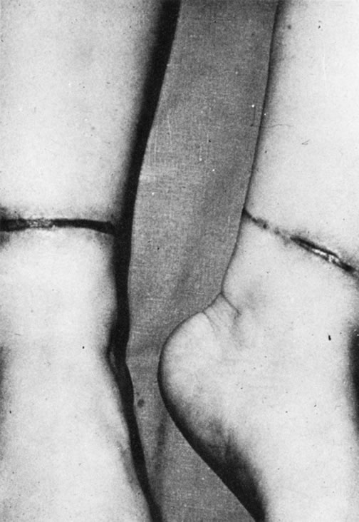
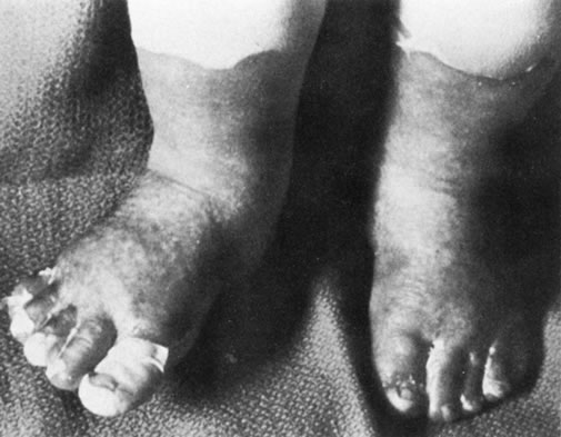
Approximately 10% of all cases of child abuse involve burns.13 The most frequent cause of burns in abused children is scalding with hot water.13,14 The buttocks and perineum are frequently burned areas. Full-thickness, symmetric burns of the hands or feet suggest strongly that the extremities were held in a hot liquid (Fig. 5). Another commonly inflicted burn is from a cigarette. These lesions are often found on the palms, soles, or abdomen and often have an excavated center with raised edges.

Fig. 5. Symmetric burns of feet from immersion in hot liquid (Courtesy of Dr. Barton D. Schmitt and the American Academy of Pediatrics.)

THE BATTERED CHILD—OPHTHALMIC MANIFESTATIONS

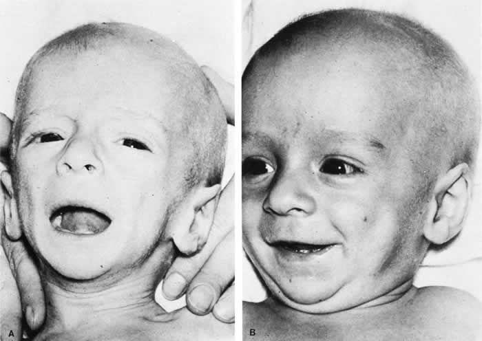
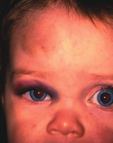
The spectrum of ocular findings in the battered child is vast. Essentially any injury to the eye or adnexa could be due to abuse. An ocular injury may be as mild as periorbital edema or as severe as a ruptured globe. Signs of bilateral ocular trauma suggest inflicted injury because accidents usually involve only one eye. Perhaps one exception is bilateral periocular ecchymosis due to an accidental, single, central forehead trauma (Fig. 6). However, periorbital ecchymosis may also be caused by inflicted trauma with or without injury to the underlying globe. Attempts to date bruises by their color, particularly when the blood is accumulated in the loose skin of the lids, is notoriously unreliable.

Fig. 6. Accidental bruise, right forehead, causing bilateral (right greater than left) periorbital ecchymosis.

There are some eye findings that almost always indicate trauma, and others that should raise the possibility of trauma (Table 3). Eyelid, conjunctival, corneal, and scleral lacerations are always due to trauma. Iridodialysis cannot be due to causes other than trauma, although this lesion can be mimicked by congenital anterior segment malformations such as Axenfeld-Reiger anomaly. Certain vitreoretinal injuries, such as commotio retinae and avulsion of the vitreous base are traumatic injuries. If a child with any of these injuries does not have an adequate explanation, the question of abuse should be raised. The ophthalmologist may conduct a complete physical examination to look for other indicators. Consultation with a multidisciplinary child abuse team is also indicated.


Table 3. Ocular Findings In Children That Raise Concern About Trauma


Ocular FindingAlways Trauma?Possibly Child Abuse?
Eyelids
   Lacerationyesyes
   Bruisingnoyes
Conjunctiva
   Hemorrhagenoyes
   Lacerationyesyes
Cornea
   Lacerationyesyes
   Scarnoyes
Anterior Chamber
   Hyphemanoyes
   Iritisnoyes
   Iridodialysisyesyes
Lens
   Cataractnoyes
   Ectopia Lentisnoyes
Retina
   Detachmentnoyes
   Hemorrhagesnoyes
   Commotioyesyes
Vitreous
   Vitreous base avulsionyesyes
   Hemorrhagenoyes
Optic Nerve
   Atrophynoyes

Modified from Levin AL: The Ocular Findings in Child Abuse. Focal Points Clinical Modules for Ophthalmologists 26: 3–11, 1998.


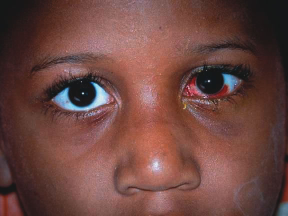
Other eye findings such as hyphema and unilateral lens subluxation are usually due to trauma, but rarely have other causes. Subconjunctival hemorrhage is a common finding in child abuse, occurring in 4% to 10% of patients.9,10 Jain and colleagues15 reported a 1.7% incidence of subconjunctival hemorrhage in newborns as a result of the normal birth process. This is transient, and subconjunctival hemorrhage beyond the first 2 weeks of life should be considered suspicious.3 One must also rule out other causes of subconjunctival hemorrhage such as thrombocytopenia. Pertussis can result in severe 360-degree bilateral subconjunctival hemorrhage. Consideration of appropriate diagnostic tests is always important in eliminating explanations other than abuse.

Other ocular signs such as cataract, iritis, retinal detachment, and optic atrophy occur frequently in the absence of trauma. In a child, however, the ophthalmologist must always consider the possibility of nonaccidental injury, especially if the finding is unilateral, history or other findings reveal inconsistencies and evidence to support a non traumatic diagnosis is absent. For example, Tseng and Keys16 discuss a case simulating congenital glaucoma in a 9-week-old physically abused infant presenting with hazy, enlarged corneas, elevated intraocular pressures, subluxated and cataractous lenses, vitreous hemorrhage, and hyphema.

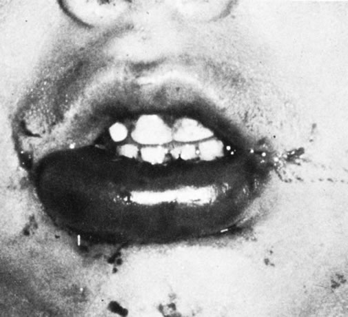
Injuries, such as hyphema or ruptured globe, may be sustained in the course of discipline, occurring “accidentally” during a belt beating (Fig. 7). An unexpected move by the child or perpetrator not in control of the implement may cause injury to the eye. Spanking, as a form of discipline, may be controversial, but the use of an implement puts a child at significant risk for injury. Injuries to a child by beating should be reported to child protection agencies, even if the injury occurs “accidentally” in the course of the beating. Such circumstances clearly indicate that the perpetrator was “out of control” when the incident occurred. It is this loss of control that is one of the main features of the perpetrators.

Fig. 7. Left subconjunctival hemorrhage and periorbital ecchymosis sustained when child moved during belt beating and was struck by belt buckle. Also present (but not visible on this image) are left hyphema and commotio retinae.

MUNCHAUSEN SYNDROME BY PROXY

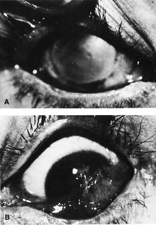
Munchausen syndrome by proxy (MSBP) is a form of physical (and emotional) child abuse that occurs when a parent, the mother in over 90% of cases, causes the appearance of an illness by the falsification of history, creation of physical findings, or manipulation of laboratory test results. The most common presentation of MSBP is covert suffocation injury causing the child to have seizures or recurrent apnea. The presenting ocular sign may include subconjunctival hemorrhage that is sometimes associated with petechia of the face and perioral or perinasal injury. The perpetrator may induce vomiting in the child or feed the child substances that cause vomiting or diarrhea. Covert poisoning is also a common form of MSBP, and cases of injection of substances ranging from insulin to feces are well known. Reported ocular manifestations have been numerous and include recurrent conjunctivitis due to instillation of chemicals into the fornices. This has even resulted in corneal scarring with bilateral legal blindness (Fig. 8). Pupil and eye movement abnormalities due to toxic topical or systemic medications, recurrent periorbital cellulitis due to injection of foreign substances around the eye, and even intravenous injection of noxious substances into a child receiving chemotherapy for retinoblastoma have been reported. As a result of recurrent unexplained symptoms, the child may be subjected to multiple invasive diagnostic tests, procedures, and hospitalizations, and the physician ordering the tests becomes an unknowing participant in the battery of the patient. Physicians must be aware of the signs of MSBP that are summarized in Table 4 and may help to reveal the diagnosis.


Table 4. Signs Raising Possibility of Munchausen Syndrome by Proxy

Disorder that is difficult to diagnose and
   • Does not respond to treatment in predictable manner
   • Does not fit into any known diagnostic category
   • Intermittent and unpredictable symptoms
   • Chronic
   • “Never seen a case like this before”
Multiple caretakers or parents dissatisfied with care
Mother overly involved with child's care
   • Speaks for the child
   • Never leaves bedside
   • Father absent or uninvolved
   • Overly bonded maternal-child relationship
Mother very social and inappropriately happy during hospitalization
   • Bonds with ward staff
   • At ease with bad news
Mother with history of factitious illness
Siblings with unexplained illness
Mother with prior access to or experience in health-care system

Modified from Levin AL: The Ocular Findings in Child Abuse. Focal Points: Clinical Modules for Ophthalmologists 26: 3–11, 1998.


Fig. 8. A. and B. Recurrent chemical injuries inflicted by the father resulted in corneal scarring and vascularization that blinded this 6-year-old girl. (Taylor D, Bentovim A: Recurrent nonaccidentally inflicted chemical eye injuries to siblings. J Pediatr Ophthalmol Strabismus 13:238, 1976.)

SHAKEN BABY SYNDROME

The triad of the SBS includes brain injury usually with hemorrhage, ocular injury, and skeletal injury. These children often have no external signs of trauma. The infant and young child are particularly vulnerable because of their relatively large head, weak cervical musculature, large size of the cranial vault in relation to the size of the brain,3 and immature, unmyelinated brain. Violent shaking causes repetitive anteroposterior and side-to-side disorganized head movement with abrupt acceleration-deceleration forces.7,17,18,19 The magnitude of acceleration-deceleration forces needed to cause brain and eye injuries in humans is extreme but not exactly quantified.20 Although in Caffey's original description, he inferred that normal play activities could cause SBS-like injuries, we now recognize that this is not the case.21

Abusive head trauma is the most common type of child abuse resulting in death,22 although it represents only 3% to 5% of all cases referred to child abuse teams.23 Assault represents more than half of all traumatic brain injury in the first year of life and 90% of brain injuries between 1and 4 years of life. The average age of SBS victim is between 5 and 10 months,22–31 with most children younger than 2 years of age.22,25,26,30 Victims up to 5 years old are rare. The mortality rate of SBS, based on studies with more than 10 patients, is approximately 8% to 61%,23 although this may be a reflection, in part, of separation or imprisonment of perpetrators after the first incident. Recidivism rates are high.

The most common perpetrators of SBS are biological fathers and biologically unrelated boyfriends of the mother.22,26,30,32,33 Babysitters, females 4.4 times more often than males,22 are the perpetrators in 4% to 20% of cases.22,26,33 Biological mothers commit this crime in 5% to 12% of cases.22,26,33 Only a minority (10% to 15%)23,33 of perpetrators confess, although it may be as high as 43%34 in fatal cases.

SHAKEN BABY SYNDROME: SYSTEMIC MANIFESTATIONS

Brain injury in SBS is common: Subdural hemorrhage is found in 10% to 93%, subarachnoid hemorrhage in 10% to 72%, posterior interhemispheric blood in 20% to 100%, stroke in 12% to 50%, intraparenchymal hemorrhage in 5% to 30% and parenchymal tears in 0 to 100%. 23,24,26,29,35 Increased intracranial pressure or cerebral edema is found in 44% to 85%.23,35 The wide variation in incidence figures reflects the nature of the study populations: findings at presentation, in survivors, or at autopsy. At autopsy, subdural hemorrhage, subarachnoid hemorrhage, and cerebral edema are the most consistent findings.21,34,36,37 Although one study using a mechanical model and autopsy investigation suggested that blunt head impact is required to generate the forces necessary to cause the brain injury of SBS,38 a wealth of clinical and pathologic investigations indicate that shaking alone can cause significant injury and even death. Perhaps calculation of forces does not allow for a full understanding as cellular and biochemical responses to shearing stress along with other factors such as anemia and hypoxia may play an important role that cannot be modeled. Violent shaking causes shearing forces that tear the bridging veins running from the cortex to the dural venous sinuses, resulting in subdural and subarachnoid hemorrhage. Shearing also causes diffuse axonal injury with secondary brain edema.39,40,41,42 The diagnosis of brain injury is usually confirmed with computed tomography (CT) scan. However, the CT scan may initially be normal or show edema without hemorrhage. Magnetic resonance imaging (MRI) may be useful to find hemorrhage not visualized on CT43 and to date the findings seen on CT.

The long-term prognosis for children with brain injury secondary to SBS is poor. In one study, only 28% of survivors had normal neurologic exams on discharge from the hospital; this figure decreased to 8% to 14% in long-term studies.22 Late findings seen on imaging studies include cerebral atrophy, hydrocephalus ex vacuo, chronic subdural effusion, and encephalomalacia. Patients may have quadriplegia, diplegia, hemiplegia, mental retardation, developmental delay; learning disability, seizures (7% to 65%), and psychiatric/behavioral issues(28% to 50%).23,24,26,30,44

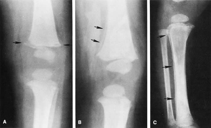
Skull fractures occur in 9% to 31% of shaken babies,23,30 with the parietal and occipital bones most affected. The characteristics of skull fractures that are highly suggestive of abuse include branching, stellate, crossing suture lines, multiple, greater than 5 mm wide, or progressively expanding fractures in a child less than 3 years of age.29 Rib fractures are the most common bone injury in SBS, and are usually posterolateral due to the perpetrator's hands grasping the child. Long bone fractures affect the tibia, forearm bones, femur, or humerus in decreasing order of prevalence. The characteristic metaphyseal fracture, which rarely occurs in young children except in the setting of abuse, results in a “corner” or “bucket handle” chip fracture at the end of the bone (Fig 9). Other injuries seen in SBS include hemorrhagic stripping of the periostium, spiral fractures, and nonsupracondylar humerus fractures—all due to shaking while the infant is held by an extremity, causing the long bones to be twisted and broken.29,30

Fig. 9. Epiphyseal-metaphyseal fractures as seen in shaken baby syndrome: stages of healing. A. Fresh fracture (less than 10 days). Note rarefaction of the bone underneath the epiphyseal plate (short arrow). B. Appearance of fracture at 10 days to 2 weeks. Note early periosteal new bone deposition (long arrows). C. Appearance of fracture at 2 to 4 weeks. Note more pronounced periosteal new bone deposition and epiphyseal-metaphyseal changes characteristic of this stage of healing. (Swischuk LE: Radiology of the skeletal system. In Ellerstein NS [ed]: Child Abuse and Neglect: A Medical Reference. New York, John Wiley & Sons)

SHAKEN BABY SYNDROME: OCULAR MANIFESTATATIONS

Retinal Hemorrhages

Retinal hemorrhage is the most common ocular manifestations of SBS. The incidence of retinal hemorrhages in SBS varies in published reports. This variation is in part due to methods of examination (ophthalmologists vs pediatricians, dilated vs undilated pupils) and the population studied. Overall, the incidence varies from 30% to 100%.18,21,30,38,51,52,53 The incidence in studies including children with abusive head trauma not due to shaking is lower than in studies only involving SBS. In postmortem studies, the incidence of retinal hemorrhages approaches 100%.34,51,54,55

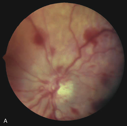
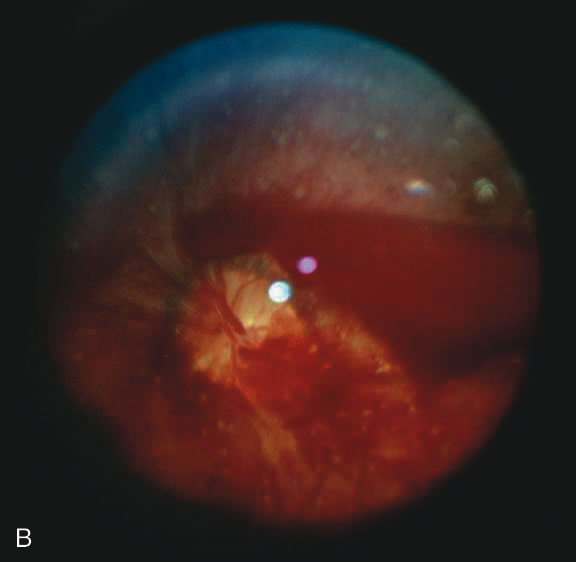
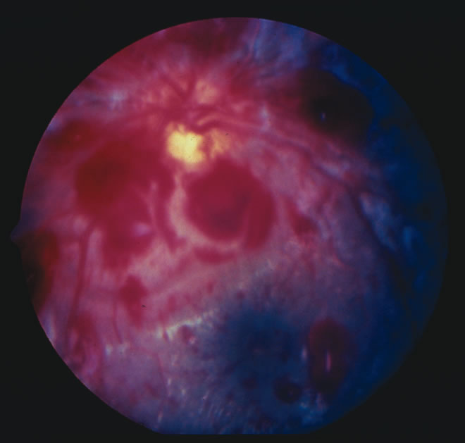
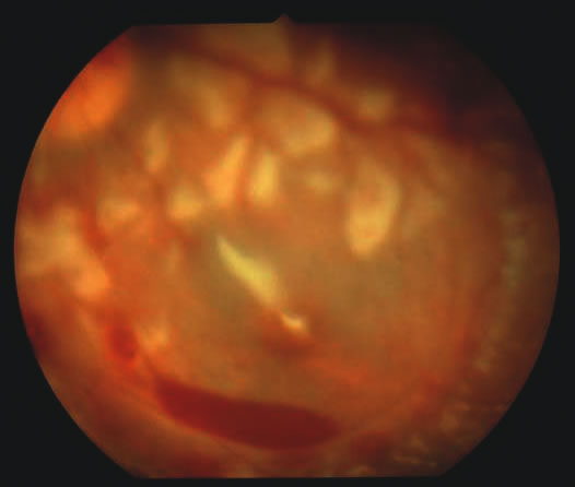
Findings range from a normal fundus to a small number of scattered intraretinal hemorrhages in the posterior pole to massive, confluent hemorrhages from the posterior pole to the ora serrata.(Fig. 10) The hemorrhages may be subretinal, deep intraretinal (dot/blot), nerve fiber layer (flame shaped) or preretinal.19,45 Intraretinal hemorrhages are more common than preretinal or subretinal hemorrhages.32,46 Preretinal hemorrhages must be distinguished from traumatic retinoschisis (see later), which has particular diagnostic significance. White-centered retinal hemorrhages, although classically associated with endocarditis, can occur in any condition that causes retinal hemorrhages, including SBS. Vitreous hemorrhage may be small to massive, and may occur secondary to escape of blood from intraretinal collections or from torn vessels.19 Although vitreous hemorrhage may occur at the time of injury, it may also be a delayed finding occurring 1 to 3 days or more after the initial trauma.47,48

Fig. 10. Retinal hemorrhages in SBS may range from a nonspecific mild posterior pole distribution (10A) to a more characteristic picture of too numerous to count, multiple, extensive intraretinal hemorrhages (in this many with white centers) with subhyaloid hemorrhage (10B, 10C)

Retinal hemorrhages may be associated with papilledema in SBS. However, papilledema is seen in less than 10% of shaken babies.35, 49 These small, flame-shaped hemorrhages on and radiating around the optic nerve are not necessarily caused by shaking and may be seen in papilledema from any cause. However, retinal hemorrhages associated with SBS may be seen on the optic disc in the absence of papilledema.50

Retinal hemorrhage is usually bilateral but may also be asymmetric or unilateral. Retinal hemorrhage can not be dated with any precision and, therefore, should not be used to help determine when the abusive event occurred.46,47,56,57,58,59 At best, generalizations may be made with wide intervals. For example, intraretinal hemorrhages do not last months, and blood in schisis cavities does not go away in days. Retinal scars or optic atrophy do not form in days.

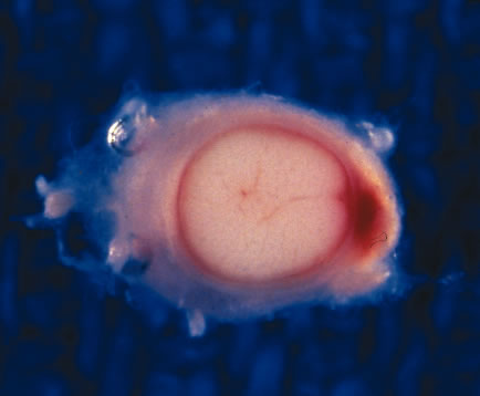
The pathophysiologic mechanisms of retinal hemorrhage in SBS are varied. Vitreous and perhaps orbital shaking is likely to be involved in most of the vitreoretinal injuries. In children, the vitreous is well attached to the retina at the macula, blood vessels, and the periphery. Shaking of an infant causes the vitreous to shake, which, in turn, applies shearing forces to the retina at points of firm attachment. These shearing forces at the macula may split the retina at any layer, causing the formation of a cystic cavity, which may be filled partially or completely with blood (Fig. 11). This traumatic retinoschisis has been well documented in abuse cases by ultrasound, electroretinogram, and pathology.34,58,60,61,62,63,64 Histopathology reveals a widening of the retinal layers or a stripping of the internal limiting membrane. The vitreous may stay adherent or detach. Clinically, to recognize this important finding in SBS, the examiner may observe a hemorrhagic or hypopigmented curvilinear edge to the schisis cavity, with or without a fold in the retina (Figure 11).50,65,66 Recognition of this edge helps distinguish retinoschisis from subhyaloid hemorrhage. However, subhyaloid hemorrhage, which may have originated from blood breaking out of a schisis cavity, may obscure the underlying schisis. It is important to follow any potentially shaken child with preretinal blood in the macula until that blood has cleared as the signs of schisis may be unmasked as the blood resorbs thus confirming the diagnosis of SBS. The retinal fold or hypopigmented line may be a complete circle or just an arc. In the long term, these patients may have surprisingly few sequelae and good vision as the cavity flattens spontaneously. There may also be findings of permanent curvilinear, hypopigmented scars or retinal folds. These provide clues to prior abuse.67

Fig. 11. Putscher retinopathy in SBS. Note circumlinear demarcation of macular retinoschisis containing small layer of blood.

There does appear to be a statistically significant higher incidence of peripheral retinal hemorrhage in SBS versus accidental head trauma. Vitreous shaking and shearing forces may be responsible because the vitreous is well attached in the periphery at the vitreous base. This theory requires further investigation. Evidence from animal models suggests that there may also be a biochemical cascade of factors involved with vascular permeability and autoregulation that may also play a role in the generation of retinal hemorrhages in response to shearing stress.68 Other factors such as hypoxia and anemia may also contribute.

Shaking may also cause changes in the orbit that contribute to the generation of retinal hemorrhages. One of the authors (AVL) has performed orbital dissection on victims of SBS and accidental head trauma finding hemorrhage within the dura proper, orbital fat, cranial nerve sheaths, and extraocular muscle in the former but rarely in the latter. Other authors have described intrascleral hemorrhage at the junction of the optic nerve and globe.69,70 These findings suggest that there may be mechanical disruption to the circle of Zinn or other vessels running to the eye and trauma to the cranial nerves that carry autonomic supply involved with vascular autoregulation. In addition, orbital injury remains the only viable theory to explain the high incidence of optic nerve injury in survivors.

Other factors may also contribute to the appearance of retinal hemorrhages in SBS, although in a minor way. Terson's syndrome is the association of intracranial blood and retinal hemorrhages well recognized in adults in particular after spontaneous subarachnoid hemorrhage or ruptured aneurysm. Optic nerve sheath hemorrhage is also often associated with Terson's syndrome34,71,72 (Fig. 12). Some have theorized that the optic nerve sheath blood is due to tracking of blood from the subarachnoid or subdural space,29,73 whereas others attribute this finding to sudden elevation of intracranial pressure from intracranial bleeding.56,74,75 It remains unclear whether optic nerve sheath hemorrhages have a role in creating retinal hemorrhages. There is significant evidence to suggest that there is not continuity between the brain and optic nerve sheath through the subdural or subarachnoid space.76 In addition, Terson's syndrome may be observed in the absence of optic nerve sheath hemorrhage or with hemorrhage confined only to the anterior optic nerve sheath. Although optic nerve sheath hemorrhage is not uncommon in SBS, Terson's syndrome in children is rare.77 Further suggesting a lesser role for Terson's syndrome in the pathogenesis of SBS retinal hemorrhage is the absence of correlations between the side of the intracranial hemorrhages and the retinal hemorrhages or between elevated intracranial pressure and retinal hemorrhages in SBS.35

Fig. 12. Optic nerve sheath hemorrhage seen on cross section of optic nerve of shaken baby at autopsy.

There remain still other theories for retinal hemorrhage in SBS that also seem to play a minor role. Some have suggested a Purtscher-like mechanism due to an increase in intrathoracic pressure when the perpetrator squeezes the child's chest. Although the characteristic white retinal patches of Purtscher retinopathy may be seen in SBS (Fig. 11), there appears to be no correlation with rib fractures,35 and the finding is very uncommon. The failure to see significant retinal hemorrhaging after the chest compressions of cardiopulmonary resuscitation in both animal models and humans also argues against a Purtscher-like mechanism.

Differential Diagnosis

The differential diagnosis of retinal hemorrhages in a child is vast. However, if one considers a child with the full spectrum of injuries seen in SBS, including brain, skeletal, and eye findings, most would not dispute that the retinal findings are due to nonaccidental injury. However, in a child with a small number of retinal hemorrhages and without retinoschisis, (Fig. 10A) the diagnosis of SBS may be less clear.

The birth process is likely the most common cause of retinal hemorrhages in newborns, varying in several reports from 11% to 31% or higher.4,15,78 These retinal hemorrhages are most common after vaginal or vacuum-assisted vaginal delivery.79–82 The exact mechanism is unknown, but there is no association with brain hemorrhages in these infants.79,81,83 Prostaglandin release may play an important role. The distribution of retinal hemorrhages secondary to the birth process may extend from the posterior pole to the ora serrata. Most often they are limited to dot, blot, or flame-type of hemorrhages but preretinal and less commonly subretinal hemorrhage may also be seen.79,81,84 Although macular hemorrhages are common,82,85 schisis has never been observed despite the examination of thousands of children. All types of retinal hemorrhages due to the birth process resolve by 6 weeks of age with the exception perhaps of deep foveal hemorrhage. The majority of hemorrhages clear much earlier. Most flame-shaped hemorrhages resolve by 3 to 5 days, with all resolved by 1 week.86,87 Any retinal hemorrhages seen beyond these dates should arouse suspicion of another cause. Retinal hemorrhages seen within the first 6 weeks of life may also be due to SBS, and additional investigations must be considered when appropriate.

Caretakers often relate histories of relatively minor head trauma as the cause of injuries that are otherwise generating the suspicion of SBS.21,34 After much study, most authors88–90 conclude that severe injury after a minor fall should raise the suspicion that the history is false and that the child has been abused. Furthermore, “most investigators agree that trivial forces such as those involving routine play, infant swings, or falls from a low height are insufficient to cause” the injuries seen in SBS.21,91 Even severe accidental head injury such as depressed skull fracture or intracranial hemorrhage does not routinely cause retinal hemorrhages.90,92,93 In compiling the available literature, retinal hemorrhage occurs in less than 3% of accidental head trauma, and then, almost always only in cases of severe, life-threatening injury.35,53,72,91,92,94,95 In the few cases of accidental head trauma with retinal hemorrhage, the bleeding is confined to the posterior pole with dot-blot, flame-shaped, or preretinal hemorrhages. Occasionally, these hemorrhages can extend to the midperiphery. But even the most severe accidental head trauma injuries do not cause the extensive retinal hemorrhages seen in SBS, except perhaps when the injury mechanism involves multiple acceleration-deceleration events such a motor vehicle accident in which the car strikes several objects or rolls consecutively resulting in death of the child. Retinochisis has not been observed after accidental head trauma, although there is one case of a child who may have suffered a severe crush injury to the head (five skull fractures with brain extrusion) and also a few adults with other causes of injury who have been observed to have paramacular folds or subinternal limiting membrane hemorrhage. These cases should not be taken to imply that in the SBS age range, macular schisis has a differential diagnosis other than SBS.

There are a variety of other causes of retinal hemorrhage in children (Table 5). However, these are usually easily identified on the basis of history, systemic physical examination, or ocular examination. The retinal hemorrhages are almost always few in number and confined to the posterior pole unless there is predisposing retinal pathology which causes the hemorrhages to be located elsewhere (e.g. at the active ridge in retinopathy of prematurity). If an examining ophthalmologist finds retinal hemorrhage in a patient with these systemic problems, the physician must also attempt to understand if the disease could be the cause of the hemorrhages or if the child has been abused.


Table 5. Retinal Hemorrhages in Childhood*


Etiology in Children <5 years of ageIdentifying Features
Accidental head traumaObvious history of trauma
Hyperviscosity/polycythemia anemiaAbnormal CBC
Hg, <5, usually with thrombocytopenia, vascular tortuosity and dilation
Cardiopulmonary resuscitation with chest compressionsHistory of CPR, very few peripapillary intra or preretinal hemorrhages
ECMOHistory of ECMO
Hypoxia/asphyxiaVascular tortuosity/dilation
Incontinentia pigmentiaSkin and dental abnormalities, peripheral avascular retina, hemorrhage peripherally only, temporal retinal traction
MalariaAbnormal blood smear
Menke's DiseaseAbnormal hair
Carbon monoxide poisoningEnvironmental exposure, clinical symptoms improve in new setting, increased carboxyhemoglobin
MeningitisAbnormal lumbar puncture
CoagulopathyAbnormal PT/PTT/INR/bleeding time
Collagen disordersHistory of head trauma, clinical systemic findings
EndocarditisWhite-centered hemorrhages, heart murmur or history of predisposing cardiac disease, abnormal echocardiogram, positive blood cultures, fever
LeukemiaAbnormal CBC
PrematurityPeripheral avascular retina with neovascularization
Cyanotic congenital heart diseaseHeart malformation, low oxygen saturation, dilated and tortuous retinal veins
Rocky Mountain spotted feverCharacteristic rash, history of exposure/tick bite
SepsisFever, positive blood culture, hypotension
Galactosemia jaundiceHepatosplenomegaly, formula/breast milk intolerance,
Glutaric aciduriaChronic macrocephaly, extrapyramidal signs on recovery, basal ganglia involvement on CT
ValsalvaUsually exclusively preretinal hemorrhage
Increased intracranial pressure intracranial pressurePapilledema, other evidence of increased
VasculitisPerivascular hemorrhage
Vitamin K DeficiencyProlonged PTT/abnormal INR, history of missed neonatal dose/malabsorption/liver disease

* Unless otherwise noted, retinal findings confined to a small number of intraretinal or preretinal hemorrhages in the posterior pole.
Identifying features not meant to be complete CBC, complete blood count; CPR, cardiopulmonary resuscitation; CT, Computed tomography; ECMO, extracorporeal membrane oxygenation; INR, International Normalized Ratio; PT, prothrombin time; PTT, partial thromboplastin time.


OTHER OCULAR INJURIES

Victims of SBS may sustain other ocular injuries either due to the acceleration-deceleration events or from concomitant blunt eye trauma. Partial or total retinal detachment may occur, likely secondary to the same types of shearing forces described earlier. Most of the reported retinal detachments have been rhegmatogenous.96–98 Macular edema may be detected, although it is uncommon.99 Although edema may suggest retinal ischemia, this should be differentiated clinically from commotio, which indicates blunt trauma to the globe. Subconjunctival hemorrhage is another rare finding in SBS,100 as is hyphema. Subconjunctival hemorrhage may be due to associated strangulation. Cranial nerve palsies may occur affecting nerves III, IV, V, VI, and VII.

OUTCOMES

Subretinal fibrosis is a late finding and represents scar tissue formation.99 Other late findings include optic atrophy,9 the circumlinear paramacular hypopigmented scars or folds due to schisis, and peripheral chorioretinal scarring. Injury to the occipital cortex is the most common cause of permanent visual loss in these patients. There are many mechanisms including infarction, contusion, shearing/laceration, diffuse axonal injury, intraparenchymal hemorrhage, and contre-coup injury.101–104

The majority of retinal hemorrhages caused by shaking with or without blunt head trauma clear without sequelae. Retinoschisis also usually has a good visual prognosis. However, preretinal hemorrhage over the fovea and vitreous hemorrhages may cause amblyopia. Although surgical evacuation is usually unnecessary, close follow-up and patching may be indicated. The incidence of blindness in long-term survivors of SBS is 15% to 28%. 24,44 An additional 15% experience other forms of visual impairment. Even children without permanent retinal damage may have visual loss secondary to brain injury. One study showed that 35% of children without retinal injury had permanent visual loss.105 Other forms of permanent visual impairment due to SBS include visual field loss, color vision impairment, decreased contrast sensitivity, and abnormal binocularity. Considering all forms of visual impairment, 67% of patients in one study of SBS were affected.30

Back to Top
SEXUAL ABUSE
Sexual abuse of children is usually different from the violent, explosive act characterizing adult rape. It is more typically a secretive act, occurring chronically, and done in a manner to avoid discovery. The abuser usually knows the child well, occupies a position of relative power, and has frequent access to the child. Secrecy is achieved by abuse of this power, perhaps along with coercive threats of retribution to the child, their family members, or even pets. The victims may be too powerless to resist, too embarrassed to bring the act to the attention of others, or confused by the act because they are too young to understand. Usually, there are no physical findings or injuries on examination, a fact that helps to keep the events concealed.

Abuse may be in the form of genital touching, orogenital contact, vaginal or anal penetration, or involving the child in pornography. Reported ophthalmic manifestations include conjunctivitis with a sexually transmitted organism, systemic infections, periocular manifestations, retinal hemorrhages, and functional visual loss. Conjunctivitis may be caused by gonorrhea, herpes simplex, or chlamydia. Periocular lesions may be due to sexual transmission of herpes simplex or molluscum contagiosum; however, these diseases are much more commonly spread in a nonsexual fashion. Pubic lice occurring on the brow or lashes may be a sexually transmitted disease. An ophthalmologist may also see patients with systemic and ocular manifestations of human immunodeficiency virus (HIV) or syphilis. Retinal hemorrhage has been reported due to presumed Valsalva's maneuver in resistance to anal penetration. Fifty percent of children who present with functional visual loss have an identifiable stressor in their lives. In some of these children, the stress factor may be sexual abuse.106 However, because functional illness in childhood is so commonly benign, further investigations as to the inciting factors are recommended only in those children with symptoms lasting more than 3 months after consulting the ophthalmologist, or substitution of one functional complaint for another over time. Collaboration with the family physician or pediatrician can be very helpful in such circumstances.

As with all forms of abuse, the ophthalmologist should have a high level of suspicion, especially when there are inconsistencies in the history or other cause for suspicion. The presence of culture positive, sexually transmitted disease from the conjunctiva should elicit consultation with the child's pediatrician or family physician. This should be followed, in turn, by a full standard sexual abuse investigation, including culturing of the anus, vagina, male urethra, and throat along with genital examination, oral examination, and a full history performed by a specialist in the field of sexual abuse. There is evidence that gonorrhea may possibly be transmitted to the conjunctiva through nonsexual means. This may also apply to chlamydia and lice, and certainly to herpes and molluscum. In the latter two instances, sexual abuse evaluation is not warranted unless there is other evidence to generate a concern. But the presence of chlamydia, gonorrhea, or pubic lice should certainly warrant a referral for full evaluation. Acquired syphilis or HIV (in the absence of other risk factors) are strong indicators of sexual contact.

Back to Top
NEGLECT AND NONCOMPLIANCE
Children whose parents or guardians put them at risk of severe, permanent visual loss because of noncompliance with keeping appointments, administering medications or complying with other treatments such as patching for amblyopia, are subjecting the child to another form of abuse. The guidelines for reporting these patients are less clear than with other forms of abuse. In addition, extenuating circumstances must be considered in all of these cases: poverty, child care issues for siblings, transportation, and lack of understanding. It may require the help of primary care physicians, social work, or community agencies for effective intervention. The dental literature has suggested the use of written contracts on the patient's chart, signed by the primary caretaker, the physician and a witness. In cases in which a chronic pattern of noncompliance or neglect is observed, and after social work or other professional intervention has been attempted without success, a statement can be placed on the chart indicating a threshold to be applied for reporting to child protection agencies. Should the parent then violate this agreement, it may be more likely that child protection authorities will intervene as parental understanding has been demonstrated.

Another form of neglect is nonorganic failure to thrive (Fig. 13). Children with this condition may become emaciated either from starvation or from failure to thrive despite the provision of food. The latter situation is believed to be due to hormonal effects brought on through poor parent-child binding and attachment. Although ocular manifestations are rare, children with corneal exposure and epithelial breakdown have been observed, largely due to lagophthalmos because they listlessly remain unmoving in their debilitated state. Physical trauma, including bruising, burns and fractures, may also accompany failure to thrive.

Fig. 13. A. This child was admitted to the hospital for failure to thrive and dehydration. B. After hospitalization, the child gained weight and the cause of failure to thrive was discovered to be child neglect. (Courtesy of Dr. Barton D. Schmitt and the American Academy of Pediatrics.)

Back to Top
EMOTIONAL ABUSE
Although there may be no direct injury to the eye, countless children are visually exposed to events they should not be allowed to witness including pornography, live sex acts, drug abuse, and violence. One only needs to watch the evening news or glance at a newspaper to be confronted with the violence that has become an intimate part of our existence. Several studies have shown the high prevalence of violence in television programming and its effect on children. Emotional abuse and neglect may also be manifested by extreme verbal aggression or belittlement. These behaviors can have long-lasting psychological morbidity for a child with effects that can be as devastating as physical abuse.
Back to Top
ROLE OF THE OPHTHALMOLOGIST
Approximately 40% of child abuse cases include some ocular manifestations.9,10 In Friendly's 54 reported cases of child abuse, the ophthalmology department was directly responsible for the admission of three patients (6%).9 It has been estimated that 4% to 6% of abused children first present to the ophthalmologist. Ophthalmologists may even experience abuse first hand when a child is threatened or struck by a frustrated parent in the doctor's waiting room. The ophthalmologist may be the first to recognize a case of maltreatment and to initiate steps for the protection and treatment of the child (Table 6). However, the care of the child should ultimately be managed by a multidisciplinary team specializing in child abuse.


Table 6. Role of the Ophthalmologist in Child Abuse

Consider abuse in the differential diagnosis of any injury.
Look for environmental, familial, and physical risk factors for abuse.
Look for ocular and systemic physical indicators of abuse.
Record a detailed history and take photographs, when available.
Stay calm, nonaccusatory, compassionate.
Seek consultation with peers and colleagues, when needed.
Report grounded suspicions to the appropriate child protection agency.
Arrange follow up as indicated to screen for long-term ocular sequelae


In cases of suspected SBS or inflicted traumatic brain injury the ophthalmologist must perform indirect ophthalmoscopy through a pharmacologically dilated pupil.27,40,42 All children younger than 5 years of age with unexplained seizures, altered mental status, hydrocephalus, cerebral atrophy, subdural effusions, near-miss sudden infant death syndrome (SIDS), or head trauma without satisfactory explanatory history should be examined in this manner . The examination should be conducted within 24 to 48 hours, if possible. If a child is brain dead or severely brain injured, dilating drops may not be needed if the pupils are fixed and dilated. Objections may be raised by the medical team to dilating drops if pupil monitoring is needed. In this circumstance, one pupil may be dilated at a time; very short-acting drops (e.g., phenylephrine 2.5% or tropicamide 0.5%) may be used or the examination may be done through an undilated pupil (especially with all-pupil indirect ophthalmoscopes). Postmortem, the cornea will remain clear for up to 72 hours, allowing for clinical examination during that time interval. Whether or not the eyes are examined by funduscopy, they should be removed and examined grossly and histopathologically in all cases of unexplained infant death. We suggest that the globe and all orbital tissues be removed en bloc through a combined anterior and intracranial approach, bathed in formalin for 72 hours, and then sectioned as a unit to look for the orbital abnormalities discussed above.

The ophthalmologist must precisely document the findings. Documentation should include a complete history and detailed written description of clinical findings, including retinal drawings. Taking time to do this well only serves to aid the ophthalmologist's memory should there be subsequent legal proceedings that may not occur for months or years. The written consultation must clearly reflect a concern about possible, or in the case of a child with macular schisis and severe multilayered retinal hemorrhages to the ora serrata in the absence of another explanation, certainty that the child is a victim of SBS. Basic coagulation studies and a complete blood count (CBC) are usually part of the investigations and the ophthalmologist may wish to make other recommendations to rule out potential entities in the differential diagnosis. When available and technically feasible, fundus photography can be invaluable as part of the clinical record. However, it must be understood that these cameras may be cost prohibitive for some centers or there may be other obstacles such as an awake, crying child or a lack of corneal clarity. The ophthalmologist's role also includes recommendations for treatment when indicated and appropriate follow-up to assess for long term vision and neurological injury.

It is mandatory in all 50 states and in most countries for a physician to report suspected cases of child abuse. The law protects the physician from liability, provided that the report was not made maliciously or negligently.45 Failure to report a suspected nonaccidental injury can result in a misdemeanor charge, as well as a civil lawsuit, against the physician, and there is legal precedent for successful prosecution of nonreporting physicians.108 An ophthalmologist can initiate the protective process by admitting the child to a hospital even if the degree of injury would not normally require hospitalization. Once the child is in the hospital and is at least temporarily protected from the potential perpetrator, an appropriate evaluation of the child's physical and developmental state is helpful. Because the ophthalmologist may not be able to personally offer the necessary evaluation, he or she should seek appropriate consultations with pediatricians, social workers, or psychiatrists. Often, a multidisciplinary child abuse team is available. Appropriate treatment programs can be effective in helping parents who are abusive to their children. Between 80% to 90% of parents can be rehabilitated and therefore provide appropriate care for their child.108 However, if the child is returned to the parents without any intervention, a high rate of recurrent child abuse (30%) will prevail.110

Back to Top
REFERENCES

1. Bergman A, Larson R, Mueller B: Changing spectrum of serious child abuse. Pediatrics 77:113, 1986

2. Kessler DB, Hyder P: Physical, sexual and emotional abuse of children. Clin Symp 43:1, 1991

3. Widom CS: The cycle of violence. Science 244:160, 1989

4. Levin A: Ocular manifestations of child abuse. Ophthalmol Clin North Am 3:249, 1990

5. Solomon T: History and demography of child abuse. Pediatrics 51:773, 1973

6. Kempe CH, Silverman FH, Steele BF et al: The battered child syndrome. JAMA 181:105, 1962

7. Caffey J: Multiple fractures in the long bones of infants suffering from chronic subdural hematoma. Am J Radiol 56:163, 1946

8. Aikman, J: Cerebral hemorrhage in infant, aged eight months: recovery. Arch Pediatr 45:56, 1928.

9. Friendly DS: Ocular manifestations of physical child abuse. Trans Am Acad Ophthalmol Otolaryngol 75:318, 1972

10. Jensen AD, Smith RE, Olson MI: Ocular clues to child abuse. J Pediatr Ophthalmol 8:270, 1971

11. Ellerstein NS: The cutaneous manifestations of child abuse and neglect. Am J Dis Child 133:906, 1979

12. Sussman SJ: Skin manifestations of the battered-child syndrome. J Pediatr 72:99, 196

13. Gillespie RW: The battered child syndrome: thermal and caustic manifestations. J Trauma 5:523, 1965

14. Ayoub C, Pfeifer D: Burns as a manifestation of child abuse and neglect. Am J Dis Child 133:910, 1979

15. Jain IS, Singh YP, Grupta SL et al: Ocular hazards during birth. J Pediatr Ophthalmol Strabismus 17:14, 1980

16. Tseng SS, Keys MP: Battered child syndrome simulation congenital glaucoma. Arch Ophthalmol 94:839, 1976

17. Caffey J: The whiplash shaken infant syndrome: Manual shaking by the extremities with whiplash-induced intracranial and intraocular bleeding, linked with residual permanent brain damage and mental retardation. Pediatrics 54:396, 1974

18. Ludwig S, Warman M: Shaken baby syndrome: a review of 20 cases. Ann Emerg Med 13:51, 1984

19. Greenwald M: The shaken baby syndrome. Semin Ophthalmol 5:202, 1990

20. Hanigan W, Peterson R, Njus G: Tin ear syndrome: rotational acceleration in pediatric head injuries. Pediatrics 80:618, 1987

21. Duhaime AC, Christian CW, Rorke LB, Zimmerman RA: Nonaccidental head injury in infants – the ‘shaken baby syndrome’. N Engl J Med 338:1822, 1998

22. Starling S, Holden J, Jenny C: Abusive head trauma: the relationship of perpetrators to their victims. Pediatrics 95:259, 1995

23. Bonnier C, Nassogne M, Errard P: Outcome and prognosis of whiplash shaken infant syndrome; late consequences after a symptom-free interval. Dev Med Child Neurol 37:943, 1995

24. Duhaime AC, Christian C, Moss E, Seidl T: Long-term outcome in infants with the shaking-impact syndrome. Pediatr Neurosurg 24:292, 1996

25. Swenson J, Levitt C: Shaken baby syndrome: diagnosis and prevention. Minn Med 80:41, 1997

26. Ewing-Cobbs L, Kramer L, Prasad M, et al: Neuroimaging, physical, and developmental findings after inflicted and noninflicted traumatic brain injury in young children. Pediatrics 102:300, 1998

27. Green MA, Lieberman G, Milroy CM, Parsons MA: Ocular and cerebral trauma in non-accidental injury in infancy: underlying mechanisms and implications for pediatric practice. Br J Ophthalmol 80:282, 1996

28. Feldman K, Brewer D, Shaw D: Evolution of the cranial computed tomography scan in child abuse. Child Abuse Negl 19:307, 1995

29. Brown J, Minns R: Non-accidental head injury, with particular reference to whiplash shaking injury and medico-legal aspects. Devel Med Child Neurol 35:849, 1993

30. Child Abuse Prevention Center: Research project on the incidence and risk factors of shaken baby syndrome in the State of Utah. 1998. (Available through e-mail at mail@dontshake.com)

31. Alexander R, Sato Y, Smith W, Bennett T: Incidence of impact trauma with cranial injuries ascribed to shaking. Am J Dis Child 144:724, 1990

32. Lancon J, Haines D, Parent A: Anatomy of the shaken baby syndrome. Anat Rec 253:13, 1998

33. Lazoritz S, Baldwin S, Kini N: The whiplash shaken infant syndrome: has Caffey's syndrome changed or have we changed his syndrome?Child Abuse Negl 21:1009, 1997

34. Betz P, Puschel K, Miltner E, et al: Morphometrical analysis of retinal hemorrhages in the shaken baby syndrome. Forensic Sci Int 78:71, 1996

35. Morad Y, Kim, YM, Armstrong, DC, Huyer D, Mian, M, Levin, AV: Correlation between retinal abnormalities and intracranial abnormalities in the Shaken Baby Syndrome. Am J Ophthalmol 134:354,2002

36. Gilliland M, Folberg R: Shaken babies—some have no impact injuries. J Forensic Sci 41:114, 1996

37. Munger CE, Peiffer RL, Bouldin TW, et al: Ocular and associated neuropathologic observations in suspected whiplash shaken infant syndrome: a retrospective study of 12 cases. Am J Forensic Med Pathol 14:193, 1993

38. Duhaime A, Gennarelli, TA, Thibault, LE, et al.: The shaken baby syndrome: a clinical, pathological and biomechanical study. J Neurosurg 66:409, 1987

39. Nashelsky M, Dix J: The time interval between lethal infant shaking and the onset of symptoms: a review of the shaken baby syndrome literature. Am J Forensic Med Pathol 6:154, 1995

40. American Academy of Pediatrics Committee on Child Abuse and Neglect: Shaken baby syndrome: inflicted cerebral trauma. Pediatrics 92:872, 1993

41. Ellis M: The pathology of fatal child abuse. Pathology 29:113, 1997

42. Wilkins B: Head injury—abuse or accident?Arch Dis Child 76:393, 1997

43. Levin AV, Magnusson MR, Rafto SE, et al.: Magnetic resonance imaging in the shaken baby syndrome. Pediatr Emerg Care 5:181, 1989

44. Fischer H, Allasio D: Permanently brain damaged: long term follow-up of shaken babies. Clin Pediatr 33:696-698, 1994

45. Friendly DS: Ocular aspects of physical child abuse. In Harley RD (ed): Pediatric Ophthalmology. Philadelphia, WB Saunders, 1983

46. Gayle MO, Kissoon N, Hered RW, Harwood-Nuss A: Retinal hemorrhage in the young child: a review of etiology, predisposed conditions, and clinical implications. J Emerg Med 13:223, 1995

47. Rao N, Smith RE, Choi JH et al: Autopsy findings in the eyes of fourteen fatally abused children. Forens Sci Int 39:293, 1988

48. Gaynon MW, Koh K, Marmour M et al: Retinal folds in the shaken baby syndrome. Am J Ophthalmol 106:423, 1988

49. Kivlin, J, Simons KB, Lazoritz S: Shaken baby syndrome. Ophthalmology 107:1246, 2000

50. Mills M: Funduscopic lesions associated with mortality in shaken baby syndrome. J AAPOS 2:67, 1998

51. Hadley MN, Sonntag VK, Rekate HL, Murphy A: The infant whiplash-shake injury syndrome: a clinical and pathological study. Neurosurgery 24:536, 1989

52. Howard M, Bell B, Uttley D: The pathophysiology of infant subdural hematoma. Br J Neurosurg 7:355, 1993

53. Johnson D, Braun D, Friendly D: Accidental head trauma and retinal hemorrhage. Neurosurgery 33:231, 1993

54. Hymel KP, Rumack CM, Hay TC, et al: Comparison of intracranial computed tomographic (CT) findings in pediatric abusive and accidental head trauma. Pediatric Radiol 27:743, 1997

55. Elner SG, Elner VM, Arnall M, Albert DM: Ocular and associated systemic findings in suspected child abuse: a necropsy study. Arch Ophthalmol 108:1094, 1990

56. Mushin A: Ocular damage in the battered-baby syndrome. Br Med J 3:402, 1971

57. Tomasi L, Rosman P: Purtscher retinopathy in the battered child syndrome. Am J Dis Child 93:1335, 1986

58. Spaide RF, Swengel RM, Scharre DW, Mein CE: Shaken baby syndrome. Am Fam Physician 41P1145 , 1990

59. Giangiacomo J, Barkett KJ: Ophthalmoscopic findings in occult child abuse. J Pediatr Ophthalmol Strabismus 22:234, 1985

60. Weinberg H, Tunnessen W: Megacephaly: heeding the head. Contemp Pediatr 13:169, 1996

61. Andrews A: Ocular manifestations of child abuse. Penn Med 99S:71, 1996

62. Dana MR, Werner MS, Viana MA, Shapiro MJ: Spontaneous and traumatic vitreous hemorrhage. Ophthalmology 100:1377, 1993

63. Bedell, A: The importance of ophthalmoscopic photographs in forensic medicine: Kodachromes. Trans Am Ophthamol Soc 53:63, 1955

64. Tyagi A, Willshaw H, Ainsworth J: Unilateral retinal hemorrhages in non-accidental injury. Lancet 349:1224, 1997

65. Massicotte SJ, Folberg R, Torczynski E, et al: Vitreoretinal traction and perimacular folds in the eyes of deliberately traumatized children. Ophthalmology 98:1124, 1991

66. Sebag J: Age related differences in the human vitreoretinal interface. Arch Ophthalmol 109:966, 1991

67. Greenwald MJ, Weiss A, Oesterle CS, Friendly DS: Traumatic retinoschisis in battered babies. Ophthalmology 93:618, 1986

68. Nagaoka T, Sakamoto T, Mori F, et al: The effect of nitric oxide on retinal blood flow during hypoxia in cats. Invest. Ophthalmol Vis Sci 43:3037, 2002

69. Elner SG, Elner VM, Arnall M, Alber, DM: Ocular and associated systemic findings in suspected child abuse: a necropsy study. Arch Ophthalmol 108:1094, 1990

70. Lin, KC, Glasgow, BJ: Bilateral periopticointrascleral hemorrhages associated with traumatic child abuse. Am J Ophthalmol 127:473, 1999

71. Budenz DL, Faarber MG, Mirchandani HG, et al: Ocular and optic nerve hemorrhages in abused infants with intracranial injuries. Ophthalmology 101:559, 1994

72. Riffenburgh R, Sathyavagiswaran L: Ocular findings at autopsy of child abuse victims. Ophthalmology 98:1519, 1991

73. Manschot W: Subarachnoid hemorrhage: intraocular symptoms and their pathogenesis. Am J Ophthalmol 38:501, 1954

74. Muller P, Deck J: Intraocular and optic nerve sheath hemorrhage in cases of sudden intracranial hypertension. J Neurosurg 41:160, 1974

75. Brinker T, Ludemann W, von Rautenfeld DB, et al: Breakdown of the meningeal barrier surrounding the intraorbital optic nerve after experimental subarachnoid hemorrhage. Am J Ophthalmol 124:373, 1997

76. Ballantyne AJ: The ocular manifestations of spontaneous subarachnoid haemorrhage. Br J Ophthalmol 27:383,1943

77. Schloff S, Mullaney PB, Armstrong DC, et al: Retinal findings in children with intracranial hemorrhage. Ophthalmology 109:1472, 2002

78. Sezen F: Retinal hemorrhages in newborn infants. Br J Ophthalmol 55:248, 1970

79. Schenker J, Gombos G: Retinal hemorrhage in the newborn. Obstet Gynecol 27:521, 1966

80. Egge K, Lyng G, Maltau J: Effect of instrumental delivery on the frequency and severity of retinal hemorrhages in the newborn. Acta Obstet Gynecol Scand 60:153, 1981

81. Levin S, Janive J, Mintz M, et al: Diagnostic and prognostic value of retinal hemorrhages in the neonate. Obstet Gynecol 155:309, 1980

82. Berkus MD, Ramamurthy RS, O'Connor PS, et al: Cohort study of silastic obstetric vacuum cup deliveries: I Safety of the instrument. Obstet Gynecol 66:503, 1985

83. Maumenee A, Hellman L, Shettles L: Factors influencing plasma prothrombin in the newborn infant. Bull Johns Hopkins Hosp 68:158, 1941

84. Sihota R, Bose S, Paul A: The neonatal fundus in maternal toxemia. J Pediatr Ophthalmol Strabismus 26:281, 1989

85. VonNoorden G, Khodadoust A: Retinal hemorrhages in newborns and organic amblyopia. Arch Ophthalmol 89:91, 1973

86. Sezen F: Retinal haemorrhage in newborn infants. Br J Ophthalmol 55:248, 1970

87. Baum J, Bulpitt C: Retinal and conjunctival haemorrhage in newborn. Arch Dis Child 45:344, 1970

88. Billmire M, Myers M: Serious head injury in infants: accident or abuse?Pediatrics 75:340, 1985

89. Shugerman RP, Paez A, Grossman DC, et al: Epidural hemorrhage: is it abuse?Pediatrics 97:664, 1996

90. Reiber G: Fatal falls in childhood: how far must children fall to sustain fatal head injury? Report of cases and review of the literature. Am J Forensic Med Pathol 14:201, 1993

91. Duhaime AC, Alario AJ, Lewander WJ, et al: Head injury in very young children: mechanism, injury types, and ophthalmologic findings in 100 hospitalized patients younger than 2 years of age. Pediatrics 90:179, 1992

92. Buys YM, Levin AV, Enzenauer RW, et al: Retinal findings after head trauma in infants and young children. Ophthalmology 99:1718, 1992

93. Wheeler D, Shope T: Depressed skull fracture in a 7month old who fell from bed. Pediatrics 100:1033, 1997

94. Plunkett J: Fatal pediatric head injuries caused by short-distance falls. Am J Forensic Med Pathol 22: 1-12, 2001

95. Levin AV: Fatal pediatric head injuries caused by short-distance falls. Am J Forensic Med Pathol 22:417, 2001

96. Kiffney GT: The eye of the battered child. Arch Ophthalmol 72:231, 1964

97. Mushin AS: Ocular damage in the battered-baby syndrome. Br Med J 3:402, 1971

98. Ober R: Hemorrhagic retinopathy in infancy: A clinicopathologic report. J Pediatr Ophthalmol Strabismus 17:17, 1980

99. Weidenthal DT, Levin DB: Retinal detachment in a battered infant. Am J Ophthalmol 81:725, 1976

100. Wong J, Wong P, Yeoh R: Ocular manifestations in shaken baby syndrome. Singapore Med J 36:391, 1995

101. Roussey M, Betremieux P, Journel H, et al: L'ophtalmologiste et les victimes de sevices. J Fr Ophtalmol 10:201, 1987

102. Dykes L: The whiplash shaken infant syndrome: what have we learned?Child Abuse Negl 10:211, 1986

103. Matthews G, Das A: Dense vitreous hemorrhages predict poor visual and neurological prognosis in infants with shaken baby syndrome. J Pediatr Ophthalmol Strabismus 33:260, 1996

104. Zimmerman RA, Bilaniuk LT, Bruce D, et al: Computed tomography of craniocerebral injury in the abused child. Radiology 130:687, 1979

105. Frank Y, Zimmerman R, Leeds N: Neurological manifestations in abused children who have been shaken. Dev Med Child Neurol 27:312, 1985

106. Hollenhorst R, Stein H: Ocular signs and prognosis in subdural and subarachnoid bleeding in young children. Arch Ophthalmol 60:187, 1958

107. Catalano, RA, Simon, JW, Krohel, GB, et al: Functional visual loss in children. Ophthalmology 93:385, 1986

108. Rao N, Smith RE, Choi JH, et al: Autopsy findings in the eyes of fourteen fatally abused children. Forens Sci Int 39:293, 1988

109. Kempe CH: A practical approach to the protection of the abused child and rehabilitation of the abusing parent. Pediatrics 51(suppl):804, 1973

110. Allen MD, Ten Besel RW, Raile RB: The battered child syndrome: medical aspects. Minn Med 51:1793, 1968

Back to Top