Chapter 40
Ophthalmic Disorders Associated With Selected Primary and Acquired Immunodeficiency Diseases
RALPH D. LEVINSON , JAMES P. DUNN and GARY N. HOLLAND
Main Menu   Table Of Contents

Search

PRIMARY IMMUNODEFICIENCY DISEASES
ACQUIRED IMMUNODEFICIENCY DISEASES
CONCLUSIONS
ACKNOWLEDGMENT
REFERENCES

Immunodeficiency diseases are complex and increasingly important problems in modern medicine,1–4 and a broad spectrum of ophthalmic disorders may be among their protean manifestations.5–9 Immunodeficiency can be primary, or it can be acquired as a consequence of other disease states or iatrogenic immunosuppression. In this chapter, selected primary and acquired immunodeficiency diseases are reviewed, with emphasis on their associated ophthalmic findings. The ophthalmic manifestations of the acquired immunodeficiency syndrome (AIDS) are highlighted.

A review of the topic at this length cannot be exhaustive; the subject of immunodeficiency diseases is far too broad and complex. Instead, the reader is provided with a general overview of the topic, on which a rational approach to the evaluation and management of ophthalmic disorders in immunodeficient patients can be based.

Back to Top
PRIMARY IMMUNODEFICIENCY DISEASES
Immunodeficiency can result from a defect in any component in the immune system: lymphocytes, phagocytic cells (mononuclear cells and polymorphonuclear leukocytes), or the complement system. The World Health Organization (WHO) has classified the primary immunodeficiency diseases into the following categories: (1) combined immunodeficiencies (with defects in both T- and B-lymphocyte function); (2) predominantly antibody deficiencies (abnormal B-lymphocyte development or defects or failure of B lymphocytes to respond to T-lymphocyte signals); (3) predominantly T-lymphocyte defects; (4) immunodeficiencies associated with other major defects; (5) complement deficiencies; and (6) congenital defects of phagocytic cell number, function, or both2 (Table 1). B-lymphocyte disorders probably account for half the primary immunodeficiency diseases; the remaining half, in descending order of frequency, are attributed to combined disorders (20%), phagocytic disorders (18%), predominantly T-lymphocyte disorders (10%), and complement disorders (2%).4 Immunodeficiency diseases, however, do not always fall into these broad functional categories. Although classically pure antibody disorders can occur, the interactive nature of the multiple arms of the immune system can result in overlap between these classifications.


Table 1. Primary Immunodeficiency Disorders

  1. Combined immunodeficiencies
    1. Severe combined immunodeficiency
      1. T-lymphocyte-deficient variant
      2. T- and B-lymphocyte-deficient variant
    2. X-linked hyper-immunoglobulin M syndrome
    3. Purine nucleoside phosphorylase deficiency
    4. Major histocompatibility complex class II deficiency
    5. CD3 deficiency
    6. Zap-70 deficiency
    7. TAP-2 deficiency

  2. Predominantly antibody deficiencies
    1. X-linked agammaglobulinemia
    2. Non-X-linked hyper-IgM syndrome
    3. Immunoglobulin heavy-chain deletions
    4. Autosomal recessive K-chain deficiency
    5. Selective immunoglobulin subclass deficiency
    6. Antibody deficiency with normal immunoglobulin
    7. Common variable immunodeficiency
    8. IgA deficiency
    9. Transient hypogammaglobulinemia of infancy
    10. Autosomal recessive agammaglobulinemia

  3. Predominantly T-lymphocyte deficiencies
    1. CD4 deficiency
    2. CD7 deficiency
    3. Interleukin-2 deficiency
    4. Multiple cytokine defect
    5. Signal transduction defect
    6. Calcium flux defect

  4. Other well-defined syndromes
    1. Wiskott-Aldrich syndrome
    2. Ataxia-telangiectasia
    3. DiGeorge's anomaly

  5. Complement deficiencies*
  6. Congenital defects of phagocytic number and/or function
    1. Severe congenital neutropenia
    2. Cyclic neutropenia
    3. Leukocyte adhesion defect
    4. Chédiak-Higashi syndrome
    5. Specific granule deficiency
    6. Schwamann's syndrome
    7. Chronic granulomatous disease
    8. Neutrophil G6PD deficiency
    9. Myeloperoxidase deficiency
    10. Interferon-γ deficiency


*Defects of almost all components have been described.
(Adapted from Rosen FS, Wedgewood RJ, Eibl M et al. Primary immunodeficiency diseases. Report of a WHO scientific group. Clin Exp Immunol 1997;109[Suppl]:1.)


Our knowledge of the abnormal genes and altered biochemical pathways that result in primary immunodeficiency diseases has increased dramatically in the last decade. More than 60 genetic loci have been identified that are associated with specific defects in the immune system.10 Abnormalities in the production of cell surface receptor molecules, cytoskeletal proteins, enzymes involved in purine and oxidative metabolism, immunoglobulins, complement components, and cytokines have been described; in addition, defects in intracellular signaling pathways and T-lymphocyte cell receptors, as well as defective rearrangements of immunoglobulin genes, have been identified. Understanding the genetics and pathophysiology of these abnormal states has provided new insights into the functioning of the immune system in health and disease.

Distinct and disparate immunologic diseases such as X-linked agammaglobulinemia, C3 deficiency, and neutropenia can produce similar clinical features, even though they are caused by unrelated pathogenic mechanisms affecting different components of the immune system. With selective defects of immune function, other intact immune mechanisms may partially compensate for the deficiency and further blur any clinical distinction between disorders. A similar phenomenon has been found in gene knock-out mice, strains that lack very specific effectors of immune function; they reveal redundancies in the immune system that, although confusing in research models, can be critical for maintaining function. Some diseases, however, do have unique clinical features that aid in establishing a diagnosis.

It has been estimated that in the general population approximately 2500 persons per million (0.25%) have primary immunodeficiencies, most of whom are male.11 Selective immunoglobulin A (IgA) deficiency is believed to be the most common primary immunodeficiency, with a reported prevalence of 1:700 in whites.2 It is estimated that agammaglobulinemia occurs in 1 of 50,000 live births and severe combined immunodeficiency in 1 of 100,000 to 1 of 500,000 live births.3

LYMPHOCYTE DISORDERS

Predominantly Antibody Defects

Defects of humoral immunity may involve all or only selected types of antibodies. Antibody deficiency alters several protective immunologic mechanisms, thus increasing the frequency, chronicity, and severity of infections caused by various bacterial, viral, and protozoan pathogens. Lack of secretory antibodies, which usually accompanies a deficiency of serum antibodies, increases host susceptibility to attachment of bacteria, viruses, and parasites to mucosal surfaces.4 Gastrointestinal (GI) and respiratory tract infections therefore predominate, and the ocular surface may be at increased risk. Systemic seeding of pathogens and violation of the blood-brain barrier may occur more readily because levels of antibodies that function in neutralization, cytotoxicity, or antibody-dependent cellular cytotoxicity reactions are low. Failure to form antibody-antigen complexes limits the actions of the innate, nonspecific immune defense mechanisms normally called into play, such as activation of the complement system with subsequent chemotaxis of phagocytic cells.

Bacterial pathogens commonly associated with antibody deficiencies include many encapsulated organisms, such as Streptococcus pneumoniae, Haemophilus influenzae, Staphylococcus aureus, and Neisseria meningitidis. Common viral agents include enteroviruses and rotaviruses. Giardia lamblia and Cryptosporidium species are considered the most common protozoa that cause disease in these patients. Infections with Pneumocystis carinii, (previously classified as a protozoan parasite but now considered to be more closely related to the fungi12), as well as infections with cryptosporidium, occur in patients with X-linked hyper-IgM syndrome.13,14

In addition to recurrent pneumonia and chronic diarrhea, infectious disorders associated with antibody deficiencies include sinusitis, otitis, viral hepatitis, paralytic poliomyelitis, arthritis related to Mycoplasma sp., cystitis, and urethritis. Meningitis and viral encephalitis are the most important complications. Ocular disorders usually involve the eyelid, conjunctiva, or cornea. In most cases, they are attributed to recurrent bacterial infection.

Sjögren's syndrome and other autoimmune diseases have been associated with immunoglobulin deficiencies,15–17 most commonly with IgA deficiency and common variable immunodeficiency. Those with primary Sjögren's syndrome may be deficient in the IgG2 subclass as well.16 Infiltration of the salivary glands by lymphocytes is observed. It is possible that xerophthalmia will be more commonly recognized as experience with immunodeficient patients increases.

Described subsequently are several immunodeficiency disorders for which there have been specific references to associated ophthalmic disorders. There is no evidence, however, that there are unique ocular changes in any of the specific antibody deficiency syndromes.

X-LINKED AGAMMAGLOBULINEMIA.

X-linked agammaglobulinemia (Bruton's agammaglobulinemia) is associated with a deficiency of a tyrosine kinase that is required for B-lymphocyte maturation; the gene for this enzyme is found on the X-chromosome. Patients with X-linked agammaglobulinemia have repeated bouts of pyogenic infections. Keratoconjunctivitis has been reported in these patients,18 but in most cases, the eyes remain unaffected.19,20 Eczema is common in this condition,21 and involvement of the eyelids might be expected.

SELECTIVE DEFICIENCY OF IMMUNOGLOBULIN ISOTYPES.

Some patients with IgG4 deficiency have multiple hordeola and chalazia, marginal corneal infiltrates, and vascularization of the limbus.22 Other cases, however, have no reported ocular findings.23

Little information is available about ophthalmic involvement with other selective antibody disorders. Selective IgM deficiency results in severe eczema, verrucae vulgaris, and recurrent bacterial infections of the skin.21 Periorbital cellulitis has been reported with this condition.3 Patients who have immunodeficiency with hyper-IgM develop severe verrucae vulgaris,21 which could involve the eyelids.

SELECTIVE IGA DEFICIENCY.

IgA deficiency is found in 1:700 whites but in only 1:18,500 Japanese.2 Ocular infection with IgA deficiency is believed to be rare.9,24 Ocular disease is more common when other immunoglobulin classes are also grossly affected.18 In cases in which low serum levels of IgA do not reflect a deficit in secretory IgA, the ocular surface may be at no increased risk for infection. It may be dangerous to attempt IgA replacement; patients with IgA deficiency may have anaphylactic reactions to IgA-containing fluids.25 Even if IgA replacement in blood were possible, it would not protect the ocular surface in most cases; IgA cannot be transported into tears without a secretory piece.

Hemorrhagic retinitis was associated with IgA deficiency in one patient, but it may actually have been the result of systemic lupus erythematosus in that situation.26 Retinal vasculitis has been described in patients with common variable immunodeficiency, which may be a related syndrome, because it is associated with low levels of serum IgA in some people.27 IgA levels in those patients were not reported, however.

An infant with an unusual phacomatosis that included findings of oculocutaneous pigmentation, neurologic abnormalities, and cutaneous vascular abnormalities also had an IgA deficiency.28 He had unilateral glaucoma and abnormal intraocular vessels, described as being similar to those found in Sturge-Weber syndrome.

COMMON VARIABLE IMMUNODEFICIENCY.

Common variable immunodeficiency is associated with decreased levels of serum IgA and IgG (and in some cases IgM). Approximately 25%; of cases are familial. The genetic defect is not known. There is an equal gender distribution. There are two peaks of disease onset, at ages 1 to 5 years and 16 to 20 years. Patients may have been immunologically normal before onset of IgA deficiency. Patients with IgA deficiency may progress to common variable immunodeficiency. This disorder resembles X-linked hypogammaglobulinemia clinically; the spectrum of associated disorders is similar, although infections are usually less severe and have later onset.3 In some cases, medications (e.g., sulfasalazine, hydantoin, and carbamazepine) have been implicated as causes.29

Franklin and associates18 reported on five patients, four of whom had conjunctivitis or keratoconjunctivitis. H. influenzae was isolated from three of these patients, one of whom also was found to be infected with S. aureus; Staphylococcus epidermidis was isolated from the fourth patient. Ocular findings are believed to be rare in this syndrome, however. Among approximately 500 cases in the United Kingdom, one patient was reported to have acute unilateral uveitis30 and another acute annular detachment of the choroid and retina with panuveitis (William J. Dinning, unpublished data). These findings may have been incidental; they could not be attributed specifically to the immunodeficiency. A patient with common variable immunodeficiency has been reported with severe corneal thinning that was thought to be sterile in both eyes.31 The problem progressed to corneal perforation in his right eye and a secondary H. influenzae endophthalmitis developed, with loss of the eye. The fellow eye was treated with a lamellar corneal graft, and the patient had 20/25 vision in that eye until his death 2 years later.

Autoimmune diseases, including Sjögren's syndrome, systemic lupus erythematosus, and rheumatoid arthritis have been associated with IgA deficiency and common variable immunodeficiency. Retinal vasculitis, presumed to be autoimmune in origin, has been reported in three patients with common variable immunodeficiency.27 Associated findings included macular edema, vitreitis, optic disc edema, and retinal neovascularization. No evidence of infection was found. Therapy with periocular injections of corticosteroids, oral prednisone, and cyclosporine appeared to be responsible for maintaining 20/40 or better vision in two patients, but visual acuity dropped to 5/200 in both eyes of the third patient.

Combined Immunodeficiencies

SEVERE COMBINED IMMUNODEFICIENCY DISEASE (SCID).

The combined immunodeficiencies are a group of disorders in which both T- and B- lymphocyte function is abnormal. The abnormalities that have been found appear to be primarily in T lymphocytes, highlighting the importance of these cells for normal B-lymphocyte function, as well as in cell-mediated immunity. Defects in the common γ chain of several cytokine receptors, in intracellular signal transduction, in Class II major histocompatibility complex (MHC) expression, in enzymes involved in purine metabolism, and in T-lymphocyte receptor gene rearrangement have all been described in patients with combined T- and B-lymphocyte immunodeficiencies.10

SCID is a designation that refers to potentially fatal, profound defects in both cellular and humoral immunity. Cases may be X-linked, autosomal recessive, or autosomal dominant. Because of the severe lack of normal immune function, patients with these disorders are at risk for overwhelming infection by multiple types of pathogens: viruses, bacteria, fungi, and protozoa. Patients usually succumb to infection during the first year of life, although bone marrow transplantation (BMT) may result in reconstitution of the immune system in some cases. Patients are particularly susceptible to normally mild viral respiratory pathogens (parainfluenza, adenovirus, respiratory syncytitial virus).4 Reports of fatal, disseminated adenoviral infection in these children have not mentioned the cornea or conjunctiva among the sites of disease, however. Perhaps because of the rapidly fatal nature of SCID, morbidity from opportunistic infections of the eye has not been among the commonly reported features of these syndromes. Infection with herpes simplex virus (HSV), type 1, including ocular involvement, has been reported in an infant with SCID, but no details were given.32 Cytomegalovirus (CMV) retinitis has been reported in three children with SCID33–35; two patients had been treated by BMT, however.33,35 Severe, disseminated CMV infections occur frequently in patients undergoing this procedure; the relationship between the retinal infection and the immunologic defects of SCID in these patients is therefore uncertain. The third patient was a 5-month-old girl who presented with meningismus and lethargy and was found to have leukocoria.34 SCID was diagnosed during the evaluation of the patient's systemic illness. White vitreous humor masses were found in both eyes, as well as retinal necrosis and retinal detachment. A fine needle biopsy of the mass in one eye revealed cells with cytoplasmic inclusion bodies consistent with CMV infection. Serologic test results were consistent with recent CMV infection in the patient and her mother. Despite treatment with intravenous (IV) ganciclovir, she died 1 month later from pneumonia; CMV and P. carinii were found in her lungs.

Nezelof's syndrome refers to the presence, in infants, of cell-mediated immune defects and an embryonic thymus, but normal circulating immunoglobulins; as such, it is a variant of SCID. There has been no consistency in the literature in the use of this term, and, therefore, it is difficult to compare reports of its manifestations. Among five patients with disorders labeled Nezelof's syndrome, Friedlander and associates36 identified S. aureus blepharitis in one and Pseudomonas aeruginosa-related blepharoconjunctivitis in one. The other patients were without apparent ophthalmic disease. A patient with Nezelof's syndrome and tuberculous meningitis did have optic atrophy among her constellation of findings.37

OTHER DISORDERS OF T AND B LYMPHOCYTES.

Bare lymphocyte syndrome consists of a group of defects that lead to defective expression of Class I or Class II MHC molecules. Candidal chorioretinitis has been reported in a patient with this disorder, but the patient had other, well-established risk factors for disseminated candidiasis, including indwelling catheters and systemic use of broad-spectrum antibiotics.38 The infection cleared with appropriate antifungal therapy; therefore, the effect, if any, of the specific immune defect on the infection cannot be assessed.

Immunodeficiencies Associated With Other Major Defects

WISKOTT-ALDRICH SYNDROME.

Wiskott-Aldrich syndrome is a sex-linked congenital immunodeficiency disease with both humoral and cellular disorders. Patients are also thrombocytopenic at birth. The involved gene, designated WASP (for Wiskott-Aldrich syndrome protein), is expressed in hematopoietic cells and is associated with the cytoskeletal reorganization of activated platelets and lymphocytes. The syndrome is characterized by deficiencies in IgM and elevations in IgA and IgE. Patients develop eczema; thrombocytopenia; and recurrent infections, including laryngitis, bronchitis, gastroenteritis, pyoderma, sinusitis, and otitis media. Infection is caused most often by pyogenic bacteria, but patients with this syndrome are also susceptible to various fungi, viruses, and protozoa. Kaposi varicelliform eruption, a disseminated form of cutaneous HSV infection, may occur. Such infections become apparent by 6 months of age, and susceptibility to infection increases with age. Death often occurs in the first decade of life because of bleeding or infection.

Several ophthalmic disorders are associated with Wiskott-Aldrich syndrome.39,40 Patients may develop ocular surface and adnexal disease, including eczema on the eyelids; blepharoconjunctivitis and pannus formation associated with molluscum contagiosum virus (MCV) lesions; and recurrent, sometimes bilateral, HSV keratitis. Recurrent periocular HSV infections have been reported.41 In many reported cases, the specific pathogens responsible for conjunctivitis are not mentioned. Marginal infiltrates and episcleritis also have been reported; these findings have been attributed to an altered immune response to infectious agents. Papilledema and oculomotor disorders have been attributed to intracranial lesions. Hemorrhage, both intraocularly and on the ocular surface, may occur. In a review of reported cases, Podos and associates39 found that ophthalmic disease (excluding eczema of the eyelids) had been mentioned in 18 of 80 cases. Fifteen patients had infections and five had bleeding of the conjunctiva, periorbital tissues, retina, or into the vitreous humor. Excision of MCV lesions by expression and curettage should be performed with caution in these patients because of the increased risk of hemorrhage.

ATAXIA-TELANGIECTASIA.

Ataxia-telangiectasia is a rare, autosomal recessive disorder characterized by neurologic abnormalities, including cerebellar ataxia, vascular changes of the conjunctiva and skin, recurrent respiratory infections, and malignancies, especially lymphomas. The gene responsible for ataxia-telangiectasia, termed ataxia-telangiectasia muted (ATM), has been mapped to chromosome 11 (11q). Its gene product is a protein kinase involved in cell cycle regulation and deoxyribonucleic acid (DNA) repair.42 Defects in DNA repair, cell cycle regulation, and chromosomal instability may play a role in the observed increase in lymphoid malignancies, as well as in abnormal development of the thymus.

Patients have alterations in both cellular and humoral immune functions; markedly decreased IgA and IgE levels are often present. The disease is progressive, with gradual loss of immunologic and neurologic function. Patients may become mentally retarded. They usually die as a result of this disease before 20 years of age.

Conjunctival vascular changes and eye movement disorders constitute the ophthalmic manifestations of this syndrome.43–45 Dilated venules and irregular vascular segments in the horizontal exposure area give the eye an injected appearance. These findings appear in most people with ataxia-telangiectasia between the ages of 2 and 7 years. These vascular changes have no functional importance. Similar vascular changes occur on the nose, ears, and extremities.

The disorder is associated with various eye movement disorders involving systems that stabilize images on the retina.46 Abnormalities include difficulties in fixation, pursuit, and convergence; nystagmus; an increased delay time for initiation of voluntary horizontal and vertical saccades; hypometria of voluntary saccades (an abnormality in the initiation of the fast component of involuntary saccades induced by optokinetic or vestibular stimuli, with deviation of the eyes in the direction of the slow component, rather than in the direction of the fast component, as in normal study subjects); and decreased slow component velocity of optokinetic nystagmus.44,46 Those affected may have head thrusts when making horizontal refixations, in an attempt to redirect the eyes for fixation on objects of interest; such thrusts are identical to those seen in people with congenital oculomotor apraxia. Visual acuity, pupillary reaction, and fundi are normal.

Other Lymphocyte Disorders

There are many other incompletely characterized disease states that include immunodeficiency among their findings. Those diseases with defects in cell-mediated immunity make the host susceptible to a broad array of infectious diseases caused by bacteria, viruses, fungi, and protozoa. They include opportunistic infections with organisms that generally are commensal to nonimmunocompromised hosts, such as P. carinii and Candida albicans. Devastating, disseminated viral infections by members of the herpesvirus family, adenovirus, rotavirus, enteroviruses, parainfluenza virus, and respiratory syncytial virus can occur. Atypical mycobacterial infections are common and may be widely disseminated.

CHRONIC MUCOCUTANEOUS CANDIDIASIS.

Persons with chronic mucocutaneous candidiasis may have subtle defects both in cell-mediated immunity and in specific antibody responses to Candida sp.47,48 These individuals have severe skin and mucous membrane infections, which can include ulcerative candidal blepharitis and candidal keratoconjunctivitis.49 Photophobia from keratopathy may be the first symptom of disease, occurring at an early age. Findings may include fine punctate erosions of the upper cornea, superficial pannus, and superficial stromal infiltration with scarring. Easty9 believes that these findings are due to bacterial, rather than fungal, infections, but organisms are rarely identified.

These immunologic defects rarely result in candidemia or systemic candidiasis, however. Although people with this disorder have not been reported to develop endogenous candidal chorioretinitis or endophthalmitis in the absence of other predisposing factors, deep tissue infection, including central nervous system (CNS) involvement, has been reported.50–52

Chronic mucocutaneous candidiasis may occur as an isolated disorder, or it may be one of multiple disorders in several different syndromes. Endocrine disorders are common, being seen in perhaps half of patients.53 In 1962, Gass54 reported that keratoconjunctivitis was a frequent initial finding in young children with multiple endocrine deficiency, autoimmune disease, and candidiasis syndrome, a disorder characterized by idiopathic hypoparathyroidism and adrenal insufficiency. Organisms were isolated from the conjunctiva in only one of these cases; it was suggested that the conjunctival disease was an immunologic reaction to fungal antigen. Wagman and associates55 reported a series of 16 patients with the syndrome, 4 of whom had keratitis, anterior stromal vascularization, and scarring. A report of two siblings with this disorder mentioned the additional findings of eyelash and eyebrow loss in both, and small diffuse keratic precipitates in one.56 Wong and Kirkpatrick57 reported clinical improvement in one patient with this syndrome, who was treated with allogeneic lymphocytes and administration of transfer factor and amphotericin B.

Chronic mucocutaneous candidiasis is also part of a syndrome that includes keratitis, ichthyosis, and deafness.58 The precise immunologic defect has not been identified, and evidence of chronic candidiasis has been found in only approximately 20% of cases. Corneal vascularization apparently occurs in most patients with this syndrome, and loss of eyelashes and eyebrows is common. Cowan and associates59 described a family in which three siblings had defects in lymphocyte function and antibody production that they attributed to a defect in biotin metabolism. Clinical disorders included CNS dysfunction and severe mucocutaneous candidiasis, with blepharoconjunctivitis and corneal ulceration. Although candidal infections usually are associated with defects in cellular immunity, antibody defects, such as occurred in these patients, may play a role in their development as well.49

PRIMARY PHAGOCYTIC DYSFUNCTION

Neutrophils, monocytes, and eosinophils comprise the nonspecific phagocyte system, providing immunologic defense against pyogenic bacteria, such as S. aureus, and fungi, such as Aspergillus sp. To accomplish this role, phagocytes are capable of many sequential functions, including response to chemotaxis, adhesion, opsonization, ingestion, and intracellular microbial lysis. Defects in one or more of these functions can result in chronic or recurrent infections that are refractory to antibiotic therapy. Targets of infection include skin, mucous membranes, lung, bone, lymph node, and components of the reticuloendothelial system.

Disorders of phagocytic function can occur in combination with other immune defects. Wheeland and associates60 described a father and son with chronic purulent blepharitis and secondary corneal ulceration who had abnormal neutrophil function and cellular immunodeficiency. Their disease did not fit other well-established syndromes.

Chronic Granulomatous Disease

Chronic granulomatous disease (CGD) is an inherited disorder characterized biochemically by an inability of phagocytes to use molecular oxygen for generation of important microbicidal byproducts, such as hydrogen peroxide, hydroxyl radicals, and superoxide anions, thus making those affected people susceptible to recurrent life-threatening infections. X-linked recessive and autosomal recessive inheritances are seen. The genetic heterogeneity of the disease primarily reflects different mutations in the genes for the four subunits of reduced nicotinamide adenine dinucleotide phosphate (NADPH) oxidase. The diagnosis can be pursued by in vitro testing of phagocyte function, including measurements of the respiratory burst by chemiluminescence or by measuring the ability of an individual's phagocytes to kill catalase-positive bacteria in vitro.

CGD is characterized by recurrent and often chronic infections involving the lungs, lymph nodes, skin and soft tissues, liver, GI tract, bone, and brain. The pathogens involved most often in the disease are S. aureus, Aspergillus sp., Chromobacterium violaceum, Pseudomonas cepacia, and Nocardia sp. Most of the pathogens are catalase-positive microorganisms. Catalase-positive microbes destroy the hydrogen peroxide they generate. CGD phagocytes are defective in hydrogen peroxide generation and are deprived of an alternative source of hydrogen peroxide by which their antimicrobial function could be maintained. Infections with catalase-negative microbes, such as S. pneumoniae, are rare in people with CGD.

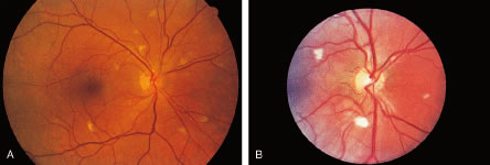
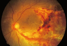
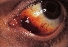
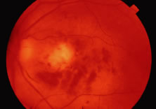
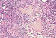
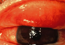
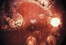
The ocular signs of CGD fall into two groups: recurrent blepharokeratoconjunctivitis and chorioretinal lesions61–64 (Fig. 1). Focal, pigmented chorioretinal lesions appear to be a common finding of CGD. Goldblatt and associates64 examined 38 patients with CGD and 36 of their family members. They found that nine of 38 affected children had chorioretinal lesions, as did three asymptomatic carriers of the gene.64 The lesions are usually perivascular or peripapillary in distribution and can progress to large areas of chorioretinal atrophy.62–64 They are most often described as punched-out chorioretinal scars. Goldblatt and associates64 described small white foveal lesions similar to drusen in some patients, but the typical chorioretinal lesions have not involved the central macula. Visual field defects may occur, corresponding to the location of chorioretinal scars.63 Histopathologic studies of the chorioretinal lesions show foci of granulomatous inflammation of the choroid and sclera.65 The granulomatous inflammation has been seen to extend through Bruch's membrane,65 and areas of Bruch's membrane may be absent.66 Pigment-filled macrophages in the retina and choroid are similar to those found elsewhere in the body.66 Chorioretinal scars with glial proliferation and areas of retinal pigment epithelium (RPE) hypertrophy and hyperplasia surrounded by atrophy of the RPE and the overlying retina are also found.66 Special stains and cultures of the reported ocular pathology and vitrectomy specimens have not revealed any infectious agents.65–67

Fig. 1. Typical fundus lesions of chronic granulomatous disease. (Photograph provided by Scott M. Whitcup. From Nussenblatt RB, Whitcup SM, Palestine AG. Uveitis: Fundamentals and Clinical Practice. St Louis: Mosby-Year Book, 1996.)

In an isolated case report,65 a 3-year-old child with CGD, who had been documented to have chorioretinal scars previously, was found to have chorioretinal lesions that appeared to be actively inflamed with an associated mild vitreitis. The lesions remained stable for several months, but after 7 months, his left eye developed a severe anterior uveitis, vitreitis, and exudative retinal detachment. A lensectomy and vitrectomy was performed. Cultures and stains of vitreous humor were negative. The presumed endophthalmitis did not respond to IV ceftazidime, nafcillin, and amphotericin B administered for 4 weeks. The eye was ultimately enucleated; cultures and stains of the enucleated eye did not reveal any infectious agents. It is not clear, however, whether there might have been an abnormal response to an infectious agent that had been eliminated by the therapy he received. Most people with CGD do not have any evidence of active inflammation or infection, however.

Chronic blepharoconjunctivitis and marginal or punctate keratitis have been reported in patients with CGD, often accompanied by both pannus formation and perilimbal infiltrates.63 These findings are believed to represent an immune reaction to staphylococcal antigens or toxins, rather than a direct invasion of the cornea by organisms. Because it is caused by a catalase-positive bacterium, staphylococcal blepharitis is often a chronic condition in patients with CGD. Regular eyelid hygiene and the selective use of topical antibiotics and corticosteroids has been used to prevent corneal neovascularization and scarring.63

Aggressive and prompt medical and surgical therapy of infections has been important for improving the prognosis of patients with CGD. Granulomata do respond to corticosteroids. The neutrophils of some patients with CGD do respond to interferon (IFN)-γ with a respiratory burst, which may depend on the production of nitric oxide rather than superoxide anions.68 Treatment of CGD with IFN-γ is associated with augmented production of nitric oxide by neutrophils.68 IFN-γ has also been used as a prophylactic agent.69 Although there are reports of cures with BMT, there have been problems with failures to engraft.70

Chédiak-Higashi Syndrome

Chédiak-Higashi syndrome (CHS) is a rare, autosomal recessive disorder, usually occurring in the children of consanguineous parents. It is characterized by partial oculocutaneous albinism, increased susceptibility to both viral and bacterial infections (with both catalase-positive and catalase-negative organisms), anemia, leukopenia, thrombocytopenia, cutaneous ulcers, and peripheral and central neurologic changes, including cerebral atrophy.71 CHS often leads to death in early childhood from infections or, less commonly, from hemorrhage.

Neutrophil abnormalities associated with CHS include neutropenia, reduced bone marrow neutrophil reserves, and reduced neutrophil chemotaxis. Abnormal granules in neutrophils and eosinophils can be seen on routine peripheral blood smears and also are present in other granule-containing cells, such as hepatocytes, renal tubular cells, melanocytes, Schwann's cells, and thyroid cells.72 Ingestion of particulate organisms occurs normally, but the rate of bacterial killing by neutrophils following ingestion is abnormal because of slow release of the abnormally large peroxidase-positive granules, leading to the delayed appearance of myeloperoxidase within phagocytic vacuoles. In addition to the deficient microbicidal activity of lysosomal degradation, natural killer cell activity also has been demonstrated to be abnormal in some cases.73 Heterozygous carriers of CHS have been identified by studies of degranulation in leukocytes.72

Children surviving past early childhood may be afflicted later with a so-called accelerated phase of the disease, characterized by lymphohistiocytic infiltration of the liver, spleen, nerves, and other tissues. Neoplastic transformation of these cells may occur. In addition to antimicrobial chemotherapy for infections, antineoplastic drugs and corticosteroids have been used to treat this accelerated phase.

Oculocutaneous albinism may be a prominent feature of CHS; an ultrastructural defect of melanosomes distinguishes this condition from true forms of albinism. Giant abnormal melanosomes, believed to be result from fusion of smaller abnormal organelles, have been reported. Hair color ranges from blonde to light brown, but most often has been reported as silver or gray.71,74 The iris is translucent with variable color. The amount of pigment in the uveal tract is variable and may be within normal limits. Histologic examination of the eyes also has revealed decreased pigmentation of the RPE, ciliary body, and choroid. These abnormal granules have been observed in monocytes and neutrophils present in the corneal limbus, iris, and choroid.71,75–79 Ultrastructural study of a conjunctival biopsy specimen from a patient with CHS revealed giant intracytoplasmic lysosomal granules in stromal fibroblasts, which are considered to be pathognomonic for CHS; thus, such a biopsy has been suggested as an ancillary diagnostic test in suspected cases of this syndrome.71

Nystagmus and photophobia generally are present, although not in all cases. During the accelerated phase of this disorder, infiltration of the optic disc and central retinal artery has been noted, and papilledema has been reported clinically.71,75–79

COMPLEMENT DEFICIENCIES

The complement system consists of approximately 20 serum proteins that are critical mediators of antigen-antibody reactions. Sequential activation of the complement proteins (complement cascade) is caused by either antibody-antigen interaction, through the so-called classic pathway, or by various stimulants, such as lipopolysaccharide, aggregated immunoglobulin, cobra venom, thrombin, or proteases through the so-called alternate pathway. Specific complement components and complexes of more than one complement protein serve different functions. C3b, for example, promotes immune adherence, phagocytosis, lymphokine production, and antibody-dependent cell-mediated cytotoxicity. C1,4 and C1,4,2,3 complexes aid in neutralization of viruses to which antibodies have attached. The completed complement cascade leads to lysis of viruses, mycoplasma, protozoa, and spirochetes by creating holes in membranes, capsules, or envelopes.

Deficiencies have been reported for each of the proteins that comprise the complement system. Complement deficiencies can result in increased susceptibilities to infections, connective tissue disorders (systemic lupus erythematosus and other autoimmune diseases), or angioedema, depending on which components are involved. For example, C2-deficient patients generally do not have problems with recurrent infections, unless other components, acting later in the cascade, are deficient as well. In contrast, deficiency of C3, which plays an integral role in both the classic and alternate pathways, results in repeated infections. Deficiencies of the later acting components (C5, C6, C7, or C8) have been linked with serious and sometimes fatal infections with N. meningitidis and Neisseria gonorrhoeae. The association between complement deficiency and autoimmune diseases, such as systemic lupus erythematosus, may relate to the host's inability to clear circulating immune complexes.

Although complement has been found in tears,80 cornea,81 aqueous humor,82 and sclera,83 ocular infections have not been reported to be prominent features of complement disorders.9 One should assume, however, that patients will be at risk for ocular infections with the same pathogens seen in nonocular sites. Experimental evidence in animal models shows that the complement system may play a role in protecting the eye against corneal infection84 and endophthalmitis85 caused by P. aeruginosa, an organism whose endotoxins activate the alternate pathway. Complement levels are increased in the vitreous humor of inflamed eyes.86 Patients with complement deficiencies who develop intraocular infections for some other reason may, therefore, have more severe disease than their immunocompetent counterparts.

Back to Top
ACQUIRED IMMUNODEFICIENCY DISEASES
Acquired immune dysfunction is more common than the primary immunodeficiency syndromes. An important cause of acquired immunodeficiency that ophthalmologists throughout the world will encounter is AIDS. Nevertheless, there are various conditions, such as malnutrition, other infections (including measles, syphilis, tuberculosis, leprosy), cancer (including Hodgkin's disease), immunosuppressive drug therapy, trauma, burns, collagen vascular diseases, diabetes, alcoholic cirrhosis, sarcoidosis, and aging, that can result in acquired immunodeficiency and may have associated ophthalmic disorders. The spectrum of disorders resulting from acquired humoral and cellular immunodeficiencies are similar to those of the primary diseases.

In many acquired immunodeficiency states, immune dysfunction does not contribute substantially to clinical manifestations. Reviewed here are several disorders in which altered host defenses lead to prominent clinical problems.

ACQUIRED IMMUNODEFICIENCY SYNDROME

AIDS was first reported in the early 1980s, and its ocular complications were described soon thereafter.87–89 It is the most severe in a spectrum of immunodeficiency states caused by infection with the human immunodeficiency virus (HIV).

Infection with HIV is characterized predominantly by T-lymphocyte deficiency. The initial step in HIV infection is binding of the envelope protein of HIV to the receptor on the host CD4+ T lymphocyte; chemokine receptors, such as CCR5 or CXCR4, act as coreceptors and are also necessary for binding. After fusion with the host cell membrane has occurred, the RNA of the virus undergoes reverse transcription, forming DNA, which enters the host cell nucleus and is integrated into the genetic material of the cell. This HIV-encoded DNA is transcribed into messenger RNA, which is then translated into viral proteins, including HIV protease, that are required for processing other HIV-encoded proteins into their functional forms. The newly assembled virus then buds from the cell surface, and new cellular infections occur. Host CD4+ T lymphocytes are depleted through a combination of increased destruction and impaired thymic production.90

The mechanisms of HIV-related disease are far too complicated to be reviewed here, but they ultimately result from dysregulation of the immune system, especially cell-mediated immunity, that occurs because of continuous CD4+ T-lymphocyte destruction and depletion. Impaired cellular immunity facilitates diseases associated with infectious agents, including both productive infections in various end organs and neoplasms that result from malignant transformation of infected cells; examples of the latter include Kaposi's sarcoma (KS), caused by human herpes virus type 8, and squamous cell carcinoma, caused by human papillomavirus. Many opportunistic infections result from reactivation of previously acquired agents of low virulence that are usually controlled by normal host immune defenses; examples include CMV, P. carinii, varicella-zoster virus, and Toxoplasma gondii. People infected with HIV seem to be predisposed to specific infectious agents (including those already noted, as well as Candida sp. and MCV) with involvement of specific organs (e.g., CMV retinitis or P. carinii pneumonia). Humoral immunity is also affected. Although HIV infection is associated with a polyclonal gammopathy, the B-lymphocyte response to new antigens is markedly impaired. In addition, production of cytokines, such as IFN-γ (which activates macrophages to kill intracellular pathogens) and interleukin-2 (which allows T-lymphocyte proliferation), is impaired.

Most patients are infected with HIV-type 1 (HIV-1). A retrovirus with lymphotropic properties similar to HIV-1 was isolated from two people in West Africa in 1986 and is called human immunodeficiency virus, type 2 (HIV-2).91 Although HIV-2 also causes AIDS, it appears to be less pathogenic than HIV-1, with a longer latency period of infection, less severe manifestations of disease, and longer survival after diagnosis of AIDS. The seroprevalence of HIV-2 is greatest in West Africa. Ocular complications of HIV-2 infection appear to be uncommon.92 For this reason, the term HIV in this chapter refers to HIV-1, unless specifically indicated otherwise.

The severity of immune dysfunction varies considerably between HIV-infected people but tends to become progressively worse over time, if untreated. Those infected with HIV can be asymptomatic; they can have nonspecific manifestations of HIV infection itself, such as lymphadenopathy; or they can have the overt manifestations of HIV-associated opportunistic infection and neoplasms. The AIDS is defined as a CD4+ T-lymphocyte count less than 200/μl (or percentage of CD4+ T-lymphocytes less than 14% of total lymphocytes) or the occurrence of one or more of a specific group of opportunistic infections and neoplasms predictive of severe immunodeficiency.93

HIV can be transmitted by one of three routes: (1) sexual intercourse; (2) percutaneous inoculation (sharing of needles by injection drug users, transfusion of contaminated blood products, accidental needle-stick injuries in health care workers); and (3) maternal-infant (vertical) transmission (in utero, during birth, or by breast-feeding). In the first two decades of the AIDS epidemic, the demographics of the disease in the United States have changed from largely a disorder of homosexual white men to one affecting a mix of homosexuals, heterosexuals, and injecting drug-users, disproportionately involving African Americans and the poor.

In the United States, over 400,000 people had died of AIDS by the year 2000, and there were over 700,000 people living with HIV infection. By 2000, over 60% of new HIV infections in 13- to 19-year-old people were occurring in women. Worldwide, over 16 million people had died, and over 33 million were infected, with over 95% of the 5.6 million adults and children who were newly infected in 1999 living in sub-Saharan Africa and other developing countries.

Currently available drugs that act against HIV include reverse transcriptase inhibitors (both nucleoside reverse transcriptase inhibitors and nonnucleoside reverse transcriptase inhibitors) and protease inhibitors.94 AIDS was initially uniformly fatal, but it can now be controlled in many patients with these medications, which are known collectively as highly active antiretroviral therapy (HAART) or potent antiretroviral therapy. These drugs, by interrupting the infection cycle of HIV, allow a partial restoration of immune function and have thereby resulted in a substantial decline in opportunistic infections. At the same time, the widespread use of potent antiretroviral therapy has resulted in new presentations of clinical disease and clinically important side effects. The cost of these drugs precludes their use in all but the wealthiest of countries. Even in the United States, the combination of intolerable side effects, drug resistance, and lack of adequate compliance with complicated treatment regimens has resulted in a long-term success rate for potent antiretroviral therapy of only 40% in some geographic areas.95

Ophthalmic disorders are among the most common manifestations of AIDS (Table 2). These manifestations include (1) noninfectious vasculopathies, (2) opportunistic infections, (3) neoplasms, (4) neuro-ophthalmic abnormalities attributable to intracranial and orbital disorders, and (5) drug-induced ocular side effects. Recognition and treatment of ocular disease are important not only because of the morbidity associated with vision loss but because ophthalmic manifestations of HIV infection often indicate progressive dysfunction of the immune system and can be the first manifestation of multisystemic diseases. Discussion of these disorders in later sections deal primarily with the manifestations of HIV infection in those people with severe immune dysfunction. The use of potent antiretroviral therapy has reduced the frequency of many HIV-related ophthalmic disorders and in some cases has altered their clinical features, course, and response to treatment.


Table 2. Ophthalmic Disorders Associated with HIV Infection

  1. Non-infectious vasculopathy
    1. Microvasculopathy
      1. Retina
        1. “HIV retinopathy” (characterized clinically by presence of cotton-wool spots, retinal hemorrhages)

      2. Conjunctiva
      3. Optic nerve

    2. Macrovasculopathy
      1. Retina
        1. Venous occlusions
        2. Arterial occlusions


  2. Infections
    1. Ocular surface and adnexal
      1. Herpes zoster ophthalmicus
      2. Herpes simplex virus keratitis
      3. Molluscum contagiosum virus dermatitis, blepharitis
      4. Microsporidial keratoconjunctivitis
      5. Crack cocaine-associated keratopathy
      6. Other

    2. Retinal and choroidal
      1. Cytomegalovirus retinitis
        1. Immune recovery uveitis

      2. Varicella zoster virus retinitis
      3. Herpes simplex virus retinitis
      4. Syphilitic uveitis/retinitis
      5. Toxoplasma gondii retinochoroiditis
      6. Pneumocystis carinii choroidopathy
      7. Mycobacterium tuberculosis choroiditis
      8. Other

    3. Orbital infections

  3. Neoplasms
    1. Eyelid/ocular surface
      1. Kaposi's sarcoma
      2. Lymphoma
      3. Squamous cell carcinoma

    2. Intraocular
      1. Lymphoma

    3. Orbit
      1. Lymphoma
      2. Kaposi's sarcoma (rare)


  4. Neuro-ophthalmologic abnormaities associated with intracranial or orbital diseases
    1. Infections
      1. Cryptococcal meningitis
      2. Intracranial toxoplasmosis
      3. Progressive multifocal leukoencephalopathy
      4. Varicella-zoster virus infection
      5. Syphilitic meningitis
      6. Other

    2. Neoplasms
      1. Intracranial lymphoma


  5. Adverse effects of systemic drugs
    1. Uveitis
      1. Cidofovir
      2. Fomivirsen
      3. Rifabutin

    2. Optic neuropathy
      1. Ethambutol

    3. Retinopathy
      1. Didanosine


Noninfectious Microvasculopathy

Noninfectious retinal lesions associated with microvascular abnormalities (cotton-wool spots, retinal hemorrhages; known as HIV retinopathy) are the most common ocular findings reported in people with HIV infection (Fig. 2). The extent and severity of these retinal findings are greater in patients with AIDS than in iatrogenically immunosuppressed people who occasionally have similar lesions.96 The presence of HIV retinopathy is related to overall immune status; asymptomatic HIV-infected patients rarely have clinically apparent lesions, but nearly all patients with advanced AIDS have some degree of HIV retinopathy.97,98 In addition to cotton-wool spots and retinal hemorrhages, clinical manifestations include retinal capillary microaneurysms, white-centered hemorrhages, telangiectasias, and capillary nonperfusion.87–89,97,99,100 Cotton-wool spots occur in the posterior pole and are seen as superficial white patches with feathery edges that sometimes obscure underlying retinal blood vessels. They are caused by stasis of axoplasmic flow within the retinal nerve fiber layer, as a result of ischemia, either because of precapillary arteriolar occlusion or reduced blood flow.101 Cotton-wool spots tend to resolve over 4 to 6 weeks. Affected patients are rarely symptomatic. Microaneurysms are found clinically in only about 20% of patients but are detected in eyes of nearly all patients with AIDS when studied by fluorescein angiography or by trypsin digestion of retinal tissue at autopsy.96,100,102 Ischemic maculopathy has been reported rarely.100 Noninfectious microvasculopathy is also the most common ocular manifestation of HIV-2 infection, and affected people have a shorter survival that HIV-2-infected people without microvasculopathy.92

Fig. 2. A, B. Cotton-wool spots in patients with AIDS.

The underlying abnormality of HIV retinopathy is disease of the retinal vasculature. Trypsin digest preparations and ultrastructural studies reveal selective loss of pericytes, microaneurysm formation, swollen endothelial cells, occluded vascular lumina, and thickened basal lamina,96,100 similar to findings in diabetic retinopathy. Fluorescent microsphere infusion studies have identified breaches in microaneurysms of 200 nm or more, even in the absence of visible hemorrhage; these defects might play a role in the passage of CMV into the retina with development of CMV retinitis.103

The cause of noninfectious microvasculopathy is unknown and likely multifactorial. HIV has been identified within retinal endothelial cells but not in amounts sufficient to account for the widespread changes seen.104 Other proposed mechanisms include deposition of circulating immune complexes,100 release of cytotoxic substances such as monokines or proteolytic enzymes104 and abnormal blood flow.101 Slowing of blood flow probably further contributes to the focal ischemia that results in periodic development of cotton-wool spots.101 The presence of both cotton-wool spots and conjunctival microvasculopathy, consisting of dilated capillaries, vessels of irregular caliber, and sludging of blood flow, has been related to elevated fibrinogen levels and red blood cell aggregation but not with levels of circulating immune complexes or immunoglobulin.101 People infected with HIV have increased neutrophil rigidity, which may also slow blood flow and contribute to microvascular damage.105 Souza Ramalho and associates106 reported a patient with HIV-2 infection and noninfectious retinal vasculitis who had elevated plasma viscosity and red blood cell filterability, both indices of abnormal blood flow. The patient's clinical and hemorheologic status improved following treatment with prednisone and chlorambucil.

Less commonly, occlusion of larger retinal blood vessels (macrovasculopathy) can occur. Both retinal artery and vein occlusions have been reported, with a prevalence greater than would be expected for age-matched controls.99,107 Hypertension and thrombotic disease are found in a high proportion of affected patients.

Other causes of noninfectious microvasculopathy, such as diabetes mellitus or hypertension, should always be considered. Patients with both diabetes mellitus and HIV disease may have particularly severe noninfectious retinopathy. Reports of hyperlipidemia, glucose intolerance, and accelerated vascular disease among patients treated with protease inhibitors108 have raised concerns that retinal vascular disease may become more frequent in HIV-infected patients. For example, lipemia retinalis has been found in a patient treated with protease inhibitors who had hyperlipidemia.109

Ocular Opportunistic Infections

CMV RETINITIS.

CMV retinitis is by far the most common intraocular infection in people with AIDS.99,110 CMV retinitis is rarely the first manifestation of AIDS111,112; most affected patients have CD4+ T-lymphocyte counts of less than 50 cells/μl at diagnosis.111,113 One study found that 85% of people with extraocular CMV infection developed CMV retinitis after a mean follow-up period of 6.4 months.114 Other risk factors for the development of CMV retinitis include high HIV levels in the blood, a previous AIDS-defining illness, homosexual activity as the route of HIV infection, lack of protease inhibitor therapy in antiretroviral regimens, high CMV levels in the blood, and CMV seropositivity.115 Up to 40% of patients with advanced HIV disease developed CMV retinitis in the era before potent antiretroviral therapy.110,111,115,116 Since the introduction of these drugs, the rate is much lower,117 probably on the order of 5% or less. The decline in frequency may reflect not only the widespread use of potent antiretroviral therapy but also the changing demographics of AIDS; heterosexual men are less likely than homosexual men to develop CMV retinitis,perhaps because homosexuals have a higher rate of pre-existing CMV infection.115 Nevertheless, CMV retinitis remains a major cause of morbidity in HIV-infected people. It is especially important to diagnose the infection early, because currently available therapy is usually effective for controlling retinal lesions and because treatment of systemic CMV infection may favorably affect concurrent HIV replication.

CMV retinitis may begin with infection of retinal vascular endothelial cells, with subsequent spread to adjacent glial and neuronal cells as well as to RPE,118 although early autopsy studies failed to identify infected endothelium in all cases.100 It is also possible that infection results from diapedesis of CMV-infected leukocytes in some cases.119

The diagnosis of CMV retinitis is based on clinical signs and symptoms.87,88,99,119,120 Vision is usually good at the time of diagnosis,113 and there is no pain or redness. People with early CMV retinitis usually have floaters and may also complain of photopsias or visual field defects, but between 10% and 75% of patients may be unaware of any symptoms,121,122 especially if the disease is unilateral.98 One study found that over half of new cases of CMV retinitis were diagnosed during routine screening eye clinic visits and that over 75% of affected patients were asymptomatic at the time of diagnosis.122 Routine dilated indirect ophthalmologic examination of patients at highest risk of CMV retinitis, based on a CD4+ T-lymphocyte count of less than between 50 and 100/μl, has been recommended.98,123

Only about 10% of people with AIDS-related CMV retinitis have clinically apparent extraocular CMV disease, although about half have a positive blood or urine cultures for CMV.113 The characteristic retinal lesion has a dry-appearing, granular border surrounding an area of retinal edema and full-thickness retinal necrosis. Findings are similar to those of CMV retinitis described in patients with iatrogenic immunosuppression before the AIDS epidemic began.124 There is a spectrum of lesion appearances; the fulminant or edematous type has prominent necrosis and hemorrhage, often along blood vessels in the posterior pole (Fig. 3). The indolent or granular type is more often found in the retinal periphery and has less dense retinal opacification and little or no hemorrhage. Many cases of CMV retinitis have features of both types. Patients who have an incomplete response to potent antiretroviral therapy may develop CMV retinitis that is less hemorrhagic and less densely opaque. Perivascular sheathing, which may be widespread, occurring in areas of the retina away from the site of necrosis, is found in between 35% and 50% of cases. Bilateral involvement occurs in 30% to 40% of cases at diagnosis.113,122,125 There are usually mild anterior chamber and vitreous inflammatory reactions, and fine keratic precipitates are often seen. Untreated lesions enlarge slowly; spread toward the fovea occurs at a median rate of only 24 μm/day and is somewhat faster toward the ora serrata.126

Fig. 3. Cytomegalovirus retinitis in a patient with AIDS; the disorder is characterized by perivascular, white, granular-appearing necrosis and hemorrhage.

Vision loss may result from primary or secondary optic nerve involvement,127,128 retinal necrosis involving the central macula, rhegmatogenous retinal detachment, retinal vein or artery occlusion, or serous macular exudation.129 Electrophysiologic and psychologic testing suggests that there is a diffuse dysfunction of the retina even when only localized, peripheral lesions are seen clinically.130

Peripapillary CMV retinitis is associated with an increased risk of CMV encephalitis.131 Early detection and treatment may reverse some of these complications and result in improved vision,132 but in most patients the goal of therapy is to prevent further vision loss in affected eyes and to reduce the risk of involvement in uninfected eyes of unilateral cases.133

CMV retinitis occurs less frequently in children with AIDS than in adults,134–136 but it is more likely to be associated with symptomatic extraocular CMV disease.137 Perhaps because children are less likely to complain of visual symptoms and are, therefore, less likely to undergo ocular examination than adults, CMV retinitis in children also more commonly presents with bilateral and posterior pole involvement.134,137 Very young children with CMV retinitis may also have high absolute CD4+ T-lymphocyte counts, because the normal CD4+ T-lymphocyte count in the first few years of life is substantially higher than in adults; however, affected children still have counts that are much lower than normal, when adjusted for age.137 Older children with CMV retinitis usually have CD4+ T-lymphocyte counts that are less than 20/μl.134

The differential diagnosis of CMV retinitis includes other necrotizing herpetic retinopathies, as described later, toxoplasmic retinochoroidopathy, candidal chorioretinitis, aspergillic retinitis, ocular cryptococcosis, endogenous bacterial endophthalmitis, syphilitic retinitis, intraocular lymphoma, and Bartonella henselae retinitis (cat-scratch disease).138 Although the distinction between these diseases is usually made clinically and additional workup is unnecessary, there are patients in whom vitrectomy or endoretinal biopsy may be useful diagnostically.139–144 In addition, concurrent ocular opportunistic infections with more than one pathogen have been reported in many people with AIDS.

There were four drugs approved by the U.S. Food and Drug Administration (FDA) as of the year 2000 for treatment of CMV retinitis: ganciclovir, foscarnet, cidofovir, and fomivirsen. The first three are available in IV forms; ganciclovir is also available in an oral preparation and as a sustained-release intraocular implant. The first three drugs act by inhibiting CMV DNA polymerase. Fomivirsen is an antisense oligonucleotide that acts by preventing the elaboration of a protein needed for infectivity of the virus; it is delivered by intravitreous injection. All these drugs are virostatic, not virucidal; discontinuation of therapy in persistently immunosuppressed patients, therefore, invariably leads to recurrence of CMV retinitis.

Ganciclovir is a nucleoside analogue. The recommended IV dose is 5 mg/kg every 12 hours for 2 to 3 weeks (induction), followed by 5 to 10 mg/kg once daily (maintenance therapy). Ganciclovir is a bone marrow suppressant; anemia, leukopenia, and thrombocytopenia are common findings associated with its therapy. Foscarnet is a pyrophosphate analogue. The recommended IV dosage is 90 mg/kg every 12 hours for 2 weeks (induction therapy), followed by 90 to 120 mg/kg once daily (maintenance therapy). Foscarnet is nephrotoxic and must be carefully dose adjusted for patients with renal insufficiency. Randomized, controlled studies have shown no therapeutic difference between ganciclovir and foscarnet; both control retinitis initially in about 90% of cases, but reactivation of retinal infection despite prolonged therapy is common, with a median time to progression of retinitis of less than 3 months. Because both IV foscarnet and ganciclovir must be given daily, a permanent indwelling catheter is placed for long-term therapy. Catheter-related infections are a major reason why other options have been developed.

Oral ganciclovir has limited bioavailability but may be effective as maintenance therapy following a course of IV induction therapy for limited periods of time.145 Commonly used doses range from 1 to 1.5 g three times daily. Side effects include GI disturbances and bone marrow suppression. Although use of oral ganciclovir as primary prophylaxis may reduce the risk of CMV infection, including retinitis,146 its use is not routinely recommended because of the cost and associated toxicity.147 An oral prodrug of ganciclovir (valganciclovir) holds the promise of providing serum drug levels comparable with those achieved with IV ganciclovir, while avoiding catheter-related complications; approval by the FDA is anticipated in 2001.

The ganciclovir implant is a sustained-release form of ganciclovir in which a 4.5-mg pellet of the drug, attached to a strut made of ethyl vinyl acetate, is placed surgically into the vitreous cavity through the pars plana and sutured to the sclera. Steady state levels of ganciclovir are reached within approximately 24 hours of placement, and intraocular drug levels are roughly four times greater than those achieved with IV ganciclovir therapy.148,149 Randomized, controlled studies have shown that the ganciclovir implant is more effective for maintaining inactivity of CMV retinitis than IV ganciclovir, with a median time to reactivation of approximately 220 days.149,150 The ganciclovir implant provides no benefit to the fellow eye; there is a substantial risk of second-eye involvement if concurrent systemic therapy is not given.150 Oral ganciclovir, as an adjunct to the placement of a ganciclovir implant in a patient with unilateral CMV retinitis, reduces the risk of second-eye involvement and extraocular disease149 and is, therefore, recommended, if there are no medical contraindications to its use.151 The long-term risk of retinal detachment is not increased in patients treated with ganciclovir implants compared with those patients treated with IV ganciclovir,149,152 although detachments may occur earlier. Surgical complications of the procedure range from temporary astigmatism and foreign body sensation to severe vision loss from hypotony, endophthalmitis, and vitreous hemorrhage.149,153 In addition, the ganciclovir implant must be replaced every 6 to 12 months in patients who remain immunosuppressed. The risk of serious surgical complications appears to be greater in patients undergoing multiple implant procedures.153,154

IV cidofovir has been proven to be effective for treatment of CMV retinitis155 but has not been compared with other anti-CMV therapies; thus, its relative efficacy has not been determined. Cidofovir is a nucleotide analogue with a long intracellular half-life, obviating the need for daily infusion or placement of a permanent, indwelling catheter for long-term therapy. The drug is given IV at a dose of 5 mg/kg once weekly for 2 weeks, followed by 5 mg/kg every 2 weeks. Cidofovir is nephrotoxic, and strict adherence to treatment guidelines is necessary. The use of oral probenecid and large amounts of IV saline for hydration at the time cidofovir is administered is mandatory.

Cidofovir does not require phosphorylation by a protein kinase produced by CMV to be taken into infected cells. Consequently, patients with low-level resistance to ganciclovir (UL 97 gene mutations) may respond to cidofovir, but high-grade ganciclovir resistance is often associated with cidofovir resistance as well, because of mutations at the level of CMV DNA polymerase.156 Resistance to anti-CMV drugs is rare before initial exposure to the drug,157 but it becomes more common with prolonged treatment.158 Resistance is associated with a worse clinical outcome, including progression of retinitis and second-eye involvement.158

Fomivirsen is an phosphorothioate antisense oligonucleotide that impairs production of an early gene product essential for CMV replication.159 The drug is given as an intravitreous injection consisting of 0.05 ml (330 μg) every 2 weeks for 2 doses, then every 4 weeks thereafter. Fomivirsen has not been compared directly with other anti-CMV therapy, but some evidence suggests that CMV retinitis resistant to other anti-CMV therapy may respond to fomivirsen. As with the ganciclovir implant, fomivirsen provides local control of retinitis only, with an associated risk of second-eye and extraocular CMV infection. Side effects of the injection include a small risk of vitreous hemorrhage or endophthalmitis, as well as uveitis, glaucoma, and cystoid macular edema. No systemic side effects have been reported.

Reactivation (relapse) of CMV retinitis with enlargement of lesions (progression) in patients who remain immunosuppressed is almost inevitable, even with continued maintenance therapy.99,133 With reactivation, there is recurrent whitening of the borders of inactive CMV retinitis scars, with centripetal spread into previously healthy retina. New lesions may also develop. Changes in lesion size may be subtle. Studies have shown that careful comparison of serial retinal photographs is a more sensitive means of detecting lesion enlargement than clinical observation only.160,161 The primary reason that reactivation occurs appears to be the limited intraocular drug delivery across the blood-retina barrier.148 Ironically, healing of CMV retinitis lesions may result in less intraocular penetration of anti-CMV therapy as the blood-retina barrier is reformed. Other causes of retinitis reactivation include development of drug resistance,162–164 noncompliance with therapy, and further waning of host immunity as HIV disease advances. Risk factors for development of new lesions in previously uninvolved eyes include positive blood and urine cultures for CMV and lower CD8+ T-lymphocyte counts.165

In the era before introduction of potent antiretroviral therapy, the time to progression (defined for study purposes as advancement of previously inactive lesion borders by 750 μm or more, as determined by a masked observer evaluating serial retinal photographs, or development of new foci of retinal infection) was on the order of 3 months or less. Retreatment with antiviral therapy at induction doses for 2 weeks, followed by higher maintenance therapy doses, was often effective at reestablishing control of the disease, but an accelerating rate of subsequent reactivations was observed.133 The use of combination antiviral therapy, such as IV foscarnet and ganciclovir, may also prolong the time to subsequent reactivation.166 Quality of life suffers when combination IV therapy is given, however.

Antiviral resistance is more likely to occur in patients who have suffered multiple reactivations. In such cases, changing from ganciclovir to foscarnet or cidofovir may be effective.162 Although data suggest that CMV DNA levels in the aqueous humor or vitreous humor167 or blood168,169 in treated patients may predict reactivation and that phenotypic or genotypic resistance may be determined by polymerase chain reaction (PCR) techniques using blood or vitreous humor isolates,158 such tests have not become as widely used in clinical practice as measurement of HIV levels in the blood and determination of antiretroviral drug resistance.170,171 Limited data suggest that patients who fail to respond to repeated induction regimens with ganciclovir or foscarnet may respond to intravitreous injections of fomivirsen.159

Before introduction of potent antiretroviral therapy, median survival following a diagnosis of CMV retinitis was 7.8 months99 to 12.4 months,172 and a reasonable goal of therapy for CMV retinitis was merely to delay advancement of lesions into the macular region, thereby preserving central vision before death. In the era of potent antiretroviral therapy, survival after the diagnosis of CMV retinitis may go on for many years,173 and more sustained control of CMV retinitis is necessary, if visual function is to be preserved.

Progression of CMV retinitis is less likely in patients being treated with anti-CMV drugs, if they are also receiving potent antiretroviral therapy, even if they do not show elevated CD4+ T-lymphocyte counts. The choice of anti-CMV therapy should, therefore, be based on a number of factors, including the location of lesions, the likelihood of the patient remaining on potent antiretroviral therapy, the response to antiretroviral agents, the presence or absence of extraocular CMV disease, the patient's overall medical condition, and patient preference.123 For example, a patient with perifoveal CMV retinitis who cannot be treated with potent antiretroviral therapy, would be a good candidate for a ganciclovir implant and oral ganciclovir therapy, whereas a patient with peripheral CMV retinitis who is expected to respond to potent antiretroviral therapy and is adherent to his or her medical regimen might safely and effectively be treated with systemic ganciclovir, avoiding the surgical risks of a ganciclovir implant.151

Rhegmatogenous retinal detachment is an important cause of vision loss in people with CMV retinitis. About 3% have detachments at the time of retinitis diagnosis, and the risk increases over time, affecting about one third of eyes with CMV retinitis per year, or approximately 50% per person-year.174–176 The risk of retinal detachment increases with active lesions, larger lesion size,174,175 and involvement of the anterior retina near the vitreous base.175 Older people and those with lower Karnofsky scores and lower CD4+ T-lymphocyte counts are also more likely to develop retinal detachments. Use of the ganciclovir implant does not increase the long-term risk of retinal detachment compared with systemic therapy only.151,152

Although large holes, horseshoe tears, and giant tears can be seen in patients with CMV retinitis and retinal detachments, most retinal detachments result from multiple, small, ill-defined holes within necrotic retina or at the junction of necrotic and normal retina. The difficulty in visualizing these small holes and the relative lack of inflammation in patients with CMV retinitis, which probably contributes to poor adhesions between the neurosensory retina and the RPE, help to account for the low success rate with conventional retinal detachment surgical techniques, such as scleral buckling alone. Despite studies indicating that laser retinopexy alone or vitrectomy with gas tamponade, laser, and scleral buckling may be effective in some cases,177 pars plana vitrectomy with long-term silicone oil tamponade and endolaser retinopexy has been considered the definitive technique for repair of retinal detachments associated with CMV retinitis, with macular reattachment rates exceeding 90%.177,178 Adjunctive scleral buckling does not appear to improve the success rate further.178 The risk of severe proliferative vitreoretinopathy was less than 4% of eyes in one study.177 Final vision often is decreased as a result of recurrent retinal detachment beneath the oil, corneal endothelial toxicity, epiretinal membrane formation, or cataract. In many cases, decreased central visual acuity occurs after retinal detachment repair, but an obvious reason cannot be identified on clinical examination.

Conventional phacoemulsification and posterior chamber intraocular lens implantation can be performed for silicone oil-associated cataracts, but adjustments in the intraocular lens power must be made to account for the hyperopia induced by silicone oil, depending on the style of implant used.179 Visual outcomes after cataract extraction have generally been disappointing,179 even in some cases in which no macular abnormalities were evident, raising the possibility of retinal toxicity from prolonged contact with silicone oil. Removal of silicone oil is associated with improvement in vision in some cases, suggesting that some visual symptoms in oil-filled eyes are optical and not due to toxicity.180

Immune reconstitution associated with potent antiretroviral therapy has had an enormous impact on the rate of CMV retinitis. Initiation of potent antiretroviral therapy in those at high risk for CMV disease results in significantly lower rates of CMV retinitis.117,173,181,182 Increasing CMV DNA levels in the blood are associated with an increased risk of CMV retinitis169; immune reconstitution induces a progressive decline in CMV viremia in the absence of specific anti-CMV therapy.183,184

There are differences in the presentation and clinical course of CMV retinitis in patients who are already taking antiretroviral agents. They may present with inactive CMV retinitis, indicating regression of the CMV disease as a result of immune reconstitution alone. Others may present with CMV retinitis and CD4+ T-lymphocyte counts that have risen above 200 cells/μl185 after recently beginning antiretroviral therapy; presumably the retinal infection developed before the counts rose. Patients may also have pronounced intraocular inflammation and cystoid macular edema;186 these findings were rare in patients with CMV retinitis before the introduction of potent antiretroviral therapy.

In a study of 14 consecutive patients with CMV retinitis diagnosed in 1997 or 1998, Jacobson and associates181 found that four of five patients who never received potent antiretroviral therapy either before or after the diagnosis of retinitis either died within 2 months or had active retinitis after 6 weeks of systemic anti-CMV therapy. In contrast, all five patients who responded well to potent antiretroviral therapy after the diagnosis of retinitis, as evidenced by a nondetectable plasma HIV level or a CD4+ T-lymphocyte count above 50/μl, also responded well to anti-CMV therapy and had no reactivation of retinitis over a median 15.5 months of ophthalmologic monitoring. In the remaining patients, who had received suboptimal antiretroviral therapy before diagnosis of CMV retinitis and who were changed to an effective antiretroviral regimen, two patients died within 1 month and two patients had persistent CMV retinitis despite 2 to 4 months of systemic anti-CMV therapy. Although the number of patients in this study was small, the results suggested that long-term ophthalmic outcomes for patients with CMV retinitis are dependent on the response to potent antiretroviral therapy.

Kempen and associates152 have shown that the use of potent antiretroviral therapy is associated with a 60% reduction in the rate of retinal detachment in eyes with CMV retinitis, with the greatest benefit observed among patients who developed an immunologic response. The use of potent antiretroviral therapy had a greater effect on the risk of retinal detachment than other well-established factors such as lesion size and anterior location of retinitis. It has been suggested that immune recovery results in a stronger adherence at the retina/RPE interface.187 Because potent antiretroviral therapy also reduces other risk factors for retinal detachment, such as active retinitis and increased lesion size, it may be that some retinal detachments in patients treated with potent antiretroviral therapy may be surgically corrected without the need for silicone oil.180

Numerous studies have shown that maintenance therapy can be discontinued without reactivation of retinitis in patients with CMV retinitis who are then successfully treated with potent antiretroviral therapy.188–190 CMV-specific CD4+ T-lymphocyte responses in patients with CMV disease may be restored by potent antiretroviral therapy,191 but it can take 3 to 6 months after the rise in absolute CD+ T-lymphocyte counts for the repertoire of cells with specific activity against CMV to be restored.192 Most investigators therefore recommend that the CD4+ T-lymphocyte count show a sustained elevation for at least 3 months and that the CMV retinitis be inactive before maintenance therapy is discontinued.193 It must be kept in mind, however, that the failure rate of potent antiretroviral therapy in unselected patients in clinical practice may be substantially greater than the failure rate in patients closely monitored in clinical trials.95 Failure of potent antiretroviral therapy may result from noncompliance with therapy, inability to take the medications because of intolerable side effects, or antiretroviral resistance.95,194 There is a risk of reactivation of CMV retinitis in patients who initially respond to, but who eventually fail, potent antiretroviral therapy, with a fall in CD4+ T-lymphocyte count to less than 50 cells/μl.168 The roles of HIV RNA and CMV DNA blood levels or specific tests of CMV immunity in determining which patients are at greatest risk for reactivation of CMV retinitis remain unclear. Furthermore, increases in CD4+ T-lymphocyte count alone do not completely reflect the immunologic status, and reactivation of CMV retinitis has occurred even in patients with persistently elevated counts.195 Patients who are taken off maintenance anti-CMV therapy therefore must understand the importance of strict adherence to the potent antiretroviral therapy regimens and of regular ophthalmic follow-up.

Immune recovery uveitis is a condition characterized by increased intraocular inflammation in eyes of patients with CMV retinitis, attributable to immune reconstitution.196–198 The CMV retinitis is usually, although not always, inactive.198 Although there is no universally accepted clinical definition of immune recovery uveitis, the most characteristic findings include prominent anterior chamber and vitreous humor cellular reactions. Patients with immune recovery uveitis may have optic disc edema,199 epiretinal membrane formation,199 cystoid macular edema,186 vitreomacular traction,200 and retinal neovascularization.199,201 Posterior synechiae201 and optic disc neovascularization202 have been reported less commonly. Immune recovery uveitis, which may be either acute, recurrent, or chronic, is now the leading cause of severe, new vision loss in people with AIDS and CMV retinitis.203 The macular edema may not be clinically evident; therefore, patients with CMV retinitis and unexplained vision loss should undergo fluorescein angiography.

Disease mechanisms associated with immune recovery uveitis remain poorly understood. The disease occurs only in patients receiving potent antiretroviral therapy and whose CD4+ T-lymphocyte counts have increased, usually to the range of 75/μl or higher. Immune recovery uveitis does not occur in patients with CMV retinitis who are not treated with potent antiretroviral therapy or who fail to respond to antiretroviral agents and does not occur in the uninvolved eye of patients with unilateral CMV retinitis.187 Currently available anti-CMV therapy is virostatic, not virucidal, and continued production of viral proteins can occur in CMV lesions that are clinically inactive.204,205 Immune recovery uveitis is presumably a response against these retained viral proteins. Immune recovery uveitis can also occur in patients with active CMV retinitis who have recently started potent antiretroviral therapy.185,198 In fact, some authors have suggested that a more prominent inflammatory reaction is actually a good prognostic sign for control of infection.206

A syndrome similar to immune recovery uveitis has been described in patients with CMV retinitis following organ transplantation, when doses of immunosuppressive drugs are reduced and immune function improves.207 Nussenblatt and Lane192 suggested that the development of immune recovery uveitis occurs along a spectrum of immune function. With profound immunodeficiency, people with CMV retinitis produce very little inflammation against the virus, explaining the relatively quiet appearance of eyes with active disease. As immune function improves, a much greater inflammatory reaction against viral proteins occurs, and immune recovery uveitis develops. At still greater levels of immune function, production of antigen ceases, and the inflammatory reaction resolves. Similarly heightened inflammatory reactions occur in some patients with other AIDS-related infections and neoplasms, such as tuberculosis, Mycobacterium avium complex (MAC) infection, hepatitis B and C, progressive multifocal leukoencephalopathy (PML), lymphoma, and KS, after initiation of potent antiretroviral therapy.208 Current theories do not explain why only a few patients develop immune recovery uveitis.

The reported frequency of immune recovery uveitis ranges from 0.109 per person-year197 to as much as 0.83 per person-year.187 Although this range might reflect subjective differences in interpretation of inflammatory reactions, other factors are likely involved. Cidofovir has been associated with the development of uveitis,209 and patients treated with cidofovir may be at higher risk for immune recovery uveitis than those patients treated with other drugs.197 In addition, the type of therapy used to control CMV retinitis initially and the duration of anti-CMV therapy after initiating potent antiretroviral therapy may affect the rate of immune recovery uveitis.210 It has been shown that some patients with immune recovery uveitis have detectable amounts of CMV in the blood after the start of potent antiretroviral therapy, despite clinically inactive CMV retinitis.190 The use of ganciclovir implants, which is more effective than systemic therapy at reducing intraocular virus activity,149 may reduce the subclinical production of virus or viral proteins that stimulate inflammatory reactions. Furthermore, immune recovery uveitis has been reported to occur less frequently among patients who are treated with anti-CMV drugs for long durations after initiation of potent antiretroviral therapy.187,197

Treatment of immune recovery uveitis with oral corticosteroids or periocular corticosteroid injections has been successful for reducing macular edema187,211 and optic disc neovascularization in some patients.202 In one series, nine patients with immune recovery uveitis had persistent cystoid macular edema when treated with oral acetazolamide and topical corticosteroids, but three of these patients improved following periocular corticosteroid injections.186 Spontaneous improvement in inflammation and vision within 6 weeks has also been reported198; all these patients were receiving anti-CMV therapy when they developed inflammation. A combination of continued, aggressive anti-CMV therapy and either local or systemic corticosteroid therapy should therefore be considered in patients for whom there is no medical contraindication to use of these drugs and for whom there is no spontaneous improvement in inflammation. However, even aggressive therapy may not be effective, and late complications of persistent immune recovery uveitis, such as posterior synechiae, proliferative vitreoretinopathy, and posterior subcapsular cataracts, may result in severe vision loss.199,201 Although corticosteroids and chronic inflammation are well-recognized causes of posterior subcapsular cataracts, the mechanisms of other sequelae of immune recovery uveitis remain poorly understood.

OTHER POSTERIOR SEGMENT OPPORTUNISTIC INFECTIONS.

Acute retinal necrosis (ARN) syndrome is a necrotizing retinitis that is also characterized by prominent anterior uveitis, retinal and choroidal occlusive vasculitis, vitreitis, and papillitis.212 Varicella-zoster virus is the most frequently identified cause of ARN syndrome, but HSV can cause it as well. ARN syndrome can also occur in immunosuppressed people, although it was initially reported in immunocompetent patients.213 Patients with AIDS and ARN syndrome usually have a CD4+ T-lymphocyte counts higher than 60 cells/μl and a history of dermatomal herpes zoster or HSV dermatitis.214 It has occurred in up to 17% of HIV-infected patients following the development of herpes zoster ophthalmicus.215 A mild unilateral case of ARN syndrome was reported in an 11-year-old girl with AIDS (CD4+ T-lymphocyte count of 115/μl) after she developed chickenpox.216

Diagnosis of ARN syndrome is usually made clinically. Diagnostic criteria for ARN syndrome have been established by the American Uveitis Society.217 These criteria are based on the clinical features of the disease and course of infection and do not depend on the patient's immunologic status. The umbrella term necrotizing herpetic retinopathy is recommended when the characteristics of the retinitis do not fit those of a well-defined clinical syndrome. The presence of optic neuritis, scleritis, or pain helps support, but does not establish, a diagnosis of ARN syndrome. In some cases, it may be difficult to distinguish ARN syndrome from CMV retinitis, in which case PCR techniques can be used to test vitreous humor specimens139 or endoretinal biopsy specimens obtained at the time of retinal detachment repair141 to determine the causal agent.

Because data regarding treatment are limited in patients with both AIDS and ARN syndrome and because affected patients are usually not profoundly immunosuppressed, therapy is usually based on experience in immunocompetent patients with ARN syndrome. IV acyclovir (500 mg/m2 or 10 mg/kg every 8 hours) is usually effective; IV foscarnet may be necessary in acyclovir-resistant cases. IV therapy should be continued until the lesion borders are inactive, after which time oral antiviral therapy (acyclovir 800 mg five times daily, famciclovir 500 mg three times daily, or valacyclovir 1000 mg three times daily) should be used for at least 6 months. Consideration should be given to prophylactic laser retinopexy to reduce the risk of retinal detachment, which can occur in up to two thirds of patients. The benefit of aspirin to treat vasoocclusive and ischemic manifestations of ARN syndrome remains unproved. Although prednisone may reduce the associated vitreous humor inflammation in immunocompetent patients with ARN syndrome,214 oral corticosteroids are usually contraindicated in HIV-infected patients who are severely immunosuppressed, to prevent further immunosuppression.

Cunningham and associates139 reported two patients with AIDS and HSV retinitis, confirmed by PCR assay. Both patients had rapidly progressive bilateral retinal necrosis associated with intraretinal hemorrhages and diffuse vasculitis. One patient had HSV-1-associated retinitis, characterized primarily by retinal arteriolar involvement, with marked capillary dropout and occlusion of larger arcade vessels. The other patient had HSV-2-associated retinitis, characterized by greater involvement of the retinal veins and an exudative retinal detachment. It remains unclear whether these different findings are related to the two types of HSV or were simply variations in disease presentation related to host or other factors.

Progressive outer retinal necrosis (PORN) syndrome is another variant of necrotizing herpetic retinopathy that occurs in patients with advanced HIV disease. It is nearly always caused by varicella-zoster virus218,219; although a patient with PORN syndrome caused by HSV-1 has recently been documented.142 In contrast to ARN syndrome, PORN syndrome is characterized by multifocal lesions, initial involvement of the outer retina, absence of vascular inflammation, minimal or no vitreous inflammation, and extremely rapid progression.218 A “cracked mud” perivascular pattern is characteristic, caused by early removal of necrotic debris or edema from retinal tissue adjacent to blood vessels. Lesions may begin peripherally or in the macula. Optic neuropathy may occur early in the disease course and may even precede the retinitis.220 Early vitreous hemorrhage can occur. As with other ocular opportunistic infections, the frequency of PORN syndrome has declined markedly in patients treated with potent antiretroviral therapy.221

PORN syndrome, like ARN syndrome, is usually diagnosed clinically. Various treatment regimens have been reportedly successful in anecdotal series, including combination IV acyclovir and foscarnet, combination IV ganciclovir and foscarnet, IV foscarnet with placement of a ganciclovir implant, and intravitreous injections of ganciclovir.221 In severely immunosuppressed patients, chronic antiviral therapy is necessary to prevent reactivation of disease218,222; oral acyclovir is inadequate for this purpose.

In one study, two of six patients with PORN syndrome had simultaneous varicella-zoster virus encephalitis,223 emphasizing the importance of systemic therapy in the treatment of the disease. Unfortunately, the prognosis is usually poor even in eyes treated promptly, with two thirds of patients progressing to no light perception vision within 1 month of diagnosis.218 Blindness can result from optic nerve disease, macular infection, or retinal detachment. Prophylactic laser retinopexy has been less successful for preventing retinal detachment in patients with PORN syndrome than in patients with ARN syndrome.218 Retinal detachment occurs in 70% of patients218 and requires vitrectomy and silicon oil tamponade for anatomic repair. Even if the retinitis is initially controlled, recurrence of disease while on maintenance therapy (manifesting as increased border opacification and spread of old lesions or development of new lesions) and development of disease in a previously uninfected eye is common.222 As with other ocular viral opportunistic infections, the most effective therapy may be recovery of immune function with potent antiretroviral therapy.

Ocular toxoplasmosis has been reported to occur in as many as 3% of people with AIDS in France.224 The current prevalence of ocular toxoplasmosis among HIV-infected people in the United States is much lower because of potent antiretroviral therapy and widespread use of trimethoprim-sulfamethoxazole for primary prophylaxis of intracranial toxoplasmosis. Holland and associates225 noted that ocular toxoplasmosis in HIV-infected patients may have different clinical features than in HIV-negative people. Bilateral involvement, absence of preexisting chorioretinal scars, and evidence of systemic toxoplasmosis may be found, suggesting that acquired disease rather than reactivation of congenital infections is relatively more common in HIV-infected people. Atypical presentations of ocular toxoplasmosis, such as miliary disease,226 optic neuritis,227 panophthalmitis,228 and acute unilateral toxoplasmic iridocyclitis in the absence of retinal lesions,229 have been reported. In one study, 20% of patients with ocular toxoplasmosis developed CMV retinitis during follow-up,224 and toxoplasmic retinitis can mimic CMV retinitis.140 The two infections can also occur simultaneously. Prominent vitreous and anterior chamber inflammation; relative absence of retinal hemorrhage; and the presence of a thick, densely opaque lesion with a smooth, nongranular border suggests toxoplasmosis. Serologic tests are usually not helpful for diagnosis, because many HIV-infected people test seropositive for both T. gondii and CMV. False-negative serologic test results for T. gondii infection can occur.226,228 Endoretinal biopsy140 or PCR techniques226,229 may be necessary in some cases to make a definitive diagnosis.

Treatment of ocular toxoplasmosis is similar to that used for toxoplasmosis in immunocompetent patients, except that indefinite maintenance therapy may be necessary to prevent recurrences in persistently immunosuppressed patients and oral corticosteroids are generally contraindicated in persistently immunosuppressed patients.225,226,228 Pyrimethamine and sulfadiazine are the preferred treatment, with clindamycin used in patients with allergies to sulfonamides. A delay in the initiation of appropriate therapy may result in progressive disease and blindness.226–228

Various infectious choroidopathies have been reported, albeit infrequently, in HIV-infected patients. The frequency is greater in autopsy than in clinical studies.143 Choroidal lesions tend to be localized in the choriocapillaris and are usually multifocal and bilateral. P. carinii pneumonia (PCP) was once the most common opportunistic infection in patients with AIDS. P. carinii choroidopathy has been associated with the use of aerosolized pentamidine as primary prophylaxis against PCP, presumably because it suppresses lung disease locally, but does not prevent dissemination of organisms to other organs.99,230 Affected people may have evidence of disseminated disease, such as fever, weight loss, and malaise without pulmonary symptoms. Ocular disease is characterized by bilateral multifocal choroidal lesions that do not involve the retina.230,231 Active lesions are slightly thickened, oval, or lobulated; appear yellow-orange; and range in size from several hundred to several thousand microns in diameter. Vision is not usually affected. No vitreous humor or anterior chamber cells are seen. Treatment with IV trimethoprim-sulfamethoxazole or pentamidine is usually effective. Treated lesions become flat and may regress completely but often leave well-demarcated, nonpigmented, orange scars behind. P. carinii choroidopathy has become rare with the widespread use of trimethoprim-sulfamethoxazole, dapsone, or other systemic agents as primary prophylaxis against PCP.

Cryptococcus neoformans is an uncommon cause of choroidopathy in HIV-infected people.232 There may be concurrent optic nerve involvement. Carney and associates233 suggested that ocular involvement in disseminated cryptococcosis occurs in stages, beginning with choroiditis and/or optic nerve disease and progressing to uveitis, retinitis, and endophthalmitis. IV amphotericin B is the treatment of choice.

Although the risk of tuberculosis is dramatically increased in people with AIDS, and extrapulmonary tuberculosis is more common in HIV-infected people, tuberculous choroidopathy has been reported only rarely.234 The lesions are usually small, yellow-white infiltrates that may coalesce, and there may be an associated vitreitis. Conversely, some patients present with anterior uveitis or vitreitis, but with apparently healed choroidal lesions, as a result of previous antituberculosis therapy.

Other reported causes of infectious retinopathy or choroidopathy include metastatic bacteria, MAC, B. henselae, Histoplasma capsulatum, Candida sp., and Aspergillus fumigatus. Morinelli and associates143,232 found that the organism causing infectious choroidopathy was thought to be the cause of death in most of their patients and reported mean survival of only 25 days after identification of choroidal lesions. The ophthalmologist can, therefore, play an important role in helping to diagnose disseminated infection, thereby allowing appropriate therapy to begin as soon as possible.

Although not usually considered an opportunistic infection, syphilis does occur more frequently in HIV-infected people. In one large urban study, approximately 1% of HIV-infected subjects had ocular syphilis.235 The exchange of drugs for sex and unprotected sexual activity are the major risk factors for the spread of the disease. People with HIV infection are more likely to have contracted syphilis at a less advanced stage of immunosuppression than many other secondary infections,99 presumably because of the decline in sexual activity among people with advanced AIDS.

The possibility of syphilis should always be considered in patients presenting with ocular inflammatory disease. As in the pre-AIDS era, iridocyclitis is the most common presentation,99,235 but retinitis, neuroretinitis, optic neuritis,236 and scleritis have also been reported.237 Syphilitic uveitis in HIV-infected people is often associated with neurosyphilis. Treatment with intramuscular penicillin has been associated with persistent infection and relapse of disease237; IV therapy, similar to that used to treat neurosyphilis, is therefore recommended.235,237 False-negative serum Venereal Disease Research Laboratories (VDRL) or rapid plasma reagin (RPR) titers have been found in some people who were coinfected with HIV and Treponema pallidum.237 Thus, results from more specific diagnostic tests (e.g., fluorescent treponemal antibody, absorbed) should also be obtained for diagnosis.

Intraocular inflammation, without identifiable opportunistic infections, and possibly related directly to HIV has been reported. Rosberger and associates238 reported six patients with anterior uveitis, of whom four patients also had vitreitis. The CD4+ T-lymphocyte counts in the affected patients ranged from 21 to 2118 cells/μl. None of the patients improved with topical corticosteroids, but all responded to zidovudine within 1 month, suggesting that the uveitis might be caused by HIV. Chronic multifocal retinal infiltrates have also been associated with HIV infection in the absence of other opportunistic infections.239 The CD4+ T-lymphocyte counts have ranged from 7 to 2118 cells/μl in patients with this disorder. The infiltrates have been described as clusters of white lesions smaller than 200 μm, located in the midperipheral and anterior retina; there was vitreitis in 94% and vasculitis in 21% of eyes in the reported series.239 Bilateral disease was noted in 81% of patients. Nine of the 26 reported patients improved with zidovudine alone, again suggesting a primary HIV-induced inflammation. These two studies were retrospective and the pathogenesis of the findings remains unclear. Clinicians should not attribute uveitis or retinal lesions to HIV infection alone unless other, more common, causes have been ruled out.

ADNEXAL AND OCULAR SURFACE INFECTIONS.

Adnexal and ocular surface infections are uncommon in HIV-infected patients.97,240 Various viral, bacterial, and fungal pathogens have been reported. The clinician should be aware of the clinical presentation of these infections, which may differ in frequency, appearance, course, and response to treatment from the same infections when they occur in immunocompetent patients.

MCV is a DNA poxvirus that can cause a self-limited follicular conjunctivitis in immunocompetent people. In HIV-infected people, MCV lesions tend to be multiple and more widespread on the body. Lesions may be the classic avascular, waxy, 2 to 3 mm in diameter, umbilicated papules noted in immunocompetent people, but they may also become coalescent or ulcerated. HIV-infected patients with MCV infection usually do not develop irritation, conjunctival injection, or follicles, although lesions near the eyelid margin may cause a mild keratoconjunctivitis.241 The differential diagnosis includes bacillary angiomatosis, papilloma, basal cell carcinoma, and disseminated fungal infection. Biopsy will reveal classic eosinophilic cytoplasmic inclusion bodies. Treatment includes surgical excision, chemical cautery, or cryotherapy, but recurrence in 6 to 8 weeks is frequent. Improvement in patients treated with IV cidofovir has been reported.242 Lesions may regress completely in patients with a good response to potent antiretroviral therapy.

C. neoformans may cause eyelid,243 conjunctiva,244,245 or iris nodules. Conjunctival cryptococcosis may mimic squamous cell carcinoma clinically but is easily distinguished by biopsy.245 Conjunctival disease may occur simultaneously with choroidal cryptococcosis.244

Corneal microsporidiosis is a rare parasitic disease caused by the genera Encephalitozoon or Septata. It results in chronically red, irritated eyes; decreased vision; and photophobia that results from diffuse, fine corneal epithelial infiltrates. The corneal stroma is spared. Most cases are bilateral. The diagnosis is made by conjunctival scraping or corneal biopsy; examination reveals characteristic gram-positive ovoid organisms within the cytoplasm of epithelial cells but few inflammatory cells. Topical fumagillin appears to be the most effective treatment but must be continued indefinitely.246

HSV keratitis does not appear to be more common in HIV-infected people, although it may be more prolonged and recurrences may be more frequent.247 One study suggested that herpetic dendrites occur more often in the peripheral cornea and that stromal keratitis is less likely in patients with AIDS.248 Treatment with débridement followed by topical trifluridine 1% nine times a day for 10 to 14 days is usually effective. Patients with recurrences may benefit from chronic oral acyclovir (400 mg twice daily) as secondary prophylaxis.

Herpes zoster ophthalmicus may have cutaneous, corneal, and intraocular complications. Corneal manifestations range from punctate epithelial keratitis and stromal or subepithelial infiltrates to severe peripheral ulcerative keratitis and neovascularization.249 Concurrent uveitis and cutaneous lesions are common, and PORN syndrome may develop. In some HIV-infected people, varicella-zoster virus infection of the corneal epithelium can persist for many weeks,250 in contrast to findings in immunocompetent patients with herpes zoster ophthalmicus. The corneal infection responds to topical and systemic antiviral agents.

Patients younger than 45 years who develop herpes zoster ophthalmicus should be considered for HIV testing, particularly in geographic areas with a high prevalence of HIV infection or in patients with known risk factors for HIV infection. In one study from Miami, over half of patients with herpes zoster ophthalmicus younger than 45 years had HIV infection.215 When HIV infection is present, there is a higher rate of corneal involvement and postherpetic neuralgia.251 Vision loss can result from corneal scarring, exposure keratopathy, or secondary infectious keratitis. In addition, Sellitti and associates215 found that 17% of patients with herpes zoster ophthalmicus and HIV infection developed retinitis, most of which was bilateral. Treatment of herpes zoster ophthalmicus in HIV-infected patients should be more aggressive than in immunocompetent patients; IV acyclovir (10 mg/kg every 8 hours) is recommended until the lesions are crusted, followed by prolonged oral antiviral therapy. Anterior uveitis usually responds well to topical corticosteroids. Periodic funduscopic examinations should be performed to detect any herpetic retinopathy and patients should be made aware of symptoms of retinitis.

Fungal and bacterial keratitis has been reported in HIV-infected patients but appears to be uncommon. Atypical pathogens such as C. albicans and Bacillus sp. appear to be disproportionately involved, and multiple organisms may be present.252,253 Conventional risk factors for infectious keratitis (e.g., contact lens wear, trichiasis, use of topical corticosteroids, and trauma) should be considered, but they may be absent in HIV-infected patients. Keratoconjunctivitis sicca is more common in HIV-infected patients254 and may predispose patients to corneal infection. The use of crack cocaine may cause punctate epithelial keratopathy, sterile corneal epithelial defects, and microbial keratitis (“crack eye syndrome”)255; epithelial defects may be caused by neurotrophic keratitis, direct thermal or chemical toxicity of the alkaline smoke, or eye rubbing.

Treatment of infectious keratitis should be based on results of appropriate cultures and stains, keeping in mind that multiple pathogens may be present. Treatment is often difficult because of poor adherence to treatment recommendation and inconsistent follow-up.

HIV may uncommonly result in diffuse superficial keratitis, herpes-like ulcerations, and interstitial keratitis.251 As with posterior segment disease attributed to HIV infection, other causes of keratitis should be ruled out. CMV may cause conjunctivitis. CMV retinitis is often associated with fine, diffuse, pigmented endothelial deposits; the presence of such keratic precipitates, especially in the presence of a mild iritis, is strongly suggestive of posterior segment disease and mandates a dilated funduscopic examination.

NEOPLASMS

KS is the most common ocular tumor in HIV-infected people, but ocular involvement is much less common than extraocular disease. In a study of 200 patients with AIDS seen between 1983 and 1988, only 5 patients had ocular KS.97 Up to 20% of patients with KS have ocular involvement, and ocular involvement may be the first manifestation of the disease.256 About 70% of lesions occur on the eyelids, with the conjunctiva affected less frequently.256 Orbital involvement is exceptionally rare. Homosexual men constitute the overwhelming majority of patients with KS. The rate of KS has been declining, which may reflect that homosexual men constitute a smaller portion of cases of AIDS or it may be related to the widespread use of potent antiretroviral therapy.257

Conjunctival lesions can occur on the palpebral or bulbar conjunctiva, but are most common in the inferior fornix (Fig. 4). Bilateral disease is frequent. The tumor spreads slowly and does not involve the cornea or intraocular structures. Lesions are bright to deep red or violaceous and may be mistaken initially for subconjunctival hemorrhages. The differential diagnosis also includes atypical hordeolum, pyogenic granuloma, cavernous hemangioma, and bacillary angiomatosis. Patients are usually asymptomatic or have only mild irritation, although larger lesions and those involving the eyelid margin may cause blurred vision, entropion, or trichiasis.

Fig. 4. Conjunctival Kaposi's sarcoma in a patient with acquired immunodeficiency disease.

Symptomatic patients or those who are bothered cosmetically by the lesions usually respond well to various treatments, including chemotherapy (often a combination of doxorubicin, bleomycin, and vincristine); single-dose258 or fractionated radiation therapy259; intralesional IFN-α2a; cryotherapy; or surgical excision.256,259,260 Local therapy of ocular lesions is necessary only if patients cannot tolerate systemic therapy, have failed to respond to systemic therapy, or have bulky or ocular lesions that need immediate resolution. In some cases, improvement of immune function with potent antiretroviral therapy alone may be sufficient to stabilize KS, without the need for potentially toxic chemotherapy.260 Human herpes virus 8 has recently been identified as the cause of KS.261 This finding helps explain why the use of oral ganciclovir as adjunctive therapy to the ganciclovir implant in patients with CMV retinitis reduces the risk of KS compared with findings in patients treated with the ganciclovir implant alone.149

Squamous cell carcinoma of the conjunctiva, sclera, or eyelid is increasingly common, especially in Africa,262 and ophthalmologists who detect squamous cell carcinoma in a young patient should consider the possibility of HIV infection. In contrast to squamous cell carcinoma in older immunocompetent people, HIV-associated squamous cell carcinoma is usually faster growing and more likely to infiltrate the cornea, sclera, and substantia propria.262–264 Human papillomavirus may play a permissive role in the development of squamous cell carcinoma in HIV-infected people.262 Fogla and associates265 reported conjunctival squamous cell carcinoma as the presenting sign of HIV-2 infection in one patient.

Treatment of limbal or conjunctival squamous cell carcinoma should involve total excision of the tumor with clear margins and cryotherapy to the underlying sclera. In cases of intraocular extension, exenteration should be performed.262,265

Lymphoma in HIV-infected patients is an important cause of orbital disease (see later discussion). CNS lymphoma may also be associated with intraocular disease,266 causing bilateral hypopyon267 or retinitis with vitreitis.268

NEURO-OPHTHALMIC DISORDERS

Neuro-ophthalmic disorders in people with AIDS include cranial nerve palsies, abnormal eye movements, visual field defects, papilledema, pupillary abnormalities, cortical blindness, and optic atrophy. The most common causes include cryptococcal meningitis, PML, intracranial toxoplasmosis,269 viral (HIV, CMV, varicella-zoster virus, HSV) encephalopathy, neurosyphilis, and orbital infections or tumors. Magnetic resonance imaging studies, cerebrospinal fluid analysis, and empiric therapy (e.g., for toxoplasmosis) are usually sufficient to distinguish these diseases, but in some cases a definitive diagnosis may be impossible without brain or orbital biopsy.270 Complicating the diagnostic task is the fact that up to one third of those with neurologic abnormalities have more than one coexisting intracranial disease.271 Although the published frequency of neuro-ophthalmologic complications based on series from eye clinics is less than 10%,97,99 underreporting because of dementia or coma that limits complete evaluation is likely. Autopsy studies have shown that 70% to 8% of HIV-infected people have neuropathologic changes. A case of HIV-2-associated uveomeningoencephalitis causing diplopia, decreased vision, and nystagmus has been reported.272

Papilledema is most commonly caused by cryptococcal meningitis or cerebral toxoplasmosis97,271 and is often associated with headache. Sixth cranial nerve palsy, causing horizontal diplopia, can occur. Evaluation should include contrast-enhanced computed tomography or magnetic resonance imaging. If scans are normal, lumbar puncture should be performed with appropriate studies to rule out other causes of meningitis, including syphilis, carcinoma, or lymphoma.

Cryptococcal meningitis is the most common cause of neuro-ophthalmic lesions in people with AIDS. Jabs and associates97 found that one third of their patients with AIDS and cryptococcal meningitis had neuro-ophthalmic lesions. Vision loss attributable to cryptococcosis can result from direct fungal infiltration of tissues with necrosis, adhesive arachnoiditis, or papilledema. Lipson and associates273 reported a patient with sudden, severe, bilateral vision loss without papilledema as a result of meningeal infiltration near the optic canals. Kestelyn and associates274 described a series of 80 Africans with AIDS and cryptococcal infection. Papilledema was observed in 26 (32.5%) of the cases, whereas seven people had vision loss and sixth cranial nerve palsy, and only two had optic atrophy. Four also had CMV-related retinitis. Optic neuropathy is caused by extension of cryptococcal infection from the intracranial sites of disease, whereas intraocular infection (most commonly chorioretinitis) is much less common and results from hematogenous spread of the organism.274

PML is an insidious, demyelinating disorder of the CNS caused by infection with a papovavirus designated the JC virus. Up to 4% of people with AIDS are affected. Hemiplegia or hemiparesis and disturbances in speech, gait, and cognitive functions are the most common neurologic signs. Neuro-ophthalmic manifestations occur in the most of those affected; these include progressive retrochiasmal visual field defects, supranuclear and nuclear cranial nerve palsies, occipital blindness, nystagmus, and ataxia.275,276 Diagnosis is based on characteristic clinical features in the presence of hyperintense white-matter lesions without mass effect on T2-weighted magnetic resonance imaging studies. A PCR test to detect JC virus in the blood and cerebrospinal fluid is now available. Although PML was a preterminal event in the era before potent antiretroviral therapy, with survival of only a few months in most cases, patients may now show stabilization or even improvement of neurologic symptoms, with increased survival, when treated with potent antiretroviral therapy.277

Winward and associates278 have divided HIV-associated optic neuropathies into four categories: (1) optic perineuritis, (2) papillitis, (3) retrobulbar neuritis, and (4) papilledema associated with intracranial hypertension. Perineuritis is characterized by optic disc edema in the presence of normal vision, pupils, and intracranial pressure and is most commonly associated with syphilis. Papillitis is characterized by reduced visual acuity, central scotoma, and an afferent pupillary defect. CMV is the most common cause,127 but other infections such as syphilis236 and necrotizing herpetic retinopathy213 should be considered. Optic neuropathy may be caused by varicella-zoster virus and may precede the development of necrotizing retinopathy.220 Retrobulbar neuritis may also be caused by syphilis, CMV infection, or cryptococcosis.279 Papilledema may be caused by several infectious or neoplastic processes increase intracranial pressure, the most common being cryptococcal meningitis.

Syphilitic optic neuropathy may take the form of optic neuritis,236,280 retrobulbar neuritis, neuroretinitis, or optic nerve gumma.281 Papilledema caused by concurrent meningitis and increased intracranial pressure may be found. Vision loss may be mild to severe. Treatment with IV penicillin is usually effective.

Optic neuropathy attributed directly to HIV infection has been reported but should be considered a diagnosis of exclusion. Decreased color vision and contrast sensitivity have been noted in HIV-infected patients with normal visual acuity,282 and a pattern of visual field loss consistent with optic nerve disease has also been identified in such patients.283 It remains unclear whether optic neuropathy in HIV-infected patients is caused by direct HIV infection or by microvascular changes in the optic nerve similar to those affecting the retina or is unrelated to the underlying HIV infection. If infectious, neoplastic, and drug-induced causes of optic neuropathy have been ruled out, systemic corticosteroids may be helpful in some patients.284

Orbital lesions in people with AIDS may cause optic neuropathy, proptosis, or cranial nerve palsies with diplopia. In suspected cases, the clinician should first rule out lymphoma and infections, such as invasive aspergillosis285,286 arising from the sinuses, which are the two most common causes of orbital lesions. Contrast-enhanced imaging studies and biopsy may be necessary. Malignant non-Hodgkin's lymphoma associated with AIDS tends to be high grade and aggressive.287 Both small, noncleaved (Burkitt's lymphoma) and large cell immunoblastic types of lymphoma have been identified. Orbital involvement is rare.240,288 Affected patients may have slow onset of mild, unilateral proptosis or a more rapid onset of proptosis and marked eyelid swelling. Computed tomography studies may demonstrate bony erosion and displacement of the globe, which are features not found in cases of orbital non-Hodgkin's lymphoma without HIV infection. Concurrent paranasal sinus, CNS, or intraocular involvement with hypopyon formation267 has been reported. Immunohistochemical staining of biopsy specimens is diagnostic. The development of high-grade non-Hodgkin lymphoma in HIV-infected patients meets the revised U.S. Centers for Disease Control and Prevention criteria for the diagnosis of AIDS.93 The prognosis for people with AIDS-related non-Hodgkin's lymphoma is poor, with a 90% mortality rate at 2 years.

Orbital infections are uncommon and usually result from contiguous sinus involvement in those with advanced HIV disease.285 Clinical findings may be atypical, without impairment of ocular motility or visual acuity.286 Orbital infections may be life threatening. A high index of suspicion should be maintained and prompt diagnostic biopsy undertaken. Orbital cellulitis resulting from extrascleral extension of toxoplasmic panophthalmitis has been reported, although T. gondii organisms were not identified in orbital tissue.228

OCULAR DRUG TOXICITY

Ocular toxicity from medications used to treat ocular or systemic diseases in patients with HIV infection is uncommon but can threaten vision. IV cidofovir has been associated with the development of a nongranulomatous anterior uveitis in a substantial proportion of patients.209 Cochereau and associates289 examined 16 eyes in 10 patients with cidofovir-associated uveitis. All eyes had inactive CMV retinitis, and all patients were taking protease inhibitors. Those authors found anterior uveitis in all eyes, low intraocular pressure in 94% of eyes, posterior synechiae in 75% of eyes, and vitreitis in 50% of eyes. The finding of posterior synechiae is more common in cidofovir-associated uveitis than in immune recovery uveitis. Hypotony is a poor prognostic sign, and irreversible vision loss can occur in affected patients if cidofovir is continued; however, most patients respond well to treatment with topical corticosteroids and cycloplegic agents, with temporary discontinuation or reduction in the dose of cidofovir. Profound hypotony with ciliary body detachment has been managed with vitrectomy and silicon oil tamponade.290

Rifabutin, a semisynthetic drug related to rifamycin that is used to prevent or treat MAC infections, can cause a fulminant uveitis with hypopyon291 that can mimic metastatic bacterial endophthalmitis or human leukocyte antigen HLA-B27-associated anterior uveitis. One or both eyes may be affected. Vitreous humor involvement is usually only mild, and posterior uveitis and chorioretinitis are usually absent. The cause of rifabutin-associated uveitis is unknown, but the concurrent use of fluconazole or clarithromycin, which elevate serum rifabutin levels, appears to be a risk factor. Diagnosis is based on the history, clinical findings, and a negative workup for other causes, including infections. The uveitis usually responds quickly to cessation of the drug and the use of intensive topical corticosteroid therapy and cycloplegic agents. Because other drugs are also effective for preventing or treating MAC infections, rifabutin is no longer commonly used.

Intravitreous injections of fomivirsen can cause uveitis, glaucoma, and RPE damage, with nyctalopia and permanent visual field loss.292 Vision-limiting complications are uncommon at currently used doses.

Other drug-induced ocular complications include ethambutol-induced optic neuropathy97 and peripheral pigmented lesions of the retina caused by didanosine.293 Possible drug-induced phospholipidosis of the cornea, resembling microsporidial keratoconjunctivitis, has been reported in patients being treated for AIDS-related opportunistic infections.294 Findings resolved with reduction in dosage of acyclovir and ganciclovir, but the role of these drugs in the pathogenesis of the keratopathy could not be confirmed. These patients were seen before the introduction of potent antiretroviral therapies.

IATROGENIC IMMUNOSUPPRESSION

Patients who receive radiation therapy or immunosuppressive drug treatment for neoplasia or prevention of allograft rejection are at risk for development of many of the infections that occur in patients with AIDS.

ORGAN TRANSPLANTATION AND OCULAR COMPLICATIONS

Before the AIDS epidemic, renal transplant recipients were the most common group to develop CMV retinitis.124,295 The prevalence of this infection is much lower in allograft recipients, however, than among people with AIDS; the reported rate has been between 0.6%; and 3.4% in renal, liver, and heart transplant recipients.296–298 Infection is most common among those with prolonged CMV viremia.299 In these people, the most effective treatment for CMV infection is to stop or reduce the dose of immunosuppressive drugs that are being administered. The current frequency of opportunistic infections in transplant patients may be lower than previously reported because more selective immunosuppressive therapy is now used to prevent allograft rejections. There has been an overall drop in the rate of CMV infections in cardiac transplant recipients during the 2 months after surgery from 15% in patients receiving azathioprine and prednisone to 3% in patients receiving cyclosporine.300

Other disseminated infections with ocular involvement after organ transplantation have been reported more rarely. Ocular infections after organ transplantation have included HSV retinitis301; H. capsulatum infection of the choroid302; Aspergillus sp. endophthalmitis297,303,304; cryptococcal choroiditis305,306; Pseudallescheria boydii endophthalmitis307; severe, necrotizing T. gondii retinochoroiditis308; candidal chorioretinitis; HSV keratitis; and varicella-zoster virus panophthalmitis.297

People who have undergone organ transplantation may have other ocular complications. Diabetic retinopathy and vascular occlusions are seen but are related to the underlying disease rather than immunosuppression.309 Cataract formation probably relates to corticosteroid use,296,309 although some investigators have considered that corticosteroids alone are not responsible.310

Posttransplantation lymphoproliferative disorder (PTLD) is found in 2% to 3% of patients who have undergone solid organ transplantation and it may have ocular manifestations. PTLD is thought to be caused by the host's inability to regulate abnormal proliferation of lymphoid tissue, stimulated by Epstein-Barr virus infection. Manifestations can range from mild polyclonal proliferation of lymphocytes to malignant lymphoma. Cho and associates311 reviewed the ophthalmic manifestations of several children with PTLD who developed ocular lesions. They included iris nodules; chronic, low-grade anterior chamber cellular reactions; and subretinal mass lesions. PTLD can respond to reduction of immunosuppressive drug therapy and possibly to acyclovir or other antiviral agents, in some cases.

OPPORTUNISTIC INFECTIONS IN HEMATOLOGIC MALIGNANCIES

CMV retinitis has been associated with both Hodgkin's disease and non-Hodgkin's lymphoma and may be difficult to discriminate from intraocular lymphoma in some cases.312,313 CMV retinitis has been confirmed by PCR techniques in patients with both Hodgkin's disease313 and non-Hodgkin's lymphoma.314 Although the retinal findings may be typical for CMV retinitis, as seen in other diseases,315 an atypical presentation suggestive of bilateral ARN syndrome may occur.314 Initial treatment for CMV retinitis is identical to that described for other immunosuppressed patients. If, however, patients respond successfully to chemotherapy for the hematologic malignancy and it can be discontinued, maintenance therapy may not be necessary, and the retinitis may remain inactive without further antiretroviral therapy after immune function improves.315

Necrotizing herpetic retinopathy has been reported in a patient with Richter's syndrome undergoing chemotherapy.316 Richter's syndrome is an aggressive malignant lymphomatous transformation in a patient with chronic lymphocytic leukemia (Fig. 5). DNA from HSV, varicella-zoster virus, and CMV was identified in vitreous humor by PCR techniques. It is possible that the viral DNA was present in leukocytes associated with the inflammatory response, and that the immunodeficient state allowed increased viral replication without all three viruses necessarily being involved in the retinal necrosis. The retinopathy responded to IV ganciclovir.

Fig. 5. Necrotizing herpetic retinopathy in a patient with Richter's syndrome. The specific virus was not identified, but the retinopathy responded to intravenous ganciclovir therapy. (From Levinson RD, Hooks JJ, Wang Y et al. Triple viral retinitis diagnosed by polymerase chain reaction of the vitreous biopsy in a patient with Richter syndrome. Am J Ophthalmol 1998;126:732.)

Other opportunistic infections have been reported rarely in patients being treated with chemotherapy or radiation therapy for hematologic malignancies. A Nocardia sp. choroidal abscess was reported in one patient with chronic lymphocytic leukemia317 and candidal keratitis leading to endophthalmitis in another.318 A patient undergoingchemotherapy for acute myeloid leukemia developed an A. fumigatus endophthalmitis.319

TOXOPLASMOSIS AND IMMUNOSUPPRESSION

Patients receiving immunosuppressive therapy have developed severe multifocal or diffuse forms of ocular toxoplasmosis in rare cases. Nicholson and Wolchok320 reported a woman with a lymphoproliferative disorder of unknown type who developed widespread bilateral retinal necrosis while receiving prolonged systemic corticosteroid therapy. Early lesions were primarily perivascular in location, suggesting that organisms reached the eye through the bloodstream. Hoerni and associates321 reported two patients with Hodgkin's disease who developed focal inflammatory chorioretinal lesions consistent with T. gondii infection after being treated with chemotherapeutic agents. Yeo and associates322 reported a patient with lymphoma treated by irradiation and chemotherapy who developed a focal retinochoroidal lesion in one eye that spread to involve the entire posterior pole. As in many people with AIDS who develop ocular toxoplasmosis, none of these people had pre-existing retinochoroidal scars. They probably had new ocular infections, in contrast to most patients with active ocular toxoplasmosis who are believed to have reactivation of encysted organisms that remain in retinochoroidal scars.

It is well-known that treatment of recurrent ocular toxoplasmosis with corticosteroid therapy alone may increase the severity of the infection. There is little evidence, however, that immunosuppression will cause reactivation of disease in a patient with retinochoroidal scars.323 Studies in a nonhuman primate model suggest that cellular immunodeficiency alone cannot reactivate encysted organisms in the eye.324 If organisms are acquired; newly disseminated to the eye; or reactivated by other, as yet undetermined, factors, immunosuppression may then allow proliferation of trophozoites with development of severe disease.

GRAFT-VERSUS-HOST DISEASE

BMT from an allogeneic (or syngeneic monozygotic twin) donor is an accepted therapy for aplastic anemia, for many lymphohematopoietic neoplasms (e.g., acute myelogenous leukemia and acute lymphocytic leukemia), and for some primary immunodeficiencies. Despite the fact that most patients receive bone marrow grafts from donors matched at the major histocompatibility loci, many develop graft-versus-host disease (GVHD), a serious and sometimes fatal complication that occurs when immunocompetent cells derived from the bone marrow donor attack the new host's skin, GI tract, liver, and other tissues that can include the ocular surface.325,326 Ocular problems often resolve with improvement or resolution of GVHD. GVHD also can occur in patients with combined immunodeficiency who receive blood transfusions, unless the blood is irradiated before infusion. Patients with GVHD have profound defects in immune function.

Acute GVHD occurs in the early weeks following BMT. It is characterized by a maculopapular skin rash, hepatic cholestasis, and diarrhea. Chronic GVHD generally occurs 100 days or more after BMT and can occur either de novo or following a protracted period of acute GVHD. Manifestations include sclerodermatoid or lichenoid skin involvement; myositis and fasciitis resulting in contractures; oral disease (inflammatory mucositis or fibrosing sialoadenitis resembling Sjögren's syndrome); and liver disease with elevated levels of alkaline phosphatase, transaminases, and bilirubin.

Ocular disorders associated with GVHD include conjunctival GVHD, dry eyes, opportunistic infections, cataracts, microvasculopathy, retinal hemorrhages, and drug toxicity. The ocular involvement that occurs with acute GVHD can be classified according to the nature and severity of conjunctival and corneal changes327 (Table 3). Stage 1 disease may resolve without sequelae. Stage 2 or higher is seen in 12% of patients with acute GVHD and is associated with more severe systemic disease and a poor prognosis.


Table 3. Classification System for Ocular Involvement in Graft-Versus-Host Disease

  Stage 1. Conjunctival hyperemia only.
  Stage 2. Chemosis or serosanguinous exudation.
  Stage 3. Peudomembranous conjunctivits.
  Stage 4. Corneal epithelial sloughing.

(Adapted from Jabs DA, Wingard J, Green WR et al. The eye in bone marrow transplant. III. Conjunctival graft-vs-host disease. Arch Ophthalmol 1989;107:1343.)


Ocular involvement occurred in 11% of patients with chronic GVHD in one series.327 The ocular disorders associated with GVHD include opportunistic infections, but not all disorders can be attributed to infection. Both prospective and retrospective studies of ocular complications after BMT have revealed the most common ocular finding to be keratoconjunctivitis sicca.328,329 A prospective study of 45 patients who underwent BMT revealed that profound dry eye syndrome and associated keratinization of the ocular surface was related to acute GVHD.329 It has been postulated that lacrimal gland stasis is the cause of dry eyes in acute GVHD330 (Fig. 6), whereas an inflammatory infiltrate may be seen in chronic disease.331 Most cases of sicca syndrome respond well to topical lubricants, punctal occlusion, or tarsorrhaphy.

Fig. 6. Lacrimal gland stasis, with accumulation of material filling and distending ductules. (hematoxylin eosin, original magnification × 150) in a patient with acute graft-versus-host disease. (From Jabs DA, Hirst LW, Green WR et al. The eye in bone marrow transplantation. II. Histopathology. Arch Ophthalmol 1983;101:585.)

Course epitheliopathies, persistent epithelial defects, and sloughing of the corneal epithelium are sometimes erroneously assumed to be manifestations of dry eyes in patients with GVHD. These changes can occur even in the absence of dryness and are thought to be a direct result of GVHD (Fig. 7). Patients with sloughing of the corneal epithelium may respond to soft contact lens wear, conjunctival flaps, or conjunctival homografts. Topical cyclosporine332 was thought to be useful in five patients, and topical retinoic acid333 in one patient with ocular manifestations of GVHD, but there have been no controlled studies to confirm these effects.

Fig. 7. Conjunctivitis with pseudomembrane formation and corneal epitheliopathy in a patient with acute graft-versus-host disease and stage 4 ocular involvement.

Ocular infections with bacteria after BMT are uncommon, perhaps in part because of routine, intensive antibiotic use when there is any evidence of infection after the procedure. Fungal endophthalmitis was found after BMT in only 1.5% of patients in one report; viral (CMV or varicella-zoster virus) or toxoplasmic retinochoroiditis was even less common.334 Fungal infections tend to occur in the first 4 months after BMT, whereas viral and parasitic infections occur later.334 Intraocular infections may have atypical features, and vitreous or endoretinal biopsy may be necessary to make a definitive diagnosis.334 Early relapse of leukemia may simulate fungal endophthalmitis.334

Cataracts develop in 10% to 23% of patients after BMT.335–338 Even though GVHD alone may be a risk factor for cataract formation, the medications used in conjunction with BMT, in particular corticosteroids, as well as total body irradiation, are probably more important factors.336–338 The use of fractionated total body irradiation has markedly reduced the frequency of cataracts after BMT.337

An occlusive retinal microvasculopathy has been described in 4% to 10% of patients after BMT.326,334,339 It appears to be more common in patients treated for leukemia, and there have been suggestions that it may be related to the use of total body irradiation, cyclosporine, or cytarabine hydrochloride; in one series, however, it was related most strongly with GVHD.334,339,340 Optic disc edema may be seen after BMT and has been thought to be an idiosyncratic reaction to cyclosporine use in some cases.341 Other causes of optic disc edema, such as meningitis, intracerebral hemorrhage, and relapse of leukemia in the CNS should be considered in the differential diagnosis.

Other ocular findings of GVHD include cicatricial lagophthalmos, ectropion following lichenification of the skin of the eyelids, pseudomembranous conjunctivitis (see Fig. 7), corneal ulcers, iritis, and histologic evidence of choroiditis.328,329,342 Subconjunctival, retinal, and vitreous hemorrhage may occur early in the posttransplant period, as a result of induced bone marrow aplasia, but the risk of vision loss is low.334

Ocular problems do not usually develop in patients who survive BMT without developing severe GVHD, but patients with chronic GVHD and dry eye have a low probability of recovery of normal tear function.329 Continued treatment of dry eye is therefore usually necessary.

Back to Top
CONCLUSIONS
One cannot easily summarize information about ophthalmic disease and immunodeficiency states; the immune system is too complex and disorders of immune function too diverse to discuss the eye of the “immunodeficient host” in general terms. Furthermore, in past reports there has been no consensus on the definitions for different syndromes; many were described on the basis of similar phenotypes before modern immunologic techniques were used to identify specific alterations in immune function. It is hard, therefore, to interpret and compare various reports; the difficulty with which immunodeficiency states are classified and the overlap in their clinical manifestations make it hard to assign a specific set of expected ocular disorders to many of the syndromes. Several broad concepts do evolve from a review of the subject, however, and are discussed subsequently.

Ophthalmic disorders in patients with immunodeficiency diseases, like other manifestations of these syndromes, fall into five categories: (1) opportunistic infections, (2) neoplasms, (3) disorders related to the primary disease but not a result of the immunodeficient state per se, (4) autoimmune disease, and (5) complications (usually infections) arising from treatment of the primary disorder. Opportunistic infections are the most serious complications. Some are specific for a given immunodeficiency disease, but, in most cases, opportunistic infections are common to several syndromes. The common predisposing immune functional defect in these syndromes usually is not known. It is hoped that advances in the study of immunogenetics may provide a better understanding of these defects.

The eyelids and ocular surface are the most common sites for development of ophthalmic infections. In a mixed group of patients with various immunodeficiency diseases, Friedlaender and associates36 found that the rate of ocular surface infection was increased when compared with findings in a control population but that the flora did not differ between groups. The most common pathogen was S. aureus. Blepharitis was more common among patients with predominantly B-lymphocyte disorders than in patients with predominantly T-lymphocyte disorders. Many patients had positive cultures but no evidence of disease, indicating the importance of natural, nonspecific defenses, such as an intact epithelium, for prevention of ocular infections. With the eye's unique anatomic and physiologic features, ocular infections will not necessarily parallel their nonocular counterparts in spectrum or frequency.

There is a definite association between certain ocular infections and immunosuppression; CMV retinitis, for example, is not seen in immunocompetent hosts. It is often stated that certain other infections, such as C. albicans chorioretinitis and HSV retinitis, are also causally associated with immunosuppression, but there has been little substantiation for these claims. It has not been well established that these infections occur at higher rates in immunodeficient patients than in immunocompetent patients in the absence of other risk factors. Host defenses against Candida sp. infections involve a complex interaction between cellular and humoral immune mechanisms, but the influence of immunosuppression on the development of candidal chorioretinitis remains poorly understood. Neutrophils from patients receiving immunosuppressive drugs for malignancy or after organ transplantation have impaired candidacidal activity.49 In vitro studies show that hydrocortisone does not alter neutrophilic fungicidal activity.49 Immunodeficiency or previous use of immunosuppressive drugs, however, have not been among the prominent predisposing factors for intraocular candidal infections in most reported series. Among 38 patients with candidemia studied by Parke and associates,343 none who developed candidal endophthalmitis had received immunosuppressive drugs and only one had received systemic corticosteroids. Conversely, of those who did not develop eye disease, 29% had received systemic corticosteroids and 18% had received immunosuppressive drugs. Only one of six patients with candidal chorioretinitis observed by Griffin and associates344 had received systemic corticosteroids. In the review of 76 cases of candidal chorioretinitis by Edwards and coworkers,345 only 8%; were known to have received immunosuppressive drugs; only 54%; were known to have received corticosteroids, and in many cases their use followed the development of infection. In contrast, Donahue and associates346 performed a prospective study of 118 patients with candidemia, in which 9.3% had chorioretinitis. Immunosuppression, defined as neutropenia, history of organ transplantation, use of corticosteroids, or use of cancer chemotherapy, was present in 10 (91%) of 11 patients with chorioretinitis but was present in only 40 (48%) of 83 patients without chorioretinitis, which was a statistically significant difference. Neutropenia and diabetes mellitus were not risk factors for the development of candidal chorioretinitis. The patients were not further stratified by immunosuppressive and other risk factors, however. It is likely that candidal chorioretinitis develops only in patients predisposed to candidemia by GI surgery, prolonged broad-spectrum antibiotic use, seeding of the bloodstream by IV drug use or indwelling IV catheters, or other factors that broach natural defenses and allow fungi access to vascular spaces. If fungi reach the eye in these cases, immunosuppression may facilitate the development of severe infection. Immunosuppression alone, however, probably does not increase substantially the risk of fungi reaching the eye through the bloodstream, even if there is extensive mucocutaneous disease. Thus, as the use of immunosuppressive drugs becomes more widespread, and as prophylactic antifungal treatment of patients with candidemia becomes more common, immunosuppressed patients may constitute an increasing proportion of patients with candidal chorioretinitis, as in the series reported by Donahue and associates,346 even though immunosuppression was not the factor precipitating infection.

Severe localized and disseminated HSV infections occur in patients with certain primary (Wiskott-Aldrich syndrome, atopic eczema) and acquired (burns, malnutrition, measles, immunosuppressive drugs) immunodeficiency states.347–349 The immunologic defect common to these various disorders is not known. Herpetic keratitis in patients with Wiskott-Aldrich syndrome and atopic eczema may be severe and prolonged.6 HSV keratitis also has been reported in patients on immunosuppressive drug therapy after renal transplantation,6 but whether its frequency in this group is higher than in the general population is not known. Conversely, although ARN syndrome caused by HSV has occurred in patients receiving immunosuppressive drug therapy,301 it has more often been reported in patients without well-established immunodeficiency syndromes. Inapparent alterations in immune function cannot be ruled out as an underlying factor common to all patients, but it is possible that host resistance and susceptibility to herpetic retinal infection may exist at the cellular level, unrelated to the immune response.350 When infections do become established, by whatever predisposing factors may be present, it can be anticipated that they will be more severe and more difficult to treat in an immunodeficient patient.

Neoplasms, especially lymphoproliferative disorders, are common in immunodeficient patients. There is an unusually high proportion of B-cell lymphomas among lymphoreticular malignancies in patients with primary immunodeficiencies, as opposed to unselected populations. There have been several proposed mechanisms for this association. It may be attributable to chromosomal instability, related to the underlying genetic defect in primary immunodeficiency diseases. Other possibilities include defective immunologic surveillance, defective immune response to oncogenic viruses (such as Epstein-Barr virus), or chronic overstimulation and proliferation of responsive cells to antigens. As previously mentioned, recent evidence suggests that KS is caused by human herpes virus, type 8.

Some ophthalmic disorders that occur in association with immunodeficiency states appear to be independent effects of the same common cause, although the mechanisms by which the ophthalmic disorders develop are obscure. Examples include the vascular changes and neuro-ophthalmic abnormalities of ataxia-telangiectasia, the oculocutaneous albinism of Chédiak-Higashi syndrome, and possibly the fundus lesions of CGD. Although these findings are not the result of opportunistic infections, they help to identify and categorize the patient's immunologic disorder. Careful examination of the eye, even in the absence of symptoms, is therefore warranted to assist in diagnosis.

Autoimmune disorders including Sjögren syndrome, systemic lupus erythematosus, rheumatoid arthritis, and possibly retinal vasculitis have been associated with immunodeficiency syndromes but are not specific for any one disease. Not only are these autoimmune disorders important clinically but they may also give clues to the nature of autoimmune disease and immunoregulation.

Many of the published associations between ocular disorders and immunodeficiency diseases have been anecdotal. The presence of eye lesions in isolated cases does not prove causation; they may be related as much to treatment of the primary disease (unique exposures to pathogens by indwelling catheters, invasive procedures, or broad-spectrum antibiotics) as they are to the immunodeficient state itself. Other rare associations may simply be incidental.

There are several principles to be considered in the ophthalmic care of patients with immunodeficiency diseases. Infections should be identified early, because they will be difficult to manage in the absence of normal host defenses. Care must be taken to avoid factors that might increase the risk of infection, such as damage to the corneal epithelial barrier by trichiasis or dry eyes or by contaminated IV catheters that give rise to fungemia.

Infections should be treated early and aggressively with narrow-spectrum antibiotics based on sensitivity testing to avoid selection of resistant organisms or fungal pathogens.9 For the same reason, prophylactic antibiotic use is not warranted in most cases.

Patients may require prolonged therapy for control of infection. If therapy is stopped or interrupted, patients should be observed closely for signs indicating reactivation of infection. Chronic suppressive therapy may be needed for established infections.

The AIDS epidemic has provided a unique opportunity to study the relationship between ocular disease and immune function. The ophthalmic manifestations of other rare immunodeficiency disorders will be more difficult to establish. It is apparent, however, that the eye is at increased risk in patients with altered immune function and should be given special attention.

Back to Top
ACKNOWLEDGMENT
We thank William J. Dinning and Jay S. Pepose for their contributions as coauthors of the first version of the chapter, which appeared in Duane's Clinical Ophthalmology in 1989.
Back to Top
REFERENCES

1. Rosen FS, Cooper MD, Wedgwood RJ. The primary immunodeficiencies (1). N Engl J Med 1984;311:235

2. Rosen FS, Wedgwood RJ, Eibl M et al. Primary immunodeficiency diseases. Report of a WHO scientific group. Clin Exp Immunol 1997;109(Suppl 1):1

3. Buckley RH. Immunodeficiency diseases. JAMA 1987;258:2841

4. Stiehm ER, Chin TW, Haas A et al. Infectious complications of the primary immunodeficiencies. Clin Immunol Immunopathol 1986;40:69

5. Cogan DG. Immunosuppression and eye disease. First Vail lecture. Am J Ophthalmol 1977;83:777

6. Easty D. Manifestations of immunodeficiency diseases in ophthalmology. Trans Ophthalmol Soc U K 1977;97:8

7. O'Connor GR. Current concepts in ophthalmology. Uveitis and the immunologically compromised host. N Engl J Med 1978;299:130

8. Sarkies NJ, Blach RK. Ocular disease in immunosuppressed patients. Trans Ophthalmol Soc U K 1985;104(Pt 3):243

9. Easty DL. Infection in the compromised eye. Trans Ophthalmol Soc UK 1986;105(Pt 1):61

10. Ochs HD, Smith CIE, Puck JM. Primary immunodeficiency diseases. New York: Oxford University Press, 1999:6–9

11. Chandra RK. Primary and secondary immunodeficiency disorders. Edinburgh, New York: Churchill Livingstone, 1983:24

12. Cushion MT. Taxonomy, genetic organization, and life cycle of Pneumocystis carinii. Semin Respir Infect 1998;13:304

13. Levy J, Espanol-Boren T, Thomas C et al. Clinical spectrum of X-linked hyper-IgM syndrome. J Pediatr 1997;131:47

14. Kaneshiro ES, Wyder MA. C27 to C32 sterols found in Pneumocystis, an opportunistic pathogen of immunocompromised mammals. Lipids 2000;35:317

15. Lee AH, Levinson AI, Schumacher HJ. Hypogammaglobulinemia and rheumatic disease. Semin Arthritis Rheum 1993;22:252

16. Eriksson P, Almroth G, Denneberg T et al. IgG2 deficiency in primary Sjogren's syndrome and hypergammaglobulinemic purpura. Clin Immunol Immunopathol 1994;70:60

17. Itescu S. Adult immunodeficiency and rheumatic disease. Rheum Dis Clin North Am 1996;22:53

18. Franklin RM, Winkelstein JA, Seto DS. Conjunctivitis and keratoconjunctivitis associated with primary immunodeficiency diseases. Am J Ophthalmol 1977;84:563

19. Bruton OC. Agammaglobulinemia. Pediatrics 1952;9:722

20. Frenkel M, Russe HP. Retinal telangiectasia associated with hypogammaglobulinemia. Am J Ophthalmol 1967;63:215

21. Dwyer JM. Cutaneous manifestations of immunogenetic deficiency disorders. Birth Defects Orig Artic Ser 1981;17:93

22. Insler MS, Gordon RA. Absolute IgG4 deficiency and recurrent bacterial blepharokeratoconjunctivitis. Am J Ophthalmol 1984;98:243

23. Beck CS, Heiner DC. Selective immunoglobulin G4 deficiency and recurrent infections of the respiratory tract. Am Rev Respir Dis 1981;124:94

24. Addison DJ, Rahi AH. Immunoglobulin A (IgA) deficiency and eye disease. Trans Ophthalmol Soc U K 1981;101:9

25. Burks AW, Sampson HA, Buckley RH. Anaphylactic reactions after gamma globulin administration in patients with hypogammaglobulinemia. Detection of IgE antibodies to IgA. N Engl J Med 1986;314:560

26. Gallo M, Tirri G, Cotticelli L et al. Lupus-like connectivitis with severe hemorrhagic retinitis in a patient with total IgA deficiency. Z Rheumatol 1983;42:362

27. van Meurs JC, Lightman S, de Waard PW et al. Retinal vasculitis occurring with common variable immunodeficiency syndrome. Am J Ophthalmol 2000;129:269

28. de Luna ML, Barquin MA, Casas JG et al. Phacomatosis pigmentovascularis with a selective IgA deficiency. Pediatr Dermatol 1995;12:159

29. Ochs HD, Smith CIE, Puck JM. Primary immunodeficiency diseases. New York: Oxford University Press, 1999:256

30. Lever AM, Dolby JM, Webster AD et al. Chronic Campylobacter colitis and uveitis in patient with hypogammaglobulinaemia. BMJ (Clin Res Ed) 1984;288:531

31. Akpek EK, Haddad RS, Winkelstein J et al. Bilateral consecutive central corneal perforations associated with hypogammaglobulinemia. Ophthalmology 2000;107:123

32. Sibrack CD, Gutman LT, Wilfert CM et al. Pathogenicity of acyclovir-resistant herpes simplex virus type 1 from an immunodeficient child. J Infect Dis 1982;146:673

33. Boone WB, O'Reilly RJ, Pahwa S et al. Acquired CMV chorioretinitis in severe combined immunodeficiency. Clin Immunol Immunopathol 1978;9:129

34. Borne MJ, Shields JA, Shields CL et al. Bilateral viral endophthalmitis as the presenting sign of severe combined immunodeficiency. Arch Ophthalmol 1994;112:1280

35. Perren BA, Raisanen J, Good WV et al. Cytomegalovirus retinitis and optic neuritis in a child with severe combined immunodeficiency syndrome. Retina 1996;16:117

36. Friedlaender MH, Masi RJ, Osumoto M et al. Ocular microbial flora in immunodeficient patients. Arch Ophthalmol 1980;98:1211

37. Mizutani T, Kurosawa N, Matsuno Y et al. Atypical manifestations of tuberculous meningitis. Eur Neurol 1993;33:159

38. Chess J, Kaplan S, Rubinstein A et al. Candida retinitis in bare lymphocyte syndrome. Ophthalmology 1986;93:696

39. Podos SM, Einaugler RB, Albert DM et al. Ophthalmic manifestations of the Wiskott-Aldrich syndrome. Arch Ophthalmol 1969;82:322

40. Brunova B. Eye manifestations in the Wiskott-Aldrich syndrome. Cesk Oftalmol 1973;29:298

41. Saijo M, Suzutani T, Murono K et al. Recurrent aciclovir-resistant herpes simplex in a child with Wiskott-Aldrich syndrome. Br J Dermatol 1998;139:311

42. Uhrhammer N, Bay JO, Bignon YJ. Seventh International Workshop on Ataxia-Telangiectasia. Cancer Res 1998;58:3480

43. Harley RD, Baird HW, Craven EM. Ataxia-telangiectasia. Report of seven cases. Arch Ophthalmol 1967;77:582

44. Baloh RW, Yee RD, Boder E. Eye movements in ataxia-telangiectasia. Neurology 1978;28:1099

45. Greenwald MJ, Weiss A. Ocular manifestations of the neurocutaneous syndromes. Pediatr Dermatol 1984;2:98

46. Lewis RF, Lederman HM, Crawford TO. Ocular motor abnormalities in ataxia telangiectasia. Ann Neurol 1999;46:287

47. Lilic D, Calvert JE, Cant AJ et al. Chronic mucocutaneous candidiasis. II. Class and subclass of specific antibody responses in vivo and in vitro. Clin Exp Immunol 1996;105:213

48. Lilic D, Cant AJ, Abinun M et al. Chronic mucocutaneous candidiasis. I. Altered antigen-stimulated IL-2, IL-4, IL-6 and interferon-gamma (IFN-gamma) production. Clin Exp Immunol 1996;105:205

49. Edwards JJ, Lehrer RI, Stiehm ER et al. Severe candidal infections: Clinical perspective, immune defense mechanisms, and current concepts of therapy. Ann Intern Med 1978;89:91

50. Germain M, Gourdeau M, Hebert J. Case report: Familial chronic mucocutaneous candidiasis complicated by deep Candida infection. Am J Med Sci 1994;307:282

51. Grouhi M, Dalal I, Nisbet-Brown E et al. Cerebral vasculitis associated with chronic mucocutaneous candidiasis. J Pediatr 1998;133:571

52. Loeys BL, Van Coster RN, Defreyne LR et al. Fungal intracranial aneurysm in a child with familial chronic mucocutaneous candidiasis. Eur J Pediatr 1999;158:650

53. Coleman R, Hay RJ. Chronic mucocutaneous candidosis associated with hypothyroidism: A distinct syndrome? Br J Dermatol 1997;136:24

54. Gass DGM. The syndrome of keratoconjunctivitis, superficial moniliasis, idiopathic hypoparathyroidism and Addison's disease. Am J Ophthalmol 1962;54:660

55. Wagmn RD, Kazdan JJ, Kooh SW et al. Keratitis associated with the multiple endocrine deficiency, autoimmune disease, and candidiasis syndrome. Am J Ophthalmol 1987;103:569

56. Traboulsi EI, Azar DT, Jarudi N et al. Ocular findings in the candidiasis-endocrinopathy syndrome. Am J Ophthalmol 1985;99:486

57. Wong VG, Kirkpatrick CH. Immune reconstitution in keratoconjunctivitis and superficial candidiasis. The role of immunocompetent lymphocyte transfusion and transfer factor. Arch Ophthalmol 1974;92:335

58. Harms M, Gilardi S, Levy PM et al. KID syndrome (keratitis, ichthyosis, and deafness) and chronic mucocutaneous candidiasis: Case report and review of the literature. Pediatr Dermatol 1984;2:1

59. Cowan MJ, Wara DW, Packman S et al. Multiple biotin-dependent carboxylase deficiencies associated with defects in T-cell and B-cell immunity. Lancet 1979;2:115

60. Wheeland RG, Thurmond RD, Gilmore W et al. Chronic blepharitis and pyoderma of the scalp: An immune deficiency state in a father and son with hypercupremia and decreased intracellular killing. Pediatr Dermatol 1983;1:134

61. Carson MJ, Chadwick DL, Brubaker C et al. Thirteen boys with progressive septic granulomatosis. Pediatrics 1965;35:405

62. Martyn LJ, Lischner HW, Pileggi AJ et al. Chorioretinal lesions in familial chronic granulomatous disease of childhood. Am J Ophthalmol 1972;73:403

63. Palestine AG, Meyers SM, Fauci AS et al. Ocular findings in patients with neutrophil dysfunction. Am J Ophthalmol 1983;95:598

64. Goldblatt D, Butcher J, Thrasher AJ et al. Chorioretinal lesions in patients and carriers of chronic granulomatous disease. J Pediatr 1999;134:780

65. Valluri S, Chu FC, Smith ME. Ocular pathologic findings of chronic granulomatous disease of childhood. Am J Ophthalmol 1995;120:120

66. Grossniklaus HE, Frank KE, Jacobs G. Chorioretinal lesions in chronic granulomatous disease of childhood. Clinicopathologic correlations. Retina 1988;8:270

67. Rodrigues MM, Palestine AG, Macher AM et al. Histopathology of ocular changes in chronic granulomatous disease. Am J Ophthalmol 1983;96:810

68. Ahlin A, Larfars G, Elinder G et al. Gamma interferon treatment of patients with chronic granulomatous disease is associated with augmented production of nitric oxide by polymorphonuclear neutrophils. Clin Diagn Lab Immunol 1999;6:420

69. Gallin JI. Interferon-gamma in the treatment of the chronic granulomatous diseases of childhood. Clin Immunol Immunopathol 1991;61:S100

70. Ho CM, Vowels MR, Lockwood L et al. Successful bone marrow transplantation in a child with X-linked chronic granulomatous disease. Bone Marrow Transplant 1996;18:213

71. BenEzra D, Mengistu F, Cividalli G et al. Chédiak-Higashi syndrome: Ocular findings. J Pediatr Ophthalmol Strabismus 1980;17:68

72. White CJ, Gallin JI. Phagocyte defects. Clin Immunol Immunopathol 1986;40:50

73. Haliotis T, Roder J, Klein M et al. Chédiak-Higashi gene in humans I. Impairment of natural-killer function. J Exp Med 1980;151:1039

74. Kinnear PE, Jay B, Witkop CJ. Albinism. Surv Ophthalmol 1985;30:75

75. Spencer WH, Hogan MJ. Ocular manifestations of Chédiak Higashi syndrome. Am J Ophthalmol 1960;50:1197

76. Bregeat P, Dhermy P, Hamard H. Ocular manifestations of the Chédiak Higashi syndrome. Arch Ophtalmol Rev Gen Ophtalmol 1966;26:661

77. Johnson DL, Jacobson LW, Toyama R et al. Histopathology of eyes in Chédiak-Higashi syndrome. Arch Ophthalmol 1966;75:84

78. Bedoya V, Grimley PM, Duque O. Chédiak-Higashi syndrome. Arch Pathol 1969;88:340

79. Valenzuela R, Morningstar WA. The ocular pigmentary disturbance of human Chédiak-Higashi syndrome. A comparative light- and electron-microscopic study and review of the literature. Am J Clin Pathol 1981;75:591

80. Mondino BJ, Zaidman GW. Hemolytic complement in tears. Ophthalmic Res 1983;15:208

81. Mondino BJ, Brady KJ. Distribution of hemolytic complement in the normal cornea. Arch Ophthalmol 1981;99:1430

82. Mondino BJ, Rao H. Complement levels in normal and inflamed aqueous humor. Invest Ophthalmol Vis Sci 1983;24:380

83. Brawman-Mintzer O, Mondino BJ, Mayer FJ. The complement system in sclera. Invest Ophthalmol Vis Sci 1988;29:1756

84. Cleveland RP, Hazlett LD, Leon MA et al. Role of complement in murine corneal infection caused by Pseudomonas aeruginosa. Invest Ophthalmol Vis Sci 1983;24:237

85. Aizuss DH, Mondino BJ, Sumner HL et al. The complement system and host defense against Pseudomonas endophthalmitis. Invest Ophthalmol Vis Sci 1985;26:1262

86. Mondino BJ, Sidikaro Y, Sumner H. Anaphylatoxin levels in human vitreous humor. Invest Ophthalmol Vis Sci 1988;29:1195

87. Freeman WR, Lerner CW, Mines J et al. A prospective study of the ophthalmologic findings in the acquired immune deficiency syndrome. Am J Ophthalmol 1984;97:133

88. Holland GN, Gottlieb MS, Yee RD et al. Ocular disorders associated with a new severe acquired cellular immunodeficiency syndrome. Am J Ophthalmol 1982;93:393

89. Holland GN, Pepose JS, Pettit TH et al. Acquired immune deficiency syndrome. Ocular manifestations. Ophthalmology 1983;90:859

90. Finzi D, Silliciano RF. Viral dynamics in HIV-1 infection. Cell 1998;93:665

91. Clavel F, Guetard D, Brun-Vezinet F et al. Isolation of a new human retrovirus from West African patients with AIDS. Science 1986;233:343

92. Monteiro-Grillo M, Sousa AP, Galvao J et al. Clinical correlations in HIV-1 and HIV-2 infected patients. Eur J Ophthalmol 1993;3:13

93. U.S. Centers for Disease Control. 1993 revised classification system for HIV infection and expanded surveillance case definition for AIDS among adolescents and adults. MMWR Morb Mortal Wkly Rep 1992;41:1

94. Carpenter CC, Cooper DA, Fischl MA et al. Antiretroviral therapy in adults: Updated recommendations of the International AIDS Society-USA Panel. JAMA 2000;283:381

95. Lucas GM, Chaisson RE, Moore RD. Highly active antiretroviral therapy in a large urban clinic: Risk factors for virologic failure and adverse drug reactions. Ann Intern Med 1999;131:81

96. Glasgow BJ, Weisberger AK. A quantitative and cartographic study of retinal microvasculopathy in acquired immunodeficiency syndrome. Am J Ophthalmol 1994;118:46

97. Jabs DA, Green WR, Fox R et al. Ocular manifestations of acquired immune deficiency syndrome. Ophthalmology 1989;96:1092

98. Kuppermann BD, Petty JG, Richman DD et al. Correlation between CD4+ counts and prevalence of cytomegalovirus retinitis and human immunodeficiency virus-related noninfectious retinal vasculopathy in patients with acquired immunodeficiency syndrome. Am J Ophthalmol 1993;115:575

99. Jabs DA. Ocular manifestations of HIV infection. Trans Am Ophthalmol Soc 1995;93:623

100. Pepose JS, Holland GN, Nestor MS et al. Acquired immune deficiency syndrome. Pathogenic mechanisms of ocular disease. Ophthalmology 1985;92:472

101. Engstrom RG, Holland GN, Hardy WD et al. Hemorheologic abnormalities in patients with human immunodeficiency virus infection and ophthalmic microvasculopathy. Am J Ophthalmol 1990;109:153

102. Newsome DA, Green WR, Miller ED et al. Microvascular aspects of acquired immune deficiency syndrome retinopathy. Am J Ophthalmol 1984;98:590

103. Glasgow BJ. Evidence for breaches of the retinal vasculature in acquired immune deficiency syndrome angiopathy. A fluorescent microsphere study. Ophthalmology 1997;104:753

104. Faber DW, Wiley CA, Lynn GB et al. Role of HIV and CMV in the pathogenesis of retinitis and retinal vasculopathy in AIDS patients. Invest Ophthalmol Vis Sci 1992;33:2345

105. Tufail A, Holland GN, Fisher TC et al. Increased polymorphonuclear leucocyte rigidity in HIV infected individuals. Br J Ophthalmol 2000;84:727

106. Souza Ramalho P, Hormigo A, Martins R et al. Haematological changes in retinal vasculitis. Eye 1988;2(Pt 3):278

107. Park KL, Marx JL, Lopez PF et al. Noninfectious branch retinal vein occlusion in HIV-positive patients. Retina 1997;17:162

108. Carr A, Samaras K, Thorisdottir A et al. Diagnosis, prediction, and natural course of HIV-1 protease-inhibitor-associated lipodystrophy, hyperlipidaemia, and diabetes mellitus: A cohort study. Lancet 1999;353:2093

109. Eng KT, Liu ES, Silverman MS et al. Lipemia retinalis in acquired immunodeficiency syndrome treated with protease inhibitors. Arch Ophthalmol 2000;118:425

110. Hoover DR, Saah AJ, Bacellar H et al. Clinical manifestations of AIDS in the era of Pneumocystis prophylaxis. Multicenter AIDS Cohort Study. N Engl J Med 1993;329:1922

111. Jabs DA, Enger C, Bartlett JG. Cytomegalovirus retinitis and acquired immunodeficiency syndrome. Arch Ophthalmol 1989;107:75

112. Sison RF, Holland GN, MacArthur LJ et al. Cytomegalovirus retinopathy as the initial manifestation of the acquired immunodeficiency syndrome. Am J Ophthalmol 1991;112:243

113. Foscarnet-Ganciclovir Cytomegalovirus Retinitis Trial: 5. Clinical features of cytomegalovirus retinitis at diagnosis. Studies of ocular complications of AIDS Research Group in collaboration with the AIDS Clinical Trials Group. Am J Ophthalmol 1997;124:141

114. Verbraak FD, van den Horn GJ, van der Meer JT et al. Risk of developing CMV retinitis following non-ocular CMV end organ disease in AIDS patients. Br J Ophthalmol 1998;82:748

115. Gallant JE, Moore RD, Richman DD et al. Incidence and natural history of cytomegalovirus disease in patients with advanced human immunodeficiency virus disease treated with zidovudine. The Zidovudine Epidemiology Study Group. J Infect Dis 1992;166:1223

116. Pertel P, Hirschtick R, Phair J et al. Risk of developing cytomegalovirus retinitis in persons infected with the human immunodeficiency virus. J Acquir Immune Defic Syndr 1992;5:1069

117. Jabs DA, Bartlett JG. AIDS and ophthalmology: A period of transition. Am J Ophthalmol 1997;124:227

118. Read RW, Zhang JA, Ishimoto SI et al. Evaluation of the role of human retinal vascular endothelial cells in the pathogenesis of CMV retinitis. Ocul Immunol Inflamm 1999;7:139

119. Holland GN, Tufail A, Jordan MC. Cytomegalovirus diseases. In Pepose JS, Holland GN, Wilhelmus KR (eds). Ocular Infection & Immunology. St Louis: Mosby-Year Book, 1996:1088–1128

120. Henderly DE, Freeman WR, Smith RE et al. Cytomegalovirus retinitis as the initial manifestation of the acquired immune deficiency syndrome. Am J Ophthalmol 1987;103:316

121. Baldassano V, Dunn JP, Feinberg J et al. Cytomegalovirus retinitis and low CD4+ T-lymphocyte counts. N Engl J Med 1995;333:670

122. Wohl DA, Pedersen S, van der Horst CM. Routine ophthalmologic screening for cytomegalovirus retinitis in patients with AIDS. J Acquir Immune Defic Syndr 2000;23:438

123. Whitley RJ, Jacobson MA, Friedberg DN et al. Guidelines for the treatment of cytomegalovirus diseases in patients with AIDS in the era of potent antiretroviral therapy: Recommendations of an international panel. International AIDS Society-USA. Arch Intern Med 1998;158:957

124. Egbert PR, Pollard RB, Gallagher JG et al. Cytomegalovirus retinitis in immunosuppressed hosts. II. Ocular manifestations. Ann Intern Med 1980;93:664

125. Dunn JP, Jabs DA. Cytomegalovirus retinitis in AIDS: Natural history, diagnosis, and treatment. AIDS Clin Rev 1995;99

126. Holland GN, Shuler JD. Progression rates of cytomegalovirus retinopathy in ganciclovir-treated and untreated patients. Arch Ophthalmol 1992;110:1435

127. Gross JG, Sadun AA, Wiley CA et al. Severe visual loss related to isolated peripapillary retinal and optic nerve head cytomegalovirus infection. Am J Ophthalmol 1989;108:691

128. Grossniklaus HE, Frank KE, Tomsak RL. Cytomegalovirus retinitis and optic neuritis in acquired immune deficiency syndrome. Report of a case. Ophthalmology 1987;94:1601

129. Gangan PA, Besen G, Munguia D et al. Macular serous exudation in patients with acquired immunodeficiency syndrome and cytomegalovirus retinitis. Am J Ophthalmol 1994;118:212

130. Latkany PA, Holopigian K, Lorenzo-Latkany M et al. Electroretinographic and psychophysical findings during early and late stages of human immunodeficiency virus infection and cytomegalovirus retinitis. Ophthalmology 1997;104:445

131. Bylsma SS, Achim CL, Wiley CA et al. The predictive value of cytomegalovirus retinitis for cytomegalovirus encephalitis in acquired immunodeficiency syndrome. Arch Ophthalmol 1995;113:89

132. Patel SS, Rutzen AR, Marx JL et al. Cytomegalovirus papillitis in patients with acquired immune deficiency syndrome. Visual prognosis of patients treated with ganciclovir and/or foscarnet. Ophthalmology 1996;103:1476

133. Foscarnet-Ganciclovir Cytomegalovirus Retinitis Trial. 4. Visual outcomes. Studies of Ocular Complications of AIDS Research Group in collaboration with the AIDS Clinical Trials Group. Ophthalmology 1994;101:1250

134. Du LT, Coats DK, Kline MW et al. Incidence of presumed cytomegalovirus retinitis in HIV-infected pediatric patients. J AAPOS 1999;3:245

135. Hennis HL, Scott AA, Apple DJ. Cytomegalovirus retinitis. Surv Ophthalmol 1989;34:193

136. Livingston PG, Kerr NC, Sullivan JL. Ocular disease in children with vertically acquired human immunodeficiency virus infection. J AAPOS 1998;2:177

137. Baumal CR, Levin AV, Read SE. Cytomegalovirus retinitis in immunosuppressed children. Am J Ophthalmol 1999;127:550

138. Davis JL. Differential diagnosis of CMV retinitis. Ocul Immunol Inflamm 1999;7:159

139. Cunningham EJ, Short GA, Irvine AR et al. Acquired immunodeficiency syndrome-associated herpes simplex virus retinitis. Clinical description and use of a polymerase chain reaction-based assay as a diagnostic tool. Arch Ophthalmol 1996;114:834

140. Elkins BS, Holland GN, Opremcak EM et al. Ocular toxoplasmosis misdiagnosed as cytomegalovirus retinopathy in immunocompromised patients. Ophthalmology 1994;101:499

141. Freeman WR, Wiley CA, Gross JG et al. Endoretinal biopsy in immunosuppressed and healthy patients with retinitis. Indications, utility, and techniques. Ophthalmology 1989;96:1559

142. Kashiwase M, Sata T, Yamauchi Y et al. Progressive outer retinal necrosis caused by herpes simplex virus type 1 in a patient with acquired immunodeficiency syndrome. Ophthalmology 2000;107:790

143. Morinelli EN, Dugel PU, Riffenburgh R et al. Infectious multifocal choroiditis in patients with acquired immune deficiency syndrome. Ophthalmology 1993;100:1014

144. Rutzen AR, Ortega-Larrocea G, Dugel PU et al. Clinicopathologic study of retinal and choroidal biopsies in intraocular inflammation. Am J Ophthalmol 1995;119:597

145. Drew WL, Ives D, Lalezari JP et al. Oral ganciclovir as maintenance treatment for cytomegalovirus retinitis in patients with AIDS. Syntex Cooperative Oral Ganciclovir Study Group. N Engl J Med 1995;333:615

146. Spector SA, McKinley GF, Lalezari JP et al. Oral ganciclovir for the prevention of cytomegalovirus disease in persons with AIDS. Roche Cooperative Oral Ganciclovir Study Group. N Engl J Med 1996;334:1491

147. U.S. Centers for Disease Control. 1999 USPHS/IDSA guidelines for the prevention of opportunistic infections in persons infected with human immunodeficiency virus. U.S. Public Health Service (USPHS) and Infectious Diseases Society of America (IDSA). MMWR Morb Mortal Wkly Rep 1999;48:1

148. Arevalo JF, Gonzalez C, Capparelli EV et al. Intravitreous and plasma concentrations of ganciclovir and foscarnet after intravenous therapy in patients with AIDS and cytomegalovirus retinitis. J Infect Dis 1995;172:951

149. Martin DF, Kuppermann BD, Wolitz RA et al. Oral ganciclovir for patients with cytomegalovirus retinitis treated with a ganciclovir implant. Roche Ganciclovir Study Group. N Engl J Med 1999;340:1063

150. Musch DC, Martin DF, Gordon JF et al. Treatment of cytomegalovirus retinitis with a sustained-release ganciclovir implant. The Ganciclovir Implant Study Group. N Engl J Med 1997;337:83

151. Martin DF, Dunn JP, Davis JL et al. Use of the ganciclovir implant for the treatment of cytomegalovirus retinitis in the era of potent antiretroviral therapy: Recommendations of the International AIDS Society-USA panel. Am J Ophthalmol 1999;127:329

152. Kempen JH, Jabs DA, Dunn JP et al. Retinal detachment risk in cytomegalovirus retinitis related to the acquired immunodeficiency syndrome. Arch Ophthalmol 2000;119:33

153. Lim JI, Wolitz RA, Dowling AH et al. Visual and anatomic outcomes associated with posterior segment complications after ganciclovir implant procedures in patients with AIDS and cytomegalovirus retinitis. Am J Ophthalmol 1999;127:288

154. Martin DF, Ferris FL, Parks DJ et al. Ganciclovir implant exchange. Timing, surgical procedure, and complications. Arch Ophthalmol 1997;115:1389

155. Parenteral cidofovir for cytomegalovirus retinitis in patients with AIDS: The HPMPC peripheral cytomegalovirus retinitis trial. A randomized, controlled trial. Studies of Ocular complications of AIDS Research Group in Collaboration with the AIDS Clinical Trials Group. Ann Intern Med 1997;126:264

156. Smith IL, Cherrington JM, Jiles RE et al. High-level resistance of cytomegalovirus to ganciclovir is associated with alterations in both the UL97 and DNA polymerase genes. J Infect Dis 1997;176:69

157. Liu W, Shum C, Martin DF et al. Prevalence of antiviral drug resistance in untreated patients with cytomegalovirus retinitis. J Infect Dis 2000;182:1234

158. Jabs DA, Martin BK, Forman S et al. Mutations conferring ganciclovir resistance in a cohort of patients with AIDS and cytomegalovirus retinitis. J Infect Dis 2001;183:333

159. de Smet MD, Meenken CJ, van den Horn GJ. Fomivirsen-a phosphorothioate oligonucleotide for the treatment of CMV retinitis. Ocul Immunol Inflamm 1999;7:189

160. Flores-Aguilar M, Munguia D, Besen G et al. Clinical versus fundus photographic evaluation of the status of cytomegalovirus retinitis in AIDS patients. Retina 1996;16:363

161. Clinical vs photographic assessment of treatment of cytomegalovirus retinitis. Foscarnet-Ganciclovir Cytomegalovirus Retinitis Trial Report 8. Studies of Ocular Complications of AIDS Research Group, AIDS Clinical Trials Group. Arch Ophthalmol 1996;114:848

162. Dunn JP, MacCumber MW, Forman MS et al. Viral sensitivity testing in patients with cytomegalovirus retinitis clinically resistant to foscarnet or ganciclovir. Am J Ophthalmol 1995;119:587

163. Liu W, Kuppermann BD, Martin DF et al. Mutations in the cytomegalovirus UL97 gene associated with ganciclovir-resistant retinitis. J Infect Dis 1998;177:1176

164. Cytomegalovirus (CMV) culture results, drug resistance, and clinical outcome in patients with AIDS and CMV retinitis treated with foscarnet or ganciclovir. Studies of Ocular Complications of AIDS (SOCA) in collaboration with the AIDS Clinical Trial Group. J Infect Dis 1997;176:50

165. Holbrook JT, Davis MD, Hubbard LD et al. Risk factors for advancement of cytomegalovirus retinitis in patients with acquired immunodeficiency syndrome. Studies of Ocular Complications of AIDS Research Group. Arch Ophthalmol 2000;118:1196

166. Combination foscarnet and ganciclovir therapy vs monotherapy for the treatment of relapsed cytomegalovirus retinitis in patients with AIDS. The Cytomegalovirus Retreatment Trial. The Studies of Ocular Complications of AIDS Research Group in Collaboration with the AIDS Clinical Trials Group. Arch Ophthalmol 1996;114:23

167. Smith IL, Macdonald JC, Freeman WR et al. Cytomegalovirus (CMV) retinitis activity is accurately reflected by the presence and level of CMV DNA in aqueous humor and vitreous. J Infect Dis 1999;179:1249

168. Torriani FJ, Freeman WR, Macdonald JC et al. CMV retinitis recurs after stopping treatment in virological and immunological failures of potent antiretroviral therapy. AIDS 2000;14:173

169. Tufail A, Moe AA, Miller MJ et al. Quantitative cytomegalovirus DNA level in the blood and its relationship to cytomegalovirus retinitis in patients with acquired immune deficiency syndrome. Ophthalmology 1999;106:133

170. Gallant JE. Strategies for long-term success in the treatment of HIV infection. JAMA 2000;283:1329

171. Perrin L, Telenti A. HIV treatment failure: Testing for HIV resistance in clinical practice. Science 1998;280:1871

172. Mortality in patients with the acquired immunodeficiency syndrome treated with either foscarnet or ganciclovir for cytomegalovirus retinitis. Studies of Ocular Complications of AIDS Research Group, in collaboration with the AIDS Clinical Trials Group. N Engl J Med 1992;326:213

173. Palella FJ, Delaney KM, Moorman AC et al. Declining morbidity and mortality among patients with advanced human immunodeficiency virus infection. HIV Outpatient Study Investigators. N Engl J Med 1998;338:853

174. Freeman WR, Friedberg DN, Berry C et al. Risk factors for development of rhegmatogenous retinal detachment in patients with cytomegalovirus retinitis. Am J Ophthalmol 1993;116:713

175. Jabs DA, Enger C, Haller J et al. Retinal detachments in patients with cytomegalovirus retinitis. Arch Ophthalmol 1991;109:794

176. Rhegmatogenous retinal detachment in patients with cytomegalovirus retinitis: The Foscarnet-Ganciclovir Cytomegalovirus Retinitis Trial. The Studies of Ocular Complications of AIDS (SOCA) Research Group in Collaboration with the AIDS Clinical Trials Group (ACTG). Am J Ophthalmol 1997;124:61

177. Davis JL, Serfass MS, Lai MY et al. Silicone oil in repair of retinal detachments caused by necrotizing retinitis in HIV infection. Arch Ophthalmol 1995;113:1401

178. Garcia RF, Flores-Aguilar M, Quiceno JI et al. Results of rhegmatogenous retinal detachment repair in cytomegalovirus retinitis with and without scleral buckling. Ophthalmology 1995;102:236

179. Meldrum ML, Aaberg TM, Patel A et al. Cataract extraction after silicone oil repair of retinal detachments due to necrotizing retinitis. Arch Ophthalmol 1996;114:885

180. Freeman WR. Retinal detachment in cytomegalovirus retinitis: Should our approach be changed? Retina 1999;19:271

181. Jacobson MA, Stanley H, Holtzer C et al. Natural history and outcome of new AIDS-related cytomegalovirus retinitis diagnosed in the era of highly active antiretroviral therapy. Clin Infect Dis 2000;30:231

182. Mitchell SM, Membrey WL, Youle MS et al. Cytomegalovirus retinitis after the initiation of highly active antiretroviral therapy: A 2 year prospective study. Br J Ophthalmol 1999;83:652

183. Deayton J, Mocroft A, Wilson P et al. Loss of cytomegalovirus (CMV) viraemia following highly active antiretroviral therapy in the absence of specific anti-CMV therapy. AIDS 1999;13:1203

184. O'Sullivan CE, Drew WL, McMullen DJ et al. Decrease of cytomegalovirus replication in human immunodeficiency virus infected-patients after treatment with highly active antiretroviral therapy. J Infect Dis 1999;180:847

185. Jacobson MA, Zegans M, Pavan PR et al. Cytomegalovirus retinitis after initiation of highly active antiretroviral therapy. Lancet 1997;349:1443

186. Cassoux N, Lumbroso L, Bodaghi B et al. Cystoid macular Edema and cytomegalovirus retinitis in patients with HIV disease treated with highly active antiretroviral therapy. Br J Ophthalmol 1999;83:47

187. Karavellas MP, Lowder CY, Macdonald C et al. Immune recovery vitritis associated with inactive cytomegalovirus retinitis: A new syndrome. Arch Ophthalmol 1998;116:169

188. Jabs DA, Bolton SG, Dunn JP et al. Discontinuing anticytomegalovirus therapy in patients with immune reconstitution after combination antiretroviral therapy. Am J Ophthalmol 1998;126:817

189. Macdonald JC, Torriani FJ, Morse LS et al. Lack of reactivation of cytomegalovirus (CMV) retinitis after stopping CMV maintenance therapy in AIDS patients with sustained elevations in CD4 T cells in response to highly active antiretroviral therapy. J Infect Dis 1998;177:1182

190. Whitcup SM, Fortin E, Lindblad AS et al. Discontinuation of anticytomegalovirus therapy in patients with HIV infection and cytomegalovirus retinitis. JAMA 1999;282:1633

191. Komanduri KV, Viswanathan MN, Wieder ED et al. Restoration of cytomegalovirus-specific CD4+ T-lymphocyte responses after ganciclovir and highly active antiretroviral therapy in individuals infected with HIV-1. Nat Med 1998;4:953

192. Nussenblatt RB, Lane HC. Human immunodeficiency virus disease: Changing patterns of intraocular inflammation. Am J Ophthalmol 1998;125:374

193. Macdonald JC, Karavellas MP, Torriani FJ et al. Highly active antiretroviral therapy-related immune recovery in AIDS patients with cytomegalovirus retinitis. Ophthalmology 2000;107:877

194. Hirsch MS, Conway B, D'Aquila RT et al. Antiretroviral drug resistance testing in adults with HIV infection: Implications for clinical management. International AIDS Society-USA Panel. JAMA 1998;279:1984

195. Petit N, Zandotti C, Riss JM et al. Relapse of CMV retinitis in an AIDS patient with high CD4 counts. J Antimicrob Chemother 1998;41:666

196. Karavellas MP, Plummer DJ, Macdonald JC et al. Incidence of immune recovery vitritis in cytomegalovirus retinitis patients following institution of successful highly active antiretroviral therapy. J Infect Dis 1999;179:697

197. Nguyen QD, Kempen JH, Bolton SG et al. Immune recovery uveitis in patients with AIDS and cytomegalovirus retinitis after highly active antiretroviral therapy. Am J Ophthalmol 2000;129:634

198. Zegans ME, Walton RC, Holland GN et al. Transient vitreous inflammatory reactions associated with combination antiretroviral therapy in patients with AIDS and cytomegalovirus retinitis. Am J Ophthalmol 1998;125:292

199. Robinson MR, Reed G, Csaky KG et al. Immune-recovery uveitis in patients with cytomegalovirus retinitis taking highly active antiretroviral therapy. Am J Ophthalmol 2000;130:49

200. Canzano JC, Reed JB, Morse LS. Vitreomacular traction syndrome following highly active antiretroviral therapy in AIDS patients with cytomegalovirus retinitis. Retina 1998;18:443

201. Karavellas MP, Song M, Macdonald JC et al. Long-term posterior and anterior segment complications of immune recovery uveitis associated with cytomegalovirus retinitis. Am J Ophthalmol 2000;130:57

202. Sanislo SR, Lowder CY, Kaiser PK. Optic nerve head neovascularization in a patient with inactive cytomegalovirus retinitis and immune recovery. Am J Ophthalmol 1998;126:318

203. Holland GN. Immune recovery uveitis. Ocul Immunol Inflamm 1999;7:215

204. Burd EM, Pulido JS, Puro DG et al. Maintenance of replicative intermediates in ganciclovir-treated human cytomegalovirus-infected retinal glia. Arch Ophthalmol 1996;114:856

205. Pepose JS, Newman C, Bach MC et al. Pathologic features of cytomegalovirus retinopathy after treatment with the antiviral agent ganciclovir. Ophthalmology 1987;94:414

206. Herbort CP, Chave JP. Cicatrization of cytomegalovirus retinitis following introduction of highly active anti-retroviral therapy: Uveitis as a possible indicator of good ocular prognosis. Graefes Arch Clin Exp Ophthalmol 1998;236:795

207. Maguire AM, Nichols CW, Crooks GW. Visual loss in cytomegalovirus retinitis caused by cystoid macular edema in patients without the acquired immune deficiency syndrome. Ophthalmology 1996;103:601

208. DeSimone JA, Pomerantz RJ, Babinchak TJ. Inflammatory reactions in HIV-1-infected persons after initiation of highly active antiretroviral therapy. Ann Intern Med 2000;133:447

209. Davis JL, Taskintuna I, Freeman WR et al. Iritis and hypotony after treatment with intravenous cidofovir for cytomegalovirus retinitis. Arch Ophthalmol 1997;115:733

210. Kuppermann BD, Holland GN. Immune recovery uveitis. Am J Ophthalmol 2000;130:103

211. Henderson HW, Mitchell SM. Treatment of immune recovery vitritis with local steroids. Br J Ophthalmol 1999;83:540

212. Duker JS, Blumenkranz MS. Diagnosis and management of the acute retinal necrosis (ARN) syndrome. Surv Ophthalmol 1991;35:327

213. Jabs DA, Schachat AP, Liss R et al. Presumed varicella zoster retinitis in immunocompromised patients. Retina 1987;7:9

214. Culbertson WW, Atherton SS. Acute retinal necrosis and similar retinitis syndromes. Int Ophthalmol Clin 1993;33:129

215. Sellitti TP, Huang AJ, Schiffman J et al. Association of herpes zoster ophthalmicus with acquired immunodeficiency syndrome and acute retinal necrosis. Am J Ophthalmol 1993;116:297

216. Friedman SM, Mames RN, Sleasman JW et al. Acute retinal necrosis after chickenpox in a patient with acquired immunodeficiency syndrome. Arch Ophthalmol 1993;111:1607

217. Holland GN. Standard diagnostic criteria for the acute retinal necrosis syndrome. Executive Committee of the American Uveitis Society. Am J Ophthalmol 1994;117:663

218. Engstrom RJ, Holland GN, Margolis TP et al. The progressive outer retinal necrosis syndrome. A variant of necrotizing herpetic retinopathy in patients with AIDS. Ophthalmology 1994;101:1488

219. Margolis TP, Lowder CY, Holland GN et al. Varicella-zoster virus retinitis in patients with the acquired immunodeficiency syndrome. Am J Ophthalmol 1991;112:119

220. Friedlander SM, Rahhal FM, Ericson L et al. Optic neuropathy preceding acute retinal necrosis in acquired immunodeficiency syndrome. Arch Ophthalmol 1996;114:1481

221. Dunn JP. Viral retinitis. Ophthalmol Clin North Am 1999;12:109

222. Johnston WH, Holland GN, Engstrom RG et al. Recurrence of presumed varicella-zoster virus retinopathy in patients with acquired immunodeficiency syndrome. Am J Ophthalmol 1993;116:42

223. Kuppermann BD, Quiceno JI, Wiley C et al. Clinical and histopathologic study of varicella zoster virus retinitis in patients with the acquired immunodeficiency syndrome. Am J Ophthalmol 1994;118:589

224. Cochereau-Massin I, LeHoang P, Lautier-Frau M et al. Ocular toxoplasmosis in human immunodeficiency virus-infected patients. Am J Ophthalmol 1992;114:130

225. Holland GN, Engstrom RG, Glasgow BJ et al. Ocular toxoplasmosis in patients with the acquired immunodeficiency syndrome. Am J Ophthalmol 1988;106:653

226. Berger BB, Egwuagu CE, Freeman WR et al. Miliary toxoplasmic retinitis in acquired immunodeficiency syndrome. Arch Ophthalmol 1993;111:373

227. Grossniklaus HE, Specht CS, Allaire G et al. Toxoplasma gondii retinochoroiditis and optic neuritis in acquired immune deficiency syndrome. Report of a case. Ophthalmology 1990;97:1342

228. Moorthy RS, Smith RE, Rao NA. Progressive ocular toxoplasmosis in patients with acquired immunodeficiency syndrome. Am J Ophthalmol 1993;115:742

229. Cano-Parra JL, Diaz-LLopis ML, Cordoba JL et al. Acute iridocyclitis in a patient with AIDS diagnosed as toxoplasmosis by PCR. Ocul Immunol Inflamm 2000;8:127

230. Shami MJ, Freeman W, Friedberg D et al. A multicenter study of Pneumocystis choroidopathy. Am J Ophthalmol 1991;112:15

231. Rao NA, Zimmerman PL, Boyer D et al. A clinical, histopathologic, and electron microscopic study of Pneumocystis carinii choroiditis. Am J Ophthalmol 1989;107:218

232. Samy CN, D'Amico DJ. Infectious choroiditis in the acquired immune deficiency syndrome. Int Ophthalmol Clin 1996;36:187

233. Carney MD, Combs JL, Waschler W. Cryptococcal choroiditis. Retina 1990;10:27

234. Blodi BA, Johnson MW, McLeish WM et al. Presumed choroidal tuberculosis in a human immunodeficiency virus infected host. Am J Ophthalmol 1989;108:605

235. Shalaby IA, Dunn JP, Semba RD et al. Syphilitic uveitis in human immunodeficiency virus-infected patients. Arch Ophthalmol 1997;115:469

236. Carter JB, Hamill RJ, Matoba AY. Bilateral syphilitic optic neuritis in a patient with a positive test for HIV. Case report. Arch Ophthalmol 1987;105:1485

237. McLeish WM, Pulido JS, Holland S et al. The ocular manifestations of syphilis in the human immunodeficiency virus type 1-infected host. Ophthalmology 1990;97:196

238. Rosberger DF, Heinemann MH, Friedberg DN et al. Uveitis associated with human immunodeficiency virus infection. Am J Ophthalmol 1998;125:301

239. Levinson RD, Vann R, Davis JL et al. Chronic multifocal retinal infiltrates in patients infected with human immunodeficiency virus. Am J Ophthalmol 1998;125:312

240. Mansour AM. Adnexal findings in AIDS. Ophthalm Plast Reconstr Surg 1993;9:273

241. Robinson MR, Udell IJ, Garber PF et al. Molluscum contagiosum of the eyelids in patients with acquired immune deficiency syndrome. Ophthalmology 1992;99:1745

242. Meadows KP, Tyring SK, Pavia AT et al. Resolution of recalcitrant molluscum contagiosum virus lesions in human immunodeficiency virus-infected patients treated with cidofovir. Arch Dermatol 1997;133:987

243. Coccia L, Calista D, Boschini A. Eyelid nodule: A sentinel lesion of disseminated cryptococcosis in a patient with acquired immunodeficiency syndrome. Arch Ophthalmol 1999;117:271

244. Muccioli C, Belfort R, Neves R et al. Limbal and choroidal Cryptococcus infection in the acquired immunodeficiency syndrome. Am J Ophthalmol 1995;120:539

245. Waddell KM, Lucas SB, Downing RG. Case reports and small case series: Conjunctival cryptococcosis in the acquired immunodeficiency syndrome. Arch Ophthalmol 2000;118:1452

246. Diesenhouse MC, Wilson LA, Corrent GF et al. Treatment of microsporidial keratoconjunctivitis with topical fumagillin. Am J Ophthalmol 1993;115:293

247. Hodge WG, Margolis TP. Herpes simplex virus keratitis among patients who are positive or negative for human immunodeficiency virus: An epidemiologic study. Ophthalmology 1997;104:120

248. Young TL, Robin JB, Holland GN et al. Herpes simplex keratitis in patients with acquired immune deficiency syndrome. Ophthalmology 1989;96:1476

249. Neves RA, Rodriguez A, Power WJ et al. Herpes zoster peripheral ulcerative keratitis in patients with the acquired immunodeficiency syndrome. Cornea 1996;15:446

250. Chern KC, Conrad D, Holland GN et al. Chronic varicella-zoster virus epithelial keratitis in patients with acquired immunodeficiency syndrome. Arch Ophthalmol 1998;116:1011

251. Ryan-Graham MA, Durand M, Pavan-Langston D. AIDS and the anterior segment. Int Ophthalmol Clin 1998;38:241

252. Hemady RK. Microbial keratitis in patients infected with the human immunodeficiency virus. Ophthalmology 1995;102:1026

253. Hemady RK, Griffin N, Aristimuno B. Recurrent corneal infections in a patient with the acquired immunodeficiency syndrome. Cornea 1993;12:266

254. Lucca JA, Farris RL, Bielory L et al. Keratoconjunctivitis sicca in male patients infected with human immunodeficiency virus type 1. Ophthalmology 1990;97:1008

255. Sachs R, Zagelbaum BM, Hersh PS. Corneal complications associated with the use of crack cocaine. Ophthalmology 1993;100:187

256. Shuler JD, Holland GN, Miles SA et al. Kaposi sarcoma of the conjunctiva and eyelids associated with the acquired immunodeficiency syndrome. Arch Ophthalmol 1989;107:858

257. Rabkin CS, Testa MA, Huang J et al. Kaposi's sarcoma and non-Hodgkin's lymphoma incidence trends in AIDS Clinical Trial Group study participants. JAIDS J Acquir Immune Defic Syndr 1999;21(Suppl 1):S31

258. Ghabrial R, Quivey JM, Dunn JJ et al. Radiation therapy of acquired immunodeficiency syndrome-related Kaposi's sarcoma of the eyelids and conjunctiva. Arch Ophthalmol 1992;110:1423

259. Dugel PU, Gill PS, Frangieh GT et al. Treatment of ocular adnexal Kaposi's sarcoma in acquired immune deficiency syndrome. Ophthalmology 1992;99:1127

260. Brun SC, Jakobiec FA. Kaposi's sarcoma of the ocular adnexa. Int Ophthalmol Clin 1997;37:25

261. Gao SJ, Kingsley L, Hoover DR et al. Seroconversion to antibodies against Kaposi's sarcoma-associated herpesvirus-related latent nuclear antigens before the development of Kaposi's sarcoma. N Engl J Med 1996;335:233

262. Lewallen S, Shroyer KR, Keyser RB et al. Aggressive conjunctival squamous cell carcinoma in three young Africans. Arch Ophthalmol 1996;114:215

263. Kim RY, Seiff SR, Howes EJ et al. Necrotizing scleritis secondary to conjunctival squamous cell carcinoma in acquired immunodeficiency syndrome. Am J Ophthalmol 1990;109:231

264. Muccioli C, Belfort RJ, Burnier M et al. Squamous cell carcinoma of the conjunctiva in a patient with the acquired immunodeficiency syndrome. Am J Ophthalmol 1996;121:94

265. Fogla R, Biswas J, Kumar SK et al. Squamous cell carcinoma of the conjunctiva as initial presenting sign in a patient with acquired immunodeficiency syndrome (AIDS) due to human immunodeficiency virus type-2. Eye 2000;14(Pt 2):246

266. Matzkin DC, Slamovits TL, Rosenbaum PS. Simultaneous intraocular and orbital non-Hodgkin lymphoma in the acquired immune deficiency syndrome. Ophthalmology 1994;101:850

267. Jan NA, Einzig AI, Suhrland MJ et al. Non-Hodgkin lymphoma in acquired immunodeficiency syndrome manifesting as bilateral hypopyon. Am J Clin Oncol 1999;22:82

268. Stanton CA, Sloan B, Slusher MM et al. Acquired immunodeficiency syndrome-related primary intraocular lymphoma. Arch Ophthalmol 1992;110:1614

269. Porter SB, Sande MA. Toxoplasmosis of the central nervous system in the acquired immunodeficiency syndrome. N Engl J Med 1992;327:1643

270. Hedges TR. Ophthalmoplegia associated with AIDS. Surv Ophthalmol 1994;39:43

271. Keane JR. Neuro-ophthalmologic signs of AIDS: 50 patients. Neurology 1991;41:841

272. Hormigo A, Bravo-Marques JM, Souza-Ramalho P et al. Uveomeningoencephalitis in a human immunodeficiency virus type 2-seropositive patient. Ann Neurol 1988;23:308

273. Lipson BK, Freeman WR, Beniz J et al. Optic neuropathy associated with cryptococcal arachnoiditis in AIDS patients. Am J Ophthalmol 1989;107:523

274. Kestelyn P, Taelman H, Bogaerts J et al. Ophthalmic manifestations of infections with Cryptococcus neoformans in patients with the acquired immunodeficiency syndrome. Am J Ophthalmol 1993;116:721

275. Ormerod LD, Rhodes RH, Gross SA et al. Ophthalmologic manifestations of acquired immune deficiency syndrome-associated progressive multifocal leukoencephalopathy. Ophthalmology 1996;103:899

276. Wein F, Francis GS, Gans MS et al. Neuro-ophthalmic findings in progressive multifocal leukoencephalopathy. Can J Ophthalmol 1998;33:270

277. De Luca A, Giancola ML, Ammassari A et al. The effect of potent antiretroviral therapy and JC virus load in cerebrospinal fluid on clinical outcome of patients with AIDS-associated progressive multifocal leukoencephalopathy. J Infect Dis 2000;182:1077

278. Winward KE, Hamed LM, Glaser JS. The spectrum of optic nerve disease in human immunodeficiency virus infection. Am J Ophthalmol 1989;107:373

279. Golnik KC, Newman SA, Wispelway B. Cryptococcal optic neuropathy in the acquired immune deficiency syndrome. J Clin Neuroophthalmol 1991;11:96

280. Zambrano W, Perez GM, Smith JL. Acute syphilitic blindness in AIDS. J Clin Neuroophthalmol 1987;7:1

281. Smith JL, Byrne SF, Cambron CR. Syphiloma/gumma of the optic nerve and human immunodeficiency virus seropositivity. J Clin Neuroophthalmol 1990;10:175

282. Quiceno JI, Capparelli E, Sadun AA et al. Visual dysfunction without retinitis in patients with acquired immunodeficiency syndrome. Am J Ophthalmol 1992;113:8

283. Sample PA, Plummer DJ, Mueller AJ et al. Pattern of early visual field loss in HIV-infected patients. Arch Ophthalmol 1999;117:755

284. Burton BJ, Leff AP, Plant GT. Steroid-responsive HIV optic neuropathy. J Neuroophthalmol 1998;18:25

285. Kronish JW, Johnson TE, Gilberg SM et al. Orbital infections in patients with human immunodeficiency virus infection. Ophthalmology 1996;103:1483

286. Vitale AT, Spaide RF, Warren FA et al. Orbital aspergillosis in an immunocompromised host. Am J Ophthalmol 1992;113:725

287. Rahhal FM, Rosberger DF, Heinemann MH. Aggressive orbital lymphoma in AIDS. Br J Ophthalmol 1994;78:319

288. Reifler DM, Warzynski MJ, Blount WR et al. Orbital lymphoma associated with acquired immune deficiency syndrome (AIDS). Surv Ophthalmol 1994;38:371

289. Cochereau I, Doan S, Diraison MC et al. Uveitis in patients treated with intravenous cidofovir. Ocul Immunol Inflamm 1999;7:223

290. Lin AP, Holland GN, Engstrom RE. Vitrectomy and silicone oil tamponade for cidofovir-associated hypotony with ciliary body detachment. Retina 1999;19:75

291. Saran BR, Maguire AM, Nichols C et al. Hypopyon uveitis in patients with acquired immunodeficiency syndrome treated for systemic Mycobacterium avium complex infection with rifabutin. Arch Ophthalmol 1994;112:1159

292. Amin HI, Ai E, McDonald HR et al. Retinal toxic effects associated with intravitreal fomivirsen. Arch Ophthalmol 2000;118:426

293. Whitcup SM, Butler KM, Caruso R et al. Retinal toxicity in human immunodeficiency virus-infected children treated with 2',3'-dideoxyinosine. Am J Ophthalmol 1992;113:1

294. Wilhelmus KR, Keener MJ, Jones DB et al. Corneal lipidosis in patients with the acquired immunodeficiency syndrome. Am J Ophthalmol 1995;119:14

295. Pollard RB, Egbert PR, Gallagher JG et al. Cytomegalovirus retinitis in immunosuppressed hosts. I. Natural history and effects of treatment with adenine arabinoside. Ann Intern Med 1980;93:655

296. Matsunami C, Hilton AF, Dyer JA et al. Ocular complications in renal transplant patients. Aust N Z J Ophthalmol 1994;22:53

297. Papanicolaou GA, Meyers BR, Fuchs WS et al. Infectious ocular complications in orthotopic liver transplant patients. Clin Infect Dis 1997;24:1172

298. Quinlan MF, Salmon JF. Ophthalmic complications after heart transplantation. J Heart Lung Transplant 1993;12:252

299. Fiala M, Chatterjee SN, Carson S et al. Cytomegalovirus retinitis secondary to chronic viremia in phagocytic leukocytes. Am J Ophthalmol 1977;84:567

300. Kahan BD. Immunosuppressive therapy with cyclosporine for cardiac transplantation. Circulation 1987;75:40

301. Uninsky E, Jampol LM, Kaufman S et al. Disseminated herpes simplex infection with retinitis in a renal allograft recipient. Ophthalmology 1983;90:175

302. Scholz R, Green WR, Kutys R et al. Histoplasma capsulatum in the eye. Ophthalmology 1984;91:1100

303. Hunt KE, Glasgow BJ. Aspergillus endophthalmitis. An unrecognized endemic disease in orthotopic liver transplantation. Ophthalmology 1996;103:757

304. Keating MR, Guerrero MA, Daly RC et al. Transmission of invasive aspergillosis from a subclinically infected donor to three different organ transplant recipients. Acta Ophthalmol (Copenh) 1996;109:1119

305. Agarwal A, Gupta A, Sakhuja V et al. Retinitis following disseminated cryptococcosis in a renal allograft recipient. Efficacy of oral fluconazole. Acta Ophthalmol 1991;69:402

306. Biswas J, Gopal L, Sharma T et al. Recurrent cryptococcal choroiditis in a renal transplant patient: Clinicopathologic study. Retina 1998;18:273

307. Pfeifer JD, Grand MG, Thomas MA et al. Endogenous Pseudallescheria boydii endophthalmitis. Clinicopathologic findings in two cases. Arch Ophthalmol 1991;109:1714

308. Singer MA, Hagler WS, Grossniklaus HE. Toxoplasma gondii retinochoroiditis after liver transplantation. Retina 1993;13:40

309. Jayamanne DG, Porter R. Ocular morbidity following renal transplantation. Nephrol Dial Transplant 1998;13:2070

310. Shun-Shin GA, Ratcliffe P, Bron AJ et al. The lens after renal transplantation. Br J Ophthalmol 1990;74:267

311. Cho A, Holland GN, Glasgow BJ et al. Ocular involvement in patients with post-transplant lymphoproliferative disorder. Arch Ophthalmol 2001;119:183

312. Derzko-Dzulynsky LA, Berger AR, Berinstein NL. Cytomegalovirus retinitis and low-grade non-Hodgkin's lymphoma: Case report and review of the literature. Am J Hematol 1998;57:228

313. Nasir MA, Jaffe GJ. Cytomegalovirus retinitis associated with Hodgkin's disease. Retina 1996;16:324

314. Akpek EK, Kent C, Jakobiec F et al. Bilateral acute retinal necrosis caused by cytomegalovirus in an immunocompromised patient. Am J Ophthalmol 1999;127:93

315. Kawai Y, Kawai R, Nakayama T et al. Successful treatment of cytomegalovirus retinitis in a patient with malignant lymphoma: A case report and review of the literature. Int J Hematol 1999;69:256

316. Levinson RD, Hooks JJ, Wang Y et al. Triple viral retinitis diagnosed by polymerase chain reaction of the vitreous biopsy in a patient with Richter syndrome. Am J Ophthalmol 1998;126:732

317. Phillips WB, Shields CL, Shields JA et al. Nocardia choroidal abscess. Br J Ophthalmol 1992;76:694

318. Ansari EA, McVerry BA. Consecutive keratitis and Candida endophthalmitis in an immunocompromised patient with chronic lymphocytic leukaemia. Eye 1997;11(Pt 6):943

319. Follows GA, Hutchinson C, Martin A et al. Aspergillus fumigatus endophthalmitis in a patient with acute myeloid leukaemia. Clin Lab Haematol 1999;21:143

320. Nicholson DH, Wolchok EB. Ocular toxoplasmosis in an adult receiving long-term corticosteroid therapy. Arch Ophthalmol 1976;94:248

321. Hoerni B, Vallat M, Durand M et al. Ocular toxoplasmosis and Hodgkin's disease: Report of two cases. Arch Ophthalmol 1978;96:62

322. Yeo JH, Jakobiec FA, Iwamoto T et al. Opportunistic toxoplasmic retinochoroiditis following chemotherapy for systemic lymphoma. A light and electron microscopic study. Ophthalmology 1983;90:885

323. Morhun PJ, Weisz JM, Elias SJ et al. Recurrent ocular toxoplasmosis in patients treated with systemic corticosteroids. Retina 1996;16:383

324. Holland GN, O'Connor GR, Diaz RF et al. Ocular toxoplasmosis in immunosuppressed nonhuman primates. Invest Ophthalmol Vis Sci 1988;29:835

325. Slavin RE, Santos GW. The graft versus host reaction in man after bone marrow transplantation: Pathology, pathogenesis, clinical features, and implication. Clin Immunol Immunopathol 1973;1:472

326. Jabs DA. Ocular complications of bone marrow transplant. In Pepose JS, Holland GN, Wilhelmus KR (eds). Ocular Infection & Immunology. St Louis: Mosby-Year Book, 1996:426–434

327. Jabs DA, Wingard J, Green WR et al. The eye in bone marrow transplantation. III. Conjunctival graft-vs-host disease. Arch Ophthalmol 1989;107:1343

328. Jack MK, Hicks JD. Ocular complications in high-dose chemoradiotherapy and marrow transplantation. Ann Ophthalmol 1981;13:709

329. Hirst LW, Jabs DA, Tutschka PJ et al. The eye in bone marrow transplantation. I. Clinical study. Arch Ophthalmol 1983;101:580

330. Jabs DA, Hirst LW, Green WR et al. The eye in bone marrow transplantation. II. Histopathology. Arch Ophthalmol 1983;101:585

331. Sale GE, Shulman HM, Schubert MM et al. Oral and ophthalmic pathology of graft versus host disease in man: Predictive value of the lip biopsy. Hum Pathol 1981;12:1022

332. Kiang E, Tesavibul N, Yee R et al. The use of topical cyclosporin A in ocular graft-versus-host-disease. Bone Marrow Transplant 1998;22:147

333. Murphy PT, Sivakumaran M, Fahy G et al. Successful use of topical retinoic acid in severe dry eye due to chronic graft-versus-host disease. Bone Marrow Transplant 1996;18:641

334. Coskuncan NM, Jabs DA, Dunn JP et al. The eye in bone marrow transplantation. VI. Retinal complications. Arch Ophthalmol 1994;112:372

335. Suh DW, Ruttum MS, Stuckenschneider BJ et al. Ocular findings after bone marrow transplantation in a pediatric population. Ophthalmology 1999;106:1564

336. Tichelli A, Gratwohl A, Egger T et al. Cataract formation after bone marrow transplantation. Ann Intern Med 1993;119:1175

337. Dunn JP, Jabs DA, Wingard J et al. Bone marrow transplantation and cataract development. Arch Ophthalmol 1993;111:1367

338. Deeg HJ, Leisenring W, Storb R et al. Long-term outcome after marrow transplantation for severe aplastic anemia. Blood 1998;91:3637

339. Bernauer W, Gratwohl A, Keller A et al. Microvasculopathy in the ocular fundus after bone marrow transplantation. Ann Intern Med 1991;115:925

340. Lopez PF, Sternberg PJ, Dabbs CK et al. Bone marrow transplant retinopathy. Am J Ophthalmol 1991;112:635

341. Avery R, Jabs DA, Wingard JR et al. Optic disc edema after bone marrow transplantation. Possible role of cyclosporine toxicity. Ophthalmology 1991;98:1294

342. Franklin RM, Kenyon KR, Tutschka PJ et al. Ocular manifestations of graft-vs-host disease. Ophthalmology 1983;90:4

343. Parke DW, Jones DB, Gentry LO. Endogenous endophthalmitis among patients with candidemia. Ophthalmology 1982;89:789

344. Griffin JR, Pettit TH, Fishman LS et al. Blood-borne Candida endophthalmitis. A clinical and pathologic study of 21 cases. Arch Ophthalmol 1973;89:450

345. Edwards JJ, Foos RY, Montgomerie JZ et al. Ocular manifestations of Candida septicemia: Review of seventy-six cases of hematogenous Candida endophthalmitis. Medicine 1974;53:47

346. Donahue SP, Greven CM, Zuravleff JJ et al. Intraocular candidiasis in patients with candidemia. Clinical implications derived from a prospective multicenter study. Ophthalmology 1994;101:1302

347. Nahmias AJ, Roizman B. Infection with herpes-simplex viruses 1 and 2. 3. N Engl J Med 1973;289:781

348. Naraqi S, Jackson GG, Jonasson O et al. Prospective study of prevalence, incidence, and source of herpesvirus infections in patients with renal allografts. J Infect Dis 1977;136:531

349. Whitley RJ. Herpesvirus infections in the immunocompromised host: Diagnosis and management. Adv Exp Med Biol 1986;202:95

350. Pepose JS, Whittum-Hudson JA. An immunogenetic analysis of resistance to herpes simplex virus retinitis in inbred strains of mice. Invest Ophthalmol Vis Sci 1987;28:1549

Back to Top