Chapter 39
Perinatal Ophthalmology
SHARON LEHMAN
Main Menu   Table Of Contents

Search

ANTENATAL OPHTHALMOLOGY
PREGNANCY
TERATOGENICITY
OCULAR TERATOGENS
TERATOGENICITY OF OCULAR THERAPEUTIC AGENTS
BIRTH TRAUMA
LOW-BIRTH-WEIGHT INFANTS
OPHTHALMIA NEONATORUM PROHYLAXIS
MYDRIATIC EYE DROPS
VITAMIN E
PHOTOTHERAPY FOR HYPERBILIRUBINEMIA
DEVELOPMENT OF VISUAL FUNCTION
MORPHOLOGIC CONSIDERATIONS
REFERENCES
ACCOMMODATION
VISUAL ACUITY
FORM PERCEPTION
BINOCULAR VISION
ACKNOWLEDGMENTS
REFERENCES

ANTENATAL OPHTHALMOLOGY
Certain ocular conditions, such as dacryocele, congenital cataract, and retinoblastoma may be detected on prenatal ultrasound. The family with a known genetic risk of ocular disease may have concerns about the future of the child's health. Antenatal ophthalmology is the prenatal diagnosis and counseling of patients with or at risk for ophthalmologic abnormalities. Education of the family may occur prior to the stressful time of delivery. The family with a remote history of ocular disease may benefit from an update on current treatment options. Information about the disease and treatment options may aid in a family's decision concerning early delivery for a life-threatening condition, such as retinoblastoma, found on prenatal ultrasound. The antenatal consultation allows for prompt evaluation and treatment after birth.

Some studies show an increased rate of chromosomal abnormalities in the population of infants born after in vitro fertilization. A rate of ocular abnormalities in infants born after in vitro fertilization was found to be 26% in one study. Abnormalities included refractive errors, anisometropia, and strabismus. Ocular structural abnormalities included Coats' disease, congenital cataract, coloboma, hypoplastic optic nerve, optic atrophy, congenital glaucoma, and retinoblastoma. Early detection and treatment of some of these conditions is essential for a good outcome. Careful attention to the ocular structures during prenatal ultrasound and in the early postnatal period is indicated in high-risk groups.1

Back to Top
PREGNANCY
The hormonal changes that occur during normal pregnancy produce ocular effects.2 Changes in levels of adrenocorticotropic hormone, corticoids, gonadotropic hormone, estrogens, progesterones, melanocyte-stimulating hormone, relaxin, and others affect salt and water retention, membrane properties, vascular integrity, pigmentation, and tissue laxity.

Intraocular pressure decreases during the second half of pregnancy, returning to previous levels approximately 2 months postpartum.3 This effect may be mediated through sex hormones affecting episcleral venous pressure or relaxin increasing facility of outflow.4–6 Ocular rigidity does not change.3

Corneal physiology is altered, yielding some edema with a minimal increase in thickness and secondary decrease in sensitivity.7,8 Refractive changes may occur. Because of these alterations, contact lenses may pose a particular problem.

Hormonally induced pigmentation produces melasma, also known as the mask of pregnancy. Krukenberg's spindles may occur.9 Results of ocular tension outflow studies remain normal, and the spindles usually show resolution in late pregnancy. Whether or not these hormonal stimuli increase the incidence or alter the course of malignant melanoma of the choroid is unclear, and there is no evidence that termination of pregnancy is beneficial.10,11

Central serous retinopathy has occurred during successive pregnancies, with remission after delivery or abortion, suggesting a hormonal influence.12 Amniotic fluid embolization may produce retinal arteriolar occlusion.13

Ptosis may occur in association with normal pregnancy. The pathophysiology is unknown. In the unusual case that does not resolve, excellent surgical correction has been obtained with small levator resections.14

Spontaneous carotid-cavernous fistulas and pituitary hypertrophy with resultant visual field changes may occur during pregnancy and be etiologically related to it.2,15 The incidence of pseudotumor cerebri does not increase in pregnancy.16

Toxemia of pregnancy is a secondary type of diastolic hypertension with associated proteinuria and edema occurring during the third trimester. It is divided into a preeclamptic stage, without seizures, and an eclamptic stage, with seizures. Retinal changes may result in visual blurring and scotomata. Initial preeclamptic changes of segmental retinal arteriolar narrowing occur first in the nasal periphery and spread toward the optic disc, followed by generalized arteriolar attenuation.17 Retinal findings, however, fail to distinguish normal from mild preeclampsia.18 Retinal hemorrhages, cotton-wool spots, intraretinal edema, ischemic optic neuropathy, papilledema, choroidal and retinal detachment, and transient cortical blindness may occur, especially with eclampsia.18–20 The severity and progression of retinal arteriolar changes correlate with fetal mortality and have been used as a determinant for termination of pregnancy.21 Children of toxemic mothers may show retinal changes similar to their mothers' retinal findings.22

The serous choroidal and retinal detachments are frequently bilateral. They may rarely occur without retinal arteriolar changes present.23 Leakage into the subretinal space may be related to choroidal vascular abnormalities, retinal pigment epithelial defects, or both.17,23–25 Reattachment usually occurs between 1 and 3 weeks postpartum, and the prognosis for visual return is favorable.26 Premature termination of pregnancy should be considered when retinal detachments are not responsive to medical control of toxemia.23

Diabetic retinopathy may be exacerbated by pregnancy.2,27 Background retinopathy should be carefully observed during pregnancy because a proliferative phase may ensue. Early photocoagulation for proliferative changes appears to improve the prognosis, especially if treatment precedes conception. Because the long-term visual prognosis does not appear altered and early termination of pregnancy may not affect the visual outcome, termination of pregnancy for proliferative retinopathy is seldom indicated.2,28

Therapy of any ocular condition during pregnancy must consider possible adverse fetal effects.29 These include abortifacient and teratogenic effects and certain transient physiologic alterations. An example of the latter is the lowering of fetal pseudocholinesterase levels through the transplacental action of echothiophate iodide eye drops used for the treatment of glaucoma. Data concerning transient physiologic effects of most therapeutic agents are few. All agents should be used cautiously and only with clear indications.

Back to Top
TERATOGENICITY
A teratogen is an agent that by acting during the embryonic or fetal period produces morphologic or functional malformations that become apparent postnatally. Most known teratogens are drugs, environmental chemicals, infectious agents, radiation, or deficiency states.

Approximately 3% to 5% of all neonates have a congenital anomaly requiring medical attention, and approximately one third of these conditions are life-threatening.30–32 Congenital anomalies are the single largest cause of infant mortality, the second largest cause of death between ages 1 and 4 years, and the third largest cause of death between ages 5 and 14 years.33 It is estimated that almost half of the children in hospitals were admitted because of prenatally acquired defects.30 The specific contribution of teratogenicity to this enormous morbidity and mortality is unknown.

The mechanism of action of teratogenic agents is varied.34 These agents may act directly on the mother by interfering with the supply of vital nutrients, disturbing enzyme systems, or altering placental function. More commonly, they cross the placenta and directly affect the embryo or fetus. Malformations may result from disturbances in cell interactions and differentiation, cell biochemistry, cell migration, and vascularization inhibition pathways. Malformation production is dependent not only on the teratogenic potential of a substance but also on the time of exposure, dose, maternal factors, and embryo or fetal susceptibility.

The most vulnerable period for malformation production is during organogenesis. Organogenesis of the human eye occurs between 24 and 40 days' gestation.35 The developmental processes occurring at the time of insult are likely to be affected, producing sequential malformations. Thus, a rubella viremia in the first trimester often results in cataracts, glaucoma, and retinopathy.36 The eyes are usually spared in the congenital rubella complex if viremia occurs after major organogenesis is complete.37

Dose and the maternal immune state, patterns of enzyme detoxification, and placental status determine embryo and fetal exposure to these agents.38 Certain teratogens exhibit their effects at low doses, whereas a defined level is necessary for others.39 Except for radiation and radiomimetic agents, few direct data are available for humans.

Because it controls morphologic and biochemical development, the genotype of the embryo/fetus is a major determinant of teratogenic susceptibility.34 For example, thalidomide produced no teratogenic effects in rats and mice and was then marketed.40 Certain inbred strains of experimental animals have a much higher susceptibility to specific teratogens than do others of the same species.30,34 Perhaps similar multifactorial mechanisms are operative in the human phenytoin, trimethadione, and fetal alcohol syndromes, in which certain malformations are statistically increased yet their production is still not common.

Much of our knowledge of teratogenic agents comes from work with experimental animals. Because of biochemical, placental, and developmental differences, teratogenicity in animals does not imply teratogenicity in humans, nor does safe use in animals imply safe use in humans.31

Conditions necessary for establishing teratogenicity are convincing evidence of contact at critical developmental periods, reproducible epidemiologic data, and consistent set of defects.41

The literature on teratogenicity is vast; a number of excellent reviews and catalogs are available.30,32,42–48 More than 600 agents are known to be teratogenic for experimental animals, yet fewer than 25 are known to produce malformations in humans.

A detailed history of possible teratogenic exposure is indicated in the investigation of all birth defects. Any defect with a possible teratogenic etiology should be reported to the National Institute of Environmental Health Sciences' Environmental Teratology Information Center. Their in-depth listings are available through the National Library of Medicine (Toxline, Toxlit, Toxnet).

Caution should be exercised in exposing any woman of childbearing age to potential teratogenic agents. Much of the vulnerable period of organogenesis has usually occurred before pregnancy is diagnosed.

Back to Top
OCULAR TERATOGENS
A number of researchers have extensively reviewed drugs that are commonly administered to pregnant women and environmental ocular teratogenic chemicals to which pregnant women might be exposed.49–51

SUGGESTIVE OCULAR TERATOGENICITY IN HUMANS

Certain agents are suggestive ocular teratogens in humans but lack the criteria for the establishment of definite teratogenicity. The suggestion is based on known teratogenicity in experimental animals, with or without isolated reports in humans, and the known pharmacologic effects of these agents. Rare human exposure, nonuniformity of postexposure anomalies, or failure to document effects prospectively or statistically casts some doubt on the ocular teratogenicity of these agents in humans. Still, to risk teratogenic exposure, patients must have an absolutely vital need for the agent.

Suggestive ocular teratogens include phenothiazines, chloroquine, quinine, anticonvulsants, coumarin derivatives, cancer chemotherapeutic agents, and various “street drugs.” Maternal hyperthermia is also a suggestive teratogen. The Prospective Collaborative Perinatal Project revealed a greatly increased risk for cataracts after first-trimester boric acid, iodide, and phenylpropanolamine exposure; this association was previously unsuspected and not correlated experimentally. A greater than fivefold risk of coloboma formation after sulfisoxazole exposure was also noted.42

Phenothiazine, Chloroquine, and Quinine

In utero exposure to phenothiazine and chloroquine has resulted in retinotoxic effects in experimental animals,52,53 with case reports in humans.54,55 Chloroquine-induced anophthalmia and microphthalmia have been noted in rats. Quinine exposure in utero has been associated with congenital glaucoma and ganglion cell toxicity resulting in optic atrophy.56–58

Large-scale studies have demonstrated no increased risk of ocular malformation with in utero exposure to phenothiazines, chloroquine, or quinine.42,59–61

Anticonvulsants

Anticonvulsant medication is necessary in approximately 1 in 200 pregnancies.62 Certain major malformations such as cleft lip and palate and congenital heart disease may be more frequent in offspring of epileptic women taking anticonvulsants. A host of other anomalies including ophthalmic abnormality are found.

A constellation of abnormalities has been found in infants exposed to phenytoin inutero.63–65 A pattern of abnormality sufficient to be ascribed to the phenytoin is found in 5% to 10% of infants exposed. Another 30% demonstrate some of the abnormalities. The original features of the fetal hydantoin syndrome are listed in Table 1.


Table 1. Frequency of Abnormalities in Thirty-Five Children Exposed to Hydantoins Prenatally*


AbnormalityNo. Affected/No. ObservedPercent/Affected
Growth and performance  
Developmental or mental deficiency   5/1338
Prenatal growth deficiency  5/2619
Postnatal growth deficiency  7/2726
Microcephaly10/3529
Craniofacial abnormalities  
Ridging of metopic suture12/3534
Strabismus  4/3511
Ptosis of eyelid  7/3520
Ocular hypertelorism  8/3523
Inner epicanthal folds16/3546
Broad and/or depressed nasal bridge23/3566
Cleft lip and/or palate 2/35  6
Limb abnormalities  
Nail and/or distal phalangeal hypoplasia§ 12/3534
Fingerlike thumb  5/3514
Positional limb deformities (calcaneovalgus deformity, pes cavus)  2/35  6
Other abnormalities  
Cardiac defect||   3/35  9
Umbilical hernias  9/3526
Inguinal hernia  6/3517
Bifid or shawl scrotum (males)  5/1533

*  The number of patients with each abnormality includes only those patients for whom a definite decision could be made. In nine instances, historical information on birth length was not available; eight children were evaluated in the neonatal period and postnatal growth could not be assessed.
  Includes only children 4 years of age or older. Among the remaining 22 younger children, 3 show developmental delay.
  Includes one child with cleft lip and palate and one with submucous cleft and bifid uvula. One other child had only a bifid uvula.
§  Excludes mild or equivocal degrees.
|| Includes one case of atrial septal defect, ventricular septal defect, and pulmonic stenosis.
(Used with permission from: Hanson JW, Myrianthopoulos NC, Sedgwick et al; Risks to offspring of women treated with hydantoin anticonvulsants, with emphasis on the fetal hydantoin syndrome. J Pediatr 89:662, 1976.)


Ophthalmologic findings include hypertelorism, epicanthic folds, ptosis, strabismus, persistent hyperplastic primary vitreous, optic nerve coloboma, and optic nerve hypoplasia (Fig. 1).63,64,66–69

Fig. 1. Top. Facial features of the fetal hydantoin syndrome; note broad, flat nasal bridge, epicanthal folds, mild hypertelorism, strabismus, and wide mouth with prominent upper lip. Bottom. Hypoplasia of distal phalanges and nails (Used with permission from: Hanson JW: Fetal hydantoin syndrome. Teratology 13:185, 1976.)

Glaucoma, prominent iris vessels, defective lacrimal apparatus, microphthalmia, retinoschisis, uveal and retinal coloboma, and retinal dysplasia have also been reported.67,69–73

Whether or not in utero exposure to phenytoin increases the likelihood of mental deficiency or learning disability remains unresolved.74,75 There may be an increased incidence of malignancies, especially of the neural crest, such as neuroblastoma.76

Trimethadione produces a pattern of malformation as shown in Table 2.77,78 Epicanthus and V-shaped eyebrows are frequent features (Fig. 2). Strabismus and myopia may be present.


Table 2. The Fetal Trimethadione Syndrome.


Common FeaturesLess Common Abnormalities
Mild mental retardationIntrauterine growth retardation
Speech difficultyShort stature
V-shaped eyebrowsMicrocephaly
EpicanthusCardiac anomaly (septal defect, tetralogy of Fallot)
Low-set, backward-sloped ears with anteriorly folded helixOcular anomaly (strabismus, myopia)
Palatal anomaly (high arched and/or cleft)Hypospadias
Irregularities of teethInguinal hernia
 Simian crease

(Used with permission from: Zackai EH, Mellman WJ, Neiderer B,et al: The fetal trimethadione syndrome. J Pediatr 87:280, 1975)


Fig. 2. Left. Facial features of the fetal trimethadione syndrome; note broad nasal bridge and V-shaped eyebrows. Right. Helix abnormalities. (Used with permission from: Zackai EH, Mellman WJ, Neiderer B, et al: The fetal trimethadione syndrome. J Pediatr 87:280, 1975.)

The features of the fetal hydantoin and trimethadione syndromes have significant overlap. Similar anomalies have been reported with other antiepileptic drugs including primidone,79 carbamazepine,80 and valproic acid.81 This general increase in abnormalities may be contributed to by individual drugs, a common teratogenic mechanism of drug metabolites, a common genetic predisposition to epilepsy and the malformation either through close linkage or etiologic mechanism, or deficiency states. Evidence that genetically determined levels of the enzyme epoxide hydrolase determine risk of anomaly helps explain many findings. This enzyme detoxifies common oxidative metabolites of antiepileptic drugs. Its level helps to explain individual susceptibility, familial recurrences, heteropaternal fraternal twin discordances, and the similarity of abnormality produced by various drugs.82

An alternative mechanism may involve interference in folic acid metabolism by valproate, carbamezepine, phenytoin, and phenobarbital.83, 84

The risks to the fetus must be carefully explained to prospective mothers with epilepsy. It is hoped that enzyme assays will prove useful in better determining individual susceptibility to teratogenic effects.82 Because combination therapy is particularly hazardous, the minimal dose of a single control drug is recommended.64

A number of the ophthalmologic disorders found in children with fetal anticonvulsant exposure are potentially amblyogenic. Early ophthalmologic screening is suggested in this group of children.

Coumarin Derivatives

An association exists between the use of coumarin derivatives (Coumadin, dicumarol, sodium-warfarin, phenindione) in the first trimester pregnancy and a phenocopy of the autosomal dominant Conradi-Hünermann syndrome, which is a form of chondrodysplasia punctata (dysplastic stippled epiphyses).85–87 As with Conradi's syndrome, cataracts are reported.88

Midtrimester use leads to a high incidence of central nervous system abnormality including optic atrophy and hydrocephalus.88–91

Microphthalmos, posterior embryotoxon/mesodermal dysgenesis, and hypertelorism have also been reported.91–93 Exposure to warfarin between 8 and 12 weeks' gestation resulted in an infant with the Dandy-Walker malformation, anterior segment dysgenesis (Peter's anomaly), and agenesis of the corpus callosum.93

Congenital defects following the use of heparin have not been reported. Heparin, however, similar to Coumadin, carries other substantial risks to a fetus.90,91

Cancer Chemotherapeutic Agents

Through their mechanism of action, cancer chemotherapeutic agents are potent teratogens.30 All classes of these agents, including antibiotics (actinomycin D), metaphase inhibitors (vincristine, vinblastine, colchicine), alkylating agents (nitrogen mustard, tretamine, busulfan, cyclophosphamide), the antimetabolites (6-amine-nicotinamide, 6-mercaptopurine, idoxuridine, azaserine, cytosine arabin-oside, methotrexate), and procarbazine have been shown in experimental animals to result in major ocular abnormalities after in utero exposure. The abnormalities include anophthalmia, microphthalmia, and malformations of the lids, optic cup, cornea, lens, and retina.49,94–97 Although the agents are highly suggestive, the low fertility rate of women usually on multiple agents has made specific teratogenic detection difficult. Cloudy corneas were noted after in utero human exposure to busulfan (Myleran).98

Street Drugs

Abusers of street drugs (cocaine, narcotics, marijuana, benzodiazepines, barbiturates, lysergic acid diethylamide [LSD], amphetamines, and others) usually use multiple drugs of unknown potency and purity, have poor prenatal care and a high incidence of other health problems, and frequently are malnourished.99 The teratogenic potential of these drugs is therefore difficult to assess. For all these drugs, the issue of congenital malformation production has been raised but has not been substantiated.48,99

Cocaine use leads to a high incidence of abruptio placentae.100 Neonatal cerebral infarcts and dilated/tortuous iris vessels are reported.101,102 Microphthalmia with persistent hyperplastic primary vitreous in one eye and a retinopathy of prematurity (ROP)-like picture in the contralateral eye in a mother who smoked “crack” cocaine throughout pregnancy is reported.103 She additionally abused marijuana, alcohol, and tobacco.103

Benzodiazepine has been reported to produce characteristic facies, growth aberrations, and central nervous system abnormality resembling the fetal alcohol syndrome.104

LSD, an apparent potent mutagen, may have teratogenic potential resulting in ocular abnormalities.105 Abnormalities of lens development have been induced in mice.106 Anophthalmia, microphthalmia, optic atrophy, persistent hyperplastic primary vitreous (PHPV), retinal detachment, and retinal dysplasia with intraocular cartilage have been reported in humans.107–109

Hyperthermia

Maternal hyperthermia of 102.2°F (39°C) or greater lasting for 1 or more days during the first trimester may result in a pattern of central nervous system dysfunction and facial dysmorphogenesis.110,111 Mental deficiency with altered muscle tone and increased deep tendon reflexes is characteristic. Facial features include microphthalmia, micrognathia, midfacial hypoplasia, palatal and lip clefts, and ear anomalies. Microphthalmia was found in more than 50% of those exposed between 4 and 7 weeks' gestation. Möbius' syndrome has also been attributed to maternal hyperthermia.112

DEFINITE OCULAR TERATOGENICITY IN HUMANS

Definite human ocular teratogenicity has been demonstrated for retinoids, thalidomide, folate antagonists, organic mercurials, ethanol, radiation, and infectious agents.

Retinoids

Long recognized as teratogenic in experimental animals, retinoids have now been shown to produce malformations in humans.113,114 Isotretinoin (Accutane) (13-cis-retinoic acid) is an oral synthetic vitamin A derivative useful in the treatment of cystic acne.115 It has been marketed in the United States since 1982. Exposure during the first trimester leads to a high incidence of anomaly.113,116 Affected infants typically exhibit defects of the central nervous system, head and face (particularly microtia/anotia, micrognathia, cleft palate and a flat depressed nasal bridge), heart (especially cotruncal and aortic arch anomaly), thymus, and ocular structures.113,116 Central nervous system abnormality includes microcephaly, hydrocephalus hydranencephaly, posterior fossa cysts, hypoplastic cerebellum, decreased cortical tissue, and calcifications.113,116–119 Ocular effects include microphthalmia, hypertelorism, antimongoloid slant of the palpebral fissures similar to Treacher Collins syndrome, and cortical blindness.116,117,119,120 Facial palsy is not uncommon.113

Another vitamin A congener, etretinate, introduced in the United States in 1986, is effective against psoriasis. Its teratogenic effects are similar to isotretinoin. Unlike isotretinoin, however, etretinate is stored in adipose tissue and released into the circulation for a long time even after treatment has been stopped. Teratogenic effects have been induced even 1 year after discontinuing the medication.121 No safe interval from the time of drug cessation has been established.

Thalidomide

The introduction of thalidomide led to a dramatic increase in a particular malformation complex; with withdrawal of this drug from the market, this complex has virtually disappeared. Few agents represent such clear-cut teratogens in humans. Thalidomide, introduced in 1956, was in widespread use in Europe but never released in the United States. In 1961, an increased incidence of severe limb deformities was recognized.122,123 The drug was withdrawn in the same year. Ocular involvement occurred in 25% of children exposed. Teratogenicity of thalidomide occurs between 20 and 36 days after fertilization. Ocular abnormalities include uveal colobomas, pigmentary retinopathy, microphthalmos, glaucoma, ptosis, facial nerve palsy, aberrant lacrimation, pupillary abnormalities, and strabismus. Rarer eye movement abnormalities such as Möbius' and Duane syndromes are also reported.124

Folic Acid Antagonists

Folic acid antagonists are used as anticancer chemotherapeutic and antimicrobial agents. Members of this class have been shown to be potent abortifacients.125 Aminopterin (4-aminopteroylglutamic acid) produces proptosis and hypertelorism owing to abnormalities of cranial ossification.126–129 Methotrexate has produced similar abnormalities.130,131

Organic Mercurials

Organic mercurials enter the environment as industrial wastes and through their use as fungicides. After introduction into the environment, concentration in seafood and grain-eating animals presents potential toxic sources. Toxic doses produce a neurologic disorder (Minamata disease) characterized by visual field constriction, decreased visual acuity leading to blindness, tremors, deafness, dementia, and death.132–134In utero exposure produces a similar condition, often of greater severity and permanence than that found in the mother.135–138 Blindness has resulted from in utero exposure.139

The commonly used preservative thimerosal is a mercury-containing fungicide. Very high dose thimerosal, typically applied to pregnant rabbits, resulted in fetal tissue accumulation without morphologic teratogenic effect.140

Ethanol

Ethanol used excessively during pregnancy results in a fetal malformation complex known as fetal alcohol syndrome (Figs. 3, 4, and 5) First described in 1967 and reported in 127 infants in 1968, its major features are listed in Table 3.141–143 Important criteria for diagnosis include low birth weight, growth retardation, central nervous system dysfunction, microcephaly, and characteristic facies.


Table 3. Common Abnormalities in the Fetal Alcohol Syndrome


AbnormalityNo. Affected/No. Observed (%)
Growth and performance 
Prenatal growth deficiency*38/39(97)
Postnatal growth deficiency*37/38(97)
Microcephaly*38/41(93)
Developmental delay or mental deficiency*31/35(89)
Fine motor dysfunction28/35(80)
Craniofacial 
Short palpebral fissures 35/38(92)
Midfacial hypoplasia26/40(65)
Epicanthal folds20/41(49)
Limb 
Abnormal palmar creases20/41(49)
Joint abnormalities (mostly minor)17/41(41)
Other 
Cardiac defect (mostly septal defects)20/41(49)
External genital abnormalities (minor)13/41(32)
Hemangiomas (mostly small, raised, strawberry angiomas)12/41(29)
Ear anomalies (minor)  9/41(22)

* 2 SDs or more below the normal for age: equivalent to below the 2.5 percentile.
Judging from standards of Chouke KS. The epicanthus or Mongolian fold in Caucasian children. Am J Phys Anthropol 13:255, 1929.
(Used with permission from: Hanson JW, Jones KL, Smith DW: Fetal alcohol syndrome: Experience with 41 patients. JAMA 235:1458, 1976. Copyright © 1976, American Medical Association)


Fig. 3. Infant with severe microphthalmos secondary to fetal alcohol syndrome.

Fig. 4. Computed tomography scan of infant with severe microphthalmos secondary to fetal alcohol syndrome.

Fig. 5. Segmental corneal opacification consistent with Peter's anomaly seen in fetal alcohol syndrome.

Problems in development range from behavior and learning difficulties with normal intelligence to significant mental retardation. The fetal alcohol syndrome accounts for a significant percentage (8% to 17%) of children with developmental delay.144,145

The incidence of the full syndrome varies in the range of 1:300 to 1:750 of all live births.145 In children born to “drinking” alcoholic mothers, the incidence of the full syndrome is in the range of 30%.145,146 These drinking alcoholic mothers were those who reported drinking 1 ounce or more of absolute alcohol per day during pregnancy or before pregnancy recognition or reported drinking 45 or more drinks per month.146 A lesser percentage of children were affected among mothers who drank less than this. No level of alcohol consumption is known to be safe.

That the full-blown syndrome occurs in a minority of significantly exposed infants suggests genetic influences in susceptibility or detoxification. Whether alcohol itself or an oxidative metabolite such as acetaldehyde is the direct teratogen is unclear.147

The spectrum of ocular findings in the fetal alcohol syndrome suggests a risk of harmful effects on the eyes at any time in gestation.145

Horizontally short palpebral fissures constitute a hallmark of this condition. Palpebral fissure size as related to gestational age is shown in Figure 6.148 Palpebral fissure size in Hispanics and blacks at 3 days of age was found to be slightly larger than in Caucasians but well within the guidelines given in Figure 6.149 The horizontal shortening of the palpebral fissures is primarily the result of a marked increase in the intercanthal distances between the medial canthi with normal interpupillary distance (primary telecanthus).150

Fig. 6. Palpebral fissure size in newborns. The large points represent the mean value and 2 SD from the mean for each age group. (Used with permission from: Jones KL, Hanson JW, Smith DW: Palpebral fissure size in newborn infants. J Pediatr 92:787, 1978.)

Other frequent (between one quarter and one half affected) ocular features include blepharoptosis, epicanthal folds, strabismus, retinal vascular tortuosity (both arterial and venous), and optic disc hypoplasia.145,150–152

Less frequent ocular findings are myopia, long eyelashes, microphthalmia, anterior segment dysgenesis (including unilateral/bilateral Peter's and Axenfeld's anomalies), steep corneal curvature, cataract (may be unilateral), and persistent hyaloid.145,150–152

A mouse model suggests that the anterior segment anomalies result from acute insult to the optic primordia at a very specific time that corresponds in humans to the third week after fertilization.153

Radiation

Radiation is the best-studied teratogen. In utero radiation exposure of experimental animals produces cataracts, microphthalmia, anophthalmia, and coloboma in a high percentage of cases when 10 to 100 rad are delivered during sensitive development periods. These data have been summarized by Brent.154 Retinal cellular deficiency results from in utero loss of radiosensitive neuroblasts in monkeys exposed to 200 to 300 rad.155

Knowledge of the human teratogenicity of radiation has been obtained after atomic bomb explosions and therapeutic or diagnostic x-ray exposure. No increased incidence of ocular abnormalities (including cataracts) occurred among in utero survivors of the atomic explosions in Hiroshima and Nagasaki.156

Therapeutic radiation of 500 rad or more in the first trimester (especially 3 to 11 weeks' gestation) is associated with frequent microphthalmia, pigment degeneration of the retina, and cataracts.157 Because of these effects, therapeutic abortion has been recommended for fetuses receiving as little as 10 rad of radiation between the 18th day and the end of the first trimester.155

Diagnostic radiation of 5 rad or less in utero has not been statistically shown to produce ocular abnormalities.158 However, iris atrophy and coloboma have been reported with this dose.159

Infections

Rubella, cytomegalovirus, toxoplasmosis, mumps, Venezuelan equine encephalitis virus, syphilis, parvovirus B19, varicella, and perhaps herpes simplex and the acquired immunodeficiency syndrome160 produce human ocular teratogenicity.

Back to Top
TERATOGENICITY OF OCULAR THERAPEUTIC AGENTS
Ophthalmologists must be aware of the teratogenic potential of any therapeutic agent prescribed for women of childbearing age. Fortunately, few agents likely to be used in the treatment of ocular disorders have known teratogenic potential in humans. Topical ocular instillation of substances with teratogenic potential has resulted in abnormalities in experimental animals.161,162

Tetracycline produces staining of deciduous teeth after the fourth month of pregnancy. With administration close to term, the crowns of the permanent teeth may be stained.163

Pyrimethamine (Daraprim), a potent antifolate useful in the treatment of toxoplasmosis, has teratogenicity in experimental animals, resulting in limb defects.164 Because other antifolates are potent human teratogens, human teratogenicity is suggestive.165

Several cytotoxic agents, including chlorambucil and Cytoxan (occasionally used for ocular immune disorders) and the antiviral antimetabolite idoxuridine, are highly suggestive human teratogens.94 Chlorambucil exposure has resulted in ureter and kidney anomalies, and Cytoxan exposure has resulted in limb deformity. Topical idoxuridine has produced exophthalmos and limb anomalies in the offspring of rabbits.161

Ocular therapeutic agents with definite or suggestive teratogenicity only in animals include steroids and acetazolamide. Topical steroids induce cleft palate, cataract, and cerebral and cardiac anomalies in susceptible animals.162,166,167 Acetazolamide produces postaxial forelimb deformities in susceptible strains.168

Back to Top
BIRTH TRAUMA
Trauma to the globe and ocular adnexa may occur during labor and delivery as a result of pressure against the bony outlet, forceps compression, or vacuum extraction.169 Lacerations of periocular structures during delivery by sharp obstetric instruments are reported (Fig. 7).170,171

Fig. 7. Periocular ecchymosis and eyelid lacerations after Caesarean section.

Puncture by amniocentesis needles or monitoring electrodes may also produce such trauma.172,173 Injury to virtually every anatomic component of the globe and ocular adnexa has been described.174 These range from clinically insignificant subconjunctival hemorrhage to total luxation of the globe.175 The following description is limited to clinically significant injuries that occur with some frequency.

Corneal opacification may be caused by direct epithelial and stromal injury or may be secondary to rupture of Descemet's membrane. Fortunately, both types are usually unilateral. Direct epithelial and stromal injury is usually associated with evidence of lid trauma and subconjunctival hemorrhage. It clears rapidly, usually by 1 week of age.

Descemet's membrane ruptures result from deformation of the globe, most frequently because of direct compression by delivery forceps.176,177 The ruptures appear as curvilinear, frequently vertical, often crescentic striae, and consist of scroll-like detachments. Overlying stromal opacification may or may not be present at birth. As endothelial regeneration and Descemet's membrane reduplication occur, clearing of the cornea ensues. High corneal astigmatism and myopia may result from Descemet's breaks and, if uncorrected, may be amblyogenic.178 Decompensation with gutatta may result in overlying opacification decades later.179

Intraocular hemorrhage is frequently observed in neonates, most commonly as retinal hemorrhage. Hyphema and intravitreal hemorrhage are rarely encountered, usually after significant direct forceps or vacuum extraction trauma. Congenital hyphema has occurred after normal spontaneous deliveries.180 Intravitreal hemorrhage has been reported in the coagulation dysfunction associated with protein C deficiency.181

Retinal hemorrhage occurs with a reported frequency ranging from 2.6% to 50%.182,183 Intraretinal hemorrhages (flame-shaped, dot-blot, and larger dot-blot with white centers) are most commonly seen in the posterior pole but can be seen anteriorly (Fig. 8). Subhyaloid and subretinal hemorrhages occur much less frequently.

Fig. 8. Multiple round and white centered retinal hemorrhages in shaken baby syndrome.

A number of factors are likely responsible but birth trauma is the principal etiologic agent.183–185 Hemorrhages may result from compressive congestion of the vessels of the neck or from cavernous sinus congestion resulting from head compression. Therefore, they are more common in primiparas, with nuchal cords, or after delivery assisted by forceps or vacuum extraction. They are uncommon after cesarean section. Hypoxic vascular damage, neonatal vascular fragility, and various dysfunctions of coagulation (vitamin K, platelets, prothrombin) have also been implicated. These latter factors may be operative in premature neonates, who have a relatively high incidence of retinal hemorrhage.184 Vitamin E therapy for neonates may increase the incidence of these hemorrhages.186

Large round hemorrhages are more frequent after neck or head compression. The superficial flame-shaped type is more commonly associated with hypoxia but is often present without such a history. Subhyaloid hemorrhages may be associated with subarachnoid or subdural hemorrhage.187

Neonatal retinal hemorrhages often resolve within a few days to 3 weeks. One recent study documented that all neonatal retinal hemorrhages resolve by 4 weeks. Retinal hemorrhages in infants older than 4 weeks of age should suggest the possibility of other etiologies including nonaccidental trauma. A clinical work-up should be conducted to rule out serious central nervous system components of shaken baby syndrome and other evidence of abuse (rib and long bone fracture).188 Localized pigmentary changes may occur, but visual sequelae are exceedingly uncommon. The incidence of retinal hemorrhage has been correlated with central nervous system hemorrhage.186 No significant correlation between retinal hemorrhages and later cognitive development of the child has been noted, however.183

Neurologic injury is a common sequela of birth trauma.189 Of ophthalmologic importance are various intracranial and peripheral nerve lesions. Intracranial bleeds and contusion with or without skull fracture produce an array of visual defects, including optic atrophy, field deficits, and cortical blindness.190

Traumatic peripheral nerve palsies result from pressure, stretch, or avulsion. A history of pressure of the head or shoulders against the bony canal, direct forceps pressure, or shoulder stretching during breech delivery is often elicited.

Peripheral facial nerve injury may result in an inability to close the eye on the affected side. Appropriate ocular lubricants or protective contact lenses are indicated. Unless necessary, moisture chambers and tarsorrhaphy are best avoided, because they may be amblyogenic. Most infants recover within days to weeks.191 Electrodiagnostic nerve excitability tests, nerve conduction latencies, chronaxy and strength duration curves, and electromyograms are useful in determining the nerve's status.192 Physical therapy, electrical stimulation, or surgical decompression may be indicated. Peripheral facial nerve injury must be differentiated from other causes of facial nerve palsy.

Brachial plexus palsy may occur after shoulder dystocia, especially with breech delivery.192 In addition to motor root injury, sympathetic denervation may occur, resulting in Horner's syndrome with heterochromia. Neonatal Horner's syndrome may also be due to cervical and mediastinal neuroblastoma.193

Congenital sixth-nerve palsies may be produced by a rise in intracranial pressure during labor and delivery. They are usually transient, resolving within 6 weeks, but may be permanent.194 Surgical intervention to achieve binocularity should be considered after 6 months if no improvement occurs.

The relationship of closed head birth trauma to fourth-nerve palsies is speculative. Because they are related to posture, signs of fourth-nerve palsy are rarely observed during the neonatal period. The frequency of occurrence and rate of spontaneous resolution of fourth-nerve palsies are therefore unknown.

Ptosis may result from birth trauma. Crawford195 found that 15 of 62 cases of traumatic ptosis resulted from birth trauma. A difficult labor or forceps delivery had occurred in all 15 cases. The mechanism is uncertain. Direct levator injury is most likely, although nerve injury is possible. Unless the pupil is occluded, surgical intervention should be deferred for at least 3 years because this condition may improve spontaneously.

Congenital eversion of the eyelids may result from an anatomic predisposition coupled with increased orbital pressure and a neonate's grimacing during delivery. A lack of fusion between the orbital septum and levator palpebrae aponeurosis is suspected. Keeping the conjunctiva moist and taping the lids in a normal position usually lead to resolution. Surgery may be necessary.196

Back to Top
LOW-BIRTH-WEIGHT INFANTS
Low birth weight (less than 2500 g) signals a host of potential problems to a pediatrician. Low birth weight results from a shortened gestation, intrauterine growth retardation, or a combination of the two.

Estimation of gestational age is obstetrically based on the menstrual history, evaluation of known physical milestones during pregnancy, ultrasonographic and x-ray parameters, and the analysis of various chemical constituents of amniotic fluid. Gestational age estimates of neonates are made from various physical and neurodevelopmental attributes that appear predictably at known gestational ages.197,198

Involution of the anterior portion of the tunica vasculosa lentis (pupillary membrane) occurs predictably with gestational age.199 Direct ophthalmoscopic examination of the pupillary membrane within the first 24 to 48 hours of life, aided by dilating the pupil, allows an arbitrary grading that is highly correlated with gestational age (Fig. 9). This assessment is also valid for most infants who are small for gestational age (SGA).200 Its value in multiple pregnancy has been questioned.201 Abnormal persistence of the pupillary membrane and asymmetry between the eyes occur with congenital toxoplasmosis, rubella, cytomegalovirus, and herpes simplex infection.202

Fig. 9. Grading system for assessment of gestational age by examination of the anterior vascular capsule of the lens. (Used with permission from: Hittner HM, Hirsch NJ, Rudolph AJ: Assessment of gestational age by examination of the anterior vascular capsule of the lens. J Pediatr 91:455, 1977.)

Premature infants have a higher incidence of central nervous system dysfunction resulting from metabolic imbalance, hypoxemia, intraventricular hemorrhage, and other insults. Sequelae include optic pathway disturbances such as optic atrophy and cortical blindness. Cerebral palsy is more common and frequently associated with strabismus.203

Even without central nervous system dysfunction or ROP, the incidence of strabismus and refractive errors (especially myopia and astigmatism) is higher in premature neonates.204 Transient lens changes appearing as clusters of small vacuoles at the lens periphery occur at the apexes of the Y suture.205

Other major ophthalmic considerations for premature neonates, such as ROP, vitamin E, and phototherapy for hyperbilirubinemia, are discussed elsewhere in this text.

Neonates with weight below the tenth percentile for gestational age are considered SGA. Similar terms include small-for-dates, pseudopremature, and intrauterine growth retarded. In the United States, approximately one third of all infants whose birth weight is less than 2500 g are SGA and not premature.206

Factors that influence intrauterine growth adversely can be divided into three categories: maternal, placental, and fetal. Maternal causes include low prepregnancy weight, poor nutrition, hypertension, preeclampsia, diabetes, congenital heart disease, sickle cell disease, autoimmune disease, drug ingestion, and multiple gestation. Any type of placental insufficiency such as chronic abruption, anatomic abnormalities or TORCH infection (villitis) may cause decreased intrauterine growth. Fetal factors involve chromosomal abnormalities, syndromes (Cornelia de Lange), metabolic disorders (galactosemia), and congenital infection (TORCH).207

Back to Top
OPHTHALMIA NEONATORUM PROHYLAXIS
Ophthalmia neonatorum is conjunctivitis occurring in the first month of life. Prophylaxis is aimed at the etiologic agent. The causes include Chlamydiatrachomatis, Streptococcus viridans, Staphylococcus aureus, Haemophilus influenzae, group D Streptococcus, Moraxella catarrhalis, Escherichia coli, other gram-negative rods, and Neisseria gonorrhoeae. N. gonorrhoeae is currently responsible for less than 1% of neonatal conjunctivitis in developed countries. The decreased incidence is the result of better prenatal screening and ocular prophylaxis for newborns.208

Crede209 introduced instillation of 1% solution of silver nitrate in the eyes of an infant at birth, which significantly reduced the incidence of ophthalmia neonatorum.

Concentration of silver nitrate solution caused by evaporation caused severe chemical injury in the past. Use of sealed ampules has helped avoid the harmful concentration but chemical conjunctivitis occurs with the 1% solution as well.210

Both 0.5% erythromycin and 1% tetracycline ointments have been shown to be as effective as silver nitrate in preventing gonococcal ophthalmia. These agents are less irritating and are not associated with chemical conjunctivitis. Silver nitrate is still recommended in areas where there is significant penicillinase-producing N. gonorrhoeae. Infants born to mothers with untreated gonococcal infection should have one dose of ceftriaxone (25 to 50 mg/kg) or cefotaxime (100 mg/kg) administered as well as topical prophylaxis.208

A 2.5% ophthalmic solution of povidone-iodine may be an effective agent for prophylaxis against ophthalmia neonatorum. More studies are needed prior to its use. Its decreased cost, low toxicity, and ease of availability make it an attractive alternative.211 No commercial preparation is yet available.

Chlamydial trachomatis is a common cause of neonatal conjunctivitis. It is a benign ocular condition but may be associated with pneumonia and long-term respiratory problems. Because prophylaxis may mask the infection and delay diagnosis, it is important to screen pregnant women and treat them for infection to prevent perinatal infection.212–214

Efficacy of topical antibiotics and silver nitrate against ophthalmia neonatorum caused by C. trachomatis is unclear. Some evidence exists the 2.5% povidone-iodine solution may be a more effective agent for prophylaxis against chlamydial infection.211

Nongonococcal nonchlamydial ophthalmia neonatorum is effectively prevented by silver nitrate, povidone-iodine, and, probably, erythromycin.

Current recommendations for ocular prophylaxis call for the use of 1% silver nitrate, 0.5% erythromycin, or 1% tetracycline ointment placed in the conjunctival sac. None of the agents should be flushed from the eyes. Infants born via cesarean section should also receive prophylaxis since infection by the ascending route can occur. Prophylaxis should be given as expediently after birth as possible, however, delaying for as long as 1 hour has not been shown to change efficacy. Longer delays have not been studied.208

Back to Top
MYDRIATIC EYE DROPS
Mydriatic ophthalmic solutions are used in infants for diagnostic fundoscopic examinations. Premature and SGA infants are most sensitive to the pharmacologic effects and may exhibit adverse systemic manifestations. Factors responsible for the increased sensitivity include the greater dose-to-body weight ratio, poor autonomic regulatory mechanisms, and immature detoxification systems.

Phenylephrine is an α-adrenergic agent. Only the 2.5% ophthalmic solution should be used in low-birth-weight infants. Adverse effects may include a negative influence on respiratory parameters in infants with bronchopulmonary dysplasia, systemic hypertension, and tachycardia.214–217

Cyclopentolate is an anticholinergic agent that inhibits the sphincter pupillae muscle. Concern about possible systemic side effects in low-birth-weight infants limits the use of cyclopentolate to the 0.5% concentration. Side effects include fever, tachycardia, abnormal thermoregulation, delayed gastric emptying, and decreased gastrointestinal secretions. The gastrointestinal side effects may predispose the infant to paralytic ileus and necrotizing enterocolitis.218

Tropicamide is an anticholinergic agent that inhibits the sphincter pupillae muscle. Concern about systemic side effects in low-birth-weight infants limits the use of tropicamide to the 0.5% concentration. Systemic side effects are similar to those with cyclopentolate.219 A commercially available combination ophthalmic preparation containing phenylephrine 1% and cyclopentolate 0.2% produces adequate mydriasis without significant systemic effect.216

Back to Top
VITAMIN E
Vitamin E is the oldest recognized biologic antioxidant. Along with a number of other nutrients and enzyme systems, vitamin E checks the damage produced by superoxide free radicals.220,221 The transition from the intrauterine to the extrauterine environment is marked by relative hyperoxemia. The hyperoxemia produced in the treatment of the respiratory distress syndrome especially encourages the formation of superoxide radicals. Defective antioxidant mechanisms may contribute to retinal and pulmonary toxicity.

Although body stores and serum levels of vitamin E are low at birth, serum levels usually rise to normal adult levels within several days of feeding of human milk or commercially available formulas.221,222 Deficiency of vitamin E in preterm infants may lead to a hemolytic anemia with edema and thrombocytosis, which may be exacerbated by iron supplementation. Supplemental vitamin E, in amounts above that found in human milk or commercial (vitamin E-fortified) formulas, may not be necessary to prevent anemia.222,223

ROP has been postulated to be the result of oxidant injury. Proposed mechanisms include oxidant injury to forming immature retinal capillaries either directly or through flow changes secondary to vasoconstriction.224,225 Another proposed mechanism involves the oxygen-induced formation of spindle cell gap junctions, which remove those cells from vasoformation.226 Vitamin E has also been shown to affect coagulation parameters, decrease production of the potent vasoconstrictor and platelet aggregator thromboxane, favor the production of the vasodilator prostacyclin, and act as an anti-inflammatory agent.227 Any of these functions could be affected by vitamin E deficiency.

The role of vitamin E supplementation of premature infants in the prevention or treatment of ROP is currently undetermined. Some studies have suggested a protective role of vitamin E in lessening the incidence of ROP,228 the severity of ROP,226,229,230 or the progression of ROP.228 Despite the theoretical advantages and the studies to the contrary, there is no conclusive statistical evidence that vitamin E either reduces the incidence or severity of ROP.231 Nor is there evidence that vitamin E prevents other ophthalmic sequelae, such as myopia, hyperopia, anisometropia, or amblyopia,232 in premature neonates.

Direct lung parenchymal injury by oxygen is likely to be responsible for neonatal bronchopulmonary dysplasia (BPD). Administration of vitamin E during the acute phase of the idiopathic respiratory distress syndrome has been reported to minimize the development of BPD.233 The efficacy of vitamin E for the treatment of BPD, however, has not been confirmed.234

The risk of intraventricular hemorrhage seems to be lowered by vitamin E treatment of prematures.231 The overall effect of vitamin E on intracerebral hemorrhage or neurologic sequelae is unclear.231

Vitamin E supplementation to pharmacologic levels (5 mg/dL) has been reported to increase the incidence of necrotizing enterocolitis and sepsis in prematures,228 but most studies have failed to confirm this result.231 An intravenous vitamin E preparation, E-Ferol, consisting of the acetate ester of dl-α-tocopherol in a polysorbate vehicle, resulted in a clinical syndrome consisting of ascites, liver and renal failure, thrombocytopenia, and death among low-birth-weight infants in 1983 to 1984.235 Whether the tocopherol or the polysorbate vehicle of the E-Ferol was responsible is unclear. Oral preparations of vitamin E designed to achieve adult physiologic levels (1 to 3 mg/dL) may be irregularly absorbed in very-low-birth-weight neonates, resulting in higher pharmacologic (>3.5 mg/dL) levels.236

The early achievement and maintenance of physiologic levels of vitamin E appear to be free of side effects and may have beneficial effects regarding intraventricular hemorrhage, ROP, and BPD.237 This level is best achieved through normal nutritional sources or oral vitamin supplements. Vitamin E is a component of intravenous multivitamin supplements that may be used.237

Back to Top
PHOTOTHERAPY FOR HYPERBILIRUBINEMIA
Neonatal jaundice is commonly treated by the photoreaction of bilirubin to products that are more readily excreted and do not result in brain damage.238–240 Intermittent or continuous illumination for 1 to 10 days is used as necessary. Phototherapy units use either white “daylight” or blue 440- to 470-nm fluorescent light tubes.239–243 The blue spectrum is most effective because it closely matches the absorption spectrum of bilirubin.242,243 Maximum decline in serum bilirubin levels occurs when infants are exposed to an irradiance of 1851 μW/cm2 (approximately 165 foot-candles) in the blue range (spectral irradiance 23 μW/cm2 nm).243 This is the order of magnitude of irradiance produced by most commercial units.241,242 Increasing irradiance over this saturation point does not significantly improve the efficacy of phototherapy.

Phototherapy units and other light sources of similar intensity and wavelength produce photoreceptor damage in experimental animals when used for periods similar to those used to treat human infants.244–247 Bilateral ocular occlusion is, therefore, recommended during phototherapy. Neither short-term nor long-term effects on fundus appearance or photoreceptor function, as measured by electroretinography and visual acuity, have been demonstrated in humans when phototherapy using bilateral ocular occlusion was used.248–250 Continuous 6-day exposure to a lesser fluorescent source (90 foot-candles) without ocular occlusion produced no photoreceptor dysfunction detectable by visual acuity or electroretinography.251

Bilateral ocular occlusion and dark rearing of experimental animals near birth result in changes in the visual cortex and abnormalities of ocular alignment.252–256 One study reported a higher incidence of strabismus for a small group of children bilaterally occluded for phototherapy.257 A larger study demonstrated no increase in the incidence of strabismus or loss of stereoacuity on long-term follow-up of children bilaterally occluded for phototherapy as infants.258

Accidental incomplete occlusion of one eye may not only lead to retinotoxic effects but may also be amblyogenic for the totally occluded eye. Kittens monocularly occluded during the fourth and fifth weeks for only a few days develop permanent amblyopia.259 The existence or timing of a similar critical period in humans is suspected but not known with certainty.260 Mask or eye-pad bilateral total ocular occlusion should be ensured.

Back to Top
DEVELOPMENT OF VISUAL FUNCTION
Knowledge of visual function and its development in the very young has wide-ranging clinical implications. It is necessary for the understanding of many psychologic and social developmental events. It is important for timely detection of, intervention in, and follow-up of pathologic conditions that interrupt and may permanently disrupt visual development.

Underlying the cognitive recognition of the visual realm is a complex set of incompletely understood requisite, interrelated components. The function of these requisite components depends on the development of anatomic and neurophysiologic mechanisms. Oculomotor coordination, image formation, transmission, reception, integration, and cognition all are necessary for detail, color, form, movement, and spatial relationship appreciation.

Knowledge of the function and development of these requisites in neonates and infants must be determined by nonverbal means. Attention and response parameters that also require interpretation are thus imposed.

Technical advances and improved methodologies have aided in the understanding of the development of these requisite components. The component and level of the component that a particular method assesses require careful scrutiny. Extrapolation to cognitive perception is often unwarranted. Knowledge of these components does, however, provide clinically useful clues toward understanding neonatal and infant visual perception and its development.

Back to Top
MORPHOLOGIC CONSIDERATIONS
The length of the eye increases from approximately 17 mm at birth to 24 mm in adulthood. More than half of this sagittal growth occurs during the first year of life.261,262 Expansion of the posterior segment accounts for a disproportionate amount of the eye's growth after birth.263

The corneal diameter at birth is in the range of 9 to 10.5 mm, with a mean of 9.8 mm.264,265 In preterm neonates, the range is usually 7.5 to 9.2 mm, with a mean of 8.2.265 The corneal curvature is very steep at birth, with a mean of 47 to 51 D at term and even steeper preterm.262,266,267 The corneal curvature lessens significantly in the first few months.262,266 The lens also loses significant power (mean decrease 8 D) during the first year.262

The retina is generally well developed at term, with adult-like electroretinographic rod and cone responses.268 The foveal region, however, is immature at birth, reaching almost adult morphology by 4 months of age.268–270 Full anatomic foveal maturity may not be reached until 15 to 45 months of age.271

Neural morphology and myelination are immature at term. Cells in the lateral geniculate nucleus increase rapidly in size during the first 6 to 12 months, reaching adult size at age 2 years.272 Myelination of the optic nerve is not complete until 7 months, and there is continued thickening of the sheaths through age 2 years.273 The cortical net is immature and poorly myelinated at term.274

Optical clarity is present at term.275

Back to Top
REFRACTIVE STATUS
The steep corneal diameter and high lens power of a neonate are not enough to overcome the short axial length in full-term newborns, so the mean refractive error is usually a slight hyperopia.262 The spherical equivalent refractive error of full-term neonates has generally been found to be normally distributed at approximately a mean of +2 D, with a standard deviation of 2 D.276 For technical reasons, this degree of hyperopia may be more apparent than real.277 Maximum visually evoked cortical potentials (VECP) amplitudes to patterned stimuli were elicited with neonates viewing through +1 or +2 D lenses. Preferential looking (PL) was also improved with +1 or +2 D lenses.278

Longitudinal studies show a rapid trend during the first 1 to 2 months toward hyperopia of approximately 2 D greater than that present at birth.276,279 Little change occurs for the next 6 months; subsequently, hyperopia gradually diminishes during infancy and childhood.276 There is a tendency for deviant errors in neonates to move toward the mean by 6 months, although hyperopic and myopic neonates tend to remain with their respective error.280

Astigmatic errors are more common in neonates and young infants than in older children and adults.276,281,282 Cycloplegic refraction reveals a peak incidence of 25% greater than 1 D of astigmatism, primarily against the rule, at 6 to 7 months; this gradually decreases to 15% by 1 year.275,281 Dynamic retinoscopy and photorefraction techniques uncover an even larger astigmatic incidence.283,284 Photorefraction may prove to be a sensitive and reliable screen in infants.285 Longitudinal studies of infants with greater than 2 D of astigmatism between ages 3 and 6 months revealed only one quarter remaining highly astigmatic at 1 year.283 The reason for this astigmatism remains speculative.276,281

Infant visual preference for gratings oriented to be in better focus for a particular astigmatic error has been demonstrated. Correction of the astigmatic error brings the acuity back to the norm for the previously blurred gratings.283,284

Astigmatism may lead to meridional amblyopia if persistent in later infancy.286 Premature infants tend to be less hyperopic or mildly myopic and more astigmatic at birth than do full-term infants.287–289 A pattern of increasing hyperopia and decreasing astigmatism during the first weeks of life, gradually plateauing at 6 to 8 months and then slowing, shifting toward emetropia, has been noted.262,276,279,280,287,290 Premature infants also have a high incidence of anisometropia.287 The myopia of prematurity may or may not become apparent in later childhood.280,287,288 SGA infants have the refractive status of their normal-weight gestational counterparts, demonstrating that prematurity and not low birth-weight is responsible for the refractive differences.291

Back to Top
ACCOMMODATION
Accommodative ability is present in the first weeks in life and reaches adult accuracy by 9 weeks of age.276,292,293 The motor ability to accommodate large amounts is present in neonates.294

Accommodative responses in neonates are dependent on the ability to detect blur resulting from a focusing error. Because of small pupillary size and poor visual acuity, neonates have a great depth of focus. Image clarity therefore remains constant over a relatively large object distance range, and optical blur decreases vision much less than would occur in later life.295 As acuity improves and depth of focus decreases, measurable accommodative accuracy increases.292,296,297

Accommodative-convergence is demonstrable at 2 months of age.298

The accommodation mechanism does not appear to be a limiting factor in visual development.

Back to Top
VISUAL ACUITY

CLINICAL EVALUATION

Clinical assessment of central vision in neonates requires both experience and patience. Preliminary assessment should include evaluation of pupillary responses, observing alignment, noting the presence or absence of nystagmus, and demonstrating motor ability.299

Pupillary constriction to light should be present after 32 weeks postconception age.300 Neonates of less than 32 weeks' gestation may have fixed and dilated pupils.301 Pupillary constriction to darkness may be found with Leber's amaurosis, cone dystrophy, congenital stationary night blindness, congenital achromatopsia, albinism, Best's disease, optic nerve hypoplasia, dominant optic atrophy, anomalies of optic nerve development, congenital nystagmus, strabismus, and amblyopia.302

Strabismus often occurs with monocular visual loss on any basis. Nystagmus occurs between 1 and 3 months of age, when binocular visual loss prevents the establishment of the macula fixation reflex. Opacities of the ocular media and retinal and optic nerve dysfunctions may be etiologic. Once well developed, this nystagmus is permanent and sharp foveal fixation cannot be re-established, relegating vision to 20/200 (6/60) at best. It is not usually found with cortical visual impairment.

A strong light induces blinking even in the most immature infant.303 Turning toward a diffuse light falling on one side of the face usually can only be demonstrated between 32 and 36 weeks'postconception age.303

Fixation-and-following movements should be demonstrable by 3 to 4 months of age.304 Fixation and following may be elicited in most newborns if care is taken that they are alert and presented with proper stimuli. Neonates particularly fix and follow the human face or its likeness.305 The technique of testing for fixation and following should include binocular and then monocular assessment.306 In neonates, the following may be a jerky saccadic pursuit movement, which represents a series of hypometric saccades to localize the target.307

If following cannot be demonstrated, it should be verified that the motor system is intact. Range of motion and the ability to generate a saccade may be assessed by inducing vestibular nystagmus by rotating the child.308

The assessment of neonatal visual acuity is important for proper diagnostic and early intervention considerations. Because vision development is delayed in some infants who subsequently develop normal visual capability,309,310 prognostication must be done cautiously. Visual inattention may be a sign of significant neurologic sequelae such as cerebral palsy, mental retardation, and seizures, especially in preterm infants.311 Underestimating or overestimating a child's visual potential may have serious consequences.

MEASUREMENT TECHNIQUES

Techniques are now available to help assess the visual resolution capability of the very young. These techniques include VECP, optokinetic nystagmus (OKN), and PL responses. Each of these demonstrates a dramatic improvement in “acuity” during the first year of life (Fig. 10).312–317

Fig. 10. Visual acuity (resolution) as a function of age according to three techniques: optokinetic nystagmus (OKN), preferential looking (PL), and visually evoked cortical potential (VECP). (Used with permission from: Fulton AB, Hansen RM, Manning KA: Measuring visual acuity in infants. Surv Ophthalmol 25:325, 1981.)

That each of these techniques yields somewhat different results is not surprising. The different stimuli used and response parameters measured determine the particular visual function and level being assessed. Each of these techniques is clinically useful for evaluating visual resolution in the very young.

Inconsistency in visual acuities determined by preferential looking technique and VECP has been demonstrated. Children with developmental delay may have poorer performance on preferential looking techniques. Severity of developmental delay correlates with greater disparity.318 Repeat testing using the same technique is necessary to document information used in a child's management.

Visually Evoked Cortical Potentials

By presenting a resoluble stimulus to a very young infant while monitoring and computer analyzing the resultant occipital cortical responses, an acuity function is obtained.313,316,319 Various stimuli, including flashed checkerboards, alternating checkerboards, and gratings, have been used.

Acuity function estimates are made by an analysis of the computer-averaged waveforms and amplitudes. This is variously accomplished by comparing various waves with data from optically blurred adults, determining the smallest stimulus pattern producing a measurable VECP, or extrapolating to zero-response amplitude a series of waves produced by various stimuli. General agreement exists among the different techniques used in VECP acuity estimates.313,319

VECP acuities are in the range of 20/400 (6/120)* during the first days of life, improving to close to normal adult 20/20 (6/6) equivalent responses by 5 to 6 months (Fig. 10).278,320–323 When a more rapid spatial frequency sweep VECP technique is used, even better neonatal acuity has been found.319


*Metric equivalent given in parentheses after Snellen notation.

Considerations in assessing the significance of VECP acuities include the following: Primary reception areas and not higher cognitive areas are monitored. The small signals detected by computer averaging of many responses may not be of threshold level for higher neural processing.

Optokinetic Nystagmus

OKN is an involuntary response elicited by moving a series of objects across the visual field. OKN may be elicited in an awake neonate placed under a field of moving stripes. By varying the stripe width while observing for OKN, an acuity function is measured. Observation may be direct, by cinephotography, or may use electroretinography.324

Snellen acuity equivalents in the range of 20/300 to 20/400 (6/90 to 6/120) have generally been found for full-term newborns during the first week of life.325,326 Acuity as high as 20/150 (6/45) has been recorded in neonates but may have been the result of spurious techniques.313 Premature infants show lower acuities.327

Acuity as measured by OKN improves markedly during the first 6 months of life. Improvement from 20/400 to 20/100 (6/30) has been found to occur between 2 and 6 months postterm (Fig. 10).328

Considerations in assessing the significance of OKN acuity are that the resolution of moving targets may not be comparable to stationary ones, OKN may be based more on peripheral than central vision, and subcortical pathways may play a large part in its production.312

Preferential Looking

Neonates and infants preferentially view a perceived patterned stimulus over a homogeneous one simultaneously presented.329,330 By varying the pattern of the stimulus and observing the response, an acuity function is measured (Fig. 11). Stimuli used include various stationary stripes, photographic patterns, or oscilloscope-generated gratings designed to match a homogeneous field of equal space-average luminance. An acuity estimate for a particular age of infant is determined by the smallest stimulus pattern preferentially viewed.

Fig. 11. A. The preferential looking screen. A small shield, 36 cm in front of the screen marks the correct distance of the infant from the screen. The shield also prevents the person holding the infant from seeing the position of the stripes. The side panels keep the infant from looking around the room. B. Preferential looking screen in use. (Used with permission from: Fulton AB, Hansen RM, Manning KA: Measuring visual acuity in infants. Surv Ophthalmol 25:325, 1981.)

PL acuity may be determined with acuity cards by observing that stimulus choice is preferred as assessed by an observer who, only after presenting the stimulus and noting the response, looks at which side the stripes are on.317 The definity and correctness of the infants choice as subjectively assessed then determines the next grating presented (finer or coarser) or determines an end-point of resolution ability. This method has the advantages of being rapid (3 to 5 minutes) and improving testability. More than 80% of neonates (age 7 days or younger) were testable binocularly. At 1 month of age, 86% were testable monocularly.317 Commercial acuity cards and screens are available. Alternatively, PL acuity may be assessed by an observer who remains blind to the stimuli, attempting to choose which side the gradient is on by observing the infant's behavior through an entire series of gratings. This latter method, the forced-choice PL technique, is scored positively when the observer's choices are 75% correct. The forced-choice technique is slow, typically requiring 15 to 45 minutes, and, thus, limits testability.

Acuity estimates by PL techniques generally reveal acuities in the range of 20/400 at term; these increase to 20/150 at 6 months and 20/40 (6/12) by 1 year and approach 20/20 at age 2 (Fig. 10).317,328,331,332 Premature infants show poorer acuity than do full-term newborns. If conceptual age is considered, however, the acuities are similar for both groups.333

PL techniques may underestimate or overestimate neonatal and infant visual acuity. For neonates, behavioral and attention parameters may limit responses, and stimuli may not be optimum to elicit responses. PL measures a resolution task, not a recognition task. In older infants and children, acuity measured by PL exceeds recognition acuity (pictures or letters).334 This is especially true in the presence of amblyopia.334,335

Back to Top
FORM PERCEPTION
Neonates are able to discriminate curved shapes from straight ones as well as horizontal stripes from vertical ones.336–337 Facelike stimuli are preferentially viewed by newborns, raising the question of innate perceptual organization.315 Different domains in the brain for facial recognition are theorized: amygdala, parietal lobe, and cerebral cortex. Face processing may require interaction between multiple domains.338–340 By 1 month of age, a circle can be discriminated from a triangle.341 Dramatic changes occur at approximately 2 months of age. Discrimination of shapes within identical frames is possible. Oculomotor development is sufficient for smooth and thorough scanning of the visual realm.342

Recognition of the solidity and permanence of visualized objects is present within the first few weeks of life,343 again raising the possibility of an innate perceptual organization. Within the first few months, recognition of an object by its features rather than movement or place occurs.343 The timing of this cognition has been debated.344–346

Back to Top
BINOCULAR VISION
Binocular fixation may be present at birth. By 3 months of age, infants demonstrate consistent binocular ocular alignment to a point at or near the fovea for a range of stimulus distances.347

Fusion in infants has been demonstrated by observing ocular realignment after prism-induced monocular image shift. It develops between 4½ and 6 months of age.348 Fusion may be present even earlier, because the development of the observed motor response may be a limiting factor of the technique.

Stereopsis has been demonstrated at approximately 3 months of age, using random-element stereogram projections in a PL technique.349,350

Back to Top
ACKNOWLEDGMENTS
The author and editors wish to acknowledge the contributions of Frederick M. Wang, MD, New York, NY, author of the previous chapter. Some of the material, including illustrations, have been included in this revision.
Back to Top
REFERENCES

1. Anteby I, Cohen E, Anteby E, et al: Ocular manifestations in children born after in vitro fertilization. Arch Ophthalmol 119:1525, 2001

2. Sunness JS: The pregnant woman's eye. Surv Ophthalmol 32:219, 1988

3. Hørven I, Gjonnaess H: Corneal indentation pulse and intraocular pressure in pregnancy. Arch Ophthalmol 91:92, 1974

4. Myer EJ, Roberts CR, Leibowitz HM, et al: Influence of norethynodrel with mestranol on intraocular pressure in glaucoma: A controlled double-blind study. Arch Ophthalmol 75:771, 1966

5. Kass MA, Sears ML: Hormonal regulation of intraocular pressure. Surv Ophthalmol 22:153, 1977

6. Phillips CI, Gore SM: Ocular hypotensive effect of late pregnancy with and without high blood pressure. Br J Ophthalmol 69:117, 1985

7. Millodot M: The influence of pregnancy on the sensitivity of the cornea. Br J Ophthalmol 61:646, 1977

8. Weinreb RN, Lu A, Beeson C: Maternal corneal thickness during pregnancy. Am J Ophthalmol 105:258, 1988

9. Duncan TE: Krukenberg spindles in pregnancy. Arch Ophthalmol 91:355, 1974

10. Frenkel M, Klein HZ: Malignant melanoma of the choroid in pregnancy. Am J Ophthalmol 62:910, 1966

11. Seddon JM, MacLaughlin DT, Albert DM, et al: Uveal melanomas presenting during pregnancy and the investigation of oestrogen receptors in melanomas. Br J Ophthalmol 66:695, 1982

12. Fastenberg DM, Ober RR: Central serous choroidopathy in pregnancy. Arch Ophthalmol 101:1055, 1983

13. Chang M, Herbert WNP: Retinal arteriolar occlusions following amniotic fluid embolism. Ophthalmology 91:1634, 1984

14. Sanke RF: Blepharoptosis as a complication of pregnancy. Ann Ophthalmol 16:720, 1984

15. Duke-Elder S: System of Ophthalmology, Vol. 13, p. 857. St. Louis, CV Mosby, 1974

16. Digre KB, Varner MW, Corbett JJ: Pseudotumor cerebri and pregnancy. Neurology 34:721, 1984

17. Kenny GS, Cerasoli JR: Color fluorescein angiography in toxemia of pregnancy. Arch Ophthalmol 87:383, 1972

18. Jaffe G, Schatz H: Ocular manifestations of preeclampsia. Am J Ophthalmol 103:309, 1987

19. Beck RW, Gamel JW, Willcourt RJ, et al: Acute ischemic optic neuropathy in severe preeclampsia. Am J Ophthalmol 90:342, 1980

20. Duncan R, Hadley D, Bone I, et al: Blindness in eclampsia: CT and MR imaging. J Neurol Neurosurg Psychiatry 52:899, 1989

21. Sadovsky A, Serr DM, Landou J: Retinal changes and fetal prognosis in toxemias of pregnancy. Obstet Gynecol 8:426, 1956

22. Sihota RS, Bose S, Paul AH: The neonatal fundus in maternal toxemia. J Pediatr Ophthalmol Strabismus 26:281, 1989

23. Oliver M, Uchenik D: Bilateral exudative retinal detachment in eclampsia without hypertensive retinopathy. Am J Ophthalmol 90:792, 1980

24. Gitter KA, Hauser BP, Sarin LK, et al: Toxemia of pregnancy: An angiographic interpretation of fundus changes. Arch Ophthalmol 80:449, 1968

25. Chaine G, Attali P, Gaudric A, et al: Ocular fluorophotometric and angiographic findings in toxemia of pregnancy. Arch Ophthalmol 104:1632, 1986

26. Folk JC, Weingeist TA: Fundus changes in toxemia. Ophthalmology 88:1173, 1981

27. Phelps RL, Sakol P, Metzger B, et al: Changes in diabetic retinopathy during pregnancy correlations with regulation of hyperglycemia. Arch Ophthalmol 104:1806, 1986

28. Johnston GP: Pregnancy and diabetic retinopathy. Am J Ophthalmol 90:519, 1980

29. Samples JR, Meyer SM: Use of ophthalmic medications in pregnant and nursing women. Am J Ophthalmol 106: 616, 1988

30. Shepard TH: Teratogenicity of therapeutic agents. Curr Probl Pediatr 10:1, 1979

31. Fraser FC: Relation of animal studies to the problem in man. In Wilson JG, Fraser FC (eds): Handbook of Teratology, Vol. 1, p. 75. New York, Plenum, 1977

32. Kalter H, Warkany J: Congenital malformations. N Engl J Med 308:424, 1983

33. Rudolph RS: Vital statistics: U.S. In Rudolph AM (ed): Pediatrics, 18th ed, p. 6. Norwalk, Appleton & Lange, 1987

34. Wilson JG: Current status of teratology: General principles and mechanisms derived from animal studies. In Wilson JG, Fraser FC (eds): Handbook of Teratology, Vol. I, p. 47. New York, Plenum, 1977

35. Duke-Elder S, Cook C: Normal development. Part 1. Embryology. In Duke-Elder S (ed): System of Ophthalmology, Vol. 3, p. 29. St. Louis, CV Mosby, 1963

36. Rudolph AJ, Desmond MM: Clinical manifestations of the congenital rubella syndrome. Int Ophthalmol Clin 12:3, 1972

37. Hardy JB, McCracken GH Jr, Gilkeson MR, et al: Adverse fetal outcome following maternal rubella after the first trimester of pregnancy. JAMA 207:2414, 1969

38. Gillette JR: Factors that affect drug concentrations in maternal plasma. In Wilsonm JG, Fraser FC (eds): Handbook of Teratology, Vol. 2, p. 35. New York, Plenum, 1977

39. Wilson JG: Embryological considerations in teratology. Ann NY Acad Sci 123:219, 1965

40. Wilson JG: An animal model of human disease: Thalidomide embryopathy in primates. Comp Pathol Bull 5:3, 1973

41. Stromland K, Miller M, Cook C: Ocular teratology. Surv Ophthalmol 35:429, 1991

42. Heinonen OP, Slone D, Shapiro S: Birth Defects and Drugs in Pregnancy. Littleton, MA, Publishing Sciences Group, 1977

43. Shepard TH: Catalog of Teratogenic Agents, 6th ed. Baltimore, Johns Hopkins University Press, 1989

44. Schardein JL: Drugs as Teratogens. Cleveland, CRC Press, 1976

45. Wilson JG, Fraser FC (eds): Handbook of Teratology. New York, Plenum, 1977

46. Weinstein L (ed): Teratology and Congenital Malformations: A Comprehensive Guide to the Literature. New York, IFI/Plenum, 1976

47. Briggs GG, Freeman RK, Yaffe SJ: Drugs in Pregnancy and Lactation, 2nd ed. Baltimore, Williams & Wilkins, 1986

48. Brent RL, Beckman DA: Environmental Teratogens. Bull NY Acad Med 66:123, 1990

49. Apt L, Gaffney WL: Congenital eye abnormalities from drugs during pregnancy. In Leopold I (ed): Symposium of Ocular Therapy, p. 1. St. Louis, CV Mosby, 1974

50. Spaeth GL, Nelson LB, Beaudoin AR: Ocular teratology. In Tasman W, Jaeger EA (eds): Duane's Biomedical Foundations of Ophthalmology, Vol. 1, p. 2. Philadelphia, JB Lippincott, 1989

51. Palmer EA: Teratogenic agents. In Isenberg SJ (ed): The Eye in Infancy, p. 110. Chicago, Year Book Medical Publishers, 1989

52. Ullberg S, Lindquist NG, Sjostrand SE: Accumulation of chorioretinotoxic drugs in the fetal eye. Nature 227:1257, 1970

53. Dencker L, Lindquist NG, Ullberg S: Distribution of an I-125 labeled chloroquine analogue in a pregnant macaca monkey. Toxicology 5:255, 1975

54. Hart CW, Nautan RF: The ototoxicity of chloroquine phosphate. Arch Otolaryngol 80:407, 1964

55. Smith DW: Dysmorphology (teratology). J Pediatr 69: 1150, 1966

56. Winckel CFW: Quinine and congenital injuries of the ear and eye of the fetus. J Trop Med Hyg 51:2, 1948

57. Reed H, Briggs JN, Martin JK: Congenital glaucoma, deafness, mental deficiencies and cardiac anomaly following attempted abortion. J Pediatr 46:182, 1955

58. Richardson S: The toxic effect of quinine on the eye. South Med J 29:1156, 1936

59. Slone D, Siskind V, Heinonen OP, et al: Antenatal exposure to the phenothiazines in relation to congenital malformations, prenatal mortality rate, birth weight and intelligence quotient score. Am J Obstet Gynecol 128:486, 1977

60. Rumeau-Rouquette C, Goujard J, Huel G: Possible teratogenic effect of phenothiazines in human beings. Teratology 15:57, 1977

61. Milkovich L, den Berg BJ: An evaluation of the teratogenicity of certain antinauseant drugs. Am J Obstet Gynecol 125:244, 1976

62. Fairgrieve SD, Jackson M, Jonas P, et al: Population based, prospective study of the care of women with epilepsy in pregnancy. BMJ 321:674-5, 2000

63. Hanson JW, Myrianthopoulos NC, Sedgwick Harvey MA, et al: Risks to offspring of women treated with hydantoin anticonvulsants, with emphasis on the fetal hydantoin syndrome. J Pediatr 89:662, 1976

64. Hanson JW: Teratogen update: Fetal hydantoin effects. Teratology 33:349, 1986

65. Hanson JW, Smith DW: The fetal hydantoin syndrome. J Pediatr 87:285, 1975

66. Smith DW: Teratogenicity of anticonvulsive medications. Am J Dis Child 131:1337, 1977

67. Bartoshesky LE, Bhan I, Nagpaul K, et al: Severe cardiac and ophthalmologic malformations in an infant exposed to diphenylhydantoin in utero. Pediatrics 69:202, 1982

68. Hoyt CS, Billson FA: Maternal anticonvulsants and optic nerve hypoplasia. Br J Ophthalmol 62:3, 1978

69. Wallar PH, Genstler DE, George CC: Multiple systemic and periocular malformations associated with the fetal hydantoin syndrome. Ann Ophthalmol 10:1568, 1978

70. Wilson RS, Smead W, Char F: Diphenylhydantoin teratogenicity: Ocular manifestations and related deformities. J Pediatr Ophthalmol Strabismus 15:137, 1978

71. Hampton GR, Krepostman JI: Ocular manifestations of the fetal hydantoin syndrome. Clin Pediatr (Phila) 20:475, 1981

72. Tunnessen WW Jr, Lowenstein EH: Glaucoma associated with the fetal hydantoin syndrome. J Pediatr 89:154, 1976

73. Dabee V, Hart AG, Hurley RM: Teratogenic effects of diphenylhydantoin. Can Med Assoc J 112:75, 1975

74. Gaily E, Kantola-Sorsa E, Granström M-L: Intelligence of children of epileptic mothers. J Pediatr 113:677, 1988

75. Van Dyke DC, Hodge SE, Heide F, et al: Family studies in fetal phenytoin exposure. J Pediatr 113:301, 1988

76. Allen RW: Fetal hydantoin syndrome and malignancy. J Pediatr 105:681, 1984

77. Zackai EH, Mellman WJ, Neiderer B, et al: The fetal trimethadione syndrome. J Pediatr 87:280, 1975

78. Feldman GL, Weaver DD, Lourien EW: The fetal trimethadione syndrome: Report of an additional family and further delineation of this syndrome. Am J Dis Child 131:1389, 1977

79. Krauss CM, Holmes LB, Van Lang QCN, et al: Four siblings with similar malformations after exposure to phenytoin and primidone. J Pediatr 105:750, 1984

80. Jones KL, Lacro RV, Johnson KA, et al: Pattern of malformations in the children of women treated with carba-mazepine during pregnancy. N Engl J Med 320:1661, 1989

81. Ardinger HH, Atkin JF, Blackston D, et al: Verification of the fetal valproate syndrome phenotype. Am J Med Genet 29:171, 1988

82. Buehler BA, Delimont D, van Waes M, et al: Prenatal prediction of risk of the fetal hydantoin syndrome. N Engl J Med 322:1567, 1990

83. Smith DB, Carl GF: Interactions between folates and caramazepine or valproate in the rat. Neurology 32:965, 1982

84. Dansky LV, Rosenblatt DS, Andermann E: Mechanisms of teratogenesis: folic acid and antiepiletic therapy. Neurology 42:32, 1992

85. Kerber U, Warr OSIII , Richardson C: Pregnancy in a patient with prosthetic mitral valve: Associated with a fetal anomaly attributed to warfarin sodium. JAMA 203:223, 1968

86. Shaul WL, Emery H, Hall JG: Chondrodysplasia punctata and maternal warfarin during pregnancy. Am J Dis Child 129:360, 1975

87. Pauli RM, Madden JD, Kranzler KJ, et al: Warfarin therapy initiated during pregnancy and phenotypic chondrodysplasia punctata. J Pediatr 88:506, 1976

88. Becker MH, Genieser NB, Finegold M, et al: Chondrodysplasia punctata: Is warfarin therapy a factor?Am J Dis Child 129:356, 1975

89. Warkany J: Warfarin embryopathy. Teratology 14:205, 1976

90. Stevenson RE, Burton OM, Ferlauto GJ: Hazards of oral anticoagulants during pregnancy. JAMA 243:1549, 1980

91. Hall JG, Pauli RM, Wilson KM: Maternal and fetal sequelae of anticoagulants during pregnancy. Am J Med 68: 122, 1980

92. Harrod MJE, Sherrod PS: Warfarin embryopathy in siblings. Obstet Gynecol 57:673, 1981

93. Kaplan LC: Congenital Dandy Walker malformation associated with first trimester. Warfarin: A case report and literature review. Teratology 32:333, 1985

94. Nicholson HO: Cytotoxic drugs in pregnancy. J Obstet Gynecol Br Commonw 75:307, 1968

95. Chernoff N, Rogers JM, Alles AJ, et al: Cell cycle alternations and cell death in cyclophosphamide teratogenesis: Teratogenesis Carcinog Mutagen 9:199, 1989

96. Smith SB, Cooke CB, Yielding KL: Effects of the antioxidant butylated hydroxytoluene (BHT) on retinal degeneration induced transplacentally by a single low dosage of N-methyl-N-nitrosourea (MNV). Teratogenesis Carcinog Mutagen 8:175, 1988

97. Narbaitz R, Marino I: Experimental induction of microphthalmia in the chick embryo with a single dose of cisplatin. Teratology, 37:127, 1988

98. Diamond I, Anderson MM, McCreadies SR: Transplacental transmission of busulfan (Myleran) in mother with leukemia (production of fetal malformation and cytomegaly). Pediatrics 25:85, 1960

99. Pruitt AW, Jacobs EA, Schydlower M, et al: Committee on substance abuse: Drug-exposed infants. Pediatrics 86: 639, 1990

100. Chasnoff IJ, Burns WJ, Schnoll SH, et al: Cocaine use in pregnancy. N Engl J Med 313:666, 1985

101. Chasnoff IJ, Bussey ME, Savich R, et al: Perinatal cerebral infarction and maternal cocaine use. J Pediatr 108: 456, 1986

102. Isenberg SJ, Spierer A, Inkelis SH: Ocular signs of cocaine intoxication in neonates. Am J Ophthalmol 103:211, 1987

103. Teske MP, Trese MT: Retinopathy of prematurity-like fundus and persistent hyperplastic primary vitreous associated with maternal cocaine use. Am J Ophthalmol 103: 719, 1987

104. Laegreid L, Olegard R, Walstrom J, et al: Teratogenic effects of benzodiazepine use during pregnancy. J Pediatr 114:126, 1989

105. Long SY: Does LSD induce chromosome damage and malformations? A review of the literature. Teratology 6:75, 1972

106. Hanaway SK: LSD, effects on the developing mouse lens. Science 164:574, 1969

107. Apple DJ, Bennett TO: Multiple systemic and ocular malformations associated with maternal LSD usage. Arch Ophthalmol 92:301, 1974

108. Chan CC, Fishman M, Egbert PR: Multiple ocular anomalies associated with maternal LSD ingestion. Arch Ophthalmol 96:282, 1978

109. Margolis S, Martin L: Anophthalmia in an infant of parents using LSD. Ann Ophthalmol 12:1378, 1980

110. Pleet H, Graham JM, Smith DW: Central nervous system and facial defects associated with maternal hyperthermia at four to fourteen weeks' gestation. Pediatrics 67:785, 1981

111. Warkany J: Teratogen update: Hyperthermia. Teratology 33:365, 1986

112. Graham JM, Edwards MJ, Lipson WS, et al: Gestational hyperthermia as a cause for Möebius syndrome. Teratology 37:461, 1988

113. Rosa FW, Wilk AL, Kelsey FO: Teratogen update: Vitamin A congeners. Teratology 33:355, 1986

114. Cook CS, Sulik KK: Laminin and fibronectin in retinoid-induced keratolenticular dysgenesis. Invest Ophthalmol Vis Sci 31:751, 1990

115. Peck GL, Olsen TG, Yoder FW, et al: Prolonged remissions of cystic and conglobate acne with 13-cis-retinoic acid. N Engl J Med 300:329, 1979

116. Lammer EJ, Chen DT, Hoar RM, et al: Retinoic acid embryopathy. N Engl J Med 313:837, 1985

117. Fernhoff PM, Lammer EJ: Craniofacial features of isotretinoin embryopathy. J Pediatr 105:595, 1984

118. Lott IT, Bocian M, Pribram HW, et al: Fetal hydrocephalus and ear anomalies associated with maternal use of isotretinoin. J Pediatr 105:597, 1984

119. Hall JG: Vitamin A: A newly recognized human teratogen. Harbinger of things to come. J Pediatr 105:583, 1984

120. De La Cruz E, Sun S, Vangvanichyakorn K, et al: Multiple congenital anomalies associated with maternal isotretinoin therapy. Pediatrics 74:428, 1984

121. Lammer EJ: Embryopathy in infant conceived one year after termination of maternal etretinate. Lancet 2:1080, 1988

122. Lenz W: Thalidomide and congenital abnormalities. Lancet 1:45, 1962

123. McBride WG: Thalidomide and congenital abnormalities. Lancet 2:1358, 1961

124. Miller MT, Stromland K: Teratogen update: thalidomide: A review, with a focus on ocular findings and new potential uses. Teratology 60:306, 1999

125. Thiersch JB, Phillips FS: Effect of 4-amino-pteroylglutamic acid (aminopterin) on early pregnancy. Proc Soc Exp Biol Med 74:204, 1950

126. Thiersch JB: Therapeutic abortions with a folic acid antagonist, 4-aminopteroylglutamic acid (4-amino PGA) administered by the oral route. Am J Obstet Gynecol 63:1298, 1952

127. Warkany J, Beauadry PH, Hornstein S: Attempted abortion with 4-aminopteroglutamic acid (aminopterin): Malformation of the child. Am J Dis Child 97:274, 1959

128. Emerson DJ: Congenital malformations due to attempted abortion with aminopterin. Am J Obstet Gynecol 84:356, 1962

129. Shaw EB: Fetal damage due to maternal aminopterin ingestion: Follow-up at age 9 years. Am J Dis Child 124:93, 1972

130. Milunsky A, Graef JW, Gaynor MF: Methotrexate-induced congenital malformations. J Pediatr 72:790, 1968

131. Darab DJ, Minkoff R, Sciote J, et al: Pathogenesis of median facial clefts in mice treated with methotrexate. Teratology 36:77, 1987

132. Kurland LT, Faro SN, Siedler H: Minamata disease. World Neurol 1:370, 1960

133. Matsumoto H, Koya G, Takeuchi T: Fetal Minamata disease. J Neuropathol Exp Neurol 24:563, 1965

134. Curley A, Sedlak VA, Girling EF, et al: Organic mercury identified as a cause of poisoning in humans and hogs. Science 172:65, 1971

135. Harada M: Congenital Minamata disease: Intrauterine methylmercury poisoning. Teratology 18:285, 1978

136. Amin-Zaki L, Elhassani S, Majeed MA, et al: Intra-uterine methylmercury poisoning in Iraq. Pediatrics 54:587, 1974

137. Snyder RD: Congenital mercury poisoning. N Engl J Med 284:1014, 1971

138. Koos BJ, Longo LD: Mercury toxicity in the pregnant woman, fetus, and newborn infant. A review. Am J Obstet Gynecol 126:390, 1976

139. Amin-Zaki L, Majeed MA, Elhassani SB, et al: Prenatal methylmercury poisoning. Am J Dis Child 133:172, 1979

140. Gasset AR, Itoi M, Ishii Y, et al: Teratogenicities of ophthalmic drugs. Part II. Teratogenicities and tissue accumulation of thimerosal. Arch Ophthalmol 93:52, 1975

141. Lamache MA: Communications: Reflections sur la déscendance des alcooliques. Bull Nat Med 151:517, 1967

142. Lemoine P, Harousseau H, Borteyru JP, et al: Les enfants de parents alcooliques: Anomalies observées: A propos de 127 cas. Quest Med 25:475, 1968

143. Hanson JW, Jones KL, Smith DW: Fetal alcohol syndrome: Experience with 41 patients. JAMA 235:1458, 1976

144. Shaywitz SE, Cohen DJ, Shaywitz BA: Behavior and learning difficulties in children of normal intelligence born to alcoholic mothers. J Pediatr 96:978, 1980

145. Stromland K: Ocular involvement in the fetal alcohol syndrome. Surv Ophthalmol 31:277, 1987

146. Graham JM: Independent dysmorphology evaluations at birth and 4 years of age for children exposed to varying amounts of alcohol in utero. Pediatrics 81:772, 1988

147. Karl PI, Gordon BHJ, Lieber CS, et al: Acetaldehyde production and transfer by the perfused human placental cotyledon. Science 242:273, 1988

148. Jones KL, Hanson JW, Smith DW: Palpebral fissure size in newborn infants. J Pediatr 92:787, 1978

149. Fuch M, Iosub S, Bingol N, et al: Palpebral fissure size revisited. J Pediatr 96:77, 1980

150. Miller M, Israel J, Cuttone J: Fetal alcohol syndrome. J Pediatr Ophthalmol Strabismus 18:6, 1981

151. Miller MT, Epstein RJ, Sugar J, et al: Anterior segment anomalies associated with the fetal alcohol syndrome. J Pediatr Ophthalmol Strabismus 21:8, 1984

152. Stromland K, Hellstrom A: Fetal alcohol syndrome: An opthalmological and socioeducational perspective study. Pediatrics 97:845, 1996

153. Cook CS, Nowotny AZ, Sulik KK: Fetal alcohol syndrome: Eye malformations in a mouse model. Arch Ophthalmol 105:1576, 1987

154. Brent RL: Radiation teratogenesis. Teratology 21:281, 1980

155. Rugh R, Skaredoff L: X-rays and the monkey fetal retina. Invest Ophthalmol 8:31, 1969

156. Miller RW: Effects of ionizing radiation from the atomic bomb on Japanese children. Pediatrics 41:257, 1968

157. Dekaban AS: Abnormalities in children exposed to x-radiation during various stages of gestation: Tentative timetable of radiation injury to the human fetus. Part I. J Nucl Med 9:471, 1968

158. Greim MD, Meier P, Dobben GD: Analysis of the morbidity and mortality of children irradiated in fetal life. Radiology 88:347, 1967

159. Jacobsen L, Mellemgaard L: Anomalies of the eyes in descendants of women irradiated with small x-ray doses during age of fertility. Acta Ophthalmol 46:352, 1968

160. Marion RW, Wiznia AA, Hutcheon RG, et al: AIDS embryopathy: A new dysmorphic syndrome in children with the acquired 
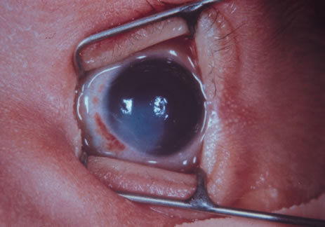
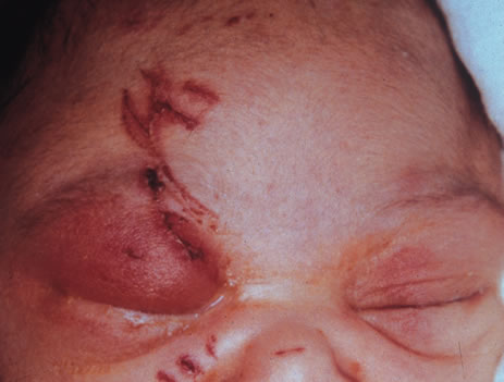
immunodeficiency syndrome. Am J Dis Child 140:638, 1986

161. Itoi M, Gefter JW, Kaneko N, et al: Teratogenicities of ophthalmic drugs. Part 1. Antiviral ophthalmic drugs. Arch Ophthalmol 93:46, 1975

162. Ballard PD, Hearnery EF, Smith MB: Induction of cleft palate in mice after ophthalmic administration of hydrocortisone. Toxicol Appl Pharmacol 34:358, 1975

163. Baden E: Environmental pathology of the teeth. In Gorlin RJ, Goldman HH (eds): Thomas' Oral Pathology, 6th ed. St. Louis, CV Mosby, 1970

164. Sullivan G, Takacs E: Comparative teratogenicity of pyrimethamine in rats and hamsters. Teratology 4:250, 1971

165. Sand BJ, Shanedling PD: Teratogenesis from Daraprim. Am J Ophthalmol 56:1011, 1963

166. Rogoyski A, Trzcinska-Dabrowska Z: Corticosteroid-induced cataract and palatoschisis in the mouse fetus. Am J Ophthalmol 68:128, 1969

167. Kasirsky G, Lombardi L: Comparative teratogenic study of various corticoid ophthalmics. Toxicol Appl Pharmacol 16:773, 1970

168. Holmes LB, Trelstad RL: The early limb deformity caused by acetazolamide. Teratology 20:289, 1979

169. Isenberg SJ: Ocular trauma. In Isenberg SJ (ed): The Eye in Infancy, p. 377. Chicago, Yearbook Medical Publishers, 1989

170. Sachs D, Levin PS, Dooley K: Marginal eyelid laceration at birth. Am J Ophthalmol 102:539, 1986

171. Harris GJ: Canalicular laceration at birth. Am J Ophthalmol 105:322, 1988

172. Isenberg SJ, Heckenlively JR: Traumatized eye with retinal damage from amniocentesis. J Pediatr Ophthalmol Strabismus 22:65, 1985

173. Thomas G, Blackwell RJ: A hazard associated with the use of spiral fetal scalp electrodes. Am J Obstet Gynecol 121:1118, 1975

174. Duke-Elder S, Mac Paul PA: Injuries, Part 1, Mechanical Injuries. In Duke-Elder S (ed): System of Ophthalmology, Vol. 14, p. 9. St. Louis, CV Mosby, 1972

175. Dutescu M, Lappe A: Luxation of the eyeball in the newborn. J Pediatr Ophthalmol 11:82, 1974

176. Hofmann RF, Paul TO, Pentelei-Molnar J: The management of corneal birth trauma. J Pediatr Ophthalmol Strabismus 18:45, 1981

177. Angell LK, Robb RM, Berson FG: Visual prognosis in patients with ruptures in Descemet's membrane due to forceps injuries. Arch Ophthalmol 99:2137, 1981

178. Stein RM, Cohen EJ, Calhoun JH, et al: Corneal birth trauma managed with a contact lens. Am J Ophthalmol 103:596, 1987

179. Spencer WH, Ferguson WJ, Shaffer RN, et al: Late degenerative changes in the cornea following breaks in Descemet's membrane. Trans Am Acad Ophthalmol Otolaryngol 70:973, 1966

180. Wu G, Behrens MM: Hyphema in the newborn: Report of a case. J Pediatr Ophthalmol Strabismus 19:56, 1982

181. Pulido JS, Lingua RW, Cristol S, et al: Protein C deficiency associated with vitreous hemorrhage in a neonate. Am J Ophthalmol 104:546, 1987

182. Duke-Elder S, Dobree JH: Diseases of the retina. In Duke-Elder S (ed): System of Ophthalmology, Vol. 10, p. 139. St. Louis, CV Mosby, 1967

183. Levin S, Janive J, Mintz M, et al: Diagnostic and prognostic value of retinal hemorrhages in the neonate. Obstet Gynecol 55:309, 1980

184. Chace RR, Merritt KK, Bellows M: Ocular findings in the newborn infant. Arch Ophthalmol 44:236, 1950

185. Von Barsewich B: Perinatal Retinal Hemorrhages. New York, Springer-Verlag, 1979

186. Rosenbaum AL, Phelps DL, Isenberg SJ, et al: Retinal hemorrhages in retinopathy of prematurity associated with tocopherol treatment. Ophthalmology 92:1012, 1985

187. Russell PA: Subdural haematoma in infancy. BMJ 2:446, 1965

188. Emerson MV, Pieramici DJ, Stoessel KM, et al: Incidence and rate of disappearance of retinal hemorrhage in newborns. Opthalmology. 108:36, 2001

189. Volpe JJ: Injuries of extracranial, cranial, intracranial, spinal cord, and peripheral nervous system structures. In VolpeJJ: Neurology of the Newborn, p. 638. Philadelphia, WB Saunders, 1987

190. Walsh TJ, Smith JL, Shipley T: Blindness in infants. Am J Ophthalmol 62:546, 1966

191. Falco NA, Eriksson E: Facial nerve palsy in the newborn: Incidence and outcome. Plast Reconstr Surg 85:1, 1990

192. Eng GD: Neuromuscular diseases. In Avery GB (ed): Neonatology: Pathophysiology and Management of the Newborn, 3rd ed, p. 1158. Philadelphia, JB Lippincott, 1987

193. Jaffe N, Cassady R, Peterson R, et al: Heterochromia and Horner syndrome associated with cervical and mediastinal neuroblastoma. J Pediatr 87:75, 1975

194. Reisner SH, Perlman M, Ben-Tovim N, et al: Transient lateral rectus muscle paresis in the newborn infant. J Pediatr 78:461, 1971

195. Crawford JS: Ptosis as a result of trauma. Can J Ophthalmol 9:244, 1974

196. Blechman B, Isenberg S: An anatomical etiology of congenital eyelid eversion. Ophthalmic Surg 15:111, 1984

197. Dubowitz LMS, Dubowitz V, Goldberg C: Clinical assessment of gestational age in the newborn infant. J Pediatr 77:1, 1970

198. Ballard JL, Novak KK, Driver M: A simplified score for assessment of fetal maturation of newly born infants. J Pediatr 95:769, 1979

199. Hittner HM, Hirsch NJ, Rudolph AJ: Assessment of gestational age by examination of the anterior vascular capsule of the lens. J Pediatr 91:455, 1977

200. Hittner HM, German WA, Rudolph AJ: Examination of the anterior vascular capsule of the lens, Part II. Assessment of gestational age in infants small for gestational age. J Pediatr Ophthalmol Strabismus 18:52, 1981

201. Mimouni F, Nissenkorn I, Wilunski E, et al: Assessment of gestational age by examination of the anterior vascular capsule of the lens: Value in multiple pregnancy (quintuplets). J Pediatr Ophthalmol Strabismus 20:27, 1983

202. Hittner HM, Speer ME, Rudolph AJ: Examination of the anterior vascular capsule of the lens. Part III. Abnormalities in infants with congenital infection. J Pediatr Ophthalmol Strabismus 18:55, 1981

203. Hiles DA, Wallar PH, MacFarlane F: Current concepts in the management of strabismus in children with cerebral palsy. Ann Ophthalmol 7:789, 1975

204. Cats BP, Tan KEWP: Prematures with and without regressed retinopathy of prematurity: Comparison of long-term (6–10 years) ophthalmological morbidity. J Pediatr Ophthalmol Strabismus 26:271, 1989

205. Alden ER, Kalina RE, Hodson WA: Transient cataracts in low birth weight infants. J Pediatr 82:314, 1973

206. Gruenwald P: Infants of low birth weight among 5,000 deliveries. Pediatrics 34:15, 1964

207. Mujsce DJ, Palmer C: Common neonatal illnesses. In HoekelmanRA, editor: Primary Pediatric Care, p. 597. Philadelphia, Mosby, 2001

208. AmericanAcademy of Pediatrics, Prevention of neonatal opthalmia. In Pickering LK (ed): 2000 Red Book: Report of the Committee on Infectious Diseases, 25th ed, p. 741. Elk Grove Village, IL, American Academy of Pediatrics, 2000

209. Crede R: Reports from the obstetrical clinic in Leipzig. Prevention of eye inflammation in the newborn. Am J Dis Child 121:3, 1971

210. Nishida H, Risemberg HM: Silver nitrate opthalmic solution and chemical conjunctivitis. Pediatrics 56:368, 1975

211. Isenberg SJ, Apt L, Wood M: A controlled trial of povidone-iodine as prophylaxis against opthalmia neonatorum. N Engl J Med 332:562, 1995

212. Alexander ER, Harrison HR: Role of Chlamydia trachomatis in perinatal infection. Rev Infect Dis 5:713, 1983

213. Weiss SG, Newcomb RW, Beem MO: Pulmonary assessment of children after chlamydial pneumonia of infancy. J Pediatr 108:659, 1986

214. Hammerschlag MR: Chlamydial infections. J Pediatr 114:727, 1989

215. Rosales T, Leake R, Isenberg S, et al: Systematic effects of mydriatics in lower-weight infants. J Pediatr Opthalmol Strabismus 18:42, 1981

216. Isenberg S, Everett S: Cardiovascular effects of myodriatics in low-birth-weight infants. J Pediatr 105:111, 1984

217. Mirmanesh SJ, Abbasi S, Bhutani VK: Alpha-adrenergic bronchoprovocation in neonates with bronchopulmonary dysplasi. J Pediatr 121:622, 1992

218. Isenberg SJ, Abrams C, Hymen PE: Effects of cyclopentliate eyedrops on gastric secretory function in pre-term infants. Opthalmology 92:698, 1985

219. Wallace DK, Steinkuller PG: Ocular medications in children. Clin Pediatr 37:645, 1998

220. Oski FA: Vitamin E: A radical defense. N Engl J Med 303:454, 1980

221. Bieri JG, Corash L, Hubbard VS: Medical uses of vitamin E. N Engl J Med 308:1063, 1983

222. Lemons JA, Maisels MJ: Vitamin E: How much is too much?Pediatrics 76:625, 1985

223. Zipursky A, Brown EJ, Watts J, et al: Oral vitamin E supplementation for the prevention of anemia in premature infants: A controlled trial. Pediatrics 79:61, 1987

224. Kushner BJ, Essner D, CohenIL , et al: Retrolental fibroplasia. Part II. Pathologic correlation. Arch Ophthalmol 95:29, 1977

225. Ashton N: Oxygen and the retinal blood vessels. Trans Ophthal Soc UK 100:359, 1980

226. Hittner HM, Rudolph AJ, Kretzer FL: Suppression of severe retinopathy of prematurity with vitamin E supplementation: Ultrastructural mechanism of clinical efficacy. Ophthalmology 91:1512, 1984

227. Johnson L: Retrolental fibroplasia. A new look at an unsolved problem. Hosp Pract 16:109, 1981

228. Johnson L, Quinn GE, Abbas S, et al: Effect of sustained pharmacologic vitamin E levels on incidence and severity of retinopathy of prematurity: A controlled clinical trial. J Pediatr 114:827, 1989

229. Johnson L, Quinn GE, Abbasi S, et al: Severe retinopathy of prematurity in infants with birth weights less than 1250 grams: Incidence and outcome of treatment with pharmacologic serum levels of vitamin E in addition to cryotherapy from 1985 to 1991. J Pediatr 127:632, 1995

230. Raju TN, Langenberg P, Bhutani V, et al: Vitamin E prophylaxis to reduce retinopathy of prematurity: a reappraisal of published trails. J Pediatr 131:844, 1997

231. Law MR, Wijewardene K, Wald NJ: Is routine vitamin E administration justified in very low-birthweight infants. Dev Med Child Neurol 32:442, 1990

232. Schaffer DB, Johnson L, Quinn GE, et al: Vitamin E and retinopathy of prematurity: Follow-up at one year. Ophthalmology 92:1005, 1985

233. Wender DF, Thulin GE, Wolken Smith GJ, et al: Vitamin E affects lung biochemical and morphologic response to hyperoxia in the newborn rabbit. Pediatr Res 15:262, 1981

234. Saldanha RL, Cepeda EE, Poland RL: The effect of vitamin E prophylaxis on the incidence and severity of bronchopulmonary dysplasia. J Pediatr 101:89, 1982

235. Arrowsmith JB, Faich GA, Tomita DK, et al: Morbidity and mortality among low birth weight infants exposed to an intravenous vitamin E product, E-ferol. Pediatrics 83:244, 1989

236. Neal PR, Erickson P, Baenziger JC, et al: Serum vitamin E levels in the very low birth weight infant during oral supplementation. Pediatrics 77:636, 1986

237. Ehrenkranz RA: Vitamin E and retinopathy of prematurity: Still controversial. J Pediatr 114:801, 1989

238. Cremer RJ, Perryman PW, Richards DH: Influence of light on the hyperbilirubinemia of infants. Lancet 1:1094, 1958

239. Brown AK, Kim MH, Wu PYK, et al: Efficacy of phototherapy in prevention and management of neonatal hyperbilirubinemia. Pediatrics 75:393, 1985

240. Ennever JF, Knox I, Denne SC, et al: Phototherapy for neonatal jaundice. In vivo clearance of bilirubin photoproducts. Pediatr Res 19:205, 1985

241. Landry RJ, Schedt PC, Hammond RW: Ambient light and phototherapy conditions of eight neonatal care units: A summary report. Pediatrics 75:393, 1985

242. Rubaltelli FF, Zanardo V,0Granati B: Effect of various phototherapy regimens on bilirubin decrement. Pediatrics 61:838, 1978

243. Tan KL: The pattern of bilirubin response to phototherapy for neonatal hyperbilirubinemia. Pediatr Res 16:670, 1982

244. Dobson V: Phototherapy retinal damage. Invest Ophthalmol 15:595, 1976

245. Messner KH, Maisels MJ, Loure-DuPree AE: Phototoxicity to the newborn primate retina. Invest Ophthalmol Vis Sci 17:1178, 1978

246. Messner KH: Light toxicity to newborn retina. Pediatr Res I2:530, 1978

247. Sykes SM, Robison WG, Waxier M, et al: Damage to the monkey retina by broad-spectrum fluorescent light. Invest Ophthalmol Vis Sci 20:425, 1981

248. Kalina RE, Forrest GL: Ocular hazards of phototherapy for hyperbilirubinemia. J Pediatr Ophthalmol 8:116, 1971

249. Dobson V, Cowett RM, Riggs LA: Long-term effects of phototherapy on visual function. J Pediatr 86:555, 1975

250. Bhupathy K, Sethupathy R, Pildes RS, et al: Electroretinography in neonates treated with phototherapy. Pediatrics 61:189, 1978

251. Dobson V, Riggs L, Siqueland ER: Electroretinographic determination of dark adaptation functions of children exposed to phototherapy as infants. J Pediatr 85:25, 1974

252. Sherman SM: Development of interocular alignment in cats. Brain Res 37:187, 1972

253. Cyander M, Berman N, Hein A: Recovery of function in cat visual cortex following prolonged deprivation. Exp Brain Res 25:139, 1976

254. Cyander M: Interocular alignment following visual deprivation in the cat. Invest Ophthalmol Vis Sci 18:726, 1979

255. Weisel TN, Hubel OH: Comparison of the effects of unilateral and bilateral eye closure on cortical unit responses in kittens. J Neurophysiol 28:1029, 1965

256. Hubel DH: The visual cortex of normal and deprived monkeys. Am Sci 67:532, 1979

257. Drew JH: Phototherapy: Short- and long-term complications. Arch Dis Child 51:454, 1976

258. Hoyt CS: The long-term visual effects of short-term binocular occlusion of at-risk neonates. Arch Ophthalmol 98:1967, 1980

259. Hubel DH, Wiesel TN: The period of susceptibility to the physiological effects of unilateral eye closure in kittens. J Physiol 206:419, 1970

260. von Noorden GK: New clinical aspects of stimulus deprivation amblyopia. Am J Ophthalmol 92:416, 1981

261. Larsen JS: The sagittal growth of the eye. Part IV. Ultrasonic measurement of the axial length of the eye from birth to puberty. Acta Ophthalmol 49:873, 1971

262. Gordon RA, Donzis P: Refractive development of the human eye. Arch Ophthalmol 103:785, 1985

263. Swan K, Wilkins JH: Extraocular muscle surgery in early infancy: Anatomical factors. J Pediatr Ophthalmol Strabismus 21:44, 1984

264. Blomdahl S: Ultrasonic measurements of the eye in the newborn infant. Acta Ophthalmol 57:1048, 1979

265. Sorsby A, Sheridan M: The eye at birth: Measurement of the principal diameters in forty-eight cadavers. J Anat 94:192, 1960

266. Inagaki Y: The rapid change of corneal curvature in the neonatal period and infancy. Arch Ophthalmol 104:1026, 1986

267. Donzis PB, Insler MS, Gordon RA: Corneal curvature in premature infants. Am J Ophthalmol 99:213, 1985

268. Horsten GPM, Winkelman JE: Electrical activity of the retina in relation to histological differentiation in infants born prematurely and at full term. Vision Res 2:269, 1962

269. Mann I: The Development of the Human Eye, p. 114. London, Cambridge University Press, 1928

270. Isenberg SJ: Macular development in the premature infant. Am J Ophthalmol 101:74, 1986

271. Hendrickson AE, Yuodelis C: The morphological development of the human fovea. Ophthalmology 91:603, 1984

272. Hickey TL: Postnatal development of the human lateral geniculate nucleus: Relationship to a critical period for the visual system. Science 198:836, 1977

273. Magoon EH, Robb RM: Development of myelin in human optic nerve and tract. Invest Ophthalmol Vis Sci 19:3, 1980

274. Conel J: The Postnatal Development of Human Cerebral Cortex, Vol. I, The Cortex of the Newborn. Cambridge, Harvard University Press, 1939

275. Ingram RM: Refraction of 1-year-old children after atropine cycloplegia. Br J Ophthalmol 63:343, 1979

276. Banks MS: Infant refraction and accommodation. Int Ophthalmol Clin 20:205, 1980

277. Glickstein M, Millodot M: Retinoscopy and eye size. Science 168:605, 1970

278. Harter MR, Deaton FK, Odom JV: Maturation of evoked potentials and visual preference in 6–45 day old infants: Effects of check size, visual acuity and refractive error. Electroencephalogr Clin Neurophysiol 42:595, 1977

279. Scharf J, Zonis S, Zeltzer M: Refraction in Israeli premature babies. J Pediatr Ophthalmol 12:193, 1975

280. Scharf J, Zonis S, Zeltzer M: Refraction in premature babies: A prospective study. J Pediatr Ophthalmol 15:48, 1978

281. Fulton AB, Dobson V, Salem D, et al: Cycloplegic refractions in infants and young children. Am J Ophthalmol 90:239, 1980

282. Sorsby A, Sheridan M, Leary GA, et al: Vision, visual acuity, and ocular refraction of young men: Findings in a sample of 1,033 subjects. BMJ 1:1394, 1960

283. Mohindra I, Held R, Gwiazda J, et al: Astigmatism in infants. Science 202:329, 1980

284. Howland HC, Sayles N: Photorefractive measurements of astigmatism in infants and young children. Invest Ophthalmol Vis Sci 25:93, 1984

285. Hsu-Winges C, Hamer RD, Norcia AM, et al: Polaroid photorefractive screening of infants. J Pediatr Ophthalmol Strabismus 26:254, 1989

286. Mitchell DE, Freeman RD, Millodot M, et al: Meridional amblyopia: Evidence for modification of the human visual system by early experience. Vision Res 13:535, 1973

287. Dobson V, Fulton AB, Manning K, et al: Cycloplegic refractions of premature infants. Am J Ophthalmol 91:490, 1981

288. Shapiro A, Yanko L, Nawratzki I, et al: Refractive power of premature children at infancy and early childhood. Am J Ophthalmol 90:234, 1980

289. Nissenkorn I, Yassur Y, Mashkowski D, et al: Myopia in premature babies with and without retinopathy of prematurity. Br J Ophthalmol 67:170, 1983

290. Gwiazda J, Scheiman M, Mohindra I, et al: Astigmatism in children: Changes in axis and amount from birth to six years. Invest Ophthalmol Vis Sci 25:88, 1984

291. Rutstein RP, Wesson MD, Gotlieb S, et al: Clinical comparison of the visual parameters in infants with intrauterine growth retardation vs. infants with normal birth weight. Am J Optom Physiol Opt 63:697, 1986

292. Banks MS: The development of visual accommodation during early infancy. Child Dev 51:646, 1980

293. Braddick O, Atkinson J, French J, et al: A photorefractive study of infant accommodation. Vision Res 19:1319, 1979

294. Haynes HM, White BL, Held R: Visual accommodation in human infants. Science 148:528, 1965

295. Powers MK, Dobson V: Effect of focus on visual acuity of human infants. Vision Res 22:521, 1982

296. Atkinson J, Braddick O, Moar K: Development of contrast sensitivity over the first three months of life in the human infant. Vision Res 17:1037, 1977

297. Salapetek P, Bechtold AG, Bushnell EW: Infant visual acuity as a function of viewing distance. Child Dev 47: 860, 1976

298. Aslin RN, Jackson RW: Accommodative-convergence in young infants: Development of a synergistic sensory motor system. Can J Psychol 33:222, 1979

299. Hoyt CS, Nickel BL, Billston FA: Ophthalmological examination of the infant: Developmental aspects. Surv Ophthalmol 26:177, 1982

300. Isenberg SJ, Dang Y, Jotlerand V: The pupils of term and preterm infants. Am J Ophthalmol 108:75, 1989

301. Isenberg SJ, Molarte A, Vazquez M: The fixed and dilated pupils of premature neonates. Am J Ophthalmol 110:168, 1990

302. Frank JW, Kushner BJ, France T: Paradoxic pupillary phenomena: A review of patients with pupillary constriction to darkness. Arch Ophthalmol 106:1564, 1988

303. Robinson RJ: Assessment of gestational age by neurological examination. Arch Dis Child 41:437, 1966

304. Nelson LB, Rubin SE, Wagner RS, et al: Developmental aspects in the assessment of visual function in young children. Pediatrics 73:375, 1984

305. Goren CC, Sarty M, Wu PYK: Visual following and pattern discrimination of facelike stimuli by newborn infants. Pediatrics 56:544, 1975

306. Friendly DS: Visual acuity assessment of the preverbal patient. In Isenberg SJ (ed): The Eye in Infancy, pp. 48–56. Chicago, Year Book Medical Publishers, 1989

307. Aslin RN, Salapatek P: Saccadic localization of peripheral targets by the very young human infant. Percept Psychophys 17:293, 1975

308. Eviatar L, Miranda S, Eviatar A, et al: Development of nystagmus in response to vestibular stimulation in infants. Ann Neurol 5:508, 1979

309. Hoyt CS, Jastrzebski G, Marg E: Delayed visual maturation in infancy. Br J Ophthalmol 67:127, 1983

310. Mellor DH, Fielder AR: Dissociated visual development: Electrodiagnostic studies in infants who are “slow to see.”Dev Med Child Neurol 22:327, 1980

311. Kivlin JD, Bodnar A, Ralston CW, et al: The visually inattentive preterm infant. J Pediatr Ophthalmol Strabismus 27:190, 1990

312. Linksz A: Visual acuity in the newborn with notes on some objective methods to determine visual acuity. Doc Ophthalmol 34:259, 1973

313. Dobson V, Teller DY: Visual acuity in human infants: Physiological studies. Vision Res 18:1469, 1978

314. Fulton AB, Hansen RM, Manning KA: Measuring visual acuity in infants. Surv Ophthalmol 25:325, 1981

315. Dobson V: Behavioral tests of visual acuity in infants. Int Ophthalmol Clin 20:233, 1980

316. Sokol S: Pattern visual evoked potentials: Their use in pediatric ophthalmology. Int Ophthalmol Clin 20:251, 1980

317. Teller DY, McDonald M, Preston K, et al: Assessment of visual acuity in infants and children: The acuity card procedure. Dev Med Child Neurol 28:779, 1986

318. Westall CA, Ainsworth JR, Buncic JR: Which ocular and neurologic conditions cause disparate results in visual acuity scores recorded with visually evoked potential and teller acuity cards?J AAPOS 4:295, 2000.

319. Norcia AM, Tyler CW: Spatial frequency sweep VEP: Visual acuity during the first year of life. Vision Res 25:1399, 1985

320. Harter MR, Suitt CD: Visually-evoked cortical responses and pattern vision in the infant: A longitudinal study. Psychon Sci 18:235, 1970

321. Marg E, Freeman DN, Peltzman P, et al: Visual acuity development in human infants: Evoked potential measurements. Invest Ophthalmol 15:150, 1976

322. Sokol S, Dobson V: Pattern reversal visually evoked potentials. Invest Ophthalmol 15:58, 1976

323. Sokol S: Measurement of infant visual acuity from pattern reversal evoked potentials. Vision Res 18:33, 1978

324. Dayton GO, Jones MH, Aiu P, et al: Developmental study of coordinated eye movements in the human infant. Part I. Visual acuity in the newborn human: A study based on induced optokinetic nystagmus recorded by electro-oculography. Arch Ophthalmol 71:865, 1964

325. Gorman JJ, Cogan DO, Gellis SS: An apparatus for grading the visual acuity on the basis of optico-kinetic nystagmus. Pediatrics 19:1088, 1957

326. Gorman JJ, Cogan DG, Gellis SS: A device for testing visual acuity in infants. Sight Sav Rev 29:80, 1959

327. Kiff RD, Lepard C: Visual responses of premature infants. Arch Ophthalmol 75:631, 1966

328. Frantz RL, Ordy JM, Udelf MS: Maturation of pattern vision in infants during the first six months. J Comp Physiol Psychol 55:907, 1962

329. Berlyne DE: The influence of the albedo and complexity of stimuli on visual fixation in the human infant. Br J Psychol 49:315, 1958

330. Frantz RL: Pattern vision in young infants. Psychol Rec 8:43, 1958

331. Teller DY, Morse R, Borton R, et al: Visual acuity for vertical and diagonal gratings in human infants. Vision Res 14:1433, 1974

332. Mayer DL, Dobson V: Assessment of vision in young children: A new operant approach yields estimates of acuity. Invest Ophthalmol Vis Sci 19:566, 1980

333. Frantz RL, Fagan JF, Miranda SB: Early visual selectivity as a function of pattern variables, previous exposure, age from birth and conception, and expected cognitive deficit. In Cohen LB, Salapatek P (eds): Infant Perception: From Sensation To Cognition, Vol. I. Basic Visual Process, p. 249. New York, Academic Press, 1975

334. Mayer DL, Fulton AB, Rodier D: Grating and recognition acuities of pediatric patients. Ophthalmology 91:947, 1984

335. Friendly DS, Jaafar MS, Morillo DL: A comparative study of grating and recognition visual acuity testing in children with anisometropic amblyopia without strabismus. Am J Ophthalmol 110:293, 1990

336. Frantz RL, Miranda SB: Newborn infant attention to form of contour. Child Dev 46:224, 1975

337. Slater A, Sykes M: Newborn infants' visual responses to square wave gratings. Child Dev 48:545, 1977

338. Grelotti DJ, Gauthier I, Schultz RT: Social interest and the development of cortical face specialization: What autism teaches us about face processing. Dev Psychobiol 40:213, 2002

339. Ballantyne AO, Trauner DA: Facial recognition in children after perinatal stroke. Neuropsychiatry Neuropsychol Behav Neurol 12:82, 1999

340. Nachson I: On the modularity of face recognition: The riddle of domain specificity. J Clin Exp Neuropsychol 17:256, 1995

341. Milewski AE: Infants' discrimination of internal and external pattern elements. J Exp Child Psychol 22:229, 1976

342. Maurer D, Lewis TL: A physiological explanation of infants' visual development. Can J Psychol 33:232, 1979

343. Bower TGR: The object in the world of the infant. Sci Am 225:30, 1971

344. Dodwell PC, Muri DW, DiFranco D: Infant perception of visually presented objects. Science 203:1138, 1979

345. Dodwell PC, Muir DW, DiFranco D: Responses of infants to visually presented objects. Science 194:209, 1976

346. Bower TGR, Dunkeld J, Wishart JG: Infant perception of visually presented objects. Science 203:1137, 1979

347. Aslin RN, Dumais ST: Binocular vision in infants: A review and theoretical framework. Adv Child Dev Behav 15:54, 1980

348. Aslin RN: Development of binocular fixation in human infants. J Exp Child Psychol 23:133, 1977

349. Fox R, Aslin RN, Shea SL, et al: Stereopsis in human infants. Science 207:323, 1980

350. Archer SM, Helveston EM, Miller KK, et al: Stereopsis in normal infants and infants with congenital esotropia. Am J Ophthalmol 101:591, 1986

Back to Top