Chapter 34
Metastatic Cancer to the Eye and Adnexa
ERIC P. SHAKIN and JERRY A. SHIELDS
Main Menu   Table Of Contents

Search

INCIDENCE
LOCATION OF METASTASIS
PRIMARY MALIGNANCY
METASTATIC TUMORS TO THE INTRAOCULAR STRUCTURES
CANCER METASTATIC TO THE CONJUNCTIVA AND SCLERA
CANCER METASTATIC TO THE EYELID
CANCER METASTATIC TO THE ORBIT
PARANEOPLASTIC SYNDROMES
EVALUATION
MANAGEMENT
PROGNOSIS
SUMMARY
REFERENCES

Cancer remains the second leading cause of death among adults in the United States. The annual incidence of cancer is 1.2 million cases/year, and the death rate is about 0.5 million cases/year.1 The most commonly encountered malignancies include gastrointestinal cancer, breast cancer, and lung cancer. The eye and adnexa may be involved by either metastasis (dissemination of a distant primary malignancy) or a secondary malignancy that has invaded the eye and adnexa from the surrounding structures. Ocular metastases are usually spread to the eye and orbit by hematogenous dissemination.2 The eye and adnexa may also be involved indirectly by effects from paraneoplastic syndromes or toxicity from chemotherapy.
Back to Top
INCIDENCE
The prevalence of metastasis to the ocular region (in patients with known cancer) ranges from as little as 0.07% to as high as 27%3–10 in a series of patients with breast cancer. There was a 4%7 rate of cancer metastatic to the eye in an autopsy series of patients who died with systemic carcinoma. This suggests that there are more than 20,000 new cases of metastatic cancer to the eye each year. Therefore, metastatic cancer to the eye is the most common intraocular malignancy in comparison to other primary intraocular malignancies.8 Furthermore, prior studies have demonstrated that there is no prior history of cancer in about 1/3 of patients who presented with an intraocular metastatic tumor11 or in about ¼ of patients with orbital metastatic tumor.9,12
Back to Top
LOCATION OF METASTASIS
Cancer metastatic to the ocular region most commonly involves the uveal tract. The isolated metastasis to the eye/orbit ratio ranges from 6:1 to 9:1 in adults.9,10,12 Simultaneous metastasis to the orbit and uvea occurs in up to 2% to 9% of patients.10,11 Metastatic cancer to the eyelid is rare. Concomitant metastasis to the central nervous system is reported in 7%11 to 45%6 of patients with metastatic breast cancer to the ocular region.
Back to Top
PRIMARY MALIGNANCY
Most metastases to the eye are carcinoma, although sarcoma, lymphoproliferative malignancies, and melanoma have been reported to metastasize to the eye. The lymphoproliferative disorders are discussed in another section. The most common primary malignancies reported in most series include breast, lung, gastrointestinal, renal, skin,11,13 and prostate cancer3–12 (Table 1). The site of the gastrointestinal primary includes esophagus,11 stomach,11 colon,11 bile duct,11 liver,14 and pancreas.11 Other less common primary sites include carcinoid tumors,15 uveal melanoma,16 testicular cancer,17 uterine cancer, cervical cancer,18 ovarian cancer, choriocarcinoma, bladder cancer, adrenal gland cancer, osteogenic sarcoma, Ewing's sarcoma,19 fibrosarcoma, neuroblastoma,20 Wilms tumor, parotid gland adenocarcinoma,21 laryngeal cancer, Merkel cell carcinoma,22 and nasopharyngeal cancer.23 In various series, the site of the primary malignancy is unknown in 9% to 17% of cases. It should be emphasized that nearly 34% of patients with intraocular metastasis have no known history of cancer at the time of ocular diagnosis.11


TABLE 1. Location of Primary Malignancy for Intraocular and Orbital Tumors


Primary SiteNumber MenNumber WomenTotal (%)Number Orbital Tumors (%)
Breast2194196 (47)18 (51)
Lung553590 (22)2 (6)
Gastrointestinal13518 (4)2 (6)
Kidney819 (2)1 (3)
Skin549 (2)1 (3)
Prostate909 (2)6 (17)
Others51116 (4) 
Unknown403373 (17)6 (17)
Total13728342036
Data for intraocular tumors from Shields CL, Shields JA, Gross NE et al: Survey of 520 eyes with uveal metastases. Ophthalmology 1997;104:1265. Data from orbital tumors from Shields CL, Shields JA, Peggs M: Metastatic tumors to the orbit. Ophthalmic Plast Reconstr Surg 1988;4:73.


Back to Top
METASTATIC TUMORS TO THE INTRAOCULAR STRUCTURES
A retrospective study reviewed 570 patients with metastatic cancer to the eye referred to the oncology service at Wills Eye Hospital over a 20-year period.11 In this study, 90% were white and 67% were female. The average age at the time of diagnosis was 58 years. The right eye was involved in 48%, the left eye was involved 52%, and there was bilateral involvement in 24% of patients.

ANTERIOR UVEA

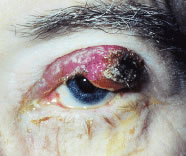
Cancer to the iris and ciliary body accounts for 4% to 11%10,11,24–26 of intraocular metastasis. Patients with cancer metastatic to the ciliary body and angle structures usually have associated injected epibulbar vessels and anterior uveitis. An iris metastasis is typically a yellow, gelatinous mass located within the iris stroma. Rarely, the iris tumor may be friable and may seed into the aqueous, producing a pseudohypopyon composed of tumor cells8 (Fig. 1). Other associated finding in those eyes with metastases to the anterior uvea include ocular hypertension, hyphema, uveitis, rubeosis, cataract, and infiltration into the surrounding structures.8,24–26 The differential diagnosis of cancer metastatic to the iris includes amelanotic melanoma, granulomatous uveitis, iris cysts, retained foreign body, and retinoblastoma in children.8,26

Fig. 1. Slit-lamp photograph of a patient with colon cancer metastatic to the iris. Note the pseudohypopyon composed of tumor cells seeding from the iris tumor.

CHOROID

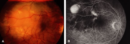
The choroid is the most common site of carcinoma metastatic to the eye (88% of eyes).6,7,10,11 Patients with choroidal metastasis usually complain of painless vision or visual field loss. About 47% of the choroidal lesions were temporal to the disc.11 Metastatic cancer to the eye was multifocal in the choroid in 28% of the eyes.11 Breast cancer was more commonly bilateral (33%) and multifocal (32%) compared with other primary neoplasms.11 There is an associated nonrhegmatogenous retinal detachment in 73% of the eyes.11 Carcinoma metastatic to the choroid is typically a placoid or dome-shaped yellow tumor of varying diameter and thickness. Multiple lesions are not infrequent (Fig. 2). The mean thickness in our series was 3 mm. Rarely a rip to the retinal pigment epithelium can develop.27 Regressed lesions are relatively flat with overlying pigment and lack of associated subretinal fluid. The differential diagnosis of choroidal metastases include amelanotic malignant melanoma, amelanotic nevus, choroidal hemangioma, choroidal osteoma, posterior scleritis, uveal effusion syndrome, Harada's disease, and central serous choroidopathy.28
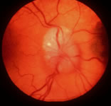
Fig. 2. Fundus photograph (A) of the left eye of a patient with breast cancer metastatic to the choroid. Several tumor nodules can be appreciated. Fluorescein angiography (B) reveals staining of the tumors in the full venous phase.

OPTIC DISC

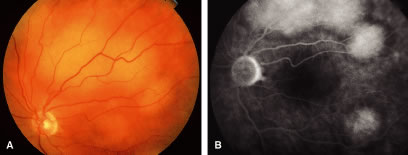
Metastatic cancer to the optic disc is rare (4.5% intraocular metastases).29 The optic disc is usually affected by direct extension of an adjacent choroidal metastatic tumor that infiltrates the optic disc (Fig. 3). Occasionally, tumor emboli enter the optic disc by hematogenous or intrathecal invasion (meningeal carcinomatosis). There is an adjacent juxtapapillary choroidal lesion in 74% of eyes.29 The associated findings include optic disc edema, venous stasis, an afferent pupillary defect, and less commonly hemorrhage or obstruction of a retinal artery or vein.8,29 The optic disc appears to be edematous, hyperemic, or atrophic in patients with meningeal carcinomatosis involving the optic nerve sheath.2,10,29 Most cases are from breast, lung, or an unknown primary malignancy. The differential diagnosis includes papilledema, optic neuritis, granuloma, optic disc drusen, and capillary hemangioma.8

Fig. 3. Fundus photograph of the right eye of a patient with breast cancer metastatic to the optic disc.

RETINA

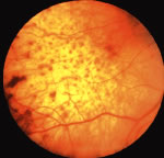
Metastatic cancer to the retina, which has rarely been reported,8,13 reaches the retina by either direct invasion from the adjacent choroid or by tumor emboli through the central retinal artery. The retinal tumors may appear as patchy yellow lesions associated with hemorrhage or exudate.

Sometimes there are overlying clumps of tumor cells in the vitreous such as in cases of cutaneous melanoma metastatic to the retina. The differential diagnosis includes cotton-wool spots, vascular obstructions, and retinitis.

VITREOUS

Metastatic cancer to the vitreous can be associated with metastasis to the retina or ciliary body.8,30 Clumps of tumor cells are visible in the vitreous. Cutaneous malignant melanoma and lymphoproliferative disorders such as large cell lymphoma are known to disseminate to the vitreous, although breast cancer and lung cancer have been associated with metastatic cells in the vitreous cavity. The differential diagnosis includes primary ocular lymphoma, vitritis, and other vitreous opacities.

Back to Top
CANCER METASTATIC TO THE CONJUNCTIVA AND SCLERA
Cancer metastatic to the conjunctiva is associated with intraocular metastasis in 1% of the eyes in our study.11 Cancers metastatic to the conjunctiva are fleshy vascular tumors8,31 (Fig. 4). Leukemia and lymphoma can also occur in the conjunctiva. The sclera was involved by direct extension of a choroidal metastasis in 11% of patients in one series.5

Fig. 4. Slit-lamp photograph of metastatic cancer to the conjunctiva. Note that this fleshy vascular tumor extends anterior to the limbus.

Back to Top
CANCER METASTATIC TO THE EYELID
Malignant tumors including the lymphoproliferative disorders metastatic to the eyelid account for 0.3% to 1.1% of eyelid neoplasms.32 Metastatic cancer to the eyelid can appear as a solitary nodule, as diffuse induration of the eyelids with infiltration of the local lymphatic drainage, or as an ulcerative lesion (Fig. 5). The tumor cells usually infiltrate the dermis or less commonly the conjunctival surface of the eyelid. Breast cancer, lung cancer, and cutaneous malignant melanoma are the most common primary malignancies to metastasize to the eyelid.32

Fig. 5. Photograph of a patient with breast carcinoma metastatic to the left upper eyelid.

Back to Top
CANCER METASTATIC TO THE ORBIT
The incidence of cancer metastatic to the orbit ranges from 2% to 9% of orbital neoplasms.9,10,12,33 Orbital metastases usually occur more commonly in women between the ages of 40 to 60 years. The presenting signs in patients with metastatic cancer to the orbit include proptosis or rarely enophthalmos; a painful, palpable mass; and ophthalmoplegia. Less common signs include vision loss, choroidal folds, or an afferent pupillary defect14 (Fig. 6). The reported primary malignancies includes breast cancer, lung cancer, gastrointestinal cancer, genitourinary tract cancers, thyroid cancer, adrenal cell cancer, and cutaneous malignant melanoma.9,10,12,33 We have reported a case of uveal malignant melanoma metastatic to the contralateral eye and orbit.16 Neuroblastoma,12,33 Ewing's sarcoma,19 and Wilms tumor12,33 have been known to metastasize to the orbit in children.

Fig. 6. Computerized axial tomography scan of head, axial section, in a patient with metastatic breast cancer to the right orbit.

Back to Top
PARANEOPLASTIC SYNDROMES
Cancer associated retinopathy (CAR syndrome, paraneoplastic retinopathy, visual paraneoplastic syndrome) was first described by Sawyer and coworkers.34 The syndrome is characterized by progressive photoreceptor degeneration with subsequent insidious vision loss over several months. The photoreceptor degeneration is a remote effect of the primary malignancy caused by a retinal toxic tumor cell product or more likely by an autoimmune response to a tumor antigen that cross-reacts with various cells in the retina such as retinal ganglion cells and photoreceptors.35–37 There are a variety of serum antibodies to retinal antigens detected in these patients. The protean symptoms include diminished visual acuity, visual obscurations, photopsia, nyctalopia, and scotoma. The associated visual field defects include ring scotoma, central scotoma, and generalized constriction of the isopters. The optic disc is usually normal. The retinal arterioles are attenuated or sheathed. Intravenous fluorescein angiography is characterized by a delay in the transit time and transmission window defects corresponding to areas of atrophy of the retinal pigment epithelium especially in a perivascular pattern. Sometimes there is associated vascular leakage. The electroretinogram and electro-oculogram are subnormal. This paraneoplastic syndrome is associated with small cell carcinoma of the lung, breast cancer, cutaneous malignant melanoma, and cervical cancer. The ocular condition may be accompanied by other neurologic, myopathic, or endocrinologic disorders. Melanoma-associated retinopathy38 is thought to be a distinct entity because of the rapid onset of nyctalopia and antibodies to retinal bipolar cells. Some cases have been associated with circumcised areas of retinal pigment epithelial (RPE) atrophy. Treatment with systemic steroids or intravenous immunoglobulins has been limited and disappointing.37,39

Bilateral diffuse uveal melanocytic proliferation (BDUMP), another paraneoplastic syndrome, is a proliferation of uveal melanocytes in response to a systemic malignancy such as ovarian, uterine, and lung cancer.40–42 Clinically, the tumors extend over large areas with a thickness of 3 to 5 mm, associated with an overlying retinal detachment. The lesions lack a p53 protein that is commonly associated with uveal malignant melanoma.41 This lends support to the theory that the lesions in BDUMP are not malignant.

Other paraneoplastic syndromes such as in the opsoclonus-myoclonus syndrome, brain stem encephalitis, or Lambert-Eaton syndrome can affect oculomotor function. Alterations of the pupil, uveitis, and optic neuritis have also been described.

Back to Top
EVALUATION
The evaluation of patients with suspected metastatic cancer to the eye or ocular adnexa should include a comprehensive systemic survey for metastatic disease, as well as a complete ocular evaluation. Other useful tests may include intravenous fluorescein angiography, ultrasonography, fine needle aspiration biopsy, and wedge biopsy. The fluorescein angiogram is useful in distinguishing neoplasms that are hyperfluorescent in contrast to metastatic lesions that are hypofluorescent in the arterial phase of the angiogram8 (Fig. 7). The ultrasound may reveal a lesion of medium reflectivity without choroidal excavation.8

Fig. 7. Fundus photograph of the right eye (A) of a patient with breast cancer metastatic to the choroid temporal to the macula and associated with overlying fluid. The actual tumor is better appreciated in the full venous phase of the fluorescein angiogram (B).

Biopsies are useful in combination with special immunohistochemical stains to help localize the primary malignancy.43Although magnetic resonance imaging (MRI), computed tomography (CT) scans, and positron emission tomography are not characteristic for these lesions, these imaging studies may reveal the extent of the tumors or other associated intracranial metastases.8,33

Back to Top
MANAGEMENT
Therapy for metastatic cancer to the eye and the adnexa is palliative. The indications for treatment include loss of vision by involvement or impending involvement of the fovea by the tumor or subretinal fluid; anterior chamber invasion; and orbital involvement associated with diplopia, pain, or optic neuropathy. If the metastatic tumor appears to be undergoing regression with chemotherapy (Fig. 8), then no other specific treatment is necessary. External beam radiotherapy is delivered to the entire posterior segment of the globe in a dose of 3000 to 4000 cGy over a 4- to 6-week period; regression occurs in 80% to 100% of patients.8,44

Fig. 8. Fundus photograph of the right eye of a patient with metastatic lung cancer to the choroid treated with radiotherapy. Note the characteristic pigment clumping (“leopard skin”) pattern.

Some solitary lesions have been treated with brachytherapy45 by a one-time application of an episcleral plaque. Transpupillary thermotherapy has also been described.46 Rarely localized resection for diagnostic purposes is performed. Rarely enucleation is necessary for intractable pain. Some authors have advocated the use of chemotherapy alone for the palliation of breast cancer metastatic to the eye.47 The adverse effects of treatment include radiation dermatitis, conjunctivitis, keratopathy, cataracts, retinopathy, uveitis, and tumor necrosis with hemorrhage. Ptosis and ophthalmoplegia occur in patients treated with vincristine. Cataracts are described in association with allopurinol, busulfan, 5-fluorouracil, methotrexate, and mitotane. Mitotane and tamoxifen are associated with retinopathy. Carmustine, tamoxifen, and vincristine may result in optic neuropathy. Interferon is associated with retinal and choroidal ischemia.48

Back to Top
PROGNOSIS
The prognosis for patients with metastatic cancer to the eye and orbit depends on the primary malignancy and the extent of metastatic disease in each patient. Generally the prognosis in patients with metastatic cancer to the eye or orbit is similar to a patient with any stage IV malignancy. Overall, the median survival from the time of ocular diagnosis to the time of death ranges from 7.4 to 10.5 months8,10,49 and 13 to 15.6 months9,33,49 in patients with metastasis to the eye and orbit, respectively. In one series, the 1-year survival rate was 57% for patients with breast cancer and 20% for patients with lung cancer metastatic to the eye.8
Back to Top
SUMMARY
Several cancers are known to metastasize to the eye and adjacent structures. Cancer metastatic to the uvea is the most common intraocular malignancy. About one third of patients with cancer metastatic to the uvea or orbit had no known prior history of malignancy. The tumors that most commonly disseminate to the uvea and orbit include breast cancer in women and lung cancer in men. The primary malignancy was not discovered in about 17% of patients with uveal metastasis.

The evaluation of patients should include a comprehensive systemic survey for the primary malignancy and metastatic disease and a complete ocular examination. Therapy for metastatic cancer to the eye and orbit is palliative. Many such tumors are controlled with chemotherapy alone, but others that are threatening vision may require radiotherapy. The long-term prognosis for survival is poor in many patients with cancer metastatic to the ocular region.

Back to Top
REFERENCES

1. Murphy GP, Lawrence W, Lenhard RE: Clinical Oncology. 4th ed. American Cancer Society, 1999

2. Albert DM, Zimmerman AW, Zeidman I: Tumor metastasis to the eye. III The fate of circulating tumor cells to the eye. Am J Ophthalmol 1967;63:733

3. Gotfredsen E: On the frequency of secondary carcinoma in the choroid. Acta Ophthalmol 1944;22:394

4. Albert DM, Rubenstein RA, Scheie HG: Tumor metastasis to the eye. Part I. Incidence in 213 adult patients with generalized malignancy. Am J Ophthalmol 1967;63:723

5. Bloch RS, Gartner S: The incidence of ocular metastatic carcinoma. Arch Ophthalmol 1971;85:673

6. Bullock JD, Yanes B: Ophthalmic manifestations of metastatic breast cancer. Ophthalmology 1980;87:961.

7. Nelson CC, Hertzberg BS, Klintworth GK: A histopathologic study of 716 unselected eyes in patients with cancer at the time of death. Am J Ophthalmol 1983;95:788

8. Shields JA, Shields CL: Intraocular Tumors: A Text and Atlas. Philadelphia: WB Saunders, 1992:207–238

9. Shields CL, Shields JA, Peggs M: Metastatic tumors to the orbit. Ophthalmic Plast Reconstr Surg 1988;4:73

10. Ferry AP, Font RL: Carcinoma metastatic to the eye and orbit Part I. A clinicopathologic study of 227 cases. Arch Ophthalmol 1974;92:276

11. Shields CL, Shields JA, Gross NE et al: Survey of 520 eyes with uveal metastases. Ophthalmology 1997;104:1265

12. Henderson JW. Orbital Tumors. Philadelphia: WB Saunders, 1973:44–494

13. Debustros S, Augsburger JJ, Shields JA et al: Intraocular metastasis from cutaneous malignant melanoma. Arch Ophthalmol 1985;103:937

14. Font RL, Maturi RK, Small RG et al: Hepatocellular carcinoma metastatic to the orbit. Arch Ophthalmol 1998;116:942

15. Fan JT, Buettner H, Bartley GB et al: Clinical features and treatment of seven patients with carcinoid tumors metastatic to the eye and orbit. Am J Ophthalmol 1994;119:211

16. Shields JA, Shields CL, Shakin EP et al: Metastasis of choroidal malignant melanoma to the contralateral choroid, orbit, and eyelid. Br J Ophthalmol 1988;72:456

17. Zech JC, Subiger L, Chiquet C et al: Testicular choriocarcinoma metastatic to the choroid. Retina 1999;19:164

18. Inoue K, Kaji NJ et al: Bilateral choroidal metastases secondary to uterocervical carcinoma of the squamous cell type. Am J Ophthalmol 2000;130:682

19. Dutton JJ, Rose JG, DeBacker CM et al: Ewing's sarcoma of the orbit. Ophthalmic Plast Reconstr Surg 2000;16:292

20. Weissgold DJ, Gragoudas ES et al: Choroidal metastasis from olfactory neuroblastoma. Retina 1999;19:459

21. Friedman SM, Margo CE, Stephenson JA: Choroidal metastasis from adenocarcinoma of the parotid gland. Retina 1999;19:457

22. Purgason PA, Hornblass A, Harrison W: Metastatic Merkel cell tumor to the eye. Ophthalmology 1991;98:1432

23. Sham JS, Choy D: Choroid metastasis from nasopharyngeal carcinoma. Eur J Surg Oncol 1991;17:384

24. Sierocki J, Charles N, Schafron KM et al: Carcinoma metastatic to the anterior ocular segment. Cancer 1980;45:2521

25. Ferry AP, Font RL: Carcinoma metastatic to the eye and orbit. A clinicopathologic correlation study of 26 patients with carcinoma metastatic to the anterior segment of the eye. Arch Ophthalmol 1975;93:472

26. Shields JA, Shields CL, Kiratli H et al: Metastatic tumors to the iris in 40 patients. Am J Ophthalmol 1995;119:422

27. Leys A: Choroidal metastasis and retinal pigment epithelial tear in a patient with small cell lung carcinoma. Retina 2000;20:216

28. Michelson JB, Stephens RF, Shields JA: Clinical conditions mistaken for metastatic cancer to the choroid. Ann Ophthalmol 1979;11:49

29. Shields JA, Shields CL, Singh AD: Metastatic neoplasms in the optic disc: The 1999 Bjerrum Lecture: Part 2. Arch Ophthalmol 2000;118:217

30. Gunduz K, Shields JA et al: Lung carcinoma metastatic to the vitreous cavity. Retina 1998;18:285

31. Kiratli H, Shields CL, Shields JA et al: Metastatic tumors to the conjunctiva: A report of 10 cases. Br J Ophthalmol 1996;80:5

32. Mansour AM, Hidayat AA: Metastatic eyelid disease. Ophthalmology 1987;94:667

33. Shields JA: Diagnosis and management of orbital tumors. Philadelphia: WB Saunders, 1989

34. Sawyer RA, Selhorst JB, Zimmerman LE et al: Blindness caused by photoreceptor degeneration as a remote effect of cancer. Am J Ophthalmol 1976;81:606

35. Keltner JA, Roth AM, Chang RS: Photoreceptor degeneration: Possible autoimmune disorder. Arch Ophthalmol 1983;101:564

36. Grunwald GB, Klein R, Simmonds MA et al: Autoimmune basis for visual paraneoplastic syndrome in patients with small cell lung carcinoma. Lancet 1985;3:658

37. Klingele TG, Burde RM, Rappazzo et al: Paraneoplastic retinopathy. J Clin Neuro-Ophthalmol 1984;4:239

38. Borkowski LM, Grover S, Fishman GA et al: Retinal findings in melanoma-associated retinopathy. Am J Ophthalmol 2001;132:273

39. Guy J, Aptsiauri N: Treatment of paraneoplastic visual loss with intravenous immunoglobulin. Arch Ophthalmol 1999;117:471

40. Barr CC, Zimmerman LE, Curtin VT et al: Bilateral diffuse melanocytic tumors associated with systemic malignant neoplasms. A recently recognized syndrome. Arch Ophthalmol 1982;100:249

41. Margo CE, Lowery RL, Kerschmann RL: Lack of p53 protein immunoreactivity in bilateral diffuse uveal melanocytic proliferation Retina 1997;17:434

42. Chahud F, Young RH, Remullah JF et al: Bilateral diffuse uveal melanocytic proliferation associated with extraocular cancer: Review of a process particularly associated with gynecologic cancers. Am J Surg Pathol 2001;25:212

43. Augsburber JJ, Shields JA: Fine needle aspiration biopsy of solid intraocular tumors: Techniques and instrumentation. Ophthalmic Surg 1984;15:34

44. Thatcher N, Thomas PRM: Choroidal metastasis from breast carcinoma. Clin Radiol 1975;26:549

45. Shields CL, Shields JA, DePotter P et al: Plaque radiotherapy in the management of uveal metastasis. Arch Ophthalmol 1997;115:203

46. Ardjomand N, Kucharczyk M, Langmannn G: Transpupillary therapy of choroidal metastases. Ophthalmologica 2001;215:241

47. Letson AD, Davidorf FH, Bruce RA: Chemotherapy for treatment of metastasis from breast carcinoma. Am J Ophthalmol 1982;93:102

48. Vizel M Oster MW: Ocular side effects of cancer chemotherapy. Cancer 1982;49:1999

49. Freedman MI, Folk JC: Metastatic tumors to the eye and orbit: Patient survival and clinical characteristics. Arch Ophthalmol 1987;107:1215

Back to Top