Chapter 32
Pregnancy and the Mother's Eye
JUSTIN C. BROWN and JANET S. SUNNESS
Main Menu   Table Of Contents

Search

EYELIDS
CONJUNCTIVA
CORNEA
INTRAOCULAR PRESSURE
LENS
EXTRAOCULAR MUSCLES
NEURO-OPHTHALMOLOGIC CHANGES
CONDITIONS ARISING DURING PREGNANCY
ORBIT
RETINAL AND CHOROIDAL DISORDERS ARISING IN PREGNANCY
OTHER RETINAL AND CHOROIDAL CHANGES DURING PREGNANCY
PREEXISTING CONDITIONS
OTHER RETINAL DISORDERS
RETINAL DIAGNOSTIC TESTING
OPHTHALMIC MEDICATIONS
REFERENCES

Pregnancy can have important effects on the normal physiology and pathophysiology of the mother's eye just as it affects other nonreproductive systems of the mother's body. Knowledge of the ocular changes that occur in association with pregnancy can help in the diagnosis and management of ocular disease in the pregnant woman. These changes also have led investigators to study the effects of various hormones on disease processes such as glaucoma.

This chapter presents information concerning the effects of pregnancy on the mother's eye. The chapter is organized into four sections. The first presents the “normal” or “physiologic” alterations seen in pregnancy in healthy eyes. This is followed by information related to ocular disease processes arising in pregnancy such as pre-eclampsia. The third section deal with the effects of pregnancy on pre-existing eye disease. These categories may overlap somewhat. For example, a pituitary microadenoma that was asymptomatic prior to pregnancy may worsen and only become symptomatic during pregnancy. Finally, information concerning the use of ophthalmic pharmacologic agents during pregnancy is discussed.

Back to Top
EYELIDS
Skin changes are a frequent and polymorphous feature of pregnancy. Chloasma is a blotchy brown discoloration that may occur around the eyelids and usually fades postpartum. Hormonal factors may play a role, either elevated levels of melanocyte stimulating hormone or estrogen and progesterone.1 Spider angiomas are common during pregnancy and can occur on the eyelids, perhaps related to high estrogen levels.1 Ptosis has been reported during and following normal pregnancies, with one patient worsening after each of three pregnancies.2–3 Most cases were unilateral. The authors suggest that the ptosis is caused by an aponeurosis defect, possibly related to increased fluid retention, hormonal effects, or the effects of labor and delivery. One study of skin changes in 60 pregnant women reported eyelid edema in three cases.4
Back to Top
CONJUNCTIVA
Changes in conjunctival blood vessels have been described toward the end of normal pregnancies. One study described granularity of the conjunctival venules, mild spasm of conjunctival arterioles, and decreased visualization of conjunctival capillaries.5 Another study reported increased vessel diameter in the second half of pregnancy.6 The conjunctival epithelium has been shown to undergo cytologic changes during pregnancy related to elevated estrogen levels.7 Vomiting during pregnancy can cause eyelid petechiae and subconjunctival hemorrhages. An increase in conjunctival pigmentation can be associated with normal pregnancy.
Back to Top
CORNEA
Corneal sensitivity has been found to be decreased in most pregnant women tested,8,9 although one study found no change in corneal sensitivity.10 The sensitivity returns to normal by 2 months postpartum.8 The corneal sensitivity change does not appear to be related to the amount of increase of corneal thickness or weight gain.9

Corneal thickness has been reported to increase during pregnancy with resolution a short time after delivery in three studies.8,11,12 The amount of increase found varied from 1 micron11 to 16 microns.12 The change appears to be present throughout pregnancy.12 Two studies found no change in corneal thickness.10,23 Possible causes of increased corneal thickness include fluid retention that is often associated with pregnancy.12

The corneal curvature has been found to increase (steepen) by one diopter on average in the second half of pregnancy, with resolution postpartum or after cessation of breastfeeding.10 However, the manifest refraction did not change in this study. A second study showed no change in corneal curvature during pregnancy.23

The changes in corneal thickness and curvature may be factors in pregnant women developing difficulty with previously comfortable contact lenses.8,10,13–15 Estimates in the literature are that 25% to 30% of contact lens wearers develop problems during pregnancy.10,15 A woman may develop contact lens intolerance during one pregnancy but not during another.10 It is not reported whether the symptoms correlate with the degree of corneal curvature and corneal thickness change. A decrease in tear production was found during the third trimester of pregnancy in about 80% of pregnant women studied.15 Other changes such as conjunctival modification and lid edema also may be factors leading to discomfort with contact lens wear.15 It is preferable to avoid prescribing a new correction for patients until several weeks postpartum if this is possible.

There is some weak evidence suggesting that pregnancy may alter corneal immune function. One case report describes bilateral herpes simplex virus (HSV) keratitis in a pregnant contact lens wearer. The authors suggest that pregnancy-associated immunosuppression and contact lens wear may have an additive effect and increase the possibility of HSV recurrence.16 A retrospective study of 3,608 corneal grafts found that pregnancy was a statistically significant univariate risk factor for graft failure.17 This suggests that pregnancy might be a risk factor for graft rejection in some patients.

One report suggests that photorefractive keratectomy (PRK) near or during the time of pregnancy may lead to suboptimal outcomes.18 Another study showed myopic regression in 12/18 (66%) and corneal haze in 10/12 (83%) of PRK eyes during pregnancy. The corneal haze and myopic regression improved in 50% of eyes after delivery.19 A third study failed to demonstrate impaired PRK outcomes after pregnancy.20

Krukenberg spindles are found more commonly in pregnant women, unassociated with other evidence of pigmentary dispersion. The spindles become smaller late in pregnancy and postpartum. It has been hypothesized that the development of Krukenberg spindles may be related to hormonal changes, and that resolution late in pregnancy relates to increased facility of outflow.21

There is one case report of a woman with bilateral keratoconus who developed corneal perforation late in pregnancy.22 The authors suggest a possible role of pregnancy in the development of perforation although this finding may be incidental.

One cross-sectional study compared 38 nonpregnant to 93 pregnant women and found no differences in refractive error, corneal curvature, corneal thickness, accommodative amplitude, AC/A ratio, and fusion break ranges between the two groups.23

Back to Top
INTRAOCULAR PRESSURE
Studies have found about a 20% decrease in intraocular pressure during pregnancy.24–30 This ocular hypotensive effect seems to increase until delivery26–30 and may persist for several months postpartum.31 A greater reduction of intraocular pressure was noted among pregnant women with baseline ocular hypertension in one study.27 Another investigation showed that the reduction in intraocular pressure was greater among multigravida than primigravida women.25

Two studies have demonstrated that intraocular pressure is higher in the peripartum period for women with third trimester hypertension or pre-eclampsia than in normotensive pregnant women.29,32 A third study failed to corroborate this association.24

Several etiologies for pregnancy-related reduction in intraocular pressure have been proposed. There is an increased facility of outflow33–36 and increased uveoscleral outflow,24 possibly related to hormonal effects. Changes in progesterone levels during pregnancy may correlate with changes in intraocular pressure.37 There may be decreased episcleral venous pressure,38,39 consistent with a general decrease of venous pressure in the upper extremities during pregnancy. Finally, there is also a small change in ocular rigidity during pregnancy.40 Aqueous production appears to remain constant during pregnancy.36,37

One case of acute angle closure glaucoma during labor has been reported. However, the patient was later believed to have had unrecognized subacute attacks prior to pregnancy.41

Back to Top
LENS
The curvature of the crystalline lens has been reported to be increased during pregnancy.42 A transient loss of accommodation has been reported during and after pregnancy.43 However, a more recent study failed to demonstrate any change in accommodative amplitude during pregnancy.23

An increase in lens autofluorescence has been reported in pregnant patients with diabetes compared with nonpregnant diabetic patients.44 This observation suggests the possibility of lenticular metabolic alterations during pregnancy.

A recent case-control study from central India45 showed that having more than three babies doubled the risk of developing bilateral cataracts in the 35- to 45-year age range. The extent to which this is related to nutritional status is not clear.

Back to Top
EXTRAOCULAR MUSCLES
Patients can present with strabismus during pregnancy. A search for pre-existing underlying conditions may help to clarify the diagnosis. For example, three otherwise healthy pregnant women developed a superior oblique palsy during pregnancy. On evaluation, they had amblyopia and decreased stereopsis, suggesting that there may have been decompensation of a latent vertical deviation.46 One healthy patient developed a transient sixth nerve palsy during pregnancy, unassociated with other changes (C. Rommel, personal communication, 1991).

Patients with hyperemesis gravidarum (pernicious vomiting in pregnancy) can develop thiamine deficiency, leading to Wernicke's encephalopathy with nystagmus and extraocular muscle palsies.47–53

Back to Top
NEURO-OPHTHALMOLOGIC CHANGES

VISUAL FIELD

A number of different studies of visual fields during pregnancy were performed in the first half of this century, motivated by findings of pituitary enlargement during pregnancy.54 Some studies found no change in most pregnant women,55 some found concentric constriction,56 and some found bitemporal constriction.57–59 These early studies found changes most prominently late in the pregnancy that generally resolved by 2 weeks postpartum. Patient fatigue and nonuniform equipment and illumination may have been factors in these findings.55 It is important to know that none of these healthy women was symptomatic in terms of visual field change.

No recent studies of visual fields in normal pregnant women have been performed. It is felt that the increase in size of the pituitary gland during pregnancy is not sufficient to cause visual field change except when there is an unusual position of the pituitary relative to the optic chiasm.54

A complaint of visual field change in a pregnant woman should not be dismissed. Rather, it should be considered an indication for evaluation for a possible disease process such as an enlarging pituitary adenoma, meningioma, or lymphocytic hypophysitis (discussed in the following).

NORMAL PITUITARY

It was reported as early as 1909 that the normal pituitary gland undergoes enlargement during pregnancy. This was confirmed by later work64 and by recent studies using magnetic resonance imaging (MRI).65,66 Beyond the first week postpartum, the pituitary rapidly returns to normal size regardless of the status of breastfeeding.66 Pregnancy-related pituitary enlargement is associated with an increased number of prolactin-secreting cells present during pregnancy.67 MRI also demonstrates increased intensity of the anterior lobe of the pituitary that is thought to be related to this increase in prolactin-secreting cells and to possible increases in endoplasmic reticulum.68 This enlargement does not lead to visual field defects. Therefore, a visual field defect in a pregnant woman should be evaluated for a possible pathologic cause.

Back to Top
CONDITIONS ARISING DURING PREGNANCY

INTRACRANIAL DISORDERS

Pituitary adenomas and meningiomas do appear to become more symptomatic and grow more rapidly during pregnancy, although intracranial tumors overall do not appear to be more frequent than in nonpregnant women.60,61 Because some of the symptoms of intracranial processes, such as nausea and vomiting, may be related to pregnancy alone, there may be a delay in diagnosing intracranial disorders. Intracranial disorders should be included in the differential diagnosis of pregnant women with visual acuity or visual field loss, persistent headaches, or oculomotor palsies,62,63 while realizing that other processes may be responsible as well.

Pituitary Adenoma

Asymptomatic pituitary adenomas are common. A recent meta-analysis estimated the prevalence of pituitary adenomas at 16.7% (14.4% in autopsy studies and 22.5% in radiologic studies).69 Historically, these tumors have been characterized based on size with microadenomas measuring less that 1 cm and macroadenomas measuring greater than or equal to 1 cm. Although pituitary adenomas are now further classified according to immunohistochemistry and functional status, the risk of visual changes in pregnant women with pituitary adenomas is related primarily to tumor size.

The availability of drugs such as bromocriptine and cabergoline to induce ovulation in women with amenorrhea and amenorrhea allow many women with pituitary adenomas to conceive.88 Pituitary adenomas can enlarge during pregnancy, probably as a result of a combination of the physiologic enlargement seen in normal pregnant women and growth of tumor. An MRI study of 17 pregnancies in women with prolactinomas reported no change in size in 45%, increased size in 27%, and decreased size in 27%.72 Another investigation estimated the risk of adenoma growth during pregnancy at 1% for microadenomas and 23% for macroadenomas.73 The critical clinical issue for the ophthalmologist is whether this enlargement is associated with visual symptoms and signs, which then warrant treatment during pregnancy.

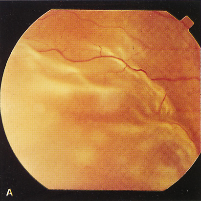
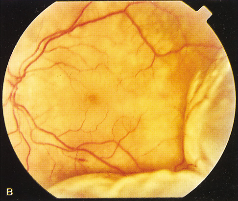
The most common visual findings are bitemporal visual field defects and visual acuity decrease (Fig. 1),78 but other visual disturbance, headaches, and rare diplopia also may be seen.74,75 The risk of a woman developing visual loss during pregnancy primarily is related to tumor size. A cohort study of 111 pregnancies in 65 women found that six of eight (75%) with macroadenomas, whereas none of 57 (0%) with microadenomas developed vision loss during pregnancy.70 A smaller study of four women with macroprolactinomas similarly showed irreversible visual field loss in three of four (75%).71 A third study found a rate of visual involvement in five of 91 (5.5%) with microadenomas and 36% of macroadenomas.76 In contrast, one study reported that only one of 14 pregnant women with large prolactinomas developed visual field defects during pregnancy.77 A final study reported that four of 18 (22%) with adenomas developed visual loss but tumors were not differentiated according to size.78

Fig. 1. Pituitary adenoma first presenting during pregnancy as a left anterior chiasmal syndrome. A. Goldmann visual fields in the sixth month of pregnancy showing bitemporal superior defects, the left (top) worse than the right (bottom). Visual acuity at this time was 20/30 OD and hand motions OS. B. Goldmann visual fields around the time of delivery show progression of the bitemporal superior defects. For each visit, the upper field is the left eye, and the lower field is the right eye. C. Goldmann visual fields performed 4 months after delivery show resolution of much of the visual field change. Visual acuity at this time was 20/15 in each eye. The patient remained stable thereafter, and had a subsequent uncomplicated pregnancy 6 years later. (Courtesy of Neil Miller, MD)

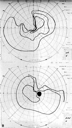
There is often spontaneous regression of the adenoma postpartum (see Fig. 1), so that the decision to treat depends on the severity of visual findings during pregnancy, and how early in pregnancy the findings developed.74 Fortunately, the dopamine agonists bromocriptine and cabergoline can cause tumor regression both within and outside of the context of pregnancy. Many studies have demonstrated the efficacy of bromocriptine for pregnancy-associated adenoma progression79–85 and there have not been any reports of adverse pregnancy outcomes related to the drug.83–85 Cabergoline also appears to be effective86,87 and safe87 during pregnancy. One case report describes the successful treatment of a thyrotropin secreting macroadenoma during pregnancy with octreotide resulting in improved visual fields, reduced tumor size, and good pregnancy outcome. Surgical therapy of symptomatic adenomas also has been successfully employed during pregnancy.80

Because the risk of symptomatic progression of a microadenoma during pregnancy is small, discontinuation of dopamine agonist therapy should be considered once pregnancy is confirmed. Treatment options for patients with macroadenomas include discontinuation of dopamine agonist therapy when pregnancy is confirmed and reinstituting therapy if the patient develops vision loss, continuous dopamine agonist therapy throughout pregnancy, and surgical debulking of the tumor prior to pregnancy.89 Decisions regarding management should be guided by an ophthalmologist, endocrinologist, obstetrician, radiologist, and possibly a neurosurgeon.

Intracranial Meningioma

Sixty-five percent of all intracranial meningiomas occur in women.192 Growth of these tumors is typically slow, producing insidious visual disturbances. In contrast, during pregnancy some meningiomas rapidly enlarge producing dramatic and relatively acute vision loss193,194 The diagnosis can be delayed because nausea, vomiting, and other symptoms caused by the tumor may be attributed to pregnancy.193 The tumors present more commonly during the second half of pregnancy.61 The predominance of meningiomas in women, their accelerated growth during pregnancy and the luteal phase of menstruation, and an association with breast cancer led to studies examining the potential role of hormones on meningioma growth.200 Subsequent studies have supported this association by demonstrating that about 70% of meningiomas express progesterone receptors and approximately 30% express estrogen receptors.201

Ocular findings can include decreased visual acuity, visual field defects that depend on the location of the tumor, oculomotor palsies, papilledema, and late optic atrophy.195–199 Symptoms may abate spontaneously after delivery and can recur during subsequent pregnancies.193,202 Hormone-responsive intracranial meningiomas should be considered in the differential diagnosis of visual disturbances during pregnancy.193 Close cooperation among ophthalmologist, neurosurgeon, obstetrician, and neonatologist are essential for an optimal outcome.

The treatment of choice during pregnancy is usually surgical excision,193 but brachytherapy has been decribed203 and external beam radiation may be considered. Antiprogesterone therapy may be effective in receptor positive cases but is unlikely to have a role during pregnancy.

Lymphocytic Hypophysitis

Lymphocytic hypophysitis (LH) or adenohypophysitis is a rare autoimmune condition that occurs most frequently in women in the last trimester of pregnancy or the early postpartum period. The most common presenting symptoms are visual field defects and headache. Most patients show signs of isolated or multiple pituitary hormone deficiencies. Adrenocorticotropin hormone (ACTH) secretion is most frequently impaired, followed by thyroid stimulating hormone (TSH) gonadotropins, growth hormone (GH), and prolactin (PRL). One-third of the cases involve hyperprolactinemia.90

One study of 16 patients found that 63% had anterior pituitary hypofunction, 56% had symptoms consistent with an expanding pituitary mass, 38% had hyperprolactinemia, 25% had associated autoimmune thyroiditis, and 19% had diabetes insipidus. Computed tomography (CT) and MRI scanning demonstrated a pituitary mass mimicking an adenoma in 83% of cases.91

Because LH can present with headache, visual field defects, hyperprolactinemia, and a pituitary mass by imaging, this disorder can be difficult to distinguish clinically from a prolactinoma. However, this differentiation is critical because a failure to recognize LH can lead to untreated pituitary insufficiency and death92 and an incorrect diagnosis of pituitary adenoma may lead to unnecessary transsphenoidal surgery. A study of 45 patients with lymphocytic hypophysitis and 806 with pituitary adenoma during pregnancy found that history and endocrinologic evaluation will usually differentiate these disorders. The spontaneous pregnancy rate was 100% in LH, whereas only 2.4% in patients with pituitary adenoma. Prolactin levels averaged 34.6 in LH compared with 393 for pituitary adenomas. Hypothyroidism and hypocortisolism were present in 57.5% of LH cases but only 2.5% of patients with pituitary adenomas. There were no distinguishing features on radiographic studies.93

The authors recommend that patients presenting to an ophthalmologist during pregnancy with signs and symptoms consistent with a pituitary disorder be co-managed with an endocrinologist, obstetrician, radiologist, and possibly a neurosurgeon.

There is strong anecdotal evidence in the form of case reports that treatment with steroids can lead to marked improvement in vision loss from LH and possibly avoid permanent pituitary insufficiency in some cases.94–98 Most cases lead to varying degrees of irreversible visual field loss or pituitary insufficiency. However, there are five reported cases of complete recovery98–102 and only one of those cases was treated with steroids.98

Little was known about the etiology of LH until recently. Serum autoantibodies directed against a 49-kDa cytosolic protein had been detected by immunoblotting in 70% of patients with biopsy proven LH. In 2002, the target pituitary autoantigen was identified as alpha-enolase103 and similar autoantigen targeting of the gamma-enolase isoform found in placental tissue was soon found.104 This established a direct link between pituitary and placental autoantigens and provides a theoretical basis for the strong predilection for LH to occur during or shortly after pregnancy. The authors are not aware of any evidence suggesting increased perinatal morbidity or mortality associated with LH.

Idiopathic Intracranial Hypertension (Benign Intracranial Hypertension or Pseudotumor Cerebri)

Idiopathic intracranial hypertension (IIH) is a syndrome of increased intracranial pressure characterized by normal brain imaging, normal cerebrospinal fluid (CSF) composition, and elevated CSF pressure. IIH occurs at the same rate in pregnant and nonpregnant women,105,108,113,114 but one study showed worsening of symptoms among nine of 11 patients with pre-existing IIH who became pregnant.106

The most common symptoms of IIH in pregnancy are headaches and visual changes. One study found that among 240 normal pregnant women, 12 developed IIH during one or more pregnancies. Ten had headaches, five had transient visual obscuration, four had reduced visual acuity, four had visual field loss, and three developed diplopia. All 12 women developed bilateral papilledema of varying severity.107

Visual outcome does not appear to differ between pregnant and nonpregnant women with IIH.105,107,108 A study of 13 pregnant IIH patients found that vision loss occurred at the same rate as age- and parity-matched controls.108 In addition, pregnancy outcomes are not adversely affected by IIH.105,109,110

The management of IIH during pregnancy includes weight control, nonketotic diet, steroids, and certain analgesics. Diuretics have been used in some cases. When medical management fails, serial lumbar punctures, optic nerve sheath fenestration, or lumboperitoneal shunt111 can be considered.105 Treatment decisions should be made in coordination with an obstetrician and possibly a neurosurgeon. Therapeutic abortion to limit progression of the disease generally is not indicated.105,111 The risk of recurrent IIH in subsequent pregnancies does not appear to be increased.109 Women who have developed IIH during pregnancy should not be advised against future pregnancies.112

Intracranial Aneurysms and Arteriovenous Malformations

Hemorrhagic cerebrovascular accidents (CVAs) are the most common neurosurgical diagnosis made in pregnancy.115 Intracranial hemorrhages are responsible for 4% to 12% of all maternal deaths133,134 and can be caused by arterial aneurysms, arteriovenous malformations (AVMs), metastatic choriocarcinoma, and disseminated intravascular coagulation (DIC).123,131 One retrospective study of 50,700 pregnancy-related admissions identified 13 hemorrhagic CVAs. Seven hemorrhages were subarachnoid and six were intracerebral. Five were caused by AVMs, three by arterial aneurysms, two by DIC, and three were idiopathic.131 Another retrospective meta-analysis study found that among 152 intracranial hemorrhages, 77% were caused by aneurysms and 23% by AVMs.134

Fifty percent of ruptured arterial aneurysms in women under the age of 40 are pregnancy-related.116 The hemodynamic and endocrinologic changes of pregnancy appear to be the cause of arterial alterations that can lead to new aneurysm formation or enlargement or rupture of a pre-existing aneurysm.115–118 Aneurysm rupture occurs more often in primiparous women115 and in the third trimester of pregnancy.115,129,131 In one study, 92% of intracranial hemorrhages occurred antepartum and 8% postpartum.134 In contrast, women with AVMs do not appear to be at a significantly increased risk of hemorrhage during pregnancy. Seven studies have reported no elevated risk of hemorrhage from AVMs during pregnancy.129,137–142

The most common presentation of a ruptured aneurysm or AVM is severe headache, seizure, or other neurologic signs from subarachnoid hemorrhage (SAH). However, enlarging aneurysms or AVMs can cause mass effects leading to cranial nerve palsies, optic nerve compression,119 or papilledema. One study reported that the possibility of focal neurologic deficit was higher among patients with intracranial AVMs than aneurysms.140 Patients with SAH can also develop subhyaloid or vitreous hemorrhage (Terson's syndrome).120 Secondary intraocular hemorrhage usually is managed conservatively,120 but vitrectomy may be indicated in bilateral or non-clearing cases.

The early diagnosis and management of a ruptured arterial aneurysm or AVM are critical in order to optimize the chances of maternal and fetal survival.116 The clinical challenge is that differentiating eclampsia from SAH can be difficult and may lead to a delay in diagnosis.116,118,121–123,134,135 The diagnosis is further complicated by the fact that pre-eclampsia and eclampsia are independent risk factors for aneurysm rupture124 so that these entities may coexist. Clinicians should maintain a high level of suspicion for intracranial aneurysm when pregnant women present with headache, seizure, altered consciousness, focal neurologic signs, visual changes, or diplopia. Urgent MRI and magnetic resonance angiography scanning is the current diagnostic modality of choice because there is no radiation risk or side effects of contrast media.132

The management of intracranial aneurysms generally is the same for pregnant as nonpregnant patients117,118 and neurosurgical considerations take precedence over obstetric considerations.117,137,144,145 When SAH occurs, endovascular obliteration,125,126,143 neurosurgical clipping,127 or stereotactic radiosurgery143 of the aneurysm or AVM may be carried out in order to avoid recurrent hemorrhage and secondary ischemia.115 One study of AVMs recommended that patients at high operative risk be managed conservatively until after delivery.143 Another study from 1990 reported that surgical management of aneurysms, but not AVMs, was associated with improved maternal and fetal survival. Taken together, these studies suggest that the threshold for neurosurgical intervention might be lower for aneurysms than AVMs, although this decision should be made by the neurosurgeon. The risk of rebleeding in the same pregnancy was 27% among AVMs in one study.136 Visual field and optic nerve changes can resolve over time following surgery.119,130

Once an aneurysm or AVM has been treated successfully, pregnancy can be allowed to progress to term. Most obstetricians prefer vaginal delivery in these cases,118,125 although one study reported a bias toward caesarian section among patients with AVM.143 Fetal mortality is not increased among mothers with intracranial aneurysms provided that the mother survives.128 Women with arterial aneurysms or AVMs are not advised against future pregnancy in most cases.129

Cerebral Venous and Sinus Thrombosis

Cerebral venous and sinus thrombosis (CVST) in the pregnant and puerperal state occurs with an incidence of 10 to 20 per 100,000 deliveries and accounts for 5% to 20% of all cerebral venous thrombosis.162 The signs and symptoms are nonspecific, which can lead to a potentially fatal delay in diagnosis.169,171 A retrospective study of 138 cases found that 74 (54%) presented in the first 7 days of the postpartum period.170 The most common signs and symptoms were fever (62%), headache (48%), seizure (46%), hemiplegia (43%), altered consciousness (41%), and papilledema (35%).170 Another retrospective study of 25 cases reported similar presenting clinical signs, which included headache (96%), focal neurologic deficits (60%), papilledema (43%), and seizure (40%).169 Magnetic resonance venography (MRV) is the preferred diagnostic modality, and treatment is with heparin anticoagulation.

The increased incidence of intracranial venous thrombosis is attributed to the hypercoagulable state of pregnancy,172,173 inherited protein C172,174,175 and S deficiencies,172,176,177 and eclampsia.178 A prospective study found that 78% of patients with venous thrombosis during pregnancy and the puerperium demonstrated activated protein C resistance (factor 5 Leiden).174 Three case reports have found associated protein S deficiency.172,176,177 There is one case report of transverse sinus thrombosis associated with eclampsia.178 The authors suggest a causative mechanism but this association may be incidental. It is recommended that causes of inherited hypercoagulability be investigated in all women who develop CVST during pregnancy or puerperium.

Mortality from CVST is less than 20%.171,179 The visual prognosis generally is good with blindness occurring because of late optic atrophy in two of 77 (3%) patients in one retrospective study.180 Women with pregnancy-related CVST should not be discouraged from future pregnancies.171,181 In one retrospective study, among 39 women with a history of CVST during pregnancy, 14 had 22 subsequent pregnancies resulting in 19 births and one elective and two spontaneous abortions. Five patients were prophylactically treated with low-dose heparin.181 In another study, among 77 women with pregnancy-related CVST there was no recurrence of CVST during subsequent pregnancies and only one postpartum deep venous thrombosis occurred.180

Metastatic Choriocarcinoma

Gestational choriocarcinoma is a malignant tumor that frequently undergoes hematogenous metastasis to highly vascularized organs such as the lung, brain, and liver. The incidence of choriocarcinoma is one in 50,000 live births and one in 30 molar pregnancies.146 Intracranial choriocarcinoma metastases can present as intracerebral,148–153 subarachnoid,153–155 or subdural hemorrhages.156,157 These tumors can bleed primarily or cause arterial aneurysms148,154,156,168 or arteriovenous fistulae155 with secondary bleeding. In a retrospective analysis of 10 cases, the most common presenting symptoms were features consistent with elevated intracranial pressure and hemiparesis.147 Ophthalmic signs and symptoms may be present and can include visual acuity or visual field loss, transient visual obscuration, papilledema, and retinal hemorrhages.147,154 It is important to consider this rare tumor in cases of young women presenting with stroke symptoms because it is potentially curable.149

Amniotic Fluid or Air Embolism

Amniotic fluid embolism is a rare but serious complication of pregnancy resulting in death in the majority of patients.160,161 Particulate matter of amniotic origin has been identified at autopsy in the brain, lungs, and kidneys.160 Focal neurologic deficits are rare162,164 but can affect the optic nerve, visual pathways, or occipital cortex. One case report describes isolated cortical blindness from air embolism that occurred following induced abortion and improved following treatment with hyperbaric oxygen.163

Carotid-Cavernous Fistulae

There are reports of five carotid-cavernous fistulas developing or worsening during pregnancy.165–168 One spontaneous fistula improved without intervention after the mother aborted the pregnancy in the twelfth week.165 A second spontaneous fistula worsened 3 weeks after a normal delivery before improving without intervention.165 One carotid-cavernous fistula was caused by metastatic choriocarcinoma.168 A traumatic fistula that had resolved 4 years prior to pregnancy recurred causing two antepartum and one postpartum hemorrhages. The authors propose that pregnancy-related hormonal and hemodynamic changes caused the recurrence and hemorrhage.167 However, the paucity of reports about carotid-cavernous fistulae during pregnancy suggest that this association may be incidental

OPTIC NERVE

Multiple Sclerosis and Optic Neuritis

Multiple sclerosis (MS) is a common cause of optic neuritis and is the most common chronic disabling neurologic disease affecting women of childbearing age.211 Pregnancy has a protective effect on MS with fewer and less severe relapses, especially during the third trimester.211–215 However, the relapse rate increases in the first 3 months following delivery before returning to pre-pregnancy rates.211–215 Breastfeeding and epidural analgesia have no deleterious effect on relapse rates.211,213,214 The Pregnancy and Multiple Sclerosis study prospectively followed 254 women with MS through 269 pregnancies. Compared with year prior to pregnancy, there was a reduction in the relapse rate during pregnancy, which was most marked in the third trimester. There was striking increase in the relapse rate in the first 3 months after delivery, although 72% of the women did not experience a relapse during these 3 months. After this, the rates returned to pre-pregnancy levels.213–214 Relapse rates remained at pre-pregnancy rates at the 2 year follow-up.214 A recent retrospective review of 351 women with MS confirmed the finding that the MS relapse rate decreases throughout pregnancy and increases during the puerperium.215

Overall, pregnancy has no long-term effect on MS related disability or disease course.211–214,216 Apart from methotrexate and cyclophosphamide, most drugs used routinely in the treatment of MS, including steroids, can be used safely by pregnant women.216 A study of 14 pregnant women with relapsing-remitting MS, who were treated with postpartum IVIg in an attempt to avoid postdelivery recurrences, found that none of the patients who received postpartum IVIg relapsed217 compared with the expected relapse rate of about 28%.214 Further study is needed to confirm this apparent protective effect.

Women with MS who desire children can be reassured that their infants are not at increased risk of malformations, preterm delivery, low birth weight, or infant death.211,213,214,216 Because a woman's past history of relapses may be the best indicator of future clinical course and long-term disability,212–214 women with more severe disease may wish to complete their families as soon as possible.216

A condition called “lactation optic neuritis” has been described in the first 12 months following delivery unassociated with known MS.204,205 The clinical course is similar to classic optic neuritis that occurs without lactation. It is possible that decreased immunosuppressive activity during the postpartum period induces the initial clinical manifestation of a previously unrecognized demyelinating disorder such as MS.204 The paucity of cases reported in the literature also suggests that this also could be an incidental association

ADDITIONAL PREGNANCY-RELATED OPTIC NEUROPATHIES

Devic's syndrome (neuromyelitis optica) is characterized by optic neuritis with transverse myelitis and may present during pregnancy.206,207 Severe vomiting in pregnancy (hyperemesis gravidarum) can lead to optic neuritis.208 One case of ischemic optic neuropathy has been reported in association with severe pre-eclampsia.209 A case of bilateral idiopathic optic neuritis with permanent visual loss recently was reported in association with pregnancy.210 One case of pregnancy-related papillophlebitis in a woman with no identifiable coagulation defect has been reported.349

OTHER NEURO-OPHTHALMOLOGIC DISORDERS

Cortical Vision Loss in Pre-Eclampsia and Eclampsia

Pre-eclampsia and eclampsia are the most common causes of pregnancy-related focal neurologic deficits. These deficits are characteristically of sudden onset and resolve over time. The precise pathogenesis of these strokelike focal deficits remains poorly understood.162,164 Transient visual changes occur in up to 25% of patients182 and cortical blindness is a rare but dramatic finding. A prospective study of 15 women who developed pre-eclampsia/eclampsia-related cortical blindness found complete resolution over 4 hours to 8 days.183 Another retrospective analysis of 19 cases of eclampsia reported transient cortical blindness in two patients with complete recovery in both.184

The etiology of cortical vision loss remains controversial. Many authors have proposed a mechanism of cerebral vasospasm with ischemic injury,183,185,186 whereas others suggest that increased capillary permeability leads to hydrostatic edema.187–189 Except for rare cases with persistent neurologic deficits and neuroradiologic abnormalities, the reversibility of vision loss argues against true ischemic necrosis.162 Recent studies have demonstrated cerebral vasospasm by angiography,185 transcranial color-coded sonography,185 and MRA188 with concurrent MRI,185,188 and single photon emission computed tomography187 findings consistent with cerebral edema. This suggests that cerebral vasospasm leads to relative ischemia and changes in regional blood flow that cause reversible edema. Hypercalcemia190 and inflammatory cytokines of placental origin191 recently have been proposed as putative inciting factors.

Horner's Syndrome

Horner's syndrome has been reported as a complication of epidural anesthesia for labor. Although not uncommon in women in labor, this complication is virtually unknown in patients outside of the context of labor.218 The incidence of Horner's syndrome was estimated in a prospective study of 200 women who received epidural analgesia during labor. The incidence was 1.3% among 150 who delivered vaginally and 4% among 50 patients who underwent caesarian section.221 There are two case reports of trigeminal nerve palsy following epidural anesthesia for labor.219,223

The etiology seems to be cephalad spread of local anesthesia, sometimes via unexplained routes and with surprising selective targeting effect.219 The propensity in pregnant women is believed to be caused by pregnancy-related anatomic and physiologic changes of the epidural space.219 In one case report, an epidurogram after Horner's syndrome developed from epidural anesthesia demonstrated unilateral left spread ascending to upper thoracic levels with delayed spread toward the right lumbar epidural space.220 This complication generally is transient in nature but must be accompanied by close maternal and fetal monitoring because it can be associated with maternal hypotension caused by autonomic effects.222

Myasthenia Gravis

Myasthenia gravis (MG) frequently affects young women in their childbearing years. Several retrospective studies suggest that approximately 40% of women with pre-existing MG clinically worsen during pregnancy.224–229 This deterioration usually occurs during the first trimester226,229 or shortly after delivery.224,225 The authors are not aware of any studies that specifically evaluate the ophthalmic signs and symptoms of MG during pregnancy but suspect that they would approximate overall disease activity.

The rate of neonatal myasthenia gravis (NMG) after delivery varies widely in the literature from 6% to 50%.225,228–231 The incidence of NMG seems to be more common among infants born to mothers with puerperal infections or shorter antenatal disease duration.224 The newborns of thymectomized mothers are relatively protected.224 Two studies demonstrate good correlation between maternal antiacetylcholine receptor antibody titers and the development of NMG, so this testing may be helpful.228,231 The largest study of neonatal outcomes retrospectively evaluated 127 births of mothers with MG compared with population-wide controls. Women with MG had 8% more complications during delivery, premature rupture of membranes was three times more common, and caesarian sections were twice as common. Five children (3.9%) had severe anomalies and three of them died.232 Another study showed that perinatal mortality also was increased among mothers who remained asymptomatic during pregnancy.233 This suggests that neonatal outcomes are not only related to MG disease severity but also to effects of the underlying immunologic dysfunction on the fetus. To the authors' knowledge, perinatal mortality has not been evaluated for cases of isolated ocular myasthenia.

Treatment of MG during pregnancy requires close coordination among the obstetrician, neurologist, pediatrician, and ophthalmologist when ocular symptoms are present. Treatment during pregnancy may include immunosuppressive therapy, plasmapheresis, intravenous human immunoglobulins,225 and thymectomy.234

Ocular Migraine

The lifetime prevalence of migraine with aura (MA) is 5%, with a female to male ratio of 2 to 1.235 Visual disturbances are the most common aura phenomenon, occurring in 90% of patients with MA.235 Several retrospective and two prospective studies have demonstrated improvement of migraine during pregnancy.236–240 The largest prospective study included 49 migraineurs who became pregnant. Migraine symptoms improved in 47% during the first trimester, 85% during the second trimester, and 87% in the third trimester. Complete remission was achieved in 79% of women by the end of pregnancy. Migraine recurred in 55% during the first month after delivery but breastfeeding seemed to afford some protection.237 Retrospective studies have demonstrated that women with migraine onset at menarche and those with perimenstrual migraine are more likely to go into remission during pregnancy.238 However, this association has not been observed in prospective studies.236,237 Retrospective studies also have shown that migraine with aura appears to be overrepresented among the small number of women (4%–8%) whose migraines worsen during pregnancy.238 Migraine can begin during pregnancy in 1% to 17% of cases, often during the first trimester, and involves a higher proportion with aura.238 The positive effects of pregnancy on migraine probably are related to uniformly high and stable estrogen levels.240,241

The symptoms of headache and visual changes during pregnancy are nonspecific. The development or worsening of migrainelike symptoms during pregnancy can indicate an underlying structural lesion or disease process such as an arterial aneurysm, arteriovenous malformation, progressive meningioma, venous sinus thrombosis, or pre-eclampsia, all of which are more common during pregnancy. Therefore, appropriate diagnostic testing should be performed to rule out other organic causes of headache and visual changes.242,243

Migraine during pregnancy does not increase the risk of pregnancy-related complications such as pre-eclampsia/eclampsia, abnormal labor, fetal loss, or congenital anomalies.238 Safe and effective acute care treatments include paracetamol, opioids, and antiemetics.244 The use of triptans appears to be safe, but data are limited and they are not broadly recommended.244,245 Preventative treatments include relaxation, biofeedback, beta-blockers such as labetalol, some antidepressants, and gabapentin.244,246

Back to Top
ORBIT

THYROID ORBITOPATHY

Graves' disease occurs in approximately 1/1,000 pregnancies and the diagnosis is made following conception in approximately 50%.247 Hyperthyroidism may worsen during the first trimester248–251 but tends to improve during the second and third trimesters.252–254

The etiology of early worsening of hyperthyroidism is related to the thyroid stimulating activity of human chorionic gonadotropin (hCG).250,251 This can cause the aggravation of pre-existing Graves' disease250 or de novo hyperthyroidism called gestational transient thyrotoxicosis that is frequently associated with hyperemesis.249,251 The authors are not aware of any studies that evaluate the effect of pregnancy-related early worsening of Graves' disease on its ophthalmic manifestations, and suspect that because this early aggravation is caused by elevated hCG rather than an increase in autoantibodies against shared thyroid-orbital antigen, ophthalmologic signs and symptoms are likely unaffected.

The improvement in Graves' disease during the second and third trimesters was previously attributed to a reduction of thyroid-stimulating antibody activity.252 More recent studies have demonstrated a change in the specificity of TSH receptor antibody from stimulatory to blocking activity during pregnancy.252–254 Whether this alteration in antibody activity also improves the ophthalmic manifestations of Graves' disease has not been explored in the literature.

Graves' disease in pregnancy carries a risk of fetal thyrotoxicosis from transplacental transfer of thyroid-stimulating antibodies, or fetal hypothyroidism from antithyroid drugs or thyroid-blocking antibodies.255 The treatment options for Graves' disease include antithyroid drugs, radioablation, and surgery.256,257 Propylthiouracil (PTU) is the drug of choice for treating pregnant and breastfeeding women because of limited transfer into placenta and breast milk.256,258 Radioiodine therapy is contraindicated in pregnancy and must be completed at least 4 months prior to conception.257 Surgery has a limited role in Graves' disease but could be considered in the context of pregnancy.257

Studies evaluating the treatment of Graves' ophthalmopathy during pregnancy have not been performed. The authors recommend conservative therapy whenever possible during pregnancy. Strabismus can be managed with prisms or patching. Ocular surface symptoms and exposure keratopathy can be treated with lubricants, punctual plugs, topical steroids or cyclosporine, or tarsorrhaphy in severe cases. Compressive optic neuropathy can be managed via orbital decompression or oral steroids but external beam radiation should be avoided. The authors do not recommend eyelid or muscle surgery during pregnancy because lid position and strabismus may not remain stable postpartum.

ORBITAL TUMORS

The growth of orbital hemangiomas during pregnancy has been reported.259–261 One case report describes the accelerated growth of a primary orbital schwannoma during pregnancy.262 The etiology of pregnancy-related tumor growth has not been investigated in these cases but may be related to hormone receptor expression, analogous to meningioma growth during pregnancy.

Two cases of orbital rhabdomyosarcoma have been reported during pregnancy.263,264 One case was associated with placental metastasis.263 A case of primary orbital melanoma that presented during the first trimester of pregnancy has been reported.265

ORBITAL HEMATOMAS

Orbital hematomas can develop during labor or in the early postpartum period.266 Pain and diplopia are the most common presenting symptoms.

Back to Top
RETINAL AND CHOROIDAL DISORDERS ARISING IN PREGNANCY

PRE-ECLAMPSIA AND ECLAMPSIA

Pre-eclampsia typically develops in the second half of pregnancy and is characterized by hypertension, edema, and proteinuria. Eclampsia is pre-eclampsia with convulsions and usually occurs late in pregnancy. In healthy women, pre-eclampsia generally is seen in first pregnancies with an incidence estimated at 5%. Risk factors for pre-eclampsia include very young or advanced maternal age, multifetal pregnancy, hemolytic disease of the newborn, diabetes mellitus, chronic systemic hypertension, and renal disease. Pre-eclampsia and eclampsia place the fetus at risk because of placental vascular insufficiency. In the first half of the 20th century, severe changes in retinal arteriolar caliber were believed to reflect placental vascular insufficiency and constitute an indication for pregnancy termination. Therefore, a great deal of attention was paid to retinal findings in pre-eclampsia. With improved medical and obstetric management of hypertension and other aspects of pre-eclampsia, retinal findings are no longer used to assess this disease. In addition, retinal changes in pre-eclampsia now may be significantly less frequent than they were in the past.

Early reports gave an impressive rate of visual disturbances. Scotoma, diplopia, dimness of vision, and photopsias were noted in 25% of patients with severe pre-eclampsia and up to 50% of patients with eclampsia.267 Recent studies, discussed in the following, suggest that the rate of visual disturbance has decreased markedly with improved medical management of pre-eclampsia. Although photic stimuli may predispose to seizures in susceptible patients, the benefits of an ophthalmoscopic examination outweigh the small risk of seizure when an examination is indicated.268

Pre-eclampsia and eclampsia have been associated with a retinopathy similar to hypertensive retinopathy; serous retinal detachments; yellow, opaque retinal pigment epithelium (RPE) lesions; and cortical blindness. Arterial and venous occlusive disease also can occur and may contribute to visual loss. Early studies of retinal disorders in pre-eclampsia have been discussed in previous reviews.269

Retinopathy in Pre-Eclampsia and Eclampsia

Focal or generalized retinal arteriolar narrowing is the most common ocular change seen in pre-eclampsia, but its frequency is declining. Early studies reported arteriolar attenuation in 40% to 100% of pre-eclamptic patients.270 Recently, a retrospective study of fluorescein angiograms in pre-eclamptic patients identified normal retinal vessel caliber in 16 of 16 patients. In contrast, four of 14 patients with pre-existing chronic systemic hypertension had retinal vascular changes.271 Another study prospectively demonstrated a statistically significant difference in arteriolar caliber between 56 study participants with severe pre-eclampsia and 25 healthy controls, but no difference between 17 patients with mild pre-eclampsia and controls.272 These studies suggest that arteriolar narrowing may be more common in pregnant patients with chronic pre-existing hypertension than those with mild pre-eclampsia. The difference in the reported prevalence of retinopathy between the early and recent literature probably is related to better medical management of pre-eclampsia and its complications.

The cause of retinal arteriolar narrowing seems to be central retinal artery vasospasm suggested by increased central retinal artery blood flow velocity.273 When present, the retinal arteriolar attenuation associated with pre-eclampsia generally resolves after delivery, presumably because of normalization of central retinal artery blood flow. Other typical hypertensive retinopathy changes such as hemorrhages, cotton wool spots, lipid deposits, diffuse retinal edema, and papilledema generally are not seen in pre-eclampsia272 and should raise suspicion about additional concurrent systemic disease.

Choroidopathy in Pre-Eclampsia and Eclampsia

Choroidal dysfunction is a common ocular complication of pre-eclampsia and eclampsia that manifests clinically as serous retinal detachments or yellow RPE lesions. The serous retinal detachments usually are bilateral and bullous (Fig. 2) but occasionally are cystic.268,274 In the early 20th century, serous retinal detachments were seen in 1% of severely pre-eclamptic patients and about 10% of eclamptic patients.275,276 A more recent retrospective study evaluated 31 women with severe pre-eclampsia or eclampsia and found that 40/62 (65%) eyes had serous retinal detachments and 36/62 (58%) had RPE lesions. RPE lesions usually were located in the macular or peripapillary regions, 33/36 (92%) were solitary or grouped, and 3/36 (8%) were large and geographic. After delivery, all serous retinal detachments and RPE lesions resolved. The three eyes with geographic RPE lesions all developed significant chorioretinal atrophy.277 The apparent historical increase in the incidence of serous detachments and RPE lesions is likely owing to improved diagnostic instrumentation and ancillary testing.

Fig. 2. Serous retinal detachments related to pre-eclampsia. A and B. Fundus photographs showing bilateral bullous serous retinal detachments. (Courtesy of John Wroblewski, MD.)

The etiology of choroidal dysfunction is thought to be ischemia, based on fluorescein angiography, limited histopathologic study, the presence of Elschnig's spots on resolution,278 and indocyanine green angiography.279 This is further supported by the observation that posterior ciliary artery blood flow velocity is increased in pre-eclampsia, suggesting vasospasm.273 The primary choriocapillaris ischemia presumably leads to RPE ischemia manifest as yellow opacification or fluid pump dysfunction allowing subretinal fluid accumulation.

Although serous retinal detachment and RPE dysfunction can cause marked loss of visual acuity, these changes fully resolve postpartum and most patients return to normal vision within a few weeks. Some patients have residual RPE changes in the macula. Years later, these changes can mimic macular dystrophy or tapetoretinal degeneration.280 Rare patients may develop optic atrophy if chorioretinal atrophy is extensive.281

One study suggested that serous detachments are more specific to pre-eclampsia and eclampsia, whereas retinopathy is seen more often in pre-eclampsia superimposed on pre-existing hypertension.282 Retinopathy is more closely associated with higher levels of blood pressure than is serous detachment.283 Although retinopathy was believed to be a reflection of possible placental insufficiency and possible adverse neonatal outcome, serous retinal detachment is not an additional risk factor.284

Postpartum serous detachments have been reported in pre-eclamptic patients,285,286 and there are rare reports of exudative detachments in patients without pre-eclampsia.287,288 Although these serous retinal detachments may be mechanistically distinct, they also resolve over several weeks.

The HELLP syndrome consists of hemolysis, elevated liver enzymes, and low platelets, and it is generally associated with severe pre-eclampsia or eclampsia. Bilateral serous retinal detachments and yellow-white subretinal opacities have been noted in two patients with this disorder.289,290

CENTRAL SEROUS CHORIORETINOPATHY

Central serous chorioretinopathy (CSC) is caused by localized RPE dysfunction resulting in the accumulation of subretinal fluid. People between the ages of 20 and 50 years typically are affected, and there is an 8:1 male predominance.291 A recent retrospective case-control study involving 624 patients showed that systemic steroid use and pregnancy are the most important risk factors for CSC with odds ratios of 37 and 7, respectively.302 The hormonal or hemodynamic changes of pregnancy may predispose some women to CSC.

Many cases of CSC associated with pregnancy are reported in the medical literature.292–302 Unlike the serous retinal detachments observed in pre-eclampsia and eclampsia, CSC generally is unilateral. The women were all previously healthy and no cases were associated with pre-eclampsia or eclampsia. No patients had antecedent eye disease other than refractive error. Primiparas and multiparas were both represented. Most of the cases developed in the third trimester and all resolved spontaneously within a few months after delivery. There were no cases of significant visual sequelae.

Pregnancy-associated CSC may recur in the context or outside of subsequent pregnancy. CSC recurred in two women, always in the same eye, in subsequent pregnancies. One patient had four successive pregnancies with CSC,298 and one had two successive pregnancies complicated by CSC.299 There is also one case report of a woman developing CSC 1 month postpartum in two successive pregnancies.300 However, there is also a report of a woman with CSC in her third pregnancy who did not experience a recurrence during a subsequent pregnancy.295 Therefore, the occurrence of CSC during one pregnancy does not necessarily mean that it will recur in future pregnancies. There is a report of one patient who experienced a recurrence of CSC outside the context of pregnancy.295

There is an increased incidence of subretinal white exudates (presumed to be fibrin) in pregnancy-associated CSC (approximately 90%) compared with CSC in males and nonpregnant women (approximately 10%). One case series reported that three of four patients with pregnancy-related CSC had subretinal exudates.295 Another study found that 6/6 cases of pregnancy-related CSC had subretinal exudates compared with only 6/50 (12%) of non–pregnancy-related cases.296 A recent case report showed highly reflective subretinal material on OCT that corresponded clinically to subretinal exudates.301 The etiology of the higher prevalence of subretinal exudates in pregnancy-associated CSCR is unknown.

OCCLUSIVE VASCULAR DISORDERS

An increase in the level of clotting factors and clotting activity occurs during pregnancy.303 Several pathologic sources of thrombosis and embolic events also can occur. One review of ischemic cerebrovascular disease suggested that pregnancy is associated with a 13-fold increase in the risk of cerebral infarctions compared with nonpregnant women.303 This increased risk of vaso-occlusive disease also may manifest as retinal or choroidal vascular occlusions.

Retinal Artery Occlusion

Two cases of unilateral central retinal artery occlusion (CRAO),304,305 one case of bilateral CRAO,306 two cases of unilateral branch retinal artery occlusion (BRAO),307,308 and three cases of bilateral multiple BRAO309 have been reported in association with pregnancy and in the absence of additional risk factors. Three cases of arteriolar occlusion were associated with pre-eclampsia306,309 and one was associated with disc edema.308 Five of the eight (63%) cases occurred within 24 hours of delivery, suggesting that this is a particularly susceptible period.

One study reported that multiple retinal arteriolar occlusions were seen within 24 hours after childbirth in four women. Two patients were pre-eclamptic and required cesarean section. One of the two also had evidence of cerebral infarctions. The third patient had hypertension, pancreatitis, and premature labor. The fourth was a previously healthy 16 year old who had an oxytocin-induced labor and had a generalized seizure 2 hours after delivery. The patients reported decreased vision and all had fundus findings characterized by retinal patches characteristic of ischemia and intraretinal hemorrhages that were similar to Purtscher's retinopathy. After resolution, patients were left with focal arteriolar narrowing and optic disc pallor. The visual acuities ranged from 20/20 to 4/200 and visual field defects were compatible with the areas of occlusion. The authors suggest that complement-induced leukoemboli could have caused the retinal arteriolar occlusions.309

Nine additional cases of pregnancy-associated BRAO have been reported in the literature.310–315 However, all of these cases had significant additional risk factors for vascular occlusion. Because pregnancy is a common condition, it is difficult to know whether these cases represent true pregnancy associations, multifactorial or synergistic etiologies, or just chance occurrences. One case was associated with intramuscular progestogen therapy for a threatened abortion.310 Three cases that occurred postpartum were associated with hypercoagulability from protein C311 or protein S309,312 deficiency. Three cases were associated with thromboembolic occlusions attributed to mitral valve prolapse313 and amniotic fluid embolism.160,161 The final two cases developed BRAO in the first trimester in association with migraine headaches.314

Retinal Vein Occlusion

Retinal vein occlusion associated with pregnancy is exceedingly rare. The authors are aware of only four pregnancy-related central retinal vein occlusions (CRVO) have been reported to date315–317 and we are not aware of any branch retinal vein occlusions. A study of central retinal vein occlusions with diurnal intraocular pressure determination in young adults included a 33-year-old pregnant woman in her third trimester who had unilateral venous dilation and tortuosity with two subretinal hemorrhages and mild foveal edema.315 Another study reported the case of a 27-year-old woman who was 6 months pregnant when she developed a unilateral CRVO. The authors suggested impaired fibrinolysis after venous stasis as a possible mechanism.316 A 30-year-old woman presented in the 28th week of her second pregnancy with HELLP syndrome. She developed a unilateral CRVO 10 days after emergency caesarean section.317 The final case is that of a patient with mild bilateral CRVO that developed early in pregnancy and resolved over several months (J. Wroblewski, personal communication). The paucity of reported cases linking pregnancy to retinal vein occlusion makes the strength of this association suspect.

Disseminated Intravascular Coagulation

Disseminated intravascular coagulation (DIC) is an acute pathologic process with widespread thrombus formation in small vessels. It can occur in obstetric complications such as abruptio placentae and intrauterine fetal death that release placental thromboplastin into maternal circulation and activate the extrinsic coagulation system. This process has a tendency to occlude the posterior choroidal vessels leading to RPE ischemia, dysfunction of the retinal pigment epithelial pump mechanism, and subsequent serous retinal detachments in the macular and peripapillary regions.318–321 The development of serous retinal detachments in pregnancy, especially late pregnancy, may be an early ocular sign of DIC.321 The authors are aware of case reports of two patients with DIC causing serous retinal detachments.318,321 These detachments tend to be bilateral and symptomatic. With recovery from the systemic disorder, vision generally returns to normal with only residual pigmentary change.320,321

Thrombotic Thrombocytopenic Purpura

Thrombotic thrombocytopenic purpura (TTP) is a rare, idiopathic, acute, systemic coagulopathy characterized by platelet consumption and thrombus formation in small vessels. TTP occurs at any age with a peak incidence in the third decade of life and a female to male preponderance of 3:2. Women who are pregnant or in the postpartum period are overrepresented, making up approximately 20% of TTP patients.325,326

Visual changes occur in approximately 8% of cases322 because of thrombus formation in the choriocapillaris and secondary RPE and outer retinal ischemia. Clinical findings usually are bilateral and include serous retinal detachments, yellow spots at the level of the RPE, and localized arteriolar narrowing. Sequelae include RPE pigmentary changes and Elschnig spots with a return to baseline vision over several weeks in most cases.322–324

Aggressive treatment with plasma transfusions and plasmapheresis have improved maternal and fetal survival.325 In published reports, 10 of 11 (91%) with idiopathic TTP, 11 of 18 (61%) women who were pregnant or postpartum at their initial presentation, and 11 of 11 (100%) with congenital TTP developed active TTP during a subsequent pregnancy.326,327 A retrospective study of nine patients reported fetal mortality from preterm delivery and intrauterine death in 33%.328 Therefore, pregnant patients with a history of TTP must be closely followed in a tertiary obstetric unit with plasmapheresis available.327

AMNIOTIC FLUID EMBOLISM

Amniotic fluid embolism is a serious complication of pregnancy with high mortality, second only to pulmonary thromboembolism as a cause of death during pregnancy and the postpartum period. Those patients who survive the initial event usually develop DIC329 with the potential ocular complications described in the preceding. Two patients developed multiple branch retinal arteriolar occlusions presumably related to particulate material from the amniotic fluid.160,161 Another patient had massive blood loss from an amniotic fluid embolism leading to severe retinal and choroidal ischemia and blindness in one eye.330

UVEAL MELANOMA

Pregnancy is heralded by a hormone-dependent tendency to hyperpigmentation and well-known cutaneous changes such as chloasma and an increase in pigmentation of pre-existing nevi owing to increased levels of melanocyte-stimulating hormone in pregnancy.331 Although estrogen and progesterone may stimulate melanogenesis, there is no evidence that this can cause malignant transformation of melanocytic cells.

A case-control study found a decreased risk of uveal melanoma for women who had ever been pregnant, with an increase in protective effect with more live births. The largest effect was observed between nulliparous and parous women.332 However, others have reported a trend toward a larger than expected number of ocular melanomas presenting during pregnancy.333 There are also a number of anecdotal reports of uveal melanomas presenting or growing rapidly during pregnancy.334–338 These reports led to speculation that uveal melanoma may be hormone responsive, but two studies have failed to show any estrogen or progesterone receptor expression in ocular melanomas.337,339 It is possible that other hormones may be involved,337 or that tumor growth may be related to pregnancy-associated immune modulation.

Pregnancy-related uveal melanoma does not seem to differ histologically from uveal melanoma not associated with pregnancy. One study reported that among 10 pregnancy-related choroidal melanomas evaluated after enucleation, the tumors did not differ in cell type, mitotic activity, and other features when compared with a matched group of tumors in nonpregnant women.340

The treatment of pregnancy-associated uveal melanoma has been described in two studies. Among 16 cases in one study, 10 eyes were enucleated, four received plaque radiotherapy during or after pregnancy, and two cases were observed. Among 14 of 16 patients who elected to carry the pregnancy to term, all delivered healthy babies with no infant or placental metastases.340 Another report of eight cases demonstrated no treatment-related pregnancy complications. The authors suggest that brachytherapy is safer toward the end of pregnancy or after delivery.341

Childbearing may be associated with improved survival in choroidal melanoma. One large prospective cohort study reported that death rates from metastasis were 25% higher in nulliparous women and men, than in women who had given birth. The protective influence of parity was greatest in the first 3 years of follow-up and increased with the number of live births.342 These results contradict a small earlier study by the same group that concluded rates of metastasis were not higher among women who reported pregnancies or oral contraceptive use.343 A much smaller third study also showed similar 5-year survival between pregnant and non-pregnant women with posterior uveal melanoma.340

Back to Top
OTHER RETINAL AND CHOROIDAL CHANGES DURING PREGNANCY
Choroidal osteomas have been reported that presented during pregnancy with visual loss resulting from choroidal neovascularization.344,345 Cases of acute macular neuroretinopathy,346 Valsalva maculopathy,347 and cystoid macular edema348 have been observed in the immediate postpartum period.

The authors are aware of three cases of choriocarcinoma that metastasized to the choroid.158,159 Two of the cases presented with vision loss and pain. One case required enucleation158 and a second resolved with systemic chemotherapy alone.159

Back to Top
PREEXISTING CONDITIONS

DIABETIC RETINOPATHY

The modern medical, ophthalmologic, and obstetric management of pregnant diabetic patients has greatly improved outcomes for both the fetus and the mother. Laser photocoagulation has reduced the risk of vision loss from diabetic retinopathy, and improved glucose control has improved the likelihood of good fetal outcomes. Well-controlled blood glucose and adequate glycosylated hemoglobin (HbA1c) before conception and throughout the pregnancy may reduce the risk of spontaneous abortion,350,351 congenital anomalies, and fetal morbidity.352 A recent study suggested that the severity of diabetic retinopathy may be a significant factor in predicting adverse fetal outcomes, even after correcting for blood glucose control.353 However, another study suggested that blood glucose control may counteract adverse fetal effects associated with maternal retinopathy and nephropathy.354

Diabetic woman who may become pregnant should establish excellent glucose control before conception, because the major period of fetal organogenesis may take place before the mother is even aware that she is pregnant. In addition, a diabetic woman's retinopathy status should be evaluated and stabilized prior to conception. This is particularly important for patients with severe nonproliferative or proliferative retinopathy because scatter laser photocoagulation may reduce progression during pregnancy.355 Laser treatment of diabetic macular edema before pregnancy also may be important, although the effects of pregnancy on macula edema have not been adequately studied.

The Diabetes in Early Pregnancy (DIEP) study, a study of 155 insulin-dependent diabetic pregnant women,356 as well as the data from the Diabetic Control and Complications Trial357 and the previous data summarized by Sunness,353 all provide evidence that better metabolic control before pregnancy diminishes the progression of diabetic retinopathy. Recent studies have found a strong correlation between the glycosylated hemoglobin level in the first month and the degree of deterioration once tight metabolic control is achieved.356 Nerve fiber layer infarctions commonly are associated with the institution of tight metabolic control of chronic hyperglycemic patients. One study described the retinopathy status of 13 patients managed by insulin pump during pregnancy. Two patients who had rapid decrease in the HbA1c level developed acute ischemic changes and ultimately proliferative retinopathy.358 However, the long-term benefits of adequate blood glucose control outweigh concerns about the transient worsening of retinopathy that has been associated with the sudden imposition of tight metabolic control.357,359,360

The frequency of ophthalmic follow-up of a diabetic patient during pregnancy is determined by her baseline retinopathy status. Guidelines for eye care in diabetic patients recommend that a diabetic woman planning pregnancy within 12 months should be under the care of an ophthalmologist, undergo repeat evaluation in the first trimester, and after that at intervals dependent on the initial findings.353,361

Progression of Diabetic Retinopathy during Pregnancy

The interpretation of changes in diabetic retinopathy as being caused by pregnancy is confounded by changes related to blood glucose levels. As medical advances continue to improve glycemic control, changes in blood glucose control that occur at the onset of pregnancy will be minimized and it may be easier to obtain a more direct understanding of the role of pregnancy in the progression of diabetic retinopathy. In the meantime, several mechanisms have been proposed as possible etiologic factors contributing to diabetic retinopathy during pregnancy. Retinal hemodynamics may play an important role. The increase in cardiac output, combined with decreased peripheral vascular resistance during pregnancy,362 have been suggested as pathogenic factors for the development or progression of diabetic retinopathy. Three studies suggest that retinal hyperperfusion may exacerbate pre-existing microvascular damage.363–365 In contrast, two studies report a reduction in retinal capillary blood flow that may exacerbate ischemia and lead to retinopathy progression.366,267 Other studies have suggested a possible role for various growth factors found at increased concentrations during pregnancy such as IGF-1,368 phosphorylated IGF binding protein,369 placenta growth factor,370,371 endothelin-1,372 and fibroblast growth factor-2.373 These factors may exert additive or synergistic effects.374

Short- and Long-Term Effects of Pregnancy on Diabetic Retinopathy

Because there is a high rate of regression of retinopathy during the postpartum period, one must consider short- and long-term changes separately. The DCCT research group reported that pregnant women in the conventional treatment group were 2.9 times more likely to progress three or more levels from baseline retinopathy status than nonpregnant women. The odds ratio peaked during the second trimester and persisted as long as 12 months after delivery.375 One study of short-term effects included 16 women with no retinopathy or nonproliferative retinopathy. Progression during pregnancy was compared with progression between 6 and 15 months postpartum in the same women. The number of microaneurysms showed a rapid increase between the 28th and 35th weeks of pregnancy. Six months postpartum the number of microaneurysms decreased but in most cases remained higher than the baseline level. The number of microaneurysms remained stable over the subsequent 9-month postpartum period.376

Three other studies compared short-term progression of retinopathy between separate control groups of nonpregnant women and pregnant women over the same time period. The first compared the course of diabetic retinopathy in 93 pregnant and 98 nonpregnant women. Progression was observed in 16% of the pregnant group compared with only 6% in the nonpregnant group. Furthermore, 32% of the nonpregnant group had retinopathy at baseline, compared with only 22% of the pregnant cohort. Therefore, one might have expected more progression in the nonpregnant group because of worse baseline disease, making these findings more significant.377 A second study compared 39 nonpregnant women, 46% of whom had retinopathy at baseline, with 53 pregnant diabetic women, 57% of whom had retinopathy at baseline. In the nonpregnant group the microaneurysms remained stable, streak or blob hemorrhages appeared in three patients (8%), and no nerve fiber layer infarctions developed over 15 months. In the pregnant group, microaneurysms increased moderately, and streak and blob hemorrhages and nerve fiber layer infarctions increased markedly over the same follow-up period. One patient with nonproliferative diabetic retinopathy from the pregnant group developed proliferative retinopathy.378 In the third study, there were 133 pregnant and 241 nonpregnant women. The groups were statistically equivalent in terms of baseline retinopathy levels. Within each quartile of glycosylated hemoglobin, pregnant women had a greater tendency to have worsening of retinopathy and the nonpregnant women had a greater tendency to have improvement in their level of diabetic retinopathy during the follow-up interval.379

There are four studies concerning the long-term effects of pregnancy on diabetic retinopathy. The first included 40 women followed for 12 months postpartum. Among 19 study participants with no retinopathy at baseline, 30% developed mild nonproliferative retinopathy during the second and third trimester. By 1 year postpartum, none had clinically detectable retinopathy. Among the 21 women with retinopathy at baseline, 11 worsened during pregnancy and two developed proliferative disease. None of these 11 women had regressed to her initial retinopathy status by 1 year postpartum.380 The second study reported changes at 12 months postpartum. Among 10 patients with no initial retinopathy who developed mild retinopathy during pregnancy, half experienced total postpartum regression, 30% had partial regression, and 20% had no change. Among five patients with initial mild retinopathy who progressed to moderate nonproliferative retinopathy, 40% experienced complete regression, 40% partial regression, and 20% no regression. However, among 12 who progressed from mild initial retinopathy to severe nonproliferative retinopathy, only 17% had total regression, 58% had partial regression, and 25% had no regression. The third study compared 28 diabetic women to 17 nulliparous matched controls over a 7-year period. Only five of 26 (19.2%) of women who had been pregnant experienced progression of retinopathy compared with eight of 16 (50%) nulliparous women, suggesting that pregnancy does not affect long-term progression and may even afford a protective effect.381 A final study of 80 women who had completed at least one successful pregnancy found no increase in the risk of proliferative retinopathy later in life compared with matched controls.382

Two studies suggest that the number of prior pregnancies does not appear to be a long-term factor in the severity of retinopathy present when duration of diabetes is taken into account.383,384 In fact, a cross-sectional European study reported lower levels of retinopathy in diabetics with multiple past pregnancies compared with women matched for age and duration of diabetes.385 It is not clear if this improved status was caused by a prolonged period of tight metabolic control and better patient education, or if pregnancy confers a long-term protective effect. Another possibility involves the bias that only women with better metabolic control may have undergone the stress of multiple pregnancies.

The Role of Baseline Retinopathy Status, Duration of Diabetes, and Metabolic Control

The major determinants of the progression of diabetic retinopathy in a pregnant woman are the duration of diabetes and the degree of retinopathy at the onset of pregnancy.355,356,378,386–388 Therefore, women with diabetes are strongly encouraged to complete childbearing early in their adult life.389

The baseline level of retinopathy at conception is the major risk factor for progression of retinopathy, according to the DIEP. When a logistic regression model was used to separate the influence of diabetes duration (shorter duration is less than 15 years; longer duration is more than 15 years) from the effect of a worse baseline level of nonproliferative diabetic retinopathy, the baseline retinopathy was highly significant but the duration of retinopathy was not. Analysis of patients with moderate or more severe retinopathy in the DIEP showed deterioration (defined as a two-step or more worsening determined on the final scale of the modified Airlie House Diabetic Retinopathy Classification) in 55% of patients with shorter duration and 50% of patients with longer duration of diabetes. However, the rates of development of proliferative retinopathy were 39% of patients with longer duration of diabetes and only 18% of patients with shorter duration of diabetes. The HbA1c level at the beginning of pregnancy was used in the DIEP as a measure of metabolic control. Women with a HbA1c level of 6 standard deviations (SD) or more from the control mean had a statistically significant higher risk of progression of retinopathy compared with patients with a HbA1c baseline level within 2 SD of the control mean.356

A longitudinal analysis of the effect of pregnancy on microvascular complications in the Diabetes Control and Complications Trial (DCCT) was published recently. Pregnant women in the intensive treatment group had a 1.63-fold greater risk of retinopathy progression during pregnancy than nonpregnant women, compared with a 2.48-fold greater risk in the conventional treatment group.375

A prospective study of 179 pregnancies in 139 women with type-1 diabetes reported a 10% progression rate of retinopathy in women with duration of diabetes 10 to 19 years compared with 0% in women with duration less than 10 years. Furthermore, women with moderate to severe retinopathy experienced progression in 30% of cases compared with only 3.7% with less severe retinopathy.390

One study prospectively that followed 112 pregnant women with insulin-dependent diabetes and found an association between the severity of retinopathy and poor glycemic control before and after pregnancy. However, no such correlation was found with intensive glycemic control during pregnancy. Women who had progression of retinopathy during or after pregnancy had an average diabetes onset at age 14 years compared with 19 years in women whose retinopathy remained stable.

Baseline Retinopathy Level

The following discussion of the progression of retinopathy during pregnancy is subdivided according to the baseline level of retinopathy present. Many of the studies did not use the more recent classification recommended by the Early Treatment Diabetic Retinopathy Study (ETDRS). Whenever possible, the results have been organized according to this classification.135

No Initial Retinopathy

In an earlier meta-analysis that included 484 diabetic pregnancies with no initial retinopathy, 12% of these patients developed some background change during pregnancy, and one patient (0.2%) developed proliferative retinopathy. In 23 cases, for which postpartum follow-up was available, there was regression of the nonproliferative changes in 57%.355 The DIEP reported a 10.3% progression to mild nonproliferative diabetic retinopathy for this group of patients.356 Four other studies of eyes with no initial retinopathy reported progression rates of 0%, 7%, 26%, and 28% respectively.388,392–394

A 12-year prospective study of patients with gestational diabetes did not demonstrate an increased risk of diabetic retinopathy.395 However, retinal vascular tortuosity in gestational diabetics has been reported, and some degree of tortuosity persisted at 5 months postpartum.396 There is one case report of a previously healthy nulliparous woman with gestational diabetes diagnosed at 8 weeks gestation. Glycemic control was instituted and the patient developed bilateral proliferative retinopathy by 31 weeks gestation. The patient had a markedly elevated HbA1c at initial diagnosis, suggesting that she may have been diabetic before becoming pregnant.397 A retrospective review of 100 gestational diabetics concluded that routine examinations have little utility in these patients.398

Mild Nonproliferative Diabetic Retinopathy

In two studies that included 24 pregnant women with fewer than 10 microaneurysms and dot hemorrhages in both eyes, 8% developed additional microaneurysms and 0% developed proliferative retinopathy.355 A more recent study showed that microaneurysm counts increase during pregnancy, peak at 3 months postpartum, and then decline to baseline levels.399 The DIEP study found that 18.8% of patients with mild nonproliferative retinopathy showed a two-step progression on the modified Airlie House classification through the end of pregnancy. Only 6% progressed from mild nonproliferative to proliferative retinopathy.356 A study that included seven patients with minimal retinopathy reported progression in only one during pregnancy that improved after delivery.394

Moderate to Severe Nonproliferative Diabetic Retinopathy

The DIEP found that 54.8% of patients with moderate retinopathy showed a two-step progression on the modified Airlie House diabetic retinopathy classification and 29% developed proliferative retinopathy by the end of pregnancy.356 In addition, 25% of those who developed proliferative retinopathy had high-risk characteristics as defined by the Diabetic Retinopathy Study.400

Ten studies published before 1988 that included 259 pregnant women with nonproliferative diabetic retinopathy were summarized by Sunness. The analysis of this information showed that 47% of patients had an increase in the severity of nonproliferative changes during pregnancy. Differences in the scale of measurements of diabetic retinopathy among the studies caused wide variations in progression rates. Most of the studies included mild and moderate retinopathy in the wider group of nonproliferative retinopathy. Only 5% of patients in this analysis developed proliferative retinopathy during pregnancy.355

Four studies after 1988 reported progression rates of nonproliferative retinopathy ranging from 12% to 55%.388,392,393,401 Two of these studies reported rates of proliferative retinopathy development at 8% and 22% during pregnancy.393,401

Proliferative Retinopathy

In a meta-analysis including 122 women with proliferative retinopathy at baseline, 46% had some increase in neovascularization that developed during pregnancy.355 A more recent study reported a 63% rate of progression in eyes with proliferative retinopathy at baseline.388

Optimal treatment of proliferative disease before pregnancy reduces the risk of progression during pregnancy. In the 1988 Sunness review, those patients who had scatter laser photocoagulation before pregnancy showed a 26% rate of progression of their proliferative disease and visual loss compared with 58% of patients without prior treatment. Those patients with complete regression of proliferative disease before pregnancy did not demonstrate progression of proliferative disease during pregnancy.355 In a more recent analysis, half of the patients with proliferative disease who underwent scatter laser photocoagulation prior to pregnancy required additional scatter treatment during pregnancy. In addition, 65% of patients who had proliferative disease during pregnancy required photocoagulation postpartum. No patient had proliferative disease that did not respond to laser photocoagulation.402

Proliferative retinopathy may regress near the end of pregnancy or in the postpartum period. One study found that four out of five women who developed proliferative retinopathy during pregnancy had spontaneous regression to nonproliferative status within 2 months postpartum.380 In contrast, another study of eight women with proliferative disease reported no spontaneous regression by 3 months postpartum.388 The possibility of spontaneous regression is a factor to consider when determining if laser photocoagulation is indicated. Most retina specialists aggressively treat patients who have high-risk proliferative retinopathy;391 some retinal specialists treat one eye or both in cases that are not high risk, given the problem of rapid progression during pregnancy. After consideration of high-risk factors such as high initial HbA1c and duration of diabetes, these decisions must be made on a case-by-case basis.

Vitreous hemorrhage during labor and delivery has been reported in a few cases,403 but there is no evidence justifying performing a cesarean section on the basis of proliferative retinopathy alone, given the relative safety of vitrectomy for the treatment of nonclearing vitreous hemorrhage if necessary.355

Diabetic Macular Edema in Pregnancy

Diabetic macular edema that involves or threatens the fovea currently is treated with focal laser photocoagulation outside the context of pregnancy in order to reduce the risk of moderate vision loss. Patients who develop macular edema during pregnancy frequently have different prognoses than nonpregnant patients. Spontaneous resolution after pregnancy is a common occurrence and is associated with improvement of visual acuity more frequently than in nonpregnant patients.401

One study reported that 16 (29%) of 56 eyes of diabetic pregnant women with initial proliferative or nonproliferative retinopathy developed diabetic macular edema during pregnancy. Of these 16 eyes, 14 (88%) had improvement in visual acuity and resolution of macular edema postpartum without laser treatment.404

In general, pregnant women with diabetic macular edema should not be treated during pregnancy because of the high rate of spontaneous improvement postpartum. Possible exceptions include cases in which lipid is threatening the fovea or severe progressive macular edema develops early in pregnancy. However, more detailed and systematic study of diabetic macular edema is required to allow evidence based management recommendations.

Other Risk Factors for Diabetic Retinopathy Progression during Pregnancy

Nephropathy and systemic hypertension are additional risk factors for the progression of diabetic retinopathy during pregnancy. A well-known association exists between nephropathy and retinopathy in nonpregnant patients. One study in pregnant diabetics showed that eight of nine patients in whom macular edema developed during pregnancy had proteinuria of more than 1 g per day.405 Two studies report that elevated systolic blood pressure is a risk factor for the progression of diabetic retinopathy.388,406 Systolic blood pressure within the normal range but over 115 mm Hg has been associated with an increased risk of retinopathy progression among pregnant patients.392 The DIEP found a 1.3 odds ratio for two-step progression of retinopathy for every 10-mm Hg increase in systolic blood pressure.356

Diabetic Retinopathy and Maternal and Fetal Well-Being

Advanced diabetic retinopathy has been considered a risk factor for adverse fetal outcomes because it may reflect more widespread systemic disease. Pregnancies associated with nonproliferative diabetic retinopathy may not be at higher risk for adverse fetal outcomes.407 However, one study reported an adverse fetal outcome in 43% of 28 women with proliferative retinopathy compared with only 13% of 131 women with nonproliferative retinopathy.353 Another study of 20 pregnancies of 17 women with proliferative retinopathy reported spontaneous abortion in two cases (10%), stillbirth in one case (5%), and three had major congenital anomalies.402 A third study reported that among 60 pregnant patients with diabetes, the seven with proliferative retinopathy had a significantly higher incidence of fetal distress.408 A final study of 26 women with proliferative retinopathy reported serious neonatal morbidity in 19% and mortality in 12%.409

One prospective study of 205 women with type-1 diabetes found that low birth weight was associated with retinopathy progression. However, retinopathy progression was not associated with earlier delivery, macrosomia, respiratory distress syndrome, neonatal hypoglycemia, or neonatal death.410

Improved medical and obstetric management has improved the outcome of diabetic pregnancies. In a study of 22 pregnancies complicated by retinopathy and nephropathy in which good glycemic control was present antepartum and throughout pregnancy, there were no infant deaths and only one case of mild respiratory distress syndrome.354 A retrospective study of 482 diabetic pregnancies reported only three perinatal deaths which was statistically equivalent to that of nonpregnant deliveries over the same period.411

Two studies suggest an association between diabetic retinopathy and the development of pre-eclampsia. The first followed 683 consecutive pregnancies with type-1 diabetes and found that retinopathy was a statistically significant independent predictor of pre-eclampsia.412 The second study looked retrospectively at 65 pregnant type-1 diabetic patients and reported that deterioration of retinopathy occurred more frequently in those with pre-eclampsia (4/8) than those without pre-eclampsia (5/65).413 Perhaps central retinal artery vasospasm associated with pre-eclampsia exacerbates retinal ischemia

TOXOPLASMIC RETINOCHOROIDITIS

The likelihood of congenital toxoplasmosis occurring in the offspring of a mother with active retinochoroiditis or chorioretinal scars is often a concern. However, usually this is unfounded because congenital toxoplasmosis in the fetus results only from infection of the mother that occurs during the pregnancy itself. The presence of focal toxoplasmic retinochoroiditis or scars in a patient reflects congenital infection in essentially all cases and not new infection of the mother.414

The fetus of a woman with active retinochoroiditis or inactive scars does not seem to be at risk for contracting congenital toxoplasmosis. A study of 18 pregnant patients with active toxoplasmosis or scars, some with a high stable toxoplasmosis titer found that no infants developed congenital toxoplasmosis.415 There is one recent case report of a pregnant woman who developed a branch retinal artery occlusions adjacent to an area of active toxoplasmic retinochoroiditis.416

NONINFECTIOUS UVEITIS

Uveitis disease activity may be altered during pregnancy and the postpartum period. However, data are limited and somewhat contradictory. A retrospective evaluation of 76 pregnancies of 50 women with noninfectious uveitis has been reported. The pregnancies were associated with Vogt-Koyanagi-Harada (VKH) syndrome in 33 women, Behçet disease in 19, and idiopathic uveitis in 24. Worsening of uveitis occurred within the first 4 months of pregnancy in 49/76 (64%) pregnancies and later in pregnancy in 17 (22%). No flare-up occurred in 21 cases (28%). An early pregnancy worsening was typical of VKH and idiopathic uveitis. Postpartum worsening occurred in 38/59 cases (64%) and was characteristic of Behçet disease.417

Six patients with pre-existing VKH syndrome who improved during pregnancy have been reported.418–420 All patients had flare-ups of their disease postpartum. Sarcoid uveitis also may improve during pregnancy421 or develop de novo during the postpartum period.422 A recent prospective observational case series of four pregnant women with idiopathic uveitis reported improvement in all patients and complete regression in three.424 However, two cases of VKH syndrome arising de novo in the second half of otherwise normal pregnancies also have been reported, with full remission occurring postpartum.423

Some authors speculate that elevated endogenous free cortisol levels associated with pregnancy may suppress uveitis.421,422 A recent prospective study of four women with chronic noninfectious uveitis measured hormone and cytokine levels during pregnancy and for 6 months postpartum. In all cases, uveitis improved after the first trimester and flared postpartum. Serum estrogen and progesterone were highly elevated during pregnancy but decreased dramatically postpartum. TGF-beta levels were depressed during pregnancy. The authors conclude that pregnancy-related augmentation of hormone levels and TGF-beta may suppress uveitis during pregnancy but lead to postpartum flare-ups.425

Back to Top
OTHER RETINAL DISORDERS
The stress of labor and delivery does not appear to pose a risk for rhegmatogenous retinal detachment in high myopes. This conclusion is based on three studies. The first examined 50 women with high myopia late in pregnancy and again in the first 2 weeks postpartum and reported no postpartum changes.426 A study of 10 asymptomatic women during 19 pregnancies who gave a history of high myopia, retinal detachment, or retinal holes or lattice degeneration did not did not develop any new retinal pathology after delivery.427 The final study examined 42 high myopes and four high myopes with previous retinal detachments before and after delivery and documented no progressive retinal changes.428

Rapid growth of choroidal hemangiomas has been reported during pregnancy.429 Another case report described the development of exudative retinal detachments associated with choroidal hemangiomas during pregnancy.430 The hemangioma may regress postpartum.431 These changes have been attributed to pregnancy-related hormonal perturbations.

Two previously healthy women developed unilateral endogenous candida endophthalmitis after undergoing surgically induced abortions. One eye underwent vitrectomy and intravitreal amphotericin B injection with a final visual acuity of 20/200. The other eye had a retinal detachment with delayed diagnosis, resulting in count fingers visual acuity.432

Retinitis pigmentosa is sometimes characterized by a sudden pregnancy-associated deterioration in visual fields after a period of relative stability. It is difficult to determine whether changes are related to pregnancy or are just coincidental. Five to ten percent of women with retinitis pigmentosa who have been pregnant reported worsening during pregnancy13,433 and did not return to baseline after delivery.13 We are aware of one report in the literature of visual field deterioration during pregnancy, which resolved postpartum.434 One case of pericentral retinal degeneration that worsened during pregnancy has been reported.435

Back to Top
RETINAL DIAGNOSTIC TESTING

FLUORESCEIN ANGIOGRAPHY

Fluorescein crosses the placenta and enters the fetal circulation in humans.436 No teratogenic effects in humans have been reported to the National Registry of Drug-Induced Ocular Side Effects.13 European investigators have performed research studies involving the administration of fluorescein to 22 pregnant diabetic women and noticed no adverse effects on the fetus.437 Another study of neonatal outcomes among 105 patients who underwent fluorescein angiography (IVFA) during pregnancy showed no increased rate of adverse neonatal outcomes.438 However, this study included only 41 cases of fluorescein angiography during the first trimester, the time when teratogenic effects are more likely to take place and are more severe. Nevertheless, one survey reported that 77% of retinal specialists never perform IVFA on a patient they know is pregnant.439 In another survey, 89% of retina specialists who had seen a pregnant woman who required fluorescein angiography withheld testing out of fear of teratogenicity or lawsuit.440 Therefore, fluorescein angiography in pregnant women can be considered if the results would change the management of a vision-threatening problem and appropriate informed consent is obtained.

INDOCYANINE GREEN ANGIOGRAPHY

Indocyanine green does not cross the placenta, is highly bonded to plasma proteins, and is metabolized by the liver. Reports of only 6 cases of the use of indocyanine green angiography (ICGA) during pregnancy have been published.441,442 In a survey of 520 retina specialists, 105 had withheld ICGA out of fear of teratogenicity or lawsuit during pregnancy and only 24% thought it was safe to use ICGA in a pregnant patient. The authors suggest that current practice patterns concerning the use of ICGA in pregnancy may be unnecessarily restrictive.440 Like IVFA, we recommend that ICGA in pregnant women can be considered if the results would change the management of a vision-threatening problem and appropriate informed consent is obtained.

Back to Top
OPHTHALMIC MEDICATIONS

PHOTODYNAMIC THERAPY

The authors are aware of only one case report of adverse fetal or neonatal effects after accidental exposure to Verteporfin photodynamic therapy during the third week of pregnancy.443 Caution is recommended.

TOPICAL ANESTHETIC AND DILATING DROPS

No teratogenic effects related to the use of topical anesthetics or dilating drops have been reported. However, systemic administration of phenylephrine, atropine, and homatropine early in pregnancy has been associated with minor fetal malformations. For this reason, a review of ophthalmic medications during pregnancy considered dilating drops as relatively contraindicated.444 Fetal hypoxia has been reported late in pregnancy or during labor in women receiving parenteral phenylephrine. The underlying conditions for which these drugs were used systemically may have been the causative factors in the adverse events, not the drugs themselves. These drops may be used when there is a clear indication to do so, using pressure on the nasolacrimal sac to minimize systemic absorption.

TOPICAL STEROIDS

Topical steroids have not been noted to cause teratogenic effects, although there may be effects of systemic steroids.444 There should be a clear indication for the use of topical steroids during pregnancy.

TOPICAL ANTIBIOTICS AND ANTIVIRALS

No adverse fetal effects have been related to topical antibiotic use, but consideration should be given to the noted systemic effects of antibiotic administration. One review suggested that erythromycin and polymyxin B may be the safest antibiotics during pregnancy.444 Topical (or systemic) administration of antivirals to animals has been found to have teratogenic effects.444

GLAUCOMA MEDICATIONS

There is only limited information regarding human administration of topical glaucoma medication. There are case reports of patients treated with pilocarpine445 and with timolol446 during pregnancy without an adverse fetal outcome. A study of 10 patients exposed to latanoprost during the first trimester reported one miscarriage and nine normal neonatal outcomes. The study is too small to draw definitive safety conclusions.452 The neonate of a patient who used echothiophate iodide (phospholine iodide) was thought to have suppressed pseudocholinesterase levels for a time following delivery.447 Systemic administration of large amounts of these agents have caused teratogenic effects in animals. However, timolol is secreted and concentrated in breast milk, and therefore should not be used for nursing mothers.445

There are reports that carbonic anhydrase inhibitors are teratogenic in animals in doses similar to those of patients. There are reports of cataracts, congenital glaucoma, microphthalmia, and teratoma in infants born to women on acetazolamide.444 There also have been reports of no effect of carbonic anhydrase to the fetus;448 therefore, carbonic anhydrase inhibitors are relatively contraindicated during pregnancy.

Further discussion of the issues in glaucoma treatment of pregnant women are available in a number of articles.444,448–451,453

Back to Top
REFERENCES

1. Pritchard JA, MacDonald PC, Grant NF: Williams Obstetrics. Norwalk, CT: Appleton-Century-Crofts, 1985:188

2. Beard C: Ptosis. St. Louis: Mosby, 1981:60

3. Sanke RF: Blepharoptosis as a complication of pregnancy. Ann Ophthalmol 16:720, 1984

4. Esteve E, Sandeau L, Pierre F, et al: Physiological cutaneous signs in normal pregnancy: A study of 60 pregnant women. Ann Dermatol Venereol 121:227, 1994

5. Landesman R: Retinal and conjunctival vascular changes in normal and toxemic pregnancy. Bull NY Acad Med 31:376, 1955

6. Gaikin AV: Condition of the microcirculatory bed of the bulbar conjunctiva in physiological and pathological pregnancies. Arkh Anat Gistol Embriol 89:36, 1985

7. Vavilis D, Maloutas S, Nasioutziki M, et al: Conjunctiva is an estrogen sensitive epithelium. Acta Obstet Gynecol Scand 74:799, 1995

8. Millodot M: The influence of pregnancy on the sensitivity of the cornea. Br J Ophthalmol 81:646, 1977

9. Riss B, Riss P: Corneal sensitivity in pregnancy. Ophthalmologica 183:57, 1981

10. Park SB, Lindahl KJ, Temnycky GO, et al: The effect of pregnancy on corneal curvature. CLAO J 18:256, 1992

11. Manchester PT: Hydration of the cornea. Trans Am Opthalmol Soc 68:425, 1970

12. Weinreb RN, Lu A, Beeson C: Maternal corneal thickness during pregnancy. Am J Ophthalmol 105:258, 1988

13. Sunness JS: The pregnant woman's eye. Surv Ophthalmol 32:219, 1988

14. Sarwar M: Contact lenses and oral contraceptives. Br Med J 1:1235, 1966

15. Imafidon CO, Imafidon JE: Contact lenses in pregnancy. Br J Obstet Gynecol 99:865, 1992

16. Reddy SV, Koffler BH: Relationship of contact lenses, pregnancy, and herpes simplex virus. CLAO J. 28:105, 2002

17. The Australian Corneal Graft Registry: 1990 to 1992 report. Aust NZ J Ophthalmol 21:1, 1993

18. McCarty CA, Ng I, Waldron B, et al: Relation of hormone and menopausal status to outcomes following excimer laser photorefractive keratectomy. Invest Ophthalmol Vis Sci 37:S57, 1996

19. Sharif K: Regression of myopia induced by pregnancy after photorefractive keratectomy. J Refract Surg 13:445, 1997

20. Hefetz L, Gershevich A, Haviv D, et al: Influence of pregnancy and labor on outcome of photorefractive keratectomy. J Refract Surg 12:512, 1996

21. Duncan TE: Krukenberg spindles in pregnancy. Arch Ophthalmol 91:355, 1974

22. Lahoud S, Brownstein S, LaFlamme MY, et al: Keratoconus with spontaneous perforation of the cornea. Can J Ophthalmol 22:230, 1987

23. Manges TD, Banaitis DA, Roth N, et al: Changes in optometric findings during pregnancy. Am J Optom Physiol Opt 64:159, 1987

24. Phillips CI, Gore SM: Ocular hypotensive effect of late pregnancy with and without high blood pressure. Br J Ophthalmol 69:117, 1985

25. Qureshi IA, Xi XR, Yaqob T: The ocular hypotensive effect of late pregnancy is higher in multigravidae than in primigravidae. Graefes Arch Clin Exp Ophthalmol 238:64, 2000

26. Qureshi IA: Measurements of intraocular pressure throughout pregnancy in Pakistani women. Chin Med Sci J 12:53, 1997

27. Qureshi IA: Intraocular pressure and pregnancy: A comparison between normal and ocular hypertensive subjects. Arch Med Res 28:397, 1997

28. Pilas-Pomykalska M, Luczak M, Czajkowski J, et al: Changes in intraocular pressure during pregnancy. Klin Oczna 106:238, 2004

29. Qureshi IA, Xi XR, Wu XD: Intraocular pressure trends in pregnancy and in third trimester hypertensive patients. Acta Obstet Gynecol Scand 75:816, 1996

30. Qureshi IA: Intraocular pressure: Association with menstrual cycle, pregnancy, and menopause in apparently healthy women. Chin J Physiol 38:229, 1995

31. Horven I, Gjonnaess H, Kroese A: Blood circulation changes in the eyes and limbs with relation to pregnancy and female sex hormones. Acta Ophthalmol 54:23, 1976

32. Giannina G, Belfort MA, Abadejos P, et al: Comparison of intraocular pressure between normotensive and preeclemptic women in the peripartum period. Am J Obstet Gynecol 176:1052, 1997

33. Becker B, Friedenwald JS: Clinical aqueous outflow. Arch Ophthalmol 50:557, 1953

34. Paterson GL, Miller SJH: Hormonal influences in simple glaucoma. Br J Ophthalmol 47:129, 1963

35. Ziai N, Ory SJ, Khan AR, Brubaker RF: Beta-human chorionic ganadotropin, progesterone, and aqueous dynamics during pregnancy. Arch Ophthalmol 112:801, 1994

36. Green K, Phillips CI, Cheeks , et al: Aqueous humor flow rate and intraocular pressure during and after pregnancy. Ophthal Res 20:353, 1988

37. Ziai N, Ory SJ, Khan AR, et al: Beta-human chorionic ganadotropin, progesterone, and aqueous dynamics during pregnancy. Arch Ophthalmol 112:801, 1994

38. Horven I, Gjonnaess H, Kroese A: Blood circulation changes in the eyes and limbs with relation to pregnancy and female sex hormones. Acta Ophthalmol 54:203, 1976

39. Wilke L: Episcleral venous pressure and pregnancy. Acta Ophthalmol (Suppl Kbh) 125:40, 1975

40. Horven I, Gjonnaess H: Corneal indentation pulse and intraocular pressure in pregnancy. Arch Ophthalmol 91:92, 1974

41. Kearns PP, Dhillon BJ: Angle closure glaucoma precipitated by labour. Acta Ophthalmol 68:225, 1990

42. Imafidon CO, Imafidon JE: Contact lenses in pregnancy. Br J Obstet Gynecol 99:865, 1992

43. Sedan J: Repercussion de la vie Genitale de la Femme Sur Son Appereil Oculaire. Ann Oculist 200:163, 1967

44. Beneyto P, Alonso I, Perez TM, et al: Study of lens autofluorescence by fluorophotometry in pregnant patients with diabetes. Ophthalmology 103:822, 1996

45. Minassian DC, Mehra V, Reidy A: Childbearing and the risk of cataract in young women: An epidemiologic study in central India. Br J Ophthalmol 86:548, 2002

46. Jacobson DM: Superior oblique palsy manifested during pregnancy. Ophthalmology 98:1874, 1991

47. Nightingale S, Bates D, Heath PD, et al: Wernicke's encephalopathy in hyperemesis gravidarum. Postgrad Med J 58:558, 1982

48. Wood P, Murray A, Sinha B, et al: Wernicke's encephalopathy induced by hyperemesis gravidarum. Br J Obstet Gynaecol 90:583, 1983

49. Rabenda-Lacka K, Wilczynski J, Breborowicz GH, et al: Wernicke's encephalopathy due to hyperemesis gravidarum. Ginekol Pol 74:633, 2003

50. Togay-Isikay C, Yigit A, Mutluer N: Wernicke's encephalopathy due to hyperemesis gravidarum: an under-recognized condition. Aust NZ J Obstet Gynaecol 41:453, 2001

51. Gardian G, Voros E, Jardanhazy T. et al: Wernicke's encephalopathy induced by hyperemesis gravidarum. Acta Neurol Scand 99:196, 1999

52. Sakakibara R, Hattori T, Yasuda K, et al: Micturitional disturbance in Wernicke's encephalopathy. Neurourol Urodyn 16:111, 1997

53. Rees JH, Ginsberg L, Schapira AH: Two pregnant women with vomiting and fits. Am J Obstet Gynecol 177:1539, 1997

54. Walsh FB, Hoyt WF: Clinical Neuro-Ophthalmology. Baltimore: Williams & Wilkins, 1969:625

55. Abramowicz I: On bitemporal contraction of the visual field in pregnancy. Br J Ophthalmol 11:17, 1927

56. Johns JP: The influence of pregnancy on the visual field. Am J Ophthalmol 13:956, 1930

57. Carvill M: Bitemporal contraction of the fields of vision in pregnancy. Am J Ophthalmol 6:885, 1923

58. Finlay CE: Bitemporal contraction of visual fields in pregnancy. Arch Ophthalmol 52:50, 1923

59. Finlay CE: Visual field defects in pregnancy. Arch Ophthalmol 12:207, 1934

60. Haas JF, Janisch W, Staneczek W: Newly diagnosed primary intracranial neoplasms in pregnant women: A population-based assessment. J Neurol Neurosurg Psychiatr 49:874, 1986

61. Roelvink NA, Kamphorst W, Alphen HM, et al: Pregnancy-related primary brain and spinal tumors. Arch Neurol 44:209, 1987

62. Anderson D, Khalil MK: Meningioma and the ophthalmologist: Diagnostic pitfalls. Can J Ophthalmol 16:10, 1981

63. Wan WL, Geller JL, Felden SE, et al: Visual loss caused by rapidly progressive intracranial meningiomas during pregnancy. Ophthalmology 97:18, 1990

64. Rasmussen AT: The weight of the principal components of the normal hypophysis cererbri of the adult human female. Am J Anat 55:253, 1934

65. Gonzalez JG, Elizondo G, Saldivar D, et al: Pituitary gland growth during normal pregnancy: An in vivo study using magnetic resonance imaging. Am J Med 85:217, 1988

66. Elster AD, Sanders TG, Vines FS, et al: Size and shape of the pituitary gland during pregnancy and postpartum: measurement with MR imaging. Radiology 181:531, 1991

67. Goluboff LG, Ezrin C: Effect of pregnancy on the somatograph and the prolactin cell of the human adenohypophysis. J Clin Endocrinol 29:1533, 1969

68. Miki Y, Asato R, Okumura R, et al: Anterior pituitary gland in pregnancy: Hyperintensity at MR. Radiology 187:229, 1993

69. Ezzat S, Asa SL, Couldwell WT, et al: The prevalence of pituitary adenomas: A systematic review. Cancer 101:613, 2004

70. Kupersmith MJ, Rosenberg C, Kleinberg D: Visual loss in pregnant women with pituitary adenomas. Ann Intern Med 121:473, 1994

71. Ahmed M, Al-Dossary E, Woodhouse NJ: Macroprolactinomas with supresellar extension: Effect of bromocriptine withdrawal during one or more pregnancies. Fertil Steril 58:492, 1992

72. Badawy SZ, Marziale JC, Rosenbaum AE, et al: The long-term effects of pregnancy and bromocriptine treatment on prolactinomas: The value of radiologic studies. Early Pregnancy 3:306, 1997

73. Molitch ME: Management of prolactinomas during pregnancy. J Reprod Med 44:1121, 1999

74. Nelson PB, Robinson AG, Arher DF: Symptomatic pituitary tumor enlargement after induced pregnancy. Br J Ophthalmol 61:646, 1978

75. Gondim J, Ramos-Junior F, Pinheiro I, et al: Minimally invasive pituitary surgery in a hemorrhagic necrosis of adenoma during pregnancy. Min Invasive Neurosurg 46:173, 2003

76. Gemzell C, Wang FW: Outcome of pregnancy in women with pituitary adenoma. Fertil Steril 31:363, 1979

77. Bergh T, Nillius SJ, Enoksson P, et al: Bromocriptine-induced pregnancies in women with large prolactinomas. Clin Endocrinol 17:625, 1982

78. Takahashi A, Su JH, Motohashi T, et al: Visual and neurological disturbances accompanying with pregnancies of hyperprolactinemic patients. Nippon Sanka Fujinka Gakkai Zasshi 35:665, 1983

79. Bergh T, Nillius SJ, Enoksson P, et al: Bromocriptine-induced regression of a suprasellar extending prolactinoma during pregnancy. J Endocrinol Invest 7:133, 1984

80. Chaiamnuay S, Moster M, Katz MR, et al: Successful management of a pregnant woman with a TSH secreting pituitary adenoma with surgical and medical therapy. Pituitary 6:109, 2003

81. Dietemann JL, Portha C, Cattin F, et al: CT follow-up of microprolactinomas during bromocriptine-induced pregnancy. Neuroradiology 25:133, 1983

82. Pittini L, Minisci N, Negri P: Effective management of a suprasellar extending prolactinoma during pregnancy. Clin Exp Obstet Gynecol 14:152, 1987

83. Hoffman G, Ackermann RH, Happ J, et al: Pregnancy and hyperprolactinemia. Exp Clin Endocrinol 81:336, 1983

84. Weinstein D, Yarkoni S, Schenker JG, et al: Conservative management of suspected prolactin secreting pituitary adenoma during pregnancy. Eur J Obstet Gynecol Reprod Biol 11:305, 1981

85. Jewelewicz R, Vande Wiele RL: Clinical course and outcome of pregnancy in twenty-five patients with pituitary microadenomas. Am J Obstet Gynecol 1:339, 1980

86. Liu C, Tyrrell JB: Successful treatment of a large macroprolactinoma with cabergoline during pregnancy. Pituitary 4:179, 2001

87. Verhelst J, Abs R, Maiter D, et al: Cabergoline in the treatment of hyperprolactinemia: A study in 455 patients. J Clin Endocrinol Metab 84:2518, 1999

88. Caron P, Gerbeau C, Pradayrol L, et al: Successful pregnancy in an inertile woman with a thyrotropin-secreting macroadenoma treated with somatostatin analog (octerotide). J Clin Endocrinol Metab 81:1164, 1996

89. Molitch ME: Evaluation and management of pituitary tumors during pregnancy. Endocr Pract 2:287, 1996

90. Hashimoto K, Takao T, Makino S: Lymphocytic adenohypophysitis and lymphocytic infundibuloneurohypophysitis. Endocrinology J 44:1, 1997

91. Thodou E, Asa SL, Kontogeorgos G, et al: Clinical case seminar: Lymphocytic hypophysitis: Clinicopathological findings. J Clin Endocrinol Metab 80:2302, 1995

92. Feigenbaum SL, Martin MC, Wilson CB, et al: Lymphocytic adenohypophysitis: A pituitary mass occurring in pregnancy. Proposal for medical treatment. Am J Obstet Gynecol 164:1549, 1991

93. Pressman EK, Zeidman SM, Reddy UM, et al: Differentiating lymphocytic adenohypophysitis from pituitary adenoma in the peripartum period. J Reprod Med 40:251, 1995

94. Kerrison JB, Lee AG, Weinstein JM: Acute loss of vision during pregnancy due to a suprasellar mass. Surv Ophthalmol 41:402, 117

95. Alexiadou-Rudolf C, Hildebrandt G, Schroder R, et al: Lymphocytic adenohypophysitis mimicking a pituitary macroadenoma. Neurosurg Rev 23:112, 2000

96. Stelmach M, O'Day J: Rapid change in visual fields associated with suprasellar lymphocytic hypophysitis. J Clin Neuro-Ophthalmol 11:19, 1991

97. Tubridy N, Molloy J, Saunders D, et al: Postpartum pituitary hypophysitis. J Neuro-Ophthalmol 21:106, 2001

98. Bitton RN, Slavin M, Decker RE, et al: The course of lymphocytic hypophysitis. Surg Neurol 36:40, 1991

99. McGrail KM, Beyerl BD, Black PM, et al: Lymphocytic adenohypophysitis of pregnancy with complete recovery. Neurosurgery 20:791, 1987

100. Ozbey N, Kahraman H, Yilman L, et al: Clinical course of a pituitary macroadenoma in the first trimester of pregnancy: Probable lymphocytic hypophysitis. Int J Clin Pract 53:478, 1999

101. Godard J, Jacquet G, Angonin P, et al: Lymphocytic adenohypophysitis. Apropos of a new case. Neurochirurgie 40:372, 1994

102. Castle D, de Villers JC, Melville R: Lymphocytic adenohypophysitis. Report of a case with demonstration of spontaneous tumour regression and a review of the literature. Br J Neurosurg 2:401, 1988

103. O'Dwyer DT, Smith AI, Matthew ML, et al: Identification of the 49-kDa autoantigen associated with lymphocytic hypophysitis is alpha-enolase. J Clin Endocrinol Metab 87:752, 2002

104. O'Dwyer DT, Clifton V, Hall A, et al: Pituitary autoantibodies in lymphocytic hypophysitis target both gamma- and alpha-enolase: A link with pregnancy. Arch Physiol Biochem 110:94, 2002

105. Tang RA, Dorotheo EU, Schiffman JS, et al: Medical and surgical management of idiopathic intracranial hypertension in pregnancy. Curr Neurol Neurosci Rep 4:398, 2004

106. Katz VL, Peterson R, Cefalo RC: Pseudotumor cerebri and pregnancy. Am J Perinatol 6:442, 1989

107. Huna-Baron R, Kupersmith MJ: Idiopathic intracranial hypertension in pregnancy. J Neurol 249:1078, 2002

108. Digre KB, Varner MW, Corbett JJ: Pseudotumor cerebri and pregnancy. Neurology 34:721, 1984

109. Peterson CM, Kelly JV: Pseudotumor cerebri in pregnancy. Case reports and review of the literature. Obstet Gynecol Surv 40:323, 1985

110. Bashiri A, Mazor M, Maymon E, et al: Pseudotumor cerebri during pregnancy. Harefuah 25:397, 1996

111. Shapiro S, Yee R, Brown H: Surgical management of pseudotumor cerebri in pregnancy: Case report. Neurosurgery 37:829, 1995

112. Sureba B, Alberca R: Pregnancy and benign intracranial hypertension. An Med Interna 8:8, 1991

113. Kassam SH, Hadi HA, Fadel HE, et al: Benign intracranial hypertension in pregnancy: Current diagnostic and therapeutic approach. Obstet Gynecol Surv 38:314, 1983

114. Giuseffi V, Wall M, Siegel PZ, et al: Symptoms and disease associations in idiopathic intracranial hypertension (psudotumor cerebri): A case-control study. Neurology 41:239, 1991

115. Depret-Mosser S, Morrnier JC, Bouthors-Ducloy AS, et al: Cerebral aneurysms and pregnancy: 4 cases. J Gynecol Obstet Biol Reprod 21:947, 1992

116. Barrett JM, Van Hooydonk JE, Boehm FH: Pregnancy-related rupture of arterial aneurysms. Obstet Gynecol Surv 37:557, 1982

117. Stoodley MA, Macdonald RL, Weir BK: Pregnancy and intracranial aneurysms. Neurosurg Clin N Am 9:549, 1998

118. Mosiew A, Jakiel G, Janusz W, et al: Treatment of intracranial aneurysms during pregnancy. Ginekol Pol 72:86, 2001

119. Shutter LA, Kline LB, Fisher WS: Visual loss and a suprasellar mass complicated by pregnancy. Surv Ophthalmol 38:63, 1993

120. Shaw HE Jr, Landers MB 3rd: Vitreous hemorrhage after intracranial hemorrhage. Am J Ophthalmol 80:207, 1975

121. Roman H, Descargues G, Lopes M, et al: Subarachnoid hemorrhage due to cerebral aneurysm rupture during pregnancy. Acta Obstet Gynecol Scand 83:330, 2004

122. Singer JR, Hummelgard AB, Martin EM: Ruptured aneurysm in pregnancy. J Neurosurg Nurs 17:230, 1985

123. Biller J, Adams HP Jr: Cerebrovascular disorders associated with pregnancy. Am Fam Phys 33:125, 1986

124. de la Monte SM, Moore GW, Monk MA, et al: Risk factors for the development and rupture of intracranial berry aneurysms. Am J Med 78:957, 1985

125. Kizilkilic O, Albayram S, Adaletli I, et al: Endovascular treatment of ruptured intracranial aneurysms during pregnancy: Report of 3 cases. Arch Gynecol Obstet 268:325, 2003

126. Poitin M, de Souza Filho CB, Kothimbakam R, et al: Endovascular treatment of acutely ruptured intracranial aneurysms in pregnancy. Am J Obstet Gynecol 185:1261, 2001

127. Lynch JC, Andrade R, Pereira C: Intracranial hemorrhage during pregnancy and peurperium: Experience with fifteen cases. Arq Neuropsiquiatr 60:264, 2002

128. Minielly R, Yuzpe AA, Drake CG: Subarachnoid hemorrhage secondary to ruptured cerebral aneurysm in pregnancy. Obstet Gynecol 53:64, 1979

129. Sayegh I, Clement HJ, Gaucherand P, et al: Cerebral vascular malformations and pregnancy: Obstetrical and anesthetic management. J Gynecol Obstet Biol Reprod 31:379, 2002

130. Robinson JL, Hall CS, Sedzimin CB: Arteriovenous malformations, aneurysms, and pregnancy. J Neurosurg 41:63, 1974

131. Jaigobin C, Silver FL: Stroke and pregnancy. Stroke 31:2948, 2000

132. Shojaku H, Seto H, Kakishita M, et al: Use of MR angiography in a pregnant patient with thalamic AVM. Radiat Med 14:159, 1996

133. Newton CL, Bell SD: Arrteriovenous malformations in the pregnant patient: A case study. J Neurosci Nurs 27:109, 1995

134. Dias MS, Sekhar LN: Intracranial hemorrhages from aneurysms and arteriovenous malformations during pregnancy and the peurperium. Neurosurgery 27:865, 1990

135. Reuteler C, Seiler RW, Herrmann U: Subarachnoid hemorrhage in pregnancy. Schweiz Med Wochenschr 117:920, 1987

136. Sadasivan B, Malik GM, Lee C, et al: Vascular malformations and pregnancy. Surg Neurol 33:305, 1990

137. Perquin DA, Kloet A, Tans JT, et al: Intracranial arteriovenous malformations in pregnant women. Ned Tijdschr Geneeskd 143:497, 1999

138. Majewski T, Liebert W, Nowak S, et al: Conservative treatment of cerebral arteriovenous malformations. Follow-up study of 31 cases. Neurol Neurochir Pol 32:573, 1998

139. Fujita K, Tsunoda H, Shigemitsu S, et al: Clinical study on the intracranial arteriovenous malformation associated with pregnancy. Nippon Sanka Fujinka Gakkai Zasshi 47:1359, 1995

140. Lin LS, Shih CJ: Intracranial arteriovenous malformations in Taiwan. J Formos Med Assoc 92 (Suppl 4):169, 1993

141. Horton JC, Chambers WA, Lyons SL, et al: Pregnancy and the risk of hemorrhage from cerebral arteriovenous malformations. Neurosurgery 27:867, 1990

142. Itoyama Y, Uemura S, Ushio Y, et al: Natural course of unoperated intracranial arteriovenous malformations: A study of 50 cases. J Neurosurg 71:805, 1989

143. Trivedi RA, Kirkpatrick PJ: Arteriovenous malformations of the cerebral circulation that rupture in pregnancy. J Obstet Gynaecol 23:484, 2003

144. Lanzino G, Jensen ME, Cappelletto B, et al: Arteriovenous malformations that rupture during pregnancy: A management dilemma. Acta Neurochir 126:102, 1994

145. Finnerty JJ, Chisholm CA, Chapple H, et al: Cerebral arteriovenous malformations in pregnancy: Presentation and neurologic, obstetric, and ethical significance. Am J Obstet Gynecol 181:296, 1999

146. Fox MW, Harms RW, Davis DH: Selected neurological complications of pregnancy. Mayo Clin Proc 1990; 65:1595, 1990

147. Suresh TN, Santosh V, Shastry Kolluri VR, et al: Intracranial hemorrhage from unsuspected choriocarcinoma metastasis. Neurol India 49:231, 2001

148. Fujiwara T, Mino S, Nagao S, et al: Metastatic choriocarcinoma with neoplastic aneurysms cured by aneurysm resection and chemiotherapy. Case report. J Neurosurg 76:148, 1992

149. Gurwitt LJ, Long JM, Clark RE: Cerebral metastatic choriocarcinoma: A postpartum cause of “stroke.” Obstet Gynecol 45:583, 1975

150. van den Doel EM, van Merrienboer FJ, Tulleken CA: Cerebral hemorrhage from unsuspected choriocarcinoma. Clin Neurol Neurosurg 87:287, 1985

151. Watanabe AS, Smoker WR: Computed tomography and angiographic findings in metastatic choriocarcinoma. J Comput Assist Tomogr 13:319, 1989

152. De Seze J, Iyriboz T, Boutan-Laroze A, et al: Cerebral hemorrhage disclosing metastatic choriocarcinoma. Rev Neurol 150:304, 1994

153. Dagi TF, Maccabe JJ: Metastatic trophoblastic disease presenting as a subarachnoid hemorrhage: Report of 2 cases and review of the literature. Surg Neurol 14:175, 1980

154. Nakahara T, Nonaka N, Kinoshita K, et al: Subarachnoid hemorrhage and aneurismal change of cerebral arteries due to metastasis of chorioepithelioma. No Shinkei Geka 3:777, 1975

155. Fadli M, Lmejjati M, Amarti A, et al: Metastatic and hemorrhagic brain arteriovenous fistulae due to a choriocarcinoma. Case report. Neurochirurgie 48:39, 2002

156. Toyama K, Tanaka T, Hirota T, et al: A case report of neoplastic aneurysm due to metastatic choriocarcinoma. No Shinkei Geka 14:385, 1986

157. Cave WS: Acute, nontraumatic subdural hematoma of arterial origin. J Forensic Sci 28:786, 1983

158. Conlon MR, Collyer RT, Joseph MG, et al: Metastatic choroidal choriocarcinoma: A clinicopathological study. Can J Ophthalmol 26:321, 1991

159. Ino K, Mitsui T, Nomura S, et al: Complete remission of gestational choriocarcinoma with choroidal metastasis treated with systemic chemotherapy alone: Case report and review of literature. Gynecol Oncol 83:601, 2001

160. Chang M, Herbert WN: Retinal arteriolar occlusions following amniotic fluid embolism. Ophthalmology 91:1634, 1984

161. Kim IT, Choi JB: Occlusion of branch retinal arterioles following amniotic fluid embolism. Ophthalmologica 214:305, 2000

162. Lamy C, Sharshar T, Mas JL: Cerebrovascular disease in pregnancy and the peurperium. Rev Neurol 152:422, 1996

163. Weissman A, Peretz BA, Michaelson M, et al: Air embolism following intra-uterine hypertonic saline instillation: Treatment in a high pressure chamber; a case report. Eur J Obstet Gynecol Reprod Biol 33:271, 1989

164. Mas JL, Lamy C: Stroke in pregnancy and the peurperium. J Neurol 245:305, 1998

165. Toya S, Shiobara R, Imuzi J, et al: Spontaneous carotid-cavernous fistula during pregnancy or in the postpartum stage. Report of two cases. J Neurosurg 54:252, 1981

166. Raskind R, Johnson N, and Hance D: Carotid cavernous fistula in pregnancy. Angiology 28:671, 1977

167. Lin TK, Chang CN, Wai YY: Spontaneous intracerebral hemorrhage from occult carotid-cavernous fistula during pregnancy and peurperium. Case report. J Neurosurg 76:714, 1992

168. Weir B, Macdonald M, Mielke B: Inmtracranial vascular complications of choriocarcinomas. Neurosurgery 2:138, 1978

169. Fink JN, McAuley DL: Cerebral venous sinus thrombosis: A diagnostic challenge. Intern Med J 31:384, 2001

170. Bansal BC, Gupta RR, Prakash C: Stroke during pregnancy and puerperium in young females below the age of 40 years as a result of cerebral venous/venous sinus thrombosis. Jpn Heart J 21:171, 1980

171. Holthausen-Markou S, Gunther KD, Heidenreich W: Venous sinus thrombosis in puerperium. Z Geburtshilfe Neonatol 204:31, 2000

172. Roos KL, Pascuzzi RM, Kuharik MA, et al: Postpartum intracranial venous thrombosis associated with dysfunctional protein C and deficiency of protein S. Obstet Gynecol 76:492, 1990

173. Martin-Araguz A, Moreno-Martinez JM, Garcia de la Rocha ML, et al: Lateral sinus thrombosis in the first 3 months of pregnancy. Rev Neurol 25:234, 1997

174. Hallak M, Senderowicz J, Cassel A, et al: Activated protein C resistance (factor 5 Leiden) associated with thrombosis of pregnancy. Am J Obstet Gynecol 176:889, 1997

175. Eon B, Aknin P, Brun JP, et al: Protein C deficiency and cerebral venous thrombosis in pregnancy. Ann Fr Anesth Reanim 8:137, 1989

176. Gokcil Z, Odabasi Z, Vural O, et al: Cerebral venous thrombosis in pregnancy: The role of protein S deficiency. Acta Neurol Belg 98:36, 1998

177. Dzialo AF, Black-Schaffer RM: Cerebral venous thrombosis in young adults: 2 case reports. Arch Phys Med Rehabil 82:683, 2001

178. Hata D, Sato K, Chiba M, et al: A case of deep cerebral venous thrombosis with sudden convulsion after delivery. No To Shinkei 55:629, 2003

179. Masuhr F, Mehraein S, Einhaupl K: Cerebral venous and sinus thrombosis. J Neurol 251:11, 2004

180. Preter M, Tzourio C, Ameri A, et al: Long-term prognosis in cerebral venous thrombosis. Follow-up of 77 patients. Stroke 27:243, 1996

181. Mehraein S, Ortwein H, Busch M, et al: Risk of recurrence of cerebral venous and sinus thrombosis during subsequent pregnancy and puerperium. J Neurol Neurosurg Psychiatry 74:814, 2003

182. Rieger L, Wessig C, Bussen S, et al: Reversible loss of vision in severe pre-eclampsia: Case report and review of the literature. Z Geburtshilfe Neonatol 207:228, 2003

183. Cunningham FG, Fernandez CO, Hernandez C: Blindness associated with pre-eclampsia and eclampsia. Am J Obstet Gynecol 172:1291, 1995

184. Tank PD, Chauhan AR, Bhattacharya MS, et al: Neurological complications in eclampsia: A case series. Int J Fertil Womens Med 49:61, 2004

185. Niehaus L, Meyer BU, Hoffmann KT: Transient cortical blindness in EHP gestosis caused by cerebral vasospasm. Nervenarzt 70:931, 1999

186. Duncan R, Hadley D, Bone I, et al: Blindness in eclampsia: CT and MR imaging. J Neurol Neurosurg Psychiatry 52:899, 1989

187. Apollon KM, Robinson JN, Schwartz RB, et al: Cortical blindness in severe pre-eclampsia: Computed tomography, magnetic resonance imaging, and single-photon-emission computed tomography findings. Obstet Gynecol 95:1017, 2000

188. Na SJ, Hong JM, Park JH, et al: A case of reversible postpartum cytotoxic edema in pre-eclampsia. J Neurol Sci 221:83, 2004

189. Do DV, Rismondo V, Nguyen QD: Reversible cortical blindness in pre-eclampsia. Am J Ophthalmol 134:916, 2002

190. Kaplan PW: Reversible hypercalcemic cerebral vasoconstriction with seizures and blindness: A paradigm for eclampsia? Clin Electroencephalogr 29:120, 1998

191. Cernea D: Eclamptic amourosis. Oftalmologia 57:43, 2003

192. Donnell MS, Meyer GA, Donegan WL: Estrogen-receptor protein in intracranial meningiomas. J Neurosurg 50:499, 1979

193. Wan WL, Geller JL, Feldon SE, et al: Visual loss caused by rapidly progressive intracranial meningiomas during pregnancy. Ophthalmology 97:18, 1990

194. Lesch KP, Fahlbusch R: Analysis of estradiol and progesterone receptors in meningioma. Neurochirurgia 29:99, 1986

195. Cushing H, Eisenhardt L: Meningiomas arising from the tuberculum sellae. Arch Ophthalmol 1:1, 1929

196. Hagedoorn A: The chiasmal syndrome and retrobulbar neuritis in pregnancy. Am J Ophthalmol 20:690, 1937

197. Rucker CW, Kearns TP: Mistaken diagnoses in some cases of meningioma. Am J Ophthalmol 51:15, 1961

198. Avisar R, Davidovich S, Savir H: Meningioma of the falx cerebri, optic atrophy, and erosion of the clinoids: Coincidence or cause and effect. Ann Ophthalmol 14:378, 1982

199. Balki M, Manninen PH: Craniotomy for suprasellar meningioma in a 28-week pregnant woman without fetal heart rate monitoring. Can J Anaesth 51:573, 2004

200. Carroll RS, Brown M, Zhang J, et al: Expression of a subset of steroid receptor cofactors is associated with progesterone receptor expression in meningiomas. Clin Cancer Res 6:3570, 2000

201. Wahab M, Al-Azzawi F: Meningioma and hormonal influences. Climacteric 6:285, 2003

202. Bickerstaff ER, Small JM, Guest IA: The relapsing course of certain meningiomas in relation to pregnancy and menstruation. J Neorl Neurosurg Psychiat 21:89, 1958

203. Old SE, Burnet NG, Macfarlane R, et al: Post-partum retreatment with iridium-192 wire brachytherapy for meningioma recurring during pregnancy. Clin Oncol 13:114, 2001

204. Erkkila H, Raitta C, Iivanainen M, et al: Optic neuritis during lactation. Graefes Arch Clin Exp Ophthalmol 222:134, 1985

205. Retzloff MG, Kobylarz EJ, Eaton C: Optic neuritis with transient total blindness during lactation. Obstet Gynecol 98:902, 2001

206. Bonnet F, Mercie P, Morlat P, et al: Devic's neuromyelitis optica during pregnancy in a patient with systemic lupus erythematosus. Lupus 8:244, 1999

207. Aguilera AJ, Carlow TJ, Smith KJ, et al: Lymphocytaplasmapheresis in Devic's syndrome. Transfusion 25:54, 1985

208. Wagener H: Lesions of the optic nerve and retina in pregnancy. JAMA 103:1910, 1934

209. Beck RW, Gamel JW, Willcourt RJ, et al: Acute ischemic optic neuropathy in severe pre-eclampsia. Am J Ophthalmol 90:342, 1980

210. Suraiya MS, Norazlina B, Carmen C, et al: Bilateral optic neuritis in pregnancy. Med J Malaysia 58:771, 2003

211. Confavreux C, Hutchinson M, Hours M, et al: Multiple sclerosis and pregnancy: Clinical issues. Rev Neurol 155:186, 1999

212. Dwosh E, Guimond C, Duquette P, et al: The interaction of MS and pregnancy: A critical review. Int MS J 10:38, 2003

213. Confavreux C, Hutchinson M, Hours NM, et al: Rate of pregnancy-related relapse in multiple sclerosis. Pregnancy in multiple sclerosis group. N Engl J Med 339:285, 1998

214. Vukusic S, Hutchinson M, Hours M, et al: Pregnancy and multiple sclerosis (the PRIMS study): Clinical predictors of post-partum relapse. Brain 127:1353, 2004

215. Salemi G, Callari G, Gammino M, et al: The relapse rate of multiple sclerosis changes during pregnancy: A cohort study. Acta Neurol Scand 110:23, 2004

216. Ferrero S, Pretta S, Ragni N: Multiple sclerosis: management issues during pregnancy. Eur J Gynecol Reprod Biol 115:3, 2004

217. Orvieto R, Achiron R, Rotstein Z, et al: Pregnancy and multiple sclerosis: A 2-year experience. Eur J Obstet Gynecol Reprod Biol 82:191, 1999

218. Skaredoff MN, Datta S: Horner's syndrome during epidural anesthesia for elective caesarian section. Can Anaesth Soc 28:82, 1981

219. Sprung J, Haddox JD, Maitra-D'Cruze AM: Horner's syndrome and trigeminal nerve palsy following epidural anaesthesia for obstetrics. Can J Anaesth 38:767, 1991

220. Adam JF, Fragment W, Castiel J, et al: Claude Bernard-Horner syndrome and lumbar epidural analgesia. Ann Fr Anesth Reanim 5:74, 1986

221. Clayton KC: The incidence of Horner's syndrome during lumbar extradural for elective Caesarian section and provision of analgesia during pregnancy. Anaesthesia 38:583, 1983

222. Schachner SM, Reynolds AC: Horner syndrome during lumbar epidural analgesia for obstetrics. Obstet Gynecol 59(6 Suppl):31, 1982

223. Gazmuri RR, Ricke CA, Dagnino JA: Trigeminal nerve block as a complication of epidural anesthesia. Reg Anesth 17:50, 1992

224. Djelmis J, Sostarko M, Mayer D, et al: Myasthenia gravis in pregnancy: Report on 69 cases. Eur J Obstet Gynecol Reprod Biol 5:21, 2002

225. Botacchi AP, Majolini L, Evoli A, et al: Course and treatment of myasthenia gravis during pregnancy. Neurology 52:447, 1999

226. Chabert L, Benhamou D: Myasthenia gravis, pregnancy and delivery: A series of 10 cases. Ann Fr Anesth Reanim 23:459, 2004

227. Mitchell PJ, Bebbington M: Myasthenia gravis in pregnancy. Obstet Gynecol 80:178, 1992

228. Eymard B, Morel E, Dulac O, et al: Myasthenia and pregnancy: A clinical and immunologic study of 42 cases (21 neonatal myasthenia cases). Rev Neurol 145:696, 1989

229. Camus M, Clouard C: Myasthenia and pregnancy. Apropos of 8 cases. J Gynecol Obstet Biol Reprod 18:905, 1989

230. Tellez-Zenteno JF, Hernandez-Ronquillo L, Salinas V, et al: Myasthenia gravis and pregnancy: Clinical implications and neonatal outcome. BMC Musculoskelet Disord 5:42, 2004

231. Picone O, Audibert F, Gajdos P, et al: Myasthenia gravis and pregnancy. Report on 13 cases. J Gynecol Obstet Biol Reprod 32:654, 2003

232. Hoff JM, Daltveit AK, Gilhus NE: Myasthenia gravis: Consequences for pregnancy, delivery, and the newborn. Neurology 61:1362, 2003

233. Hoff JM, Daltveit AK, Gilhus NE: Asymptomatic myasthenia gravis inluences pregnancy and birth. Eur J Neurol 11:559, 2004

234. Ip MS, So SY, Lam WK, et al: Thymectomy in myasthenia gravis during pregnancy. Postgrad Med J 62:473, 1986

235. Rasmussen BK, Olesen J: Migraine with aura and migraine without aura: An epidemiological study. Cephalagia 12:221, 1992

236. Marcus DA, Scharff L, Turk D: Longitudinal prospective study of headache during pregnancy and postpartum. Headache 39:625, 1999

237. Sances G, Granella F, Nappi RE, et al: Course of migraine during pregnancy and postpartum: A prospective study. Cephalagia 23:197, 2003

238. Aube M: Migraine in pregnancy. Neurology 53:S26, 1999

239. Maggioni F, Alessi C, Maggino T, et al: Headache during pregnancy. Cephalagia 17:765, 1997

240. Scharff L, Marcus DA, Turk DC: Headache during pregnancy and in the postpartum: A prospective study. Headache 37:203, 1997

241. Pfaffenrath V, Rehm M: Migraine in pregnancy: What are the safest treatment options? Drug Saf 19:383, 1998

242. Welch KM: Migraine and pregnancy. Adv Neurol 64:77, 1994

243. Silberstein SD: Migraine and pregnancy. Neurol Clin 15:209, 1997

244. Marcus DA: Pregnancy and chronic headache. Expert Opin Pharmacother 3:389, 2002

245. Loder E: Safety of sumatriptan in pregnancy: A review of the data so far. CNS Drugs 17:1, 2003

246. Dey R, Khan S, Akhouri V, et al: Labetalol for prophylactic treatment of intractable migraine during pregnancy. Headache 42:642, 2002

247. Phaupradit W, Saropala N, Rajatanavin R, et al: Grave's disease complicating pregnancy. J Med Assoc Thailand 76:380, 1993

248. Lieutaud H: Pregnancy and the thyroid gland. Ann Med Intern 150:397, 1999

249. Caffrey TJ: Transient hyperthyroidism of hyperemesis gravidarum: A sheep in wolf's clothing. J Am Board Fam Pract 13:35, 2000

250. Tamaki H, Itoh E, Kaneda T, et al: Crucial role of human chorionic gonadotropin for the aggrevation of thyrotoxicosis in early pregnancy in Graves' disease. Thyroid 3:189, 1993

251. Glinoer D: Thyroid hyperfunction during pregnancy. Thyroid 8:859, 1998

252. Kung AW, Lau KS, Kohn LD: Epitope mapping of TSH receptor blocking antibodies in Graves' disease that appear during pregnancy. J Clin Endocrinol Metab 86:3647, 2001

253. Ueta Y, Fukui H, Marakami H, et al: Development of primary hypothyroidism with the appearance of blocking-type antibody to thyrotropin receptor in Graves' disease in late pregnancy. Thyroid 9:179, 1999

254. Kung AW, Jones BM. A change from stimulatory to blocking antibody activity in Graves' disease during pregnancy. J Clin Endocrinol Metab 83:514, 1998

255. Nachum Z, Rakover Y, Weiner E, et al: Graves' disease in pregnancy: prospective evaluation of a selective invasive treatment protocol. Am J Obstet Gynecol 189:159, 2003

256. Streetman DD, Khanderia U: Diagnosis and treatment of Graves' disease. Ann Pharmacother 37:1100, 2003

257. Gittoes NJ, Franklyn JA: Hyperthyroidism. Current treatment guidelines. Drugs 55:543, 1998

258. Hamburger JI: Diagnosis and management of Graves' disease in pregnancy. Thyroid 2:219, 1992

259. Zauberman H, Feinsod M: Orbital hemangioma growth during pregnancy. Acta Ophthalmol 48:929, 1970

260. Dinakar I, Naik RT, Poruhit AK, et al: Cavernous hemangioma of the orbit: A case report. Indian J Pathol Microbiol 36:318, 1993

261. Zauberman H, Feinsod M: Orbital hemangioma growth during pregnancy. Acta Ophthalmol 48:929, 1970

262. Chang BY, Moriarty P, Cunniffe G, et al: Accelerated growth of a primary orbital schwannoma during pregnancy. Eye 17:839, 2003

263. O'Day MP, Nielsen P, al-Bozom I, et al: Orbital rhabdomyosarcoma metastatic to the placenta. Am J Obstet Gynecol 171:1382, 1994

264. Olurin O: Orbital rhabdomyosarcoma in pregnancy. Cancer 24:1013, 1969

265. Rice CD, Brown HH: Primary orbital melanoma associated with orbital melanocytosis. Arch Ophthalmol 108:1130, 1990

266. Jacobson DM, Itani K, Digre KB, et al: Maternal orbital hematoma associated with labor. Am J Ophthalmol 105:547, 1988

267. Dieckmann , WJ: The Toxemias of Pregnancy. St. Louis: Mosby, 1952

268. Folk JC, Weingeist TA: Fundus changes in toxemia. Ophthalmology 88:1173, 1981

269. Sunness JS, Gass JDM, Singerman U, et al: Retinal and choroidal changes in pregnancy. In Retinal and Choroidal Manifestations of Systemic Disease. Baltimore: Williams & Wilkins, 1991

270. Wagener HP: Arterioles of the retina in toxemia of pregnancy. JAMA 101:1380, 1933

271. Schreyer P, Tzadok J, Sherman DJ, et al: Fluorescein angiography in hypertensive pregnancies. Int J Gynecol Obstet 34:127, 1991

272. Jaffe G, Schatz H: Ocular manifestations of pre-eclampsia. Am J Ophthalmol 103:309, 1987

273. Belfort MA: The effect of magnesium sulphate on blood flow velocity in the maternal retina in mild pre-eclampsia: A preliminary color flow Doppler study. Br J Obstet Gynaecol 99:641, 1992

274. Gitter HA, Heuser BP, Sarin LK, et al: Toxemia of pregnancy: An angiographic interpretation of fundus changes. Arch Ophthalmol 80:449, 1968

275. Fry WE: Extensive bilateral retinal detachment in eclampsia with complete reattachment. Arch Ophthalmol 1:609, 1929

276. Hallum AV: Eye changes in hypertensive toxemia of pregnancy. JAMA 106:1649, 1936

277. Saito Y, Tano Y: Retinal pigment epithelial lesions associated with choroidal ischemia in pre-eclampsia. Retina 19:262, 1999

278. Oliver M, Uchenik D: Bilateral exudative retinal detachment in eclampsia without hypertensive retinopathy. Am J Ophthalmol 90:792, 1980

279. Valluri S, Adelberg DA, Curtis RS, et al: Diagnostic indocyanine green in pre-eclampsia. Am J Ophthalmol 122:672, 1996

280. Gass JDM, Pautler SE: Toxemia of pregnancy: Pigment epitheliopathy masquerading as a heredomacular dystrophy. Trans Am Ophthalmol Soc 83:114, 1985

281. Fry WE: Extensive bilateral retinal detachment in eclampsia with complete reattachment. Arch Ophthalmol 1:609, 1929

282. Saito Y, Omoto T, Kidoguchi K, et al: The relationship between ophthalmoscopic changes and classification of toxemia in toxemia of pregnancy. Acta Soc Ophthalmol Jpn 94:870, 1990

283. Sadowsky A, Serr DM, Landau J: Retinal changes and fetal prognosis in the toxemias of pregnancy. Obstet Gynecol 8:426, 1956

284. Oliver M, Uchenik D: Bilateral exudative retinal detachment in eclampsia without hypertensive retinopathy. Am J Ophthalmol 90:792, 1980

285. Bos AM, van Loon AJ, Ameln JG: Serous retinal detachment in pre-eclampsia. Ned Tijdschr Geneesjd 143:2430, 1999

286. Chatwani A, Oyer R, Wong S: Postpartum retinal detachment. J Reprod Med 34:842, 1989

287. Bosco JAS: Spontaneous nontraumatic retinal detachments in pregnancy. Am J Obstet Gynecol 82:208, 1961

288. Brismar C, Schimmelpfennig W: Bilateral exudative retinal detachment in pregnancy. Acta Ophthalmol 67:699, 1989

289. Sanchez JL, Ruiz J, Nanwani K, et al: Retinal detachment in pre-eclampsia and HELLP syndrome. 78:335, 2003

290. Burke JP, Whyte I, MacEwen CJ: Bilateral serous retinal detachments in the HELLP syndrome. Acta Ophthalmol 67:322, 1989

291. Todd KC, Hainsworth DP, Lee LR, et al: Longitudinal analysis of central serous chorioretinopathy and sex. Can J Ophthalmol 37:405, 2002

292. Normalina M, Zainal M, Alias D: Central serous choroidopathy in pregnancy. Med J Malaysia 53:439, 1998

293. Khairallah M, Nouira F, Gharsallah R, et al: Central serous chorioretinopathy in a pregnant woman. J Fr Ophtalmol 19:216, 1996

294. Quillen DA, Gass DM, Brod RD, et al: Central serous chorioretinopathy in women. Ophthalmology 103:72, 1996

295. Sunness JS, Haller JA, Fine SL: Central serous chorioretinopathy and pregnancy. Arch Ophthalmol 111:360, 1993

296. Gass JD: Central serous chorioretinopathy and white subretinal exudation in pregnancy. Arch Ophthalmol 109:677, 1991

297. Fastenberg DM, Ober RR: Central serous choroidopathy in pregnancy. Arch Ophthalmol 101:1055, 1983

298. Chumbley LC, Frank RN: Central serous retinopathy and pregnancy. Am J Ophthalmol 77:158, 1974

299. Cruysberg JR, Deutman AF: Visual disturbances during pregnancy caused by central serous choroidopathy. Br J Ophthalmol 66:240, 1982

300. Bedrossian RH: Central serous retinopathy and pregnancy. Am J Ophthalmol 78:152, 1974

301. Rezai KA, Eliott D: Optical coherence tomographic findings in pregnancy-associated central serous chorioretinopathy. Graefes Arch Clin Exp Ophthalmol 242:1014, 2004

302. Haimovici R, Koh S, Gagnon DR, et al: Risk factors for central serous chorioretinopathy: A case control study. Ophthalmology 111:244, 2004

303. Wiebers DO: Ischemic cerebrovascular complications of pregnancy. Arch Neurol 42:1106, 1985.

304. Ayaki M, Yokoyama N, Furtukawa Y: Postpartum central retinal artery occlusion simulating Purtscher's retinopathy. Ophthalmologica 209:37, 1995

305. LaMonica CB, Foye GJ, Silberman L: A case of sudden CRAO and blindness in pregnancy. Obstet Gynecol. 69:433, 1987

306. Lara-Torre E, Lee MS, Wolf MA, et al: Bilateral retinal occlusion progressing to longlasting blindness in severe pre-eclampsia. Obstet Gynecol 100:940, 2002

307. Gull S, Prentice A: BRAO in pregnancy. Br J Obstet Gynaecol 101:77, 1994

308. Humayun M, Kattah J, Cupps TR, et al: Papillophlebitis and arteriolar occlusion in a pregnant woman. J Clin Neuro-Ophthalmol 12:226, 1992

309. Blodi BA, Johnson MW, Gass JD, et al: Purtscher's-like retinopathy after childbirth. Ophthalmology 97:1654, 1990

310. Lanzetta P, Crovato S, Pirrachio A, et al: Retinal arteriolar obstruction with progestin treatment of threatened abortion. Acta Ophthalmol Scand 80:667, 2002

311. Nelson ME, Talbot JF, Preston FE: Recurrent multiple branch retinal arteriolar occlusions in a patient with protein C deficiency. Graefes Arch Clin Exp Ophthalmol 227:443, 1989

312. Greven CM, Weaver RG, Owen J, Slusher MM: Protein S deficiency and bilateral branch retinal artery occlusion. Ophthalmology 98:33, 1991

313. Bergh PA, Hollander D, Gregori CA, et al: Mitral valve prolapse and thromboembolic disease in pregnancy: A case report. Int J Gynaecol Obstet 27:133, 1988

314. Brown GC, Magargal LE, Shields JA: Retinal arterial obstruction in children and young adults. Ophthalmology 88:18, 1981

315. Chew EY, Trope GE, Mitchell BJ: Diurnal intraocular pressure in young adults with central retinal vein occlusion. Ophthalmology 94:1545, 1987

316. Gabsi S, Rekik R, Gritli N, et al: Occlusion of the central retinal vein in a 6-month pregnant woman. J Fr Ophtalmol 17:350, 1994

317. Gonzalvo FJ, Abecia E, Pinilla I, et al: Central retinal vein occlusion and HELLP syndrome. Acta Ophthalmol Scand 78:596, 2000

318. Bjerknes T, Askvik J, Albrechtsen S, et al: Retinal detachment in association with pre-eclampsia and abruptio placentae. Eur J Obstet Gynecol Reprod Biol 60:91, 1995

319. Cogan DG: Fibrin clots in the choriocapillaris and serous detachment of the retina. Ophthalmologica 172:298, 1976

320. Martin VA: Disseminated intravascular coagulopathy. Trans Ophthalmol Soc UK 98:506, 1978

321. Hoines J, Buettner H: Ocular complications of disseminated intravascular coagulation (DIC) in abruptio placentae. Retina 9:105, 1989

322. Benson DO, Fitzgibbons JF, Goodnight SH: The visual system in thrombotic thrombocytopenic purpur. Ann Ophthalmol 12:413, 1980

323. Larcan A, Lambert H, Laprevote-Heully MC, et al: Acute choriocapillaris occlusions in pregnancy and peurperium. J Mal Vasc 10:213, 1985

324. Coscas G, Gaudric A, Dhermy P, et al: Choriocapillaris occlusion in Moschowitz's disease. J Fr Ophtalmol 4:101, 1981

325. Shamseddin A, Chehal A, Usta I, et al: Thrombotic thrombocytopenic purpura and pregnancy: Report of four cases and literature review. J Clin Apheresis 19:5, 2004

326. Vesely SK, Li X, McMinn JR, et al: Pregnancy outcomes after recovery from thrombotic thrombocytopenic purpura-hemolytic uremic syndrome. Transfusion 44:1149, 2004

327. Ducloy-Bouthors AS, Caron C, Subtil D, et al: Thrombotic thrombocytopenic purpura: Medical and biological monitoring in six pregnancies. Eur J Obstet Gynecol Reprod Biol 111:146, 2003

328. Castella M, Pujol M, Julia A, et al: Thrombotic thrombocytopenic purpura and pregnancy: A review of ten cases. Vox Sang 87:287, 2004

329. Sperry K: Amniotic fluid embolism. JAMA 255:2183, 1986

330. Fischbein FI: Ischemic retinopathy following amniotic fluid embolization. Am J Ophthalmol 67:351, 1969

331. Cunningham GF, MacDonald PC, Grant NF: Williams Obstetrics. Norwalk, CT: Appleton & Lange, 1993:215.

332. Holly EA, Aston DA, Ahn DK, et al: Uveal melanoma, hormonal and reproductive factors in women. Cancer Res 51:1370, 1991

333. Reese AB: Tumors of the Eye. New York: Harper & Row, 1963:366

334. Borner R, Goder G: Melanoblastoma der uvea and schwangerschaft. Klin Monatsbl Augenheilkd 149:684, 1966

335. Frenkel M, Klein HZ: Malignant melanoma of the choroids in pregnancy. Am J Ophthalmol 62:910, 1966

336. Pack GT, Scharnagel IM: The prognosis for malignant melanoma in the pregnant woman. Cancer 4:324, 1951

337. Seddon JM, MacLaughlin DT, Albert DM, et al: Uveal melanomas presenting during pregnancy and the investigation of oestrogen receptors in melanomas. Br J Ophthalmol 66:695, 1982

338. Siegel R, Amslie WH: Malignant ocular melanoma during pregnancy. JAMA 185:542, 1963

339. Foss AJ, Alexander RA, Guille MJ, et al: Estrogen and progesterone receptor analysis in ocular melanomas. Ophthalmology 102:431, 1995

340. Shields CL, Shields JA, Eagle RC, et al: Uveal melamona and pregnancy. A report of 16 cases. Ophthalmology 98:1667, 1991

341. Romanowska-Dixon B: Melanoma of choroids during pregnancy: Case report. Klin Oczna 104:395, 2002

342. Egan KM, Quinn JL, Gragoudas ES: Childbearing history associated with improved survival in choroidal melanoma. Arch Ophthalmol 117:939, 1999

343. Egan KM, Walsh SM, Seddon JM, et al: An evaluation of reproductive factors on the risk of metastases from uveal melanoma. Ophthalmology 100:1160, 1993

344. McLeod BK: Choroidal osteoma presenting during pregnancy. Br J Ophthalmol 72:612, 1988

345. Spies AK, Tietelbaum BA, Aide FK: An atypical case of choroidal osteomas. Optometry 72:322, 2001

346. Leys M, Van Slycken S, Koller J, et al: Acute macular neuroretinopathy after shock. Bull Soc Belge Ophtalmol 241:95, 1991

347. Gass JDM: Stereoscopic Atlas of Macular Diseases: A Funduscopic and Angiographic Presentation. St. Louis: Mosby, 1997:752

348. Gass JDM: Stereoscopic Atlas of Macular Diseases: A Funduscopic and Angiographic Presentation. St. Louis: Mosby, 1997:380

349. McLoone EM, Best RM: Pregnancy-related papillophlebitis. Eur J Ophthalmology 14:65, 2004

350. Bendon RW, Mimouni F, Khouri J, et al: Histopathology of spontaneous abortion in diabetic pregnancies. Am J Perinatol 7:207, 1990

351. Mills J, Simpson JL, Driscoll SG, et al: Incidence of spontaneous abortion among normal women and insulin-dependent diabetic women whose pregnancies were identified within 21 days of conception. N Engl J Med 319:1617, 1988

352. Miller E, Hare JW, Cloherty JP, et al: Elevated maternal hemoglobin A1c in early pregnancy and major congenital anomalies in infants of diabetic mothers. N Engl J Med 304:1331, 1981

353. Klein BK, Klein RK, Meuer SM, et al: Does the severity of diabetic retinopathy predict pregnancy outcome? Diabetic Compl 2:179, 1988

354. Jovanovic R, Jovanovic L: Obstetric management when normoglycemia is maintained in diabetic pregnant women with vascular compromise. Am J Obstet Gynecol 149:617, 1984

355. Sunness JS: The pregnant woman's eye. Surv Ophthalmol 32:219, 1988

356. Diabetes in Early Pregnancy Study Group: Chew EY, James LM, Metzger BE: Metabolic control and progression of retinopathy. Diabetes Care 18:631, 1995

357. The Diabetes Control and Complications Trial Research Group: The effect of intensive diabetes treatment on the progression of diabetic retinopathy in insulin-dependent diabetes mellitus. Arch Ophthalmol 113:36, 1995

358. Laatikainen L, Teramo K, Hieta-Heikurainen H, et al: A controlled study of the influence of continuous subcutaneous insulin infusion treatment on diabetic retinopathy during pregnancy. Acta Med Scand 221:367, 1987

359. Chang S, Fuhrmann M, and the Diabetes in Early Pregnancy Study Group: Pregnancy, retinopathy, normoglycemia: A preliminary analysis. Diabetes 34(suppl):39, 1985

360. KROC Collaborative Study Group: Blood glucose control and the evaluation of diabetic retinopathy and albuminuria. N Engl J Med 311:365, 1984

361. Kentucky Diabetic Retinopathy Group: Guidelines for eye care in patients with diabetes mellitus. Arch Intern Med 149:769, 1989

362. Cunningham GF, MacDonald PC, Grant NF: Williams Obstetrics. Norwalk, CT: Appleton & Lange, 1993:763

363. Chen HC, Newsom RSB, Patel V: Retinal blood flow changes during pregnancy in women with diabetes. Invest Ophthalmol Vis Sci 35:3199, 1994

364. Loukovaara S, Kaaja R, Immonen I: Macular capillary blood flow velocity by blue-field entoptoscopy in diabetic and healthy women during pregnancy and the postpartum period. Greafes Arch Clin Exp Ophthalmol 240:977, 2002

365. Loukovaara S, Harju M, Kaaja R, et al: Retinal capillary blood flow in diabetic and nondiabetic women during pregnancy and postpartum period. Invest Ophthalmol Vis Sci 44:1486, 2003

366. Hellstedt T, Kaaja R, Teramo K, et al: Macular blood flow during pregnancy in patients with early diabetic retinopathy measured by blue-field entoptic stimulation. Graefes Arch Clin Exp Ophthalmol 234:659, 1996

367. Schocket LS, Grunwald JE, Tsang AF, et al: The effect of pregnancy on retinal hemodynamics in diabetic versus nondiabetic mothers. Am J Ophthalmol 128:477, 1999

368. Lauszus FF, Klebe JG, Bek T, et al: Increased serum IGF-1 during pregnancy is associated with progression of diabetic retinopathy. Diabetes 52:852, 2002

369. Gibson JM, Westwood M, Lauszus FF, et al: Phosphorylated insulin-like growth factor binding protein 1 is increased in pregnant diabetic subjects. Diabetes 48:321, 1999

370. Khaliq A, Foreman D, Ahmed A, et al: Increased expression of placenta growth factor in proliferative diabetic retinopathy. Lab Invest 78:109, 1998

371. Spirin KS, Saghizadeh M, Lewin SL, et al: Basement membrane and growth factor gene expression in normal and diabetic human retinas. Curr Eye Res 18:490, 1999

372. Best RM, Hayes R, Hadden DR, et al: Plasma levels of endothelin-1 in diabetic retinopathy in pregnancy. Eye 13:179, 1999

373. Hill DJ, Flyvbjerg A, Arany E, et al: Increased levels of serum fibroblast growth factor-2 in diabetic pregnant women with retinopathy. J Clin Endocrinol Metab 82:1452, 1997

374. Castellon R, Hamdi HK, Sacerio I, et al: Effects of angiogenic growth factor combinations on retinal endothelial cells. Exp Eye Res 74:523, 2002

375. Diabetes Control and Complications Trial Research Group: Effect of pregnancy on microvascular complications in the diabetes control and complications trial. Diabetes Care 24:1084, 2000

376. Soubrane G, Canivet J, Coscas G: Influence of pregnancy on the evolution of background retinopathy: preliminary results of a prospective fluorescein angiography study. In Retinal Diseases. New York: Grune & Stratton, 1985:15

377. Ayed S, Jeddi A, Dagfous F, et al: Aspects evolutifs de la retinopathie diabetique pendant la grosse. J Fr Ophtalmol 15:474, 1992

378. Moloney JM, Drury MI: The effect of pregnancy on the natural course of diabetic retinopathy. Am J Ophthalmol 93:745, 1982

379. Klein BK, Mosse SE, Klein R: Effect of pregnancy on progression of diabetic retinopathy. Diabetes Care 13:34, 1990

380. Serup L: Influence of pregnancy on diabetic retinopathy. Acta Endocrinol 277:122, 1986

381. Kaaja R, Sjoberg L, Hellsted T, et al: Long-term effects of pregnancy on doabetic complications. Diabet Med 13:165, 1996

382. Hemachandra A, Ellis D, Lloyd CE, et al: The influence of pregnancy on IDDM complications. Diabetes Care 18:950, 1995

383. Klein BK, Klein R: Gravity and diabetic retinopathy. Am J Epidemiol 119:564, 1984

384. Lipman MJ, Kranias G, Bene CH, et al: The effect of multiple pregnancies on diabetic retinopathy. Ophthalmology 100:141, 1993

385. Chaturvedi N, Stephenson JM, Fuller JH: The relationship between pregnancy and long-term maternal complications in the EURODIAB IDDM complications study. Diabetic Med 18:950, 1995

386. Aiello LM, Rand LI, Briones JC, et al: Nonocular clinical risk factors in the progression of diabetic retinopathy. In Diabetic Retinopathy. New York: Thieme-Stratton, 1983:21.

387. Dibble CM, Kochenour NK, Wocley RJ, et al: Effect of pregnancy on diabetic retinopathy. Obstet Gynecol 59:699, 1982

388. Rosenn B, Miodovnik M, Kranias G, et al: Progression of diabetic retinopathy in pregnancy association with hypertension. Am J Obstet Gynecol 166:1214, 1992

389. Beetham WP: Diabetic retinopathy in pregnancy. Trans Am Ophthalmol Soc 48:205, 1950

390. Temple RC, Aldridge VA, Sampson MJ, et al: Impact of pregnancy on the progression of diabetic retinopathy in type 1 diabetes. Diabet Med 18:573, 2001

391. Early Treatment Diabetic Retinopathy Study Research Group: Fundus photographic risk factors for the progression of diabetic retinopathy. Ophthalmology 98:823, 1991

392. Berk MA, Miodovnik M, Mimouni F: Impact of pregnancy on complications of insulin-dependent diabetes mellitus. Am J Perinatol 5:359, 1988

393. Axer-Sieger R, Hod M, Fink-Cohen S, et al: Diabetic retinopathy during pregnancy. Ophthalmology 103:1815, 1996

394. Lapolla A, Cardone C, Negrin P, et al: Pregnancy does not induce or worsen retinal and peripheral nerve dysfunction in insulin-dependent diabetic women. J Diabetes Comp 12:74, 1998

395. Horvat M, MacLean H, Goldberg L, et al: Diabetic retinopathy in pregnancy: A 12-year prospective study. Br J Ophthalmol 64:398, 1980

396. Boone MI, Farber ME, Jovanovic-Peterson L, et al: Increased retinal vascular tortuosity in gestational diabetes mellitus. Ophthalmology 96:251, 1989

397. Hagay Z, Schachter M, Pollack A, et al: Development of proliferative retinopathy in gestational diabetes patient following rapid metabolic control. Eur J Obstet Gynecol Reprod Biol 57:211, 1994

398. Puza SW, Malee MP: Utilization of routine ophthalmologic examinations in pregnant diabetic patients. J Matern Fetal Med 5:7, 1996

399. Hellstedt T, Kaaja R, Teramo L, et al: The effect of pregnancy on mild diabetic retinopathy. Graefes Arch Clin Exp Ophthalmol 235:437, 1997

400. Diabetic Retinopathy Study Research Group: Four risk factors for severe visual loss in diabetic retinopathy. Arch Ophthalmol 97:654, 1979

401. Stoessel KM, Liao PM, Thompson JT, et al: Diabetic retinopathy and macular edema in pregnancy. Ophthalmology 98:146, 1991

402. Reece E, Lockwood C, Tuck S, et al: Retinal and pregnancy outcomes in the presence of diabetic proliferative retinopathy. J Reprod Med 39:799, 1994

403. Kitzmiller JL, Aiello LM, Kaldany LM, et al: Diabetic vascular disease complicating pregnancy. Clin Obstet Gynecol 24:107, 1981

404. Sinclair SH, Nesler C, Foxman B, et al: Macular edema and pregnancy in insulin-dependent diabetes. Am J Ophthalmol 97:154, 1984

405. Chang S, Fuhrmann M: The Diabetes in Early Pregnancy Study Group: Pregnancy, retinopathy, normoglycemia: A preliminary analysis. Diabetes 34:39, 1985

406. Teuscher A, Schnell H, Wilson PWF: Incidence of diabetic retinopathy and relationship to baseline plasma glucose and blood pressure. Diabetes Care 11:246, 1988

407. Rodman HM, Singerman LJ, Aiello LM, et al: Diabetic retinopathy and its relationship to pregnancy. In The Diabetic Pregnancy: A Perinatal Perspective. New York: Grune & Stratton, 1979:73

408. Sameshima H, Kai M, Kajiya S, et al: Retinopathy and perinatal outcome in diabetic pregnancy. Nippon Sanka Fujinka Gakkai Zasshi 47:1048, 1995

409. Lauszus FF, Gron PL, Klebe JG: Pregnancies complicated by diabetic proliferative retinopathy. Acta Obstet Gynecol Scand 77:814, 1998

410. McElvy SS, Demarini S, Miodovnik M, et al: Fetal weight and progression of diabetic retinopathy. Obstet Gynecol 97:587, 2001

411. Zhu L, Nakabayashi M, Takeda Y: Statistical analysis of perinatal outcomes in pregnancy complicated with diabetes mellitus. J Obstet Gynaecol Res 23:555, 1997

412. Hiilesmaa V, Suhonen L, Teramo K: Glycaemic control is associated with pre-eclampsia but not with pregnancy-induced hypertension in women with type 1 diabetes mellitus. Diabetologia 43:1534, 2000

413. Lovestam-Adrian M, Agardh CD, Aberg A, et al: Pre-eclampsia is a potent risk factor for deterioration of retinopathy during pregnancy in type 1 diabetic patients. Diabet Med 14:1059, 1997

414. Perkins ES: Ocular toxoplasmosis. Br J Ophthalmol 57:1, 1973

415. Oniki S: Prognosis of pregnancy in patients with toxoplasmic retinochoroiditis. Jpn J Ophthalmol 27:166, 1983

416. Kucukerdonmez C, Yilmaz G, Akova YA: Branch retinal artery occlusion associated with toxoplasmic chorioretinitis. Ocul Immunol Inflamm 12:227, 2004

417. Rabiah PK, Vitale AT: Noninfectious uveitis and pregnancy. Am J Ophthalmol 136:91, 2003

418. Snyder DA, Tessler HH: Vogt-Koyanagi-Harada syndrome. Am J Ophthalmol 90:69, 1980

419. Steahly LP: Vogt-Koyanagi-Harada syndrome and pregnancy. Am J Ophthalmol 22:59, 1990

420. Taguchi C, Ikeda E, Hikita N, et al: A report of two cases suggesting positive influence of pregnancy on uveitis activity. Nippon Ganka Gakkai Zasshi 103:66, 1999

421. Hyman BN. Postpartum uveitis. Ann Ophthalmol 8:677, 1976

422. Scott JS: Immunological diseases and pregnancy. Br Med J 1:1559, 1966

423. Friedman Z, Granat M, Neumann E: The syndrome of Vogt-Koyanagi-Harada and pregnancy. Metab Pediatr Ophthalmol 4:147, 1980

424. Kubicka-Trzaska A: Endogenous uveitis during pregnancy: A report of 4 cases. Klin Oczna 106:328, 2004

425. Chan CC, Reed GF, Kim Y, et al: A correlation of pregnancy term, disease activity, serum female hormones, and cytokines in uveitis. Br J Ophthalmol 88:1506, 2004

426. Neri A, Grausbord R, Kremer I, et al: The management of labor in high myopic patients. Eur J Obstet Gynecol Reprod Biol 19:277, 1985

427. Landau D, Seelenfreund MH, Tadmor O, et al: The effect of normal childbirth on eyes with abnormalities predisposing to rhegmatogenous retinal detachment. Graefes Arch Clin Exp Ophthalmol 233:598, 1995

428. Prost M: Severe mypia and delivery. Klin Oczna 98:129, 1996

429. Reese AB: Tumors of the eye, 2nd ed. New York: Harper & Row, 1963:366

430. Cohen VM, Rundle PA, Rennie IG: Choroidal hemangiomas with exudative retinal detachments during pregnancy. Arch Ophthalmol 120:862, 2002

431. Pitta C, Bergen R, Littwin S: Spontaneous regression of a choroidal hemangioma following pregnancy. Ann Ophthalmol 11:772, 1979

432. Chen SJ, Chung YM, Liu JH: Endogenous Candida endophthalmitis after induced abortion. Am J Ophthalmol 125:873, 1998

433. Yoser SL, Heckenlively JR, Friedman L, et al: Evaluation of clinical findings and common symptoms in retinitis pigmentosa. Invest Ophthalmol Vis Sci 28:112, 1987

434. Wagener H: Lesions of the optic nerve and retina in pregnancy. JAMA 103:1910, 1934

435. Hayaska S, Ugomori S, Kanamori M, et al: Pericentral retinal degeneration deteriorates during pregnancies. Ophthalmologica 200:72, 1990

436. Samples JR, Meyer SM: Use of ophthalmic medications in pregnant and nursing women. Am J Ophthalmol 106:616, 1988

437. Soubrane G, Canivet J, Coscas G: Influence of pregnancy on the evolution of background retinopathy: Preliminary results of a prospective fluorescein angiography study. Int Ophthalmol Clin 8:249, 1985

438. Greenberg F, Lewis RA: Safety of fluorescein angiography during pregnancy. Am J Ophthalmol 110:323, 1990

439. Halperin LS, Olk RJ, Soubrane G, et al: Safety of fluorescein angiography during pregnancy. Am J Ophthalmol 109:563, 1990

440. Fineman MS, Maguire JI, Fineman SW, et al: Safety of indocyanine green angiography during pregnancy: A survey of the retina, vitreous, and macula societies. Arch Ophthalmol 119:353, 2001

441. Valluri S, Adelberg DA, Curtis RS, et al: Diagnostic indocyanine green angiography in pre-eclampsia. Am J Ophthalmol 122:672, 1996

442. Iida T, Hagimura N, Otani T, et al: Choroidal vascular lesions in serous retinal detachment viewed with indocyanine green angiography. Nippon Ganka Gakkai Zasshi 100:817, 1996

443. De Santis M, Carducci B, De Santis L, et al: First case of post-conception Verteporfin exposure: pregnancy and neonatal outcome. Acta Ophthalmol Scand 82:623, 2004

444. Samples JR, Meyer SM: Use of ophthalmic medications in pregnant and nursing women. Am J Ophthalmol 106:616, 1988

445. Lustgarten JS, Podos SM: Topical timolol and the nursing mother. Arch Ophthalmol 101:1381, 1983

446. Blaul G: Lokale Betablocker wahrend der Graviditat. Klin Monatsbl Augenheilkd 179:128, 1981

447. Birks DA, Prior VJ, Silk E, et al: Echothiophate iodide treatment of glaucoma in pregnancy. Arch Ophthalmol 79:283, 1968

448. Kooner KJ, Zimmerman TJ: Antiglaucoma therapy during pregnancy. Part II. Ann Ophthalmol 20:208, 1988

449. Kooner KJ, Zimmerman TJ: Antiglaucoma therapy during pregnancy. Part I. Ann Ophthalmol 20:166, 1988

450. Spaeth GL: Glaucoma treatment and pregnancy. Arch Ophthalmol 108:1536, 1990

451. Flach AJ: Glaucoma treatment and pregnancy. Arch Ophthalmol 109:463, 1991

452. De Santis M, Lucchese A, Carducci B, et al: Latanaprost exposure in pregnancy. Am J Ophthalmol 138:305, 2004

453. Chung CY, Kwok AK, Chung KL: Use of ophthalmic medications during pregnancy. Hong Kong Med J 10:191, 2004

Back to Top