Chapter 23
Ocular Manifestations of Hematologic Diseases
RICHARD S. KAISER and GEORGE A. WILLIAMS
Main Menu   Table Of Contents

Search

DISORDERS OF HEMOSTASIS AND COAGULATION
THROMBOPHILIA TREATMENT AND ANTICOAGULANT THERAPY
DISSEMINATED INTRAVASCULAR COAGULATION
PLATELET DISORDERS
REFERENCES

DISORDERS OF HEMOSTASIS AND COAGULATION
The ocular manifestations of hematologic diseases are protean. They reflect both the high vascularity and unique vascular anatomy of the eye. Hematologic diseases include disorders of erythrocytes, leukocytes, and platelets as well as disorders of coagulation and plasma proteins. These diseases may present with ocular involvement, or ocular manifestations may arise during their course. Ocular findings also may occur as a consequence of treatment of these disorders.

In the past 10 years, substantial advances in the understanding of various hematologic disorders have been made. These advances also have resulted in better pathophysiologic comprehension of ocular manifestations of hematologic disease. This chapter discusses these manifestations, with an emphasis on thrombophilia (hypercoagulable states), coagulopathies, and disorders of the cellular elements of the blood with the exception of the hemoglobinopathies, which are discussed elsewhere in this text.

Back to Top
DISORDERS OF HEMOSTASIS AND COAGULATION
The normal hemostatic mechanism is an intricately balanced system that regulates the formation and clearance of blood clots precisely.1 A variety of stimuli including traumatic, inflammatory, and metabolic events can stimulate coagulation through the activation of complex enzymatic cascades. The biochemical integrity of these cascades is dependent on the presence and function of multiple factors, some of which are proteases responsible for the conversion of zymogens into other proteases. Other factors act as catalysts or cofactors, enhancing the enzymatic activity of various proteases. The end product of these hemostatic cascades is the formation of polymerized fibrin, which is the major component of clotted blood (Fig. 1). The sequential activation of these factors is separated into two distinct, yet interdependent, pathways: the extrinsic and the intrinsic pathways. Although these pathways involve both different activating stimuli and different factors, their end product, activated factor X, is the same. Activated factor X then stimulates the formation of thrombin, which subsequently generates fibrin monomers, which are, in turn, polymerized into fibrin strands by factor XIII. Simultaneously with the activation of the coagulation system, the fibrinolytic system also is activated, resulting in the formation of plasmin, which clears fibrin.

Fig. 1. Coagulation cascade. Prothrombin time (PT) measures the extrinsic pathway and the activated prothrombin time (aPTT) measures the intrinsic pathway. HMWK = high-molecular weight kininogen; PK = prekallikrein. (Adapted from Cecil's Textbook of Medicine.)

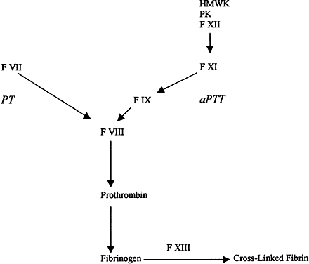
Within the extrinsic, intrinsic, and fibrinolytic cascades, there are complex autoregulatory mechanisms that tightly control their respective enzymatic activities. The interdependence of these separate pathways is perhaps best shown in disease states in which individual factors are lacking or dysfunctional as a result of either hereditary or acquired disorders. The severe hemorrhagic complications resulting from the absence of a single factor underscore the interdependence of the individual factors and pathways in the maintenance of hemostasis. These deficiencies can result in a spectrum of clinical manifestations ranging from subtle bleeding events to fulminant hemorrhagic crises. This section briefly reviews some of the coagulation disorders, both hereditary and acquired, that have been associated with ocular disease.

COAGULATION FACTOR DEFICIENCIES

The most common inherited coagulation disorders involve X-linked deficiencies of factors VIII (hemophilia A) and IX (hemophilia B), which are necessary for the generation of activated factor X by the intrinsic pathway. These disorders are clinically indistinguishable and are characterized by insidious onset of hemorrhage occurring hours to days after injury. The severity of the bleeding is correlated with the extent of the respective factor's deficiency. Patients with less than 1% of normal activity have severe hemorrhagic disease, usually presenting with complications at birth. Levels of factor activity from 1% to 5% of normal are considered moderate, and bleeding may not become apparent until the patient begins crawling or walking. Mild hemophilia with levels of from 5% to 25% of normal may not be diagnosed in patients until adolescence or after major trauma or surgery. The classic presentation of hemophilia involves pain and swelling in a weight-bearing joint because of hemarthrosis. Chronic synovitis may result, with long-term disabling sequelae. Intramuscular hematomas are common, and large hematomas may compress vital structures. Central nervous system (CNS) bleeding accounts for 25% of the deaths due to hemophilia and has a mortality rate of 34%.2 Although replacement therapy of the respective deficient factor in hemophilia has greatly improved the prognosis, it also has resulted in other severe problems such as hepatitis and acquired immunodeficiency syndrome (AIDS) from virally contaminated blood products.

Deficiencies of other factors are much less common than the hemophilias. Factor XI deficiency is an autosomal-recessive disorder with relatively minor clinical manifestations such as epistaxis, hematuria, and menorrhagia. It usually is detected when bleeding ensues after surgery. The disparity in clinical severity between factor XI deficiency and the hemophilias is explained by the ability of the extrinsic system to bypass factors XII and XI through direct activation of factor IX. Thus, an intact extrinsic pathway can compensate for dysfunction early in the intrinsic pathway.1

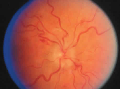
Although ocular and periocular hemorrhages may occur, particularly after trauma or surgery, ocular manifestations of hemophilia are primarily neuro-ophthalmologic in nature, resulting from CNS hemorrhage. Pupillary abnormalities, cranial nerve palsies, visual blurring, and papilledema have been described after intracranial bleeding.2,3 Repeated retinal hemorrhages and vitreous hemorrhage have been associated with factor IX deficiency.4,5

THROMBOPHILIA

The hypercoagulable state of thrombophilia is associated with both genetic and acquired defects of hemostasis that result in a predisposition to venous thrombosis through the malfunction of the normal balance between anticoagulant and procoagulant factors. Thrombophilia is a multigenic disorder resulting from both genetic and acquired disorders.6 Genetic risk factors for thrombophilia include factor V Leiden abnormality which causes activated protein C resistance, prothrombin variant 20210a, antithrombin III deficiency, protein C deficiency, and protein S deficiency. Acquired disorders causing thrombophilia include antiphospholipid antibody syndromes, hyperhomocysteinemia, and abnormal lipoprotein(a) levels.

Activated Protein C Resistance/Factor V Leiden

Protein C is a vitamin K-dependent plasma protein that acts as a physiologic anticoagulant. Protein C circulates as an inactive zymogen and is activated by enzymatic cleavage on the surface of endothelial cells by a complex of thrombin and thrombomodulin. Activated protein C is an important endogenous anticoagulant factor that inactivates the procoagulant factors V and VIII, thereby limiting the production of thrombin.7 A hypercoagulable state can result from resistance to the effects of activated protein C as measured by the activated partial thromboplastin time (PTT) assay.8 Activated protein C resistance has been shown to be the result of heterozygosity or homozygosity of a single-point mutation in the factor V gene resulting in a mutant factor V, in which arginine is replaced by glutamine at position 506.9 This is now termed factor V Leiden, which accounts for more than 80% of the cases of activated protein C resistance.10 To date, no other mutations have been associated with activated protein C resistance. Factor V Leiden is the most common genetic predisposition to venous thrombosis. It occurs in 5% to 7% of whites but is rare in nonwhite populations.6,10,11 It has been reported to be as common as occurring in up to 15% of some European populations.12

The association of activated protein C resistance and factor V Leiden with ocular thrombotic disease has been controversial. Activated protein C resistance was first reported in patients with systemic thrombophilia and central or branch retinal vein occlusions.13,14 However, other reports have failed to show a positive correlation between factor V Leiden and retinal vascular occlusions.15,16 Some reports have displayed a correlation between factor V Leiden and retinal vein occlusions. Patients with central retinal vein occlusions (CRVOs) were found to have a higher prevalence of factor V Leiden mutations compared with that of the general populations.17–19 Heterozygosity for the mutant allele has been associated with a fivefold to 10-fold increased risk of venous thrombosis and homozygosity with a 50- to 100-fold risk.20 Studies of patients with Behçet's disease found that those with ocular and systemic thromboembolic disease had a higher than normal incidence of factor V Leiden mutation.21 Overall, because of the high prevalence of factor V Leiden mutations in the general population and the small number of people with the mutation in whom venous thromboembolism develops, it seems clear that factor V Leiden carriers have thrombosis develop along with other risk factors.22 Additionally, factor V Leiden alone probably is not an important risk factor for arterial disease; however, in the presence of other risk factors, such as smoking, atherosclerosis, and other genetic defects, it may be an associated arterial risk factor.12

Prothrombin Mutations

A genetic risk factor that predisposes to venous thrombosis has been identified in the gene for prothrombin involving a mutation of nucleotide position 20210a. This mutation occurs in 1% to 2% of healthy individuals, 6% of individuals with the first onset of thrombo-embolic disease, and up to 18% of patients with unexplained familial thrombophilia. It is the second most common inherited abnormality, next to factor V Leiden, which leads to an increased risk of thrombosis. The mechanism by which prothrombin variant 20210a causes thrombophilia is unknown. Screening for this defect relies on DNA analysis. There is a strong interaction between this defect and other genetic risk factors, particularly the factor V Leiden mutation.6,10,11 Some reports suggest an association between prothrombin variant 20210a and ocular vascular disease. However, one report failed to confirm an association between prothrombin variant 20210a and central or branch retinal vein occlusions.17

Protein C Deficiency

Protein C deficiency has been associated with more than 160 mutations.23 Although homozygous deficiencies of protein C tend to result in severe thrombotic disease, heterozygous deficiencies and acquired defects result in milder thrombotic tendencies. Heterozygosity for protein C deficiency occurs in 0.3% of healthy individuals according to blood donor statistics.24 It is estimated to occur in approximately 6% of patients with inherited thrombophilia. A heterozygous deficiency of protein C confers a sevenfold increase in the risk of venous thromboembolic disease, and the risk increases with lower protein C levels.

Protein C is a vitamin K-dependent protein. Initiation of anticoagulation therapy with vitamin K antagonists may cause thrombosis in patients with protein C deficiency, which has been implicated in retinal vein occlusions and amaurosis fugax.25–27 Deficiency also has been associated with vitreous hemorrhages and hemorrhagic retinal and choroidal detachment in neonates.28

Protein S Deficiency

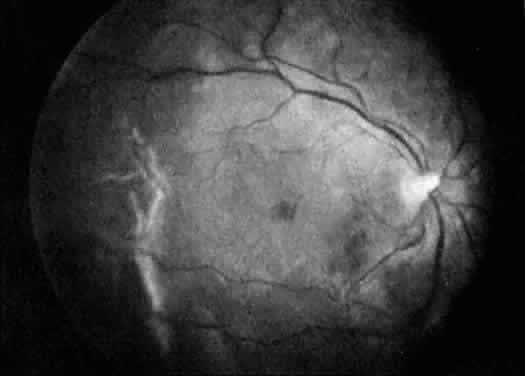
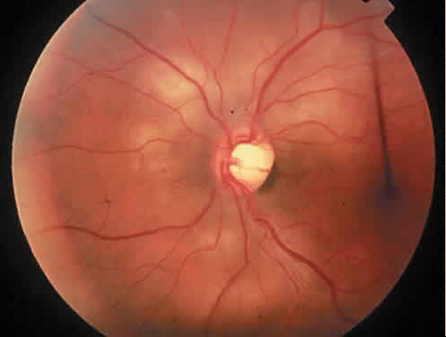
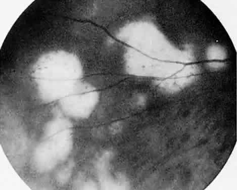
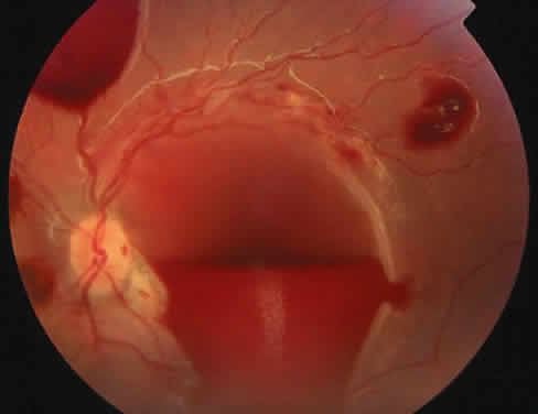
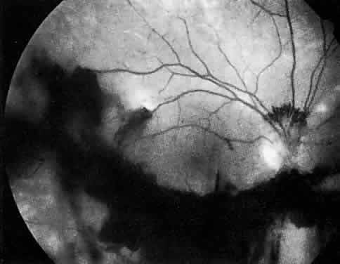
Protein S is a nonenzymatic cofactor necessary for the anticoagulant activity of activated protein C. Protein S by itself also has anticoagulant activity by forming a complex with C4b binding protein, a regulatory protein of the complement system. Protein S deficiency is inherited as an autosomal-dominant trait. Unlike protein C deficiency or antithrombin III deficiency, heterozygous protein S deficiency is not as strong a risk factor for thrombosis. Retinal artery occlusion has been described with protein S deficiency.25,29 Figure 2 shows a pregnant woman with protein S deficiency and a branch retinal artery occlusion (see Fig. 2).

Fig. 2. A 32-year-old woman during her third trimester of pregnancy with a branch retinal artery occlusion. She was found to have protein S deficiency.

Antithrombin III Deficiency

Antithrombin III is an endogenous inhibitor of thrombin and other procoagulant factors and thus is a major coagulation inhibitor. Elevated systemic thrombin-antithrombin III complexes indicate higher thrombin formation and, thus, a hypercoagulable state in the systemic circulation. Studies have shown elevated thrombin-antithrombin III complexes in patients with vein occlusions compared with those of the general population.30

Antithrombin III deficiency is inherited as an autosomal-dominant defect with heterozygosity conferring an increased risk of venous thrombosis. The prevalence of antithrombin III deficiency is increased in patients with inherited thrombophilia (4% vs. 0.02%). More than 70 specific mutations have been identified. Antithrombin III deficiency has been associated with central retinal vein occlusion and cilioretinal artery occlusion.25

Antiphospholipid Syndromesand Lupus Anticoagulant

The antiphospholipid antibody syndrome is a clinical syndrome of venous and arterial thrombotic events. Antiphospholipid antibodies consist of two different antibodies: (1) the lupus anticoagulant and (2) the anticardiolipin antibody. Although the two are similar, there are distinct clinical, laboratory, and biochemical differences. The anticardiolipin antibody antiphospholipid syndrome is more common than the lupus anticoagulant syndrome. Both of these antibodies are associated with thrombosis in connective tissue diseases such as lupus and autoimmune diseases, malignancy, AIDS, and multiple drugs.31 However, some cases of lupus anticoagulant and anticardiolipin antibody are found in otherwise healthy patients. These people are considered to have primary antiphospholipid antibody syndrome. Both syndromes may be associated with arterial and venous thrombosis, fetal loss, and thrombocytopenia. However, the anticardiolipin syndrome often is associated with both arterial and venous thrombosis, including premature coronary and cerebrovascular disease and retinal vascular disease.32 The lupus anticoagulant, although sometimes associated with arterial disease, is more often associated with venous thrombosis.31

Lupus anticoagulant is an immunoglobulin G antibody to anionic phospholipids that is related to anticardiolipin and the biologic false-positive test result for syphilis. This antibody is deemed anticoagulant because of its in vitro effects on results of coagulation tests such as the PTT test. Despite these in vitro effects, lupus anticoagulant is character-ized clinically by ocular and systemic thrombotic events.31,33 The mechanism of clinical thrombosis is uncertain, but it probably involves interference with normal coagulation mechanisms by the antibodies. It is hypothesized that the antibodies act on components of platelet membranes and vascular endothelium as well as on natural anticoagulant proteins such as protein C, protein S, and antithrombin III.32

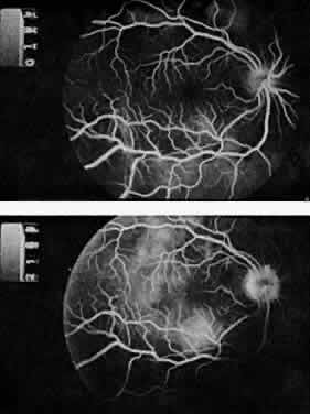
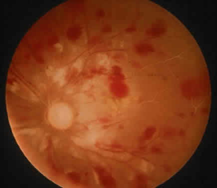
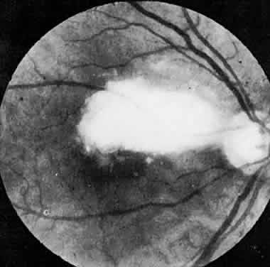
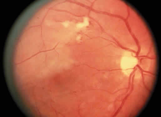
The ocular manifestations of antiphospholipid antibodies include retinal venous and arterial occlusions, amaurosis fugax, diplopia, and visual field loss.34,35 Extensive vasoocclusion, neovascularization, and vitreous hemorrhage may occur (Fig. 3). Treatment consists of photocoagulation for the neovascularization and systemic anticoagulation and immunosuppression. Vitrectomy may be required for vitreous hemorrhage. The role of systemic treatment of lupus anticoagulant in the management of ocular disease is unclear. Some investigators suggest that systemic anticoagulation be started promptly.36 The optimal duration of anticoagulation and whether antiplatelet therapy should be used are not known however. The optimal use of corticosteroids or other immunosuppressive drugs such as cyclophosphamide and azathioprine also is unknown. Two reports have found an association with the antiphospholipid syndrome and some additional retinal conditions. One report also has associated the primary antiphospholipid syndrome with central serous chorioretinopathy.37 In addition, lupus anticoagulant positivity could represent an additional risk factor for diabetic retinopathy according to a recent report.38

Fig. 3. Retinopathy associated with lupus anticoagulant with disc neovascularization and vitreous hemorrhage. (Kleiner RC, Najarian LV, Schatten S et al: Vaso-occlusive retinopathy associated with antiphospholipid antibodies [lupus anticoagulant retinopathy]. Ophthalmology 96:898, 1989.)

Lupus anticoagulant should be suspected in allcases of occlusive retinopathy without obvious causes, especially in young adults. Although the diagnosis of lupus anticoagulant is suggested by prolongation of the PTT, this test is not reliable as a screening assay. The Russell viper venom time and the tissue thromboplastin inhibition test have been advocated as sensitive and relatively specific tests for detection of lupus anticoagulant. Anticardiolipin antibodies can be measured by enzyme-linked immunosorbent assay, which can be more sensitive.39–41

Hyperhomocysteinemia

The term hyperhomocysteinemia describes a variety of disorders that cause elevated plasma levels of homocysteine, homocystine, and their metabolites. Homocysteine is an amino acid formed during the metabolism of methionine. Vitamins B12 and B6 and folate serve as cofactors in the metabolism of methionine (Fig. 4).

Fig. 4. Homocysteine pathway. Numbers indicate principal enzymes involved in hyperhomocysteinemia. (1) 5,10-methylenetretrahydrofolate reductase and (2) cystathionine-beta-synthase. (Adap-ted from Cecil's Textbook of Medicine.)

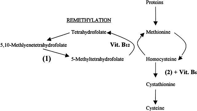
Hyperhomocysteinemia is an independent risk factor for thrombophilia and usually is caused by genetic enzymatic defects in homocysteine metabolism or nutritional deficiencies in vitamin cofactors. Congenital homocystinuria is a rare homozygous deficiency associated with marked hyperhomocysteinemia resulting in premature vascular disease. Ocular manifestations of congenital homocysteinemia include dislocation of the lens and retinal vascular occlusions. Heterozygous deficiencies of cystathionine B-synthaseIQ3 or methylenetetrahydrofolate reductase are relatively common. Nutritional deficiencies of vitamin cofactors such as folate, vitamin B12, and vitamin B6 contribute to approximately two thirds of all cases of hyperhomocysteinemia. Vitamin supplementation can normalize high homocysteine concentrations.

Overall, mild hyperhomocysteinemia occurs in 5% to 7% of the general population.42,43 Some studies have estimated that 10% of the risk of coronary artery disease in the general population is attributable to hyperhomocysteinemia.44 Ophthalmic studies have shown an increased incidence of hyperhomocysteinemia in young adults with retinal artery and vein occlusions.45 One study found that hyperhomocysteinemia is a risk factor for central retinal vein occlusions and was found in a high percentage of bilateral CRVOs (55%) and ischemic CRVOs (30%) and was associated with severe vision loss.46

Fibrinolysis

Fibrinolysis is responsible for the degradation of blood clots via plasmin. Plasmin activity is controlled by a balance between plasminogen activators and plasminogen activator inhibitors. Inhibition of plasmin activity (hypofibrinolysis) is a cause of thrombophilia. Hypofibrinolysis may occur from genetic mutations in plasminogen activator inhibitor activity or plasminogen activity.11 Some have speculated that hypercoagulability and fibrin deposition trigger fibrin(ogen)olysis, which can be hampered to some extent by elevated levels of lipoprotein(a).47 Thus, high levels of lipoprotein(a) interfere with plasminogen activation. Dysfunctional fibrinolysis has been correlated with central vein occlusion.17,47,48

THROMBOPHILIA SCREENING

Thrombophilia is a hypercoaguable state resulting from genetic or acquired abnormalities of the hemostatic mechanism that predispose to thrombosis. Multiple mechanisms contribute to thrombophilia, making it a multigenic disorder. The detection and treatment of thrombophilia are directed toward preventing life-threatening vascular disease such as deep vein thrombosis, pulmonary embolism, myocardial infarction, and cerebral vascular disease in the patient or potentially affected family members. The use of systemic anticoagulation to treat this condition remains controversial.49 There are no data to guide the ophthalmologist on the role of anticoagulation in the treatment of thrombophilia and related ocular complications.

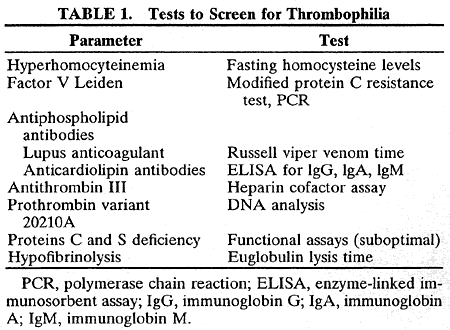
In the laboratory, the main abnormalities to search for are hyperhomocysteinemia, factor V Leiden, prothrombin variant (gene 20210a), antiphospholipid antibodies, antithrombin III, protein C and protein S deficiencies, and hypofibrinolysis (Table 1).


Detecting hyperhomocysteinemia has important clinical implications. These patients can be treated with vitamin supplementation instead of long-term anticoagulant treatment. The diagnosis of hyperhomocysteinemia can be made by measuring blood levels of homocyst(e)ine during fasting or by giving an oral dose of methionine with subsequent measurement of homocyst(e)ine blood concentrations.43

Screening tests for activated protein C resistance (factor V Leiden mutation) are easy to perform and have a high sensitivity and specificity (almost 100%). Multiple factors make screening for factor V Leiden using a PTT-based test unreliable and thus impractical.15 Screening for activated protein C with newer molecular testing is highly sensitive and specific, making molecular testing the definitive diagnostic method.6,10,11 The “modified activated protein C resistance test” is based on coagulation assays that dilute the patient's plasma sample with factor V-deficient plasma. This enables identification of patients with or without activated protein C resistance. Furthermore, the modified activated protein C resistance test has been shown to be more sensitive and specific in identifying protein C resistance in children.50 Molecular biology (polymerase chain reaction) is now only needed to confirm the factor V abnormality.12

Assays to look for prothrombin gene variant 20210a are not commonly performed. Screening for this defect relies on DNA analysis and is impractical. Information on testing positive for this genetic variant would be most beneficial in an individual with other genetic factors, particularly the factor V Leiden mutation.

Antiphospholipid antibodies can be detected by coagulation or immunologic assays. Lupus anticoagulant assays do not actually measure a titer of antibody but are functional tests. The in vitro detection methods for lupus anticoagulant appear to have no relevance to its in vivo actions. Lupus anticoagulants prolong clotting times in vitro because they agglutinate phospholipids in the plasma, thereby preventing their participation as cofactors in coagulation steps. When a patient is suspected of having lupus anticoagulant, performing a PTT test is not adequate. A more definitive test is the Russell viper venom time.31 Similarly, a PTT test usually is not prolonged in patients with anticardiolipin antibodies, and thus definitive tests such as enzyme-linked immunosorbent assays for immunoglobulin G (IgG), immunoglobulin A (IgA), and immunoglobulin M (IgM) anticardiolipin antibodies should be performed. Similarly, screening for anticardiolipin antibodies requires coagulation and immunologic assays.12

Antithrombin III deficiency is simple to detect in most coagulation laboratories using a standardized heparin cofactor assay.12 There are few variations in assay levels between patients of different ages or sex, and therefore a narrow reference range is used to determine antithrombin III abnormalities. The genetic defects of antithrombin III deficiency have been identified but do not need to be tested to identify the defect.

Screening for protein C deficiency uses clotting or amidolytic assays. There is no easy functional assay for protein C deficiency. Multiple factors may affect assay results, including age, sex, and hormonal therapy, thus causing overlap between normal subjects and heterozygotes.10,11 Molecular characterization is possible, but the large number of defects that exist makes this an impractical screening method.12

Screening for protein S deficiency relies on functional assays that are suboptimal because of high percentages of false-positive and false-negative results. Currently, most laboratories use enzyme-linked immunosorbent assays to measure free and total protein S antigen. There is no proven value of functional assays measuring protein S activity.12 Thus, the diagnosis of protein S deficiency remains difficult.

Assays to look for hypofibrinolysis (i.e., reduced level of tissue plasminogen activator or increased levels of plasminogen activator inhibitor) are not performed. There is a large variation of results, and baseline fibrinolytic value does not predict the occurrence of future thrombosis.

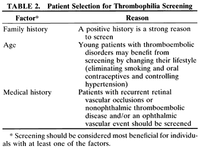
Defining which patients to screen for thrombophilia is controversial. Some general guidelines include selecting patients who have a family history of thromboembolic disease, recurrent thrombosis, an early age of onset, both arterial and venous thrombosis, or those who have thrombosis and fetal losses. It is important to rule out common acquired causes of thrombophilia such as cancer before extensive laboratory investigation. Additionally, tests should only be performed if they are going to change the treatment of the patient or if they potentially have consequences for the patient's family. Finding an abnormality rarely influences the management of an acute event. Moreover, patients who are anticoagulated, pregnant, or taking oral contraceptives have altered thrombophilia test results. These factors must be accounted for in patient screening and testing.

To investigate a patient and their family is to give specific prophylactic and therapeutic recommendations for a sustained risk for thrombosis (Table 2). For example, a symptomatic patient with multiple thrombotic events may be treated with anticoagulation for a variable duration depending on the number of risk factors detected and the clinical circumstance. Conversely, patients who are diagnosed with hyperhomocysteinemia can be treated with vitamin supplementation instead of long-term anticoagulant treatment. Another scenario in which the subject would benefit from screening is an asymptomatic family member who is found to be a carrier of a thrombophilia genetic defect. Depending on the number of risk factors, the patient may receive prophylaxis anticoagulation during high-risk situations such as pregnancy, surgery, or immobilization. A patient also may benefit from the knowledge that she is more predisposed to be hypercoaguable by changing certain aspects of her lifestyle such as stopping smoking, avoiding oral contraceptives, and tightly controlling hypertension.


There are no data to support thrombophilia screening for the general population. A combined clinical and laboratory approach, taking into account the history of the patient and the patient's family and the sensitivity and specificity of the tests, should decide which tests are most appropriate for a patient.12

Back to Top
THROMBOPHILIA TREATMENT AND ANTICOAGULANT THERAPY
Bleeding disorders associated with anticoagulant therapy probably are more common than inherited or acquired coagulation deficiencies.51 Anticoagulants are used therapeutically to prevent or limit thrombosis in a variety of conditions such as pulmonary embolism, ischemic cerebrovascular disease, cardiac disease, and postoperative immobility.

Heparin is used for acute anticoagulation, and the coumarin group of anticoagulants is used for chronic therapy. Heparin is a naturally occurring mucopolysaccharide that enhances the activity of antithrombin III, the major endogenous anticoagulant factor. In the presence of heparin, antithrombin III more effectively neutralizes thrombin and other activated coagulant factors, thereby slowing coagulation. Recently, low-molecular-weight heparins have established themselves as an important class of antithrombotic compounds. They produce a more predictable anticoagulant response and cause less bleeding than unfractionated heparin. The low-molecular-weight heparins are being used withgreater frequency because of these advantages.52 The coumarin anticoagulants interfere with vitamin K metabolism, thereby affecting production of thrombin and factors VII, IX, and X and thus slowing coagulation.1 There is a need for more potent anticoagulant drugs that are able to inactivate thrombin bound to fibrin, which may be an important trigger for clot extension at the sites of vascular injury.52 New drugs may enable more effective treatment of thrombotic diseases.

Management of thrombophilia requires anticoagulation with heparin and coumarin anticoagulants and is based only on recommendations for certain clinical scenarios and not definitive clinical trials. Patients can be classified into two general categories: (1) high risk and (2) moderate risk of thrombosis. High-risk patients require indefinite anticoagulation, and moderate-risk patients require vigorous prophylaxis during high-risk situations such as surgery, prolonged immobilization, and pregnancy. High-risk patients are those who have an episode of thrombosis plus either inherited thrombophilia or acquired thrombophilia. Additionally, patients are considered high risk if they have two episodes of documented thrombosis at different sites or if they have one episode and a positive family history for thromboembolism. Patients are considered to be at moderate risk if they are asymptomatic carriers of inherited thrombophilias or if they have acquired thrombophilia without evidence of a clinical thromboembolic episode.53 Recent reports, however, show that even the patients categorized as being at moderate risk for thromboembolic disease may benefit from long-term anticoagulation.54

The incidence of clinically significant bleeding associated with anticoagulation is dependent on the underlying primary disease process, the intensity of the anticoagulant therapy, and the duration of therapy. Minor hemorrhages such as epistaxis, hematuria, ecchymosis, and hemoptysis occur in up to 25% of anticoagulated patients. The incidence of major hemorrhages requiring transfusion or hospitalization is as high as 8%, and fatal bleeding occurs in as many as 5%.51

Anticoagulation with therapeutic agents has been implicated in various hemorrhagic ocular conditions. Isolated cases of vitreous hemorrhage have been associated with anticoagulation.55,56 However, these reports do not provide information on the type of anticoagulation used, the length of therapy, or the parameters of blood coagulation at the time of hemorrhage. Spontaneous hyphema during coumarin-type anticoagulation in both phakic and iris-fixated pseudophakic eyes also has been described.57,58 Massive subretinal and vitreous hemorrhage in age-related macular degeneration has been associated with anticoagulant therapy.59,60 These hemorrhages, which invariably result in permanent loss of vision, can present with sudden onset of severe ocular pain and elevated intraocular pressure. Histopathologic analysis suggests that the source of the hemorrhage is large choroidal vessels that vascularize into disciform scars. Anticoagulation may predispose these vessels to bleed.

Treatment of patients receiving chronic anticoagulation therapy before surgery can be difficult. Interruption of anticoagulant therapy increases the risk of thromboembolism. After warfarin therapy is discontinued, it takes several days for its antithrombotic effects to recede and, when resumed, several days to reestablish therapeutic anticoagu-lation.61 Thus, interruption of anticoagulant ther-apy increases a patient's risk of thromboembolism.A study of 50 ophthalmic surgical procedures in-cluding extracapsular cataract extraction, trabec-ulectomy, and vitreoretinal surgery on 41 anti-coagulated patients showed zero hemorrhagiccomplications with the procedures or local anesthetic placement.62 The authors concluded that ophthalmic surgery probably can be performed while the patient is therapeutically anticoagulated. Another study showed no difference in complications between patients who stopped and those who continued anticoagulant therapy before ophthalmic surgery. However, there were significantly more hemorrhagic complications when ticlopidinine hydrochloride (antiplatelet medication) was continued.63

The use of anticoagulant medication in patients with a potentially hemorrhagic ocular condition such as proliferative diabetic retinopathy or choroidal neovascularization presents a difficult management problem for both ophthalmologists and internists. The risk of ocular hemorrhage must be weighed against the potentially life-threatening complications of the systemic disease for which anticoagulant therapy is being used. Although it seems reasonable to infer an increased risk of ocular hemorrhage during anticoagulant therapy, the actual incidence of this event is unknown, and there is no evidence to support withdrawal or denial of appropriately indicated anticoagulant therapy because of fear of ocular bleeding.

Back to Top
DISSEMINATED INTRAVASCULAR COAGULATION
Disseminated intravascular coagulation (DIC) is a syndrome precipitated by various disease states that results in uncontrolled systemic coagulation and fibrinolysis. In the past, the ophthalmic literature has confused thrombotic thrombocytopenic purpura (TTP) with DIC, probably because of their similar ophthalmic and histologic findings.64 Although both entities involve thrombocytopenia, results of coagulation tests are abnormal in DIC and unaffected by TTP. DIC and TTP are now considered separate entities.

The classic ocular finding in DIC is serous retinal detachment64,65 (Fig. 5). The pathogenesis of these detachments involves choriocapillaris occlusion resulting in retinal pigment epithelial damage and subsequent loss of retinal pigment epithelial barrier and pump function. Fluorescein angiography confirms this pathophysiology, showing delayed filling of the posterior choroid with later pigment epithelial staining (Fig. 6). If the underlying DIC can be reversed, the retina may reattach, with return of vision. Other findings associated with DIC include retinal and vitreous hemorrhages.

Fig. 5. Serous retinal detachment in disseminated intravascular coagulation. (Hoines J, Buettner H: Ocular complications of disseminated intravascular coagulation [DIC] in abruptio placentae. Retina 9:107, 1989.)

Fig. 6. Delayed choroidal filling with pigment epithelial staining in disseminated intravascular coagulation. (Hoines J, Buettner H: Ocular complications of disseminated intravascular coagulation [DIC] in abruptio placentae. Retina 9:107, 1989.)

Back to Top
PLATELET DISORDERS
Platelets have a primary role in normal hemostasis.66 They form hemostatic plugs at sites of vascular disruption and also are important in the activation of the coagulation system. Platelet disorders may result in a broad spectrum of ocular manifestations ranging from hyphema to serous retinal detachments. The majority of clinically significant platelet disorders are secondary to either thrombocytopenia or platelet dysfunction syndromes.

IDIOPATHIC THROMBOCYTOPENIC PURPURA

A common cause of thrombocytopenia is accelerated platelet destruction due to either immunologic or nonimmunologic mechanisms.66 Immune-mediated mechanisms are the result of antiplatelet antibodies. These antibodies occur in idiopathic thrombocytopenic purpura (ITP) or after drug exposure.

ITP may occur acutely in children as a self-limited condition after a viral infection or, more typically, as a chronic disorder of young and middle-aged women. Autoantibodies in ITP are directed against epitopes on glycoprotein IIB/IIIA, which are the most abundant immunogenic platelet-surface glycoproteins.67 Additionally, ITP has been associated with the antiphospholipid antibody syndrome. In this disorder, antibodies are directed against complexes of proteins and negatively charged phospholipids, including exposed platelet phospholipids.68

Systemic manifestations of ITP range from petechiae to intracranial hemorrhage. Ocular manifestations of ITP include retinal hemorrhages69 (Fig. 7) and an interesting association with Graves' disease.70 The development of thrombocytopenia in Graves' disease probably involves either autoimmunity or secondary effects of thyrotoxicosis. This association should be considered in patients with Graves' disease and unexplained ocular bleeding and particularly in patients with Graves' disease undergoing ocular or orbital surgery.

Fig. 7. Retinal hemorrhages in a patient with a platelet count of 10,000/mm3.

Another type of immune-mediated thrombocy-topenia occurs after drug exposure. Certain drugsact as a hapten, and the ensuing drug-antibody com-plex then binds to the platelet, resulting in plate-let destruction.66 Quinine and quinidine are thebest studied offenders, but many others includingsulfonamides, heparin, phenytoin, diazepam, and acetaminophen have been implicated. Taking a thorough drug history is necessary to rule out drug exposure.

Drug-induced thrombocytopenia may cause various ocular bleeding events. Vitreous hemorrhage, subconjunctival hemorrhage, and postsurgical bleeding after lid surgery have been attributed to drug-induced thrombocytopenia.71,72 Bilateral serous retinal detachments and disc edema also have been described.73

Treating ITP with prednisone only produces long-term responses in fewer than 25% of patients. The morbidity of corticosteroids limits the duration of steroid treatment. Another treatment of ITP includes intravenous gamma globulin infusions, which cause an immediate increase in the platelet count. However, there are no advantages to long-term immunoglobulin therapy.74 Most patients with symptomatic ITP require splenectomy, providing approximately two thirds of patients with lasting remission.67

THROMBOTIC THROMBOCYTOPENIA PURPURA

The prototype of nonimmunologic accelerated destruction of platelets is TTP. TTP is a disease of unknown etiology that affects children or adults of any age but especially young adult females, which can prove fatal within a few weeks. The hallmark of this enigmatic entity is diffuse platelet activation and aggregation resulting in five major clinical manifestations: microangiopathic hemolytic anemia, thrombocytopenia, fever, CNS dysfunction, and renal disease.

The most common ocular manifestations of TTP include papilledema, extraocular muscle palsies, and visual field defects, which usually are secondary to concomitant CNS involvement.75 Retinal findings consist of hemorrhages, retinal vascular occlusions, and serous detachments76,77 (Figs. 8 to 10). The cause of the serous detachments appears to be focal occlusion of the choriocapillaris resulting in retinal pigment epithelial damage and blood-retinal barrier disruption.76,77 Findings on fluorescein angiography are characterized by focal areas of nonperfusion of the choriocapillaris associated with late leakage into the subretinal space76 (Fig. 11). This is consistent with histopathologic studies that show occlusion of the choriocapillaris and large choroidal vessels, presumably by fibrin, with overlying necrosis of thepigment epithelium.75,76 TTP also has been linked in one case report with Purtscher retinopathy.78

Fig. 8. Patient with thrombotic thrombocytopenic purpura and extensive retinal vascular-occlusive disease. (Courtesy of William Mieler, MD.)

Fig. 9. Fluorescein angiogram of patient with thrombotic thrombocytopenic purpura and vascular-occlusive disease. (Courtesy of William Mieler, MD.)

Fig. 10. A 42-year-old woman with thrombotic thrombocytopenic purpura and neurosensory retinal detachments. (Courtesy of Jerry Neuwirth, MD.)

Fig. 11. Fluorescein angiogram of patient with thrombotic thrombocyto-penic purpura showing late choroidalhyperfluorescence caused by focal areas of choriocapillaris nonperfusion. (Courtesy of Jerry Neuwirth, MD.)

Clinically, the development of serous retinal detachments usually is associated with exacerbations of TTP and the development of acute hypertension. Although serous retinal detachments have been described as a preterminal event, resolution of the detachments with subsequent pigment epithelial changes may occur when the underlying hypertension and thrombocytopenia are controlled.77

OTHER CAUSES OF THROMBOCYTOPENIA

Thrombocytopenia also may occur as a result of bone marrow suppression or infiltration, as in disease such as aplastic anemia, leukemia, or lymphoma. Retinal hemorrhage or vitreous hemorrhage may be more likely to occur when thrombocytopenia is accompanied by anemia.57

Platelet dysfunction syndromes may occur as congenital or acquired defects of platelet metabolism. These abnormalities can involve platelet adhesion, aggregation, or release. Various ocular findings and diseases have been associated with these disorders (Fig. 12).

Fig. 12. Patient with multilayered hemorrhages and a platelet count of 10,000. (Courtesy of Craig M. Greven, MD.)

Thrombasthenia is an autosomal-recessive disorder characterized by failure of platelets to aggregate in response to adenosine diphosphate. These platelets have abnormal binding sites for fibrinogen and fibronectin, resulting in poor platelet aggregation and clot retraction.66 Spontaneous preretinal and vitreous hemorrhages may ensue.79

Familial exudative vitreoretinopathy is a retinal vascular disorder characterized by peripheral vascular abnormalities similar in appearance to retinopathy of prematurity. Two families with the disorder have been described as having abnormal platelet aggregation in response to arachidonic acid.80 Whether this thrombocytopathy has a role in the pathogenesis of familial exudative vitreoretinopathy is unknown.

Abnormalities of platelet amino acid metabolism and protein content have been associated with some types of retinitis pigmentosa.75 A mild thrombocytopenia also has been noted in some cases of retinitis pigmentosa.77 It is unknown whether these disorders of platelet function play any part in the pathogenesis of retinitis pigmentosa.

Acquired platelet dysfunction may occur with diabetes, liver disease, uremia, or macroglobulinemia but occurs most often after ingestion of aspirin or other nonsteroidal antiinflammatory agents. These drugs affect platelet prostaglandin metabolism by inhibiting platelet cyclooxygenase, which is important in platelet aggregation and granule release. Unlike other nonsteroidal antiinflammatory agents, aspirin irreversibly acetylates cyclooxygenase at doses as low as 20 mg. This irreversible inhibition of platelet function prolongs the bleeding time for about 7 days.81

Despite the marked in vitro effects of aspirin or salicylates on platelet function, the incidence of retinal or choroidal bleeding associated with these drugs appears low.82 Massive subretinal and vitreous hemorrhage occurring in age-related macular degeneration has been associated with aspirin use.59 It is hypothesized that the aspirin may predispose choroidal neovascularization to bleed. Other ocular sequelae of these drugs involve hyphema rebleeding and postsurgical bleeding.83,84 A more recent study showed no adverse events associated with phaco-emulsification and lens implantation, both with retrobulbar anesthesia and topical anesthesia, in patients treated with aspirin.85

Acquired abnormalities of platelet function have been implicated in the pathogenesis of diabetic retinopathy. However, review of the literature produces a confusing picture of the importance of platelet dysfunction in diabetic retinopathy.86 Multiple parameters of platelet function including aggregation, platelet survival, and release of platelet factor 4 and β-thromboglobulin have been correlated with diabetic retinopathy.87 It remains uncertain, however, whether these changes represent primary events in the pathogenesis of diabetic retinopathy or are simply epiphenomena. Based on the assumption that platelet dysfunction is involved in the microangiopathy of diabetic retinopathy, the Early Treatment Diabetic Retinopathy Study examined the effect of aspirin at a daily dose of 650 mg on the course of diabetic retinopathy in almost 4000 patients.88 This study showed no effect of aspirin on progression of retinopathy or visual acuity. Perhaps more important, considering the widespread use of aspirin for cardiovascular disease, no adverse effect of aspirin was found in patients with proliferative retinopathy.88

Acquired platelet abnormalities also have been described in retinal arterial occlusive disease and acute retinal necrosis.89 Antiplatelet therapy has been recommended for both of these diseases.

Thrombocytopenia may result in bleeding complications in patients undergoing ophthalmic surgery. Bleeding is rarely encountered when the platelet count is greater than 50,000 cells/mm3. However, elevation of the platelet count should be attempted before surgery on all patients with known thrombocytopenia.90

Lymphoma

Intraocular lymphoma is a non-Hodgkin's lym-phoma (NHL) of the eye. It previously was calledreticulum cell sarcoma. The term reticulum cell sar-coma is a misnomer because these tumors areB-cell lymphomas of the diffuse large-cell type.91 The origin of the cells causing intraocular lymphoma is unknown. Most patients are middle-aged, although affliction at earlier ages has been reported.66,92 Ocular involvement usually is bilateral (80%) but often is asymmetric.93,94 Two distinct forms of ocular NHL, systemic NHL and CNS-NHL, can affect the eye.

The clinical findings of intraocular lymphoma are variable and usually nondiagnostic. In general, in patients with systemic NHL and intraocular NHL, the extent of intraocular involvement tends to parallel the severity of systemic involvement.95 Additionally, the long-term survival of patients with intra-ocular involvement from systemic NHL is poor.Patients with systemic NHL are more likely to haveuveal infiltration, whereas those with NHL-CNS generally present with vitreous infiltration.96,97 Most patients present with posterior uveitis or vitritis, but anterior uveitis and optic nerve involvement may occur.94 Other less common manifestations of intraocular lymphoma include retinal vasculitis,98 central retinal artery obstruction,99 and solitary lesions, which resemble acute retinal necrosis.100 Anterior uveitis causing episodes of elevated intraocular pressure also has been reported with NHL.101 Typically, distinctive yellow or white subpigment epithelial masses are thought to be virtually pathognomonic (Fig. 13), but nonspecific peripheral pigment changes may occur. The clinical course often is one of chronic uveitis that is poorly responsive to steroid therapy. Because of the usually nonspecific clinical findings, intraocular lymphoma should be considered in the differential diagnosis of any posterior uveitis in middle-aged individuals. Unfortunately, the diagnosis often is missed, as evidenced by the high percentage of diagnoses made at autopsy.94

Fig. 13. Lesions typical of subpigment epithelial intraocular lymphoma. (Gass JDM, Sever RJ, Gizzard WS et al: Multifocal pigment epithelial detachments by reticulum cell sarcoma. Retina 4:136, 1984.)

Patients with AIDS have a significantly higher risk of having CNS and systemic lymphomas develop. AIDS-related intraocular lymphoma is a distinct subgroup of lymphoma due to the younger mean age of presentation, a more aggressive clinical systemic and ocular course, and a poorer prognosis for survival.102 Histopathologically, most AIDS-related lymphomas are high-grade, B-cell immunoblastic lymphomas of the Burkitt's or non-Burkitt's type. Epstein-Barr virus DNA has been detected in a large percentage of AIDS-related lymphomas.103

Cytologic evaluation of the vitreous by vitreous biopsy is the benchmark for diagnosis.91 Cytology is diagnostically more accurate than lymphocyte subset analysis by immunohistology. However, the diagnosis cannot always be made with a single vitreous biopsy.91 The surgical technique used to obtain the vitreous biopsy sample affects the cytology. Cytologic details of cells taken from the vitrectomy reservoir are inferior to those of cells directly aspirated from the vitreous cavity. Thus, the diagnosis of intraocular lymphoma cannot be ruled out with a single vitreous biopsy. Additionally, the diagnostic yield of cytology specimens is lower in patients taking systemic corticosteroids because of the lytic effect on the lymphoma cells.104 In the presence of suspicious clinical findings, repeat vitreous biopsy or contralateral vitreous biopsy should be considered, as well as a complete systemic diagnostic evaluation.

Techniques for obtaining vitreous biopsies vary. Although some authors believe that vitreous aspiration with a 20-gauge needle is preferable to vitreous biopsy with an automated vitreous cutter, detrimental effects on cytologic detail can be minimized by connecting the aspiration port of the vitreous cutter to a syringe. Suction then is generated slowly and manually with the syringe while the automated vitreous cutter is used to aspirate vitreous fluid at a relatively low cutting rate of about 200 cuts per minute. By simultaneously indenting the eye to prevent hypotony, up to 1 ml of vitreous can be obtained easily without creating excessive traction on the vitreous. The low cutting rate minimizes any possible mechanical effects on the cells. The sam-ple then is given immediately to a cytopathologistfor prompt fixation, thereby avoiding any artifactinduced by the vitrectomy infusion solution. Ad-ditional diagnostic approaches to intraocular lymphoma include vitreoretinal biopsy and fine-needle aspiration biopsy. Transscleral choroidal biopsy can be used to diagnose lymphoma when other diagnostic methods fail.95

The importance of making a prompt diagnosis of ocular lymphoma is reflected in the poor survival rates for this disease. Although aggressive radiation and chemotherapy may improve survival, death usually is secondary to CNS involvement, which may precede or follow ocular involvement. A review of primary CNS lymphoma by O'Neill and Illig105 suggests that intraocular lymphoma represents a syndrome within the varied clinical manifestations of primary CNS lymphoma. In the researchers' experience with 64 cases, 11% of patients with primary CNS lymphoma had episodes of uveitis that preceded the diagnosis by months to years. This study emphasizes the importance of communica-tion between ophthalmologists and their colleaguesin the diagnosis and management of intraocular lymphoma.

Hodgkin's Lymphoma

Hodgkin's lymphoma is characterized by painless lymphadenopathy with a predisposition for opportunistic infections. Ocular involvement is uncommon but may include periphlebitis, chorioretinitis, vitritis, exudative retinal detachment, cotton-wool spots, retinal hemorrhages, and optic nerve edema (Fig. 14). These manifestations may reflect direct tumor infiltration of the eye, as evidenced by a case showing Reed-Sternberg cells in the anterior chamber, or may be secondary changes.106 Resolution of vitritis and retinitis after radiation therapy has been reported in the literature.107

Fig. 14. Fundus appearance with Hodgkin's disease. (Barr CC, Joondeph HC: Retinal periphlebitis as the initial clinical findings in a patient with Hodgkin's disease. Retina 3:25, 1983.)

Plasma Cell Dyscrasias

Plasma cell dyscrasias are characterized by malignant proliferation of B cells with subsequent overproduction of immunoglobulins (antibodies). They are classified into three major types: (1) multiple myeloma with overproduction of either IgG, IgA, immunoglobulin D (IgD), or immunoglobulin E (IgE); (2) macroglobulinemia with overproduction of IgM; and (3) light- and heavy-chain diseases. Ocular involvement may occur in any of the plasma cell dyscrasias.

Multiple myeloma is the most common plasma cell dyscrasia and is characterized by a monoclonal gammopathy and osteolytic bone lesions. Corneal involvement is uncommon but includes stromal crystalline deposits shown to be IgG by immunofluorescence. Cysts of the ciliary epithelium are found histologically in 33% to 50% of patients with multiple myeloma. The cysts are filled by a fluid with a similar immunoelectrophoretic pattern as serum. Retinal involvement includes microaneurysms, nerve fiber layer hemorrhages, infarcts, and exudative retinal detachments. The hyperviscosity syndrome is uncommon in multiple myeloma, thus immunogammopathy causes the ocular effects, not serum hyperviscosity. Macular detachments associated with and without yellow subretinal precipitates have been reported with multiple myeloma and no evidence of systemic hyperviscosity.108 Orbital and neuro-ophthalmic manifestations are caused by the mass effects of plasmacytomas. Proptosis, diplopia, choroidal folds, cranial nerve palsies, and optic nerve involvement have been reported.109

Waldenstrom's macroglobulinemia is caused by an IgM monoclonal gammopathy. Ocular manifestations usually are the result of a hyperviscosity syndrome. Hyperviscosity retinopathy also may occur in multiple myeloma, amyloidosis, and heavy-chain disease. Hyperviscosity retinopathy is related to the severity of hyperviscosity. Venous dilation, retinal hemorrhages, and retinal edema increase with increasing viscosity. In severe cases, exuda-tive detachments occur. Macular neurosensory ele-vation also has been reported in patients withWaldenstom's with simultaneous increased plasmaviscosity.108 Changes from hyperviscosity may be reversible with plasmapheresis.110

Light-chain disease accounts for a small portion of the plasma cell dyscrasias. Ocular involvement consisting of a retinal vasculopathy with capillary nonperfusion, neovascularization, and vitreous hemorrhage has been reported.111

Leukemia

Leukemia is a neoplasm of the hematopoietic system. Leukemic cells show a poor responsiveness to normal regulatory mechanisms, have a diminished capacity for normal cell differentiation, and can have malignant transformation occur at any stage of hematopoiesis. Abnormalities may affect the lymphopoietic or myelopoietic arms of hematopoiesis, resulting in a myelogenous or lymphocytic leukemia, respectively. Leukemia can occur in a rapid and aggressive form (acute) or an indolent form (chronic). These combinations account for the four broad categories of leukemia: (1) acute myelogenous leukemia (AML), (2) acute lymphocytic leukemia (ALL), (3) chronic lymphocytic leukemia (CLL), and (4) chronic myelogenous leukemia (CML). Although all types of leukemia can affect patients at almost any age, typically AML affects males older than 50 years, ALL tends to affect children younger than 15 years of age, CLL affects patients older than 50 years of age, and CML affects patients in the 5th decade with a slight male predominance.112

Ocular involvement in leukemia has long been recognized, and virtually every ocular tissue may be affected. The incidence of ocular involvement at some time during the course of leukemia ranges as high as 90%.113,114 Acute leukemias may be more likely than chronic leukemias to have ophthalmic manifestations, but this difference is not clinically significant.

Schachat and colleagues115 prospectively evaluated 120 patients with both myeloid and lymphoid leukemias. All but 4 of these patients had acute leukemia, and all patients underwent ocular examination, usually within 3 days of diagnosis of the leukemia. This work and a subsequent study by the same group reported by Guyer and associates116 have improved our understanding of the ophthalmic manifestations of leukemia substantially.

Ophthalmic manifestations of leukemia can be classified into two groups: (1) primary leukemic infiltration of tissues and (2) secondary manifestations affecting the eye. Primary leukemic ocular involvement most commonly affects the retina. Typically, leukemic retinal infiltrates are grayish-white nodules that may be associated with surrounding hemorrhages (Fig. 15).113 Clinical evidence of leukemic choroidal infiltration is rare despite the high (65% to 85%) incidence of choroidal involvement at autopsy.113 Only one of 120 patients in the study by Schachat and coworkers had clinical choroidal involvements. However, the prevalence of choroidal leukemic infiltration is related to the agonal leukocyte count and severity of systemic disease, which may explain the infrequency of clinically apparent choroidal disease. Leukemic infiltration of the choroid diminishes choriocapillaris perfusion, resulting in pigment epithelial ischemia and subsequent breakdown of pigment epithelial barrier and pump functions.117 Some reports have described retinal pigment epithelial hyperplasia and clumping. Histopathology in this situation has shown retinal and choroidal infiltration.118,119 Autopsy studies also have shown infiltrative involvement of the optic nerve in 3.6% of eyes studied.113 Clinically, optic nerve head infiltration may appear as a pale gray swelling of the optic nerve head. Clinical studies have suggested that optic nerve involvement is associated with a high frequency of CNS involvement and a poor prognosis.120,121

Fig. 15. Leukemic infiltrate in the retina in a patient with acute lymphocytic leukemia.

Secondary ocular manifestations of leukemia include subconjunctival hemorrhages, intraretinal hemorrhages, cotton-wool spots, central retinal vein obstruction, and white-centered hemorrhages (Fig. 16). White-centered retinal hemorrhages may represent small leukemic infiltrates or they may consist of platelet fibrin material or septic emboli. Thus, this finding should not be considered pathognomonic for primary leukemic involvement. Retinal neovascularization is a rare finding but has been found in the periphery and on the optic nerve in patients with leukemia. The mechanism is thought to be peripheral nonperfusion and ischemia due to increased blood viscosity. However, some have speculated that angiogenic factors from leukemic cells may play a role in this process.122,123 Overall, secondary ophthalmic manifestations of leukemia have been found in up to 39% of patients with leukemia.

Fig. 16. Retinal hemorrhages, exudates, and venous tortuosity in a patient with acute lymphocytic leukemia.

Guyer and associates116 evaluated the relationship between leukemic retinopathic lesions and hematologic parameters at the time of diagnosis. An association between intraretinal hemorrhages and thrombocytopenia was found in all patients, and in those patients with acute lymphocytic leukemia, low hematocrit levels also correlated with intraretinal hemorrhages. White-centered retinal hemorrhages were only associated with anemia in patients with acute nonlymphocytic leukemia. Cotton-wool spots were not associated with hematologic parameters. Interestingly, intraretinal hemorrhages, white-centered retinal hemorrhages, and cotton-wool spots were more common in adults than in children. This finding probably reflects an intrinsically healthier retinal vasculature in children. Patients withcentral vein occlusions tended to have markedlymphocytosis or thrombocytosis and presumed hyperviscosity.

In the study of Schachat and colleagues,115 5% of the patients who had visual acuity measurements had decreased vision. Because the ocular examinations were performed near the time of diagnosis of the leukemias, this finding suggests that some patients may present with ocular involvement as the first sign of leukemia. Stewart and associates117 reported a case of acute lymphocytic leukemia in a 12-year-old boy who presented with unilateral ocular pain and decreased vision secondary to an exudative retinal detachment. The detachment resolved and vision improved after administration of craniospinal radiation therapy, systemic chemotherapy, and intrathecal chemotherapy. Another report showed decreased vision due to bilateral preretinal layered hemorrhages as an initial presentation of acute lymphoblastic leukemia in a 5-year-old child.124

Ocular disease also may occur as a result of treatment for leukemia with bone marrow transplantation.125 A retrospective analysis of patients with pediatric bone marrow transplant showed ocular changes in 51% of patients.126 The most frequent findings included dry eye syndrome (12.5%), cataract (23%), and posterior segment complications (13.5%). Specific ocular complications of bonemarrow transplantation include opportunistic in-fections, hemorrhages, and graft-versus-host dis-ease.125,127 Herpes zoster ophthalmicus, keratitis, and fungal endophthalmitis may occur. Hemorrhagic complications include vitreous and intraretinal hemorrhages. These infectious and hemorrhagic complications are secondary to the transient pancytopenia after bone marrow transplantation. Graft-versus-host disease is a major complication of allogenic bone marrow transplantation resulting from an immunologic response of the lymphocytes of the donor marrow against the antigens of the host. Jabs and colleagues127 have described a form of conjunctivitis that serves as a marker for severe graft-versus-host disease. They classify the conjunctivitis into four stages of increasing severity: stage 1 is conjunctival hyperemia, stage 2 is hyperemia with chemosis or exudate, stage 3 is pseudomembranous conjunctivitis, and stage 4 includes the preceding plus corneal epithelial slough. Histopathologic study shows these changes to be secondary to lymphocyte migration into the basal epithelium of the conjunctiva. The importance of these conjunctival lesions is that they correlate with the severity of graft-versus-host disease and, therefore, with survival. In 19 patients with stages 2 to 4 conjunctival graft-versus-host disease, the mortality was 90%. Ophthalmologists involved in the care of patients undergoing bone marrow transplantation must be aware of these findings.

Reactive Lymphoid Hyperplasia

Reactive lymphoid hyperplasia is a rare condition characterized by lymphoid infiltration of the uvea. In a review of 19 patients, Ryan and associates128 found an age range of 30 to 94 years with a mean age of 55 years at onset of symptoms. The condition usually (90%) is unilateral and affects both sexes. Early clinical signs include iridocyclitis and multifocal creamy choroidal infiltrates.128,129 Later involvement is characterized by massive uveal thickening, angle-closure glaucoma, retinal detachment, and proptosis. Extraocular extension is common. The differential diagnosis includes choroidal melanoma, choroidal metastases, intraocular lymphoma, and the various types of inflammatory choroiditis in the early stages. Ultrasonography and computed tomographic scanning are helpful in showing extraocu-lar extension.129,130 The clinical course of benign-reactive lymphoid hyperplasia may be protracted, with the choroidal lesions remaining relatively stable for years. Treatment consists of oral corticosteroids and low-dose radiation therapy.

Anemia

Anemia may be defined as a decrease in the number of circulating erythrocytes, a decrease in the hemoglobin concentration of the blood, or a decrease in the hematocrit compared with that of normal subjects. The pathophysiologic mechanisms responsible for anemia are classified into either increased erythrocyte loss or decreased erythrocyte production. Erythrocyte loss is secondary to hemorrhage or hemolysis. Decreased erythrocyte production results from bone marrow failure associated with either diminished or ineffective erythropoiesis. The hemoglobinopathies are hemolytic anemias of great ophthalmic importance. Independent of the pathogenic mechanism, anemia may cause nerve fiber layer hemorrhages and infarcts, white-centered retinal hemorrhages, and occasional vitreous hemorrhage. The severity of the hemorrhages is related to both the degree of the anemia and the presence of accompanying thrombocytopenia.131,132 Reports have shown that fundus abnormalities do not occur until the hemoglobin levels fall below 8 g/dl132,133 (Figs. 17 and 18).

Fig. 17. A 62-year-old woman with anemia of chronic disease. Photograph shows white-centered hemorrhages, dilated tortuous retinal vasculature, and optic nerve swelling. (Courtesy of Alan Ruby, MD.)

Fig. 18. An 8-year-old boy with aplastic anemia (hemoglobin5 mg/dl; platelets 12,000 and leukopenia). (Courtesy of Alan F. Cruess, MD.)

Anemia secondary to hemorrhagic blood loss is associated with anterior ischemic optic neuropathy.134 This usually occurs in debilitated individuals who experience repeated hemorrhages. It probably is related to hypoperfusion of an already-compromised optic nerve vasculature. The visual loss is of various degrees of severity and often occurs within 48 hours of the hemorrhage.

Pernicious anemia is a disorder of erythropoiesis resulting in a megaloblastic anemia due to vitamin B12 deficiency. This deficiency is secondary to a congenital defect in the production of intrinsic factor that is necessary for proper absorption of vitamin B12 in the gut. Although there are many other causes of poor vitamin B12 absorption, only a congenital lack of intrinsic factor is considered pernicious anemia. The ocular manifestations of pernicious anemia are retinal hemorrhages and a bilateral optic neuropathy, which may precede other clinical signs. Visual loss usually occurs after age 30 but may be encountered in children. It usually is slow and progressive, with vision ranging from 20/40 to 20/200. Advanced cases are marked by eventual optic nerve atrophy.135 Visual field defects usually are central or cecocentral scotomata, but peripheral contraction may occur.136 If therapy with vitamin B is administered before optic atrophy occurs, visual recovery is good.135

Polycythemia

Polycythemia is a sustained increase in erythrocyte count, blood hemoglobin concentration, and hematocrit. Polycythemia may be secondary to a myriad of conditions causing chronic hypoxemia or elevated erythropoietin production. Polycythemia vera is a myeloproliferative disorder of middle or later life often associated with leukocytosis and thrombocytosis.

Ocular findings in polycythemia usually are secondary to thrombosis or hyperviscosity and include optic nerve atrophy, papilledema, cortical infarction with visual field defects, venous dilation, and vaso-occlusive disease such as central retinal venous occlusion.137 Milder degrees of polycythemia, such as those occurring in secondary thrombocytopenia, may have relatively subtle fundus changes such as venous tortuosity, optic nerve hyperemia, and microaneurysms.

Late in the course of polycythemia vera, there is a transition to a disease state that involves myeloid metaplasia with myelofibrosis. This is associated with extramedullary hematopoiesis (blood cell production outside the bone marrow). This process rarely occurs outside the cranium. However, there have been two cases of extramedullary hemato-poiesis invading the optic nerve sheath. On com-puted tomographic scanning, this appears as awell-demarcated retrobulbar mass along the optic nerve.138

Back to Top
REFERENCES

1. Rosenberg RD: Hemorrhagic disorders 1. Protein interactions in the clotting mechanism. In Becks WS (ed): Hematology. Cambridge, MA: Harvard Press, 1985:401

2. Eyster ME, Bill FM, Blaff PM: Central nervous system bleeding in hemophiliacs. Blood 51:1179, 1978

3. Rubenstein RA, Albert DM, Scheie HG: Ocular complications of hemophilia. Arch Ophthalmol 76:230, 1966

4. Freedman S, Puliafito CA: Peripheral retinal vascular lesions in a patient with Gaucher disease and factor XI deficiency. Arch Ophthalmol 106:1351, 1988

5. Leiba H, Ramoi B, Many A: Heredity and coagulation studies in ten families with factor XI deficiency. Br J Haematol 11:654, 1965

6. Miletich JD: Thrombophilia as a mutigenic disorder. Semin Thromb Hemost 24(Suppl 1):13, 1998

7. Gahlback B, Carlsson M, Svensson PJ: Familial thrombo-philia due to a previously unrecognized mechanism characterized by poor anticoagulant response to activated protein C: Prediction of a cofactor to activated protein C. Proc Natl Acad Sci USA 90:1004, 1993

8. Svensson PJ, Dahlbck B: Resistance to activated protein C as a basis for venous thrombosis. N Engl J Med 330:517, 1994

9. Kalafatis M, Bertina RM, Rand MD et al: Characterization of the molecular defect in factor VR506Q. J Biol Chem 343:4053, 1995

10. Bertina RM: Factor V Leiden and other coagulation factor mutations affecting thrombotic risk. Clin Chem 43:1678, 1997

11. Rao AK, Kaplan R, Sheth S: Inherited thrombophilic states. Semin Thromb Hemost 24(Suppl 1):3, 1998

12. Moerloose PD, Bounameaux HR, Mannucci PM: Screening tests for thrombophilic patients: Which tests, for which patient, by whom, when and why? Semin Thromb Hemost 24:321, 1998

13. Dhote R, Bachmayer C, Horellou MH et al: Central retinal vein thrombosis associated with resistance to activated protein C. Am J Ophthalmol 120:388, 1995

14. Greven CM, Wall AB: Peripheral retinal neovascularization and retinal vascular occlusion associated with activated protein C resistance. Am J Ophthalmol 124:687, 1997

15. Ciardella AP, Yannuzzi LA, Freund KB et al: Factor V Leiden, activated protein C resistance and retinal vein occlusion. Retina 18:308, 1998

16. Gottlieb JL, Blice JP, Mestichelli B et al: Activated protein C resistance, factor V Leiden, and central retinal vein occlusion in young adults. Arch Ophthalmol 116:577, 1998

17. Glueck CJ, Bell H, Vadlamani L et al: Heritable thrombophilia and hypofibrinolysis, possible causes of retinal vein occlusion. Arch Ophthalmol 117:43, 1999

18. Albisinni R, Coppola A, Loffredo M et al: Retinal vein occlusion and inherited conditions predisposing to thrombophilia. Thromb Haemost 80:702, 1998

19. Greiner K, Hafner G, Dick B et al: Retinal vascular occlusion and deficiencies in the protein C pathway. Am J Ophthalmol 128:69, 1999

20. Rosendaal FR, Koster T, Vandenbroucke JP et al: High risk of thrombosis in patients homozygous for factor V Leiden. Blood 85:1504, 1998

21. Verity DH, Vaughan RW, Madanat W et al: Factor V Leiden mutation is associated with ocular involvement in Behçet disease. Am J Ophthalmol 123:352, 1999

22. Tosetto A, Rodeghiero F, Martinelli I et al: Additional genetic risk factors for venous thromboembolism in carriers of the factor V Leiden mutation. Br J Ophthalmol 103:871, 1998

23. Rick ME: Protein C and protein S. JAMA 263:701, 1990

24. Tait RC, Walker ID, Reitsma PH et al: Prevalence of protein C deficiency in the healthy population. Thromb Haemost 73:87, 1995

25. Vine AK, Sammana MM: The role of abnormalities in the anticoagulant and fibrinolytic systems in retinal vascular occlusions. Surv Ophthalmol 37:283, 1993

26. Smith DB, Ens GE: Protein C deficiency: A cause of amaurosis fugax. J Neurol Neurosurg Psychiatry 50:361, 1987

27. Chung MM, Trese MT, Hong YJ: Protein C levels in retinal vein occlusions. Invest Ophthalmol Vis Sci 30:477, 1989

28. Trese MT, Ward T, Dunn S et al: Bilateral spontaneous hemorrhagic choroidal-retinal detachment secondary to intravascular thrombosis in newborns with protein C deficiency. Invest Ophthalmol Vis Sci 30:137, 1989

29. Golub EM, Sibony PA, Coller BS: Protein S deficiency associated with central retinal artery occlusion. Arch Ophthalmol 108:918, 1990

30. Illjima H, Gohdo T, Mimai M et al: Thrombin-antithrombin III complex in acute retinal vein occlusion. Am J Ophthalmol 126:677, 1998

31. Bick RL: The antiphospholipid thrombosis syndrome. Am J Clin Pathol 100:477, 1993

32. Triplett DA: Antiphospholipid protein antibodies: Laboratory detection and clinical relevance. Thrombosis Res 78:1, 1995

33. Levine SR, Crofts JW, Lesser OR et al: Visual symptoms associated with the presence of a lupus anticoagulant. Ophthalmology 95:686, 1988

34. Wiechens B, Schroder JO, Potzsch B et al: Primary antiphospholipid antibody syndrome and retinal occlusive vasculopathy. Am J Ophthalmol 123:848, 1997

35. Donders RCJM, Kappelle LJ, Derksen RH et al: Transient monocular blindness and antiphospholipid antibodies in systemic lupus erythematosus. Neurology 51:535, 1998

36. Kleiner RC, Najarian LV, Schatten S et al: Vaso-occlusive retinopathy associated with antiphospholipid antibodies (lupus anticoagulant retinopathy). Ophthalmology 96:896, 1989

37. Costen MTJ, Olson JA: Central serous chorioretinopathy may be a manifestation of the primary antiphospholipid syndrome. Br J Ophthalmol 84:667, 2000

38. Giusti C, Schiaffini R, Bosco D et al: Lupus anticoagulant positivity in insulin dependent diabetic patients: An additional risk factor in the pathogenesis of diabetic retinopathy. Br J Ophthalmol 84:531, 2000

39. Petri M, Rheinschmidt M, Whiting-O'Keefe Q et al: The frequency of lupus anticoagulant in systemic lupus erythematosus. Ann Intern Med 106:524, 1987

40. Dunn JP, Noorily SW, Petri M et al: Antiphospholipid antibodies and retinal vascular disease. Lupus 5:313, 1996

41. Levine SR, Welch KMA: Antiphospholipid antibodies. Ann Neurol 26:386, 1989

42. Biousse V, Newman NJ, Sternberg P: Retinal vein occlusion and transient monocular visual loss associated with hyperhomocystinemia. Am J Ophthalmol 124:257, 1997

43. Welch GN, Loscalzo J: Homocysteine and athero-thrombosis. N Engl J Med 338:1042, 1998

44. Boushey CJ, Beresford SA, Omenn GS et al: A quantitative assessment of plasma homocysteine as a risk factor for vascular disease: Probable benefits of increasing folic acid intakes. JAMA 274:1049, 1995

45. Wenzler EM, Adrianus J, Rademakers M et al: Hyperhomocysteinemia in retinal artery and retinal vein occlusion. Am J Ophthalmol 115:162, 1993

46. Vine AK: Hyperhomocysteinemia: A risk factor for central retinal vein occlusion. Am J Ophthalmol 129:640, 2000

47. Boandello F, Vigano S, Angelo D et al: Hypercoagulability and high lipoprotein(a) levels in patients with central retinal vein occlusion. Thromb Haemost 72:39, 1994

48. Lip PL, Blann AD, Jones AF et al: Abnormalities in haemorheological factors and lipoprotein (a) in retinal vascular occlusion: Implications for increased vascular risk. Eye 12:245, 1998

49. Kearon C, Gent M, Hirsh J et al: A comparison of three months of anticoagulation with extended anticoagulation for a first episode of idiopathic venous thromboembolism. N Engl J Med 340:901, 1999

50. Brandt G, Gruppo R, Glueck CJ et al: Sensitivity, specificity and predictive value of modified assays for activated protein C resistance in children. Thromb Haemost 79:567, 1998

51. Levine MN, Hirsh J: Hemorrhagic complications of anticoagulant therapy. Semin Thromb Hemost 12:39, 1986

52. Weitz JI: Low-molecular-weight heparins. N Engl J Med 337:688, 1997

53. Hirsh J, Prins MH, Samama M: Therapeutic agents and their practical use in thrombotic disorders. In Colman RW, Marder VJ, Salzman EW (eds): Hemostasis and Thrombosis: Basic Principles and Clinical Practice. 3rd ed. Philadelphia: Lippincott Company, 1994, pp 1543

54. Kearon C, Gent M, Hirsh J et al: A comparison of three months of anticoagulation with extended anticoagulation for a first episode of idiopathic venous thromboembolism. N Engl J Med 340:901, 1999

55. Butner RW, McPherson AR: Spontaneous vitreous hemorrhage. Ann Ophthalmol 14:268, 1982

56. Oyakawa RT, Michels RG, Blase WP: Vitrectomy for nondiabetic vitreous hemorrhage. Am J Ophthalmol 96:517, 1983

57. Koehler MP, Sholiton DB: Spontaneous hyphema resulting from warfarin. Ann Ophthalmol 15:858, 1983

58. Schiff FS: Coumadin related spontaneous hyphemas in patients with iris fixated pseudophakos. Ophthalmic Surg 16:172, 1985

59. Baba FE, Jarrett WH, Harbin TS et al: Massive hemorrhage complicating age-related macular degeneration: Clinicopathologic correlation and role of anticoagulants. Ophthalmology 93:1581, 1986

60. Kingham JD, Chen MC, Levy MH: Macular hemorrhage in the aging eye: The effect of anticoagulants. N Engl J Med 318:1126, 1988

61. Kearon C, Hirsh J: Management of anticoagulation before and after elective surgery. N Engl J Med 336:1506, 1997

62. McCormack P, Simcock PR, Tullo AB: Management of the anticoagulated patient for ophthalmic surgery. Eye 7:749, 1993

63. Saitoh AK, Saitoh A, Taniguchi H et al: Anticoagulation therapy and ocular surgery. Ophthalmic Surg Lasers 29:909, 1998

64. Cogan DO: Ocular involvement in disseminated intravascular coagulopathy. Arch Ophthalmol 93:1, 1975

65. Hoines J, Buettner H: Ocular complications of disseminated intravascular coagulation (DIC) in abruptio placentae. Retina 9:105, 1989

66. Handin R: Hemorrhagic disorders 11. Platelets and purpura. In Beck WS (ed): Hematology. Cambridge, MA: Harvard Press, 1985

67. George JN, Elharake MA, Raskob GE: Chronic idiopathic thrombocytopenic purpura. N Engl J Med 331:1207, 1994

68. Leuzzi RA, Davis GH, Cowchock FS et al: Management of immune thrombocytopenic purpura associated with the antiphospholipid antibody syndrome. Clin Exp Rheumatol 15:197, 1997

69. Rubenstein RA, Yanoff M, Albert DM: Thrombocytopenia anemia and retinal hemorrhage. Am J Ophthalmol 65:435, 1968

70. Adrouny A, Sandler RM, Carmel R: Variable presentation of thrombocytopenia in Graves' disease. Arch Intern Med 142:1460, 1982

71. Ackerman J, Goldstein M, Kanarek D: Spontaneous massive vitreous hemorrhage secondary to thrombocytopenia. Ophthalmic Surg 11:636, 1980

72. Shaw HE, Smith SW, North-Coombes JD: Quinine-induced thrombocytopenia complicating eyelid surgery. Arch Ophthalmol 105:1176, 1987

73. Klepach GL, Wray SH: Bilateral serous retinal detachment with thrombocytopenia during penicillamine therapy. Ann Ophthalmol 13:201, 1981

74. Jacobs P, Wood L, Novitzky N: Intravenous gammaglobulin has no advantages over oral corticosteroids as primary therapy for adults with immune thrombocytopenia: A prospective randomized trial. Am J Med 97:55, 1994

75. Percival SP: Ocular findings in thrombotic thrombocytopenic purpura. Br J Ophthalmol 54:73, 1970

76. Stefani FH, Brandt F, Pielsticker K: Periarteritis nodosa and thrombotic thrombocytopenic purpura with serous retinal detachment in siblings. Br J Ophthalmol 62:402, 1978

77. Lambert SR, High KA, Cotlier R et al: Serous retinal detachments in thrombotic thrombocytopenic purpura. Arch Ophthalmol 103:1172, 1985

78. Power MH, Regillo CD, Custis PH: Thrombotic thrombocytopenic purpura associated with Purtscher retinopathy. Arch Ophthalmol 115:128, 1997

79. Vaiser A, Hutton WL, Marengo-Rowe AJ et al: Retinal hemorrhage associated with thrombasthenia. Am J Ophthalmol 80:258, 1975

80. Chaudhuri PR, Rosenthal AR, Goulstine DB et al: Familial exudative vitreoretinopathy associated with familial thrombocytopathy. Br J Ophthalmol 67:755, 1983

81. Burch JW, Stanford N, Majerus PW: Inhibition of platelet prostaglandin synthetase by oral aspirin. J Clin Invest 61:314, 1978

82. Mortada A, Abboud I: Retinal hemorrhage after prolonged use of salicylates. Br J Ophthalmol 57:199, 1973

83. Paris GL, Waltuch GF: Salicylate-induced bleeding problems in ophthalmic plastic surgery. Ophthalmic Surg 13:627, 1982

84. Walsh PN, Kansu T, Savino PJ et al: Platelet coagulant activities in arterial occlusive disease of the eye. Stroke 10:589, 1979

85. Carter K, Miller K: Phacoemulsification and lens implantation in patients treated with aspirin or warfarin. J Cataract Refract Surg 24:1361, 1998

86. Colwell JA, Winocour PD, Halushka PV: Do platelets have anything to do with diabetic microvascular disease? Diabetes 32(Suppl 2):14, 1983

87. Borsey DQ, Prowse CV, Gray RS et al: Platelet coagulation factors in proliferative diabetic retinopathy. J Clin Pathol 37:659, 1984

88. Photocoagulation for diabetic macular edema: Early Treatment Diabetic Retinopathy Study Report number 1. Early Treatment Diabetic Retinopathy Study Research Group. Arch Ophthalmol 103:1796, 1985

89. Ando F, Kato M, Goto S et al: Platelet function in bilateral acute retinal necrosis. Am J Ophthalmol 96:27, 1983

90. Hay A, Olsen KR, Nicholson DH: Bleeding complications in thrombocytopenic patients undergoing ophthalmic surgery. Am J Ophthalmol 109:482, 1990

91. Char DH, Ljung B-M, Deschenes J et al: Intraocular lymphoma: Immunological and cytological analysis. Br J Ophthalmol 72:905, 1988

92. Wender A, Adar A, Maor E et al: Primary B-cell lymphoma of the eyes and brain in a 3-year-old boy. Arch Ophthalmol 112:450, 1994

93. Cooper EL, Riker JL: Malignant lymphoma of the uveal tract. Am J Ophthalmol 34:1153, 1951

94. Freeman LN, Schachat AP, Knox DL et al: Clinical features, laboratory investigations and survival in ocular reticulum cell sarcoma. Ophthalmology 94:1631, 1987

95. Gundus K, Shields JA, Shields CL et al: Transscleral choroidal biopsy in the diagnosis of choroidal lymphoma. Surv Ophthalmol 43:551, 1999

96. Ridley ME, McDonald HR, Sternberg P Jr: Retinal manifestations of ocular lymphoma. Ophthalmology 99:1153, 1992

97. Fredrick DR, Char DH, Ljung BM et al: Solitary intraocular lymphoma as an initial presentation of widespread disease. Arch Ophthalmol 107:395, 1989

98. Brown SM, Jampol LM, Cantrill HL: Intraocular lymphoma presenting as retinal vasculitis. Surv Ophthalmol 39:133, 1994

99. Gass JD, Trattler HI: Retinal artery obstruction and atheromas associated with non-Hodgkin's large cell lymphoma. Arch Ophthalmol 109:1134, 1991

100. Merchant A, Foster CS: Primary intraocular lymphoma. Int Ophthalmol 37:101, 1997

101. Letzelter N, Rouberol F, Vasselon J et al: Primary ocular lymphoma manifesting as anterior uveitis. J Fr Ophthalmol 8:881, 1999

102. Stanton CA, Sloan B, Slusher MM et al: Acquired immu-nodeficiency syndrome-related primary intraocular lym-phoma. Arch Ophthalmol 110:1614, 1992

103. Hamilton-Dutoit SJ, Pallesen G, Franzmann MB et al: AIDS-related lymphoma. Histopathology, immunophenotype, and association with Epstein-Barr virus as demonstrated by in situ nucleic acid hybridization. Am J Pathol 138:149, 1991

104. Whitcup SM, de Smet MD, Rubin BI et al: Intraocular lymphoma: Clinical and histopathologic diagnosis. Ophthalmology 100:1399, 1993

105. O'Neill BP, Illig JJ: Primary central nervous system lymphoma. Mayo Clin Prot 64:1005, 1989

106. Desforges JF, Rutherford CJ, Piro A: Hodgkin's disease. N Engl J Med 301:1212, 1979

107. Barr CC, Joondeph HC: Retinal periphlebitis as the initial clinical findings in a patient with Hodgkin's disease. Retina 3:253, 1983

108. Ho AC, Benson WE, Wong J: Unusual immunogammopathy maculopathy. Ophthalmology 107:1099, 2000

109. Knapp AJ, Gartner S, Henkind P: Multiple myeloma and its ocular manifestations. Surv Ophthalmol 31:343, 1987

110. Murphy RP: Treatment of hyperviscosity retinopathy. In Ryan SJ, Dawson AK, Little HL (eds): Retinal Diseases. New York: Grune & Stratton, 1985:247

111. Enzenhaver RJ, Stock JG, Enzenhaver RW et al: Retinal vasculopathy associated with systemic light chain deposition disease. Retina 10:115, 1990

112. Schrier SL: The leukemias and the myeloproliferative disorders. In Rubenstein E (ed): Medicine, FD. New York: Scientific American, 1984

113. Kincaid MC, Green WR: Ocular and orbital involvement in leukemia. Surv Ophthalmol 27:211, 1983

114. Leonardy NJ, Rupani M, Dent G et al: Analysis of 135 autopsy eyes for ocular involvement in leukemia. Am J Ophthalmol 109:436, 1990

115. Schachat AP, Markowitz JA, Guyer DR et al: Ophthalmic manifestations of leukemia. Arch Ophthalmol 107:697, 1989

116. Guyer DR, Schachat AP, Vitale S et al: Leukemic retinopathy: Relationship between fundus lesions and hematologic parameters at diagnosis. Ophthalmology 96:860, 1989

117. Stewart MW, Gitter KA, Cohen C: Acute leukemia pre-senting as an unilateral exudative retinal detachment. Retina 9:110, 1989

118. Wheatcroft S, Watts P, McAllister J: Leopard spot retina. Eye 7:189, 1993

119. Clayman HM, Flynn JT, Koch K et al: Retinal pigment epithelial abnormalities in leukemic disease. Am J Ophthalmol 74:416, 1972

120. Nikaido H, Mishma H, Ono H et al: Leukemic involvement of the optic nerve. Am J Ophthalmol 105:294, 1988

121. Ohkoshi K, Tsiaras WG: Prognostic importance of ophthalmic manifestations of childhood leukemia. Br J Ophthalmol 76:651, 1992

122. de Juan E, Green WR, Rice TA et al: Retinal and optic disc neovascularization associated with ocular involvement in acute lymphocytic leukemia. Retina 2:61, 1994

123. Anderson L, Amoaku WMK, Noble BA: Retinal and optic disc neovascularization in leukemia. Acta Ophthalmol 72:752, 1994

124. Dutschke K, Siebenburger H, Schrader W: Visual impairment as the initial symptom of acute lymphatic leukemia of the T-cell type. Ophthalmologe 95:832, 1998

125. Hirst LW, Jabs DA, Tutschka PJ et al: The eye in bone marrow transplantation. Part 1. Clinical study. Arch Ophthalmol 101:580, 1983

126. Suh DW, Ruttum MS, Stuckenshneider BJ et al: Ocular findings after bone marrow transplantation in a pediatric population. Ophthalmology 106:1564, 1999

127. Jabs DA, Wingard J, Green WR et al: The eye in bone marrow transplantation. Part III. Conjunctival graft-vs-host disease. Arch Ophthalmol 107:1343, 1988

128. Ryan SJ, Zimmerman LE, King FM: Reactive lymphoid hyperplasia: An unusual form of intraocular pseudotumor. Trans Am Acad Ophthalmol Otolaryngol 76:652, 1972

129. Jakobiec FA, Sacks E, Kronish JW et al: Multifocal static creamy choroidal infiltrates: An early sign of lymphoid neoplasia. Ophthalmology 94:397, 1987

130. Desroches G, Abrams GW, Gass JDM: Reactive lymphoid hyperplasia of the uvea: A case with ultrasonographic and computer tomographic studies. Arch Ophthalmol 101:725, 1983

131. Rubenstein RA, Yanoff M, Albert DM: Thrombocytopenia, anemia and retinal hemorrhage. Am J Ophthalmol 65:435, 1968

132. Merin S, Freund M: Retinopathy in severe anemia. Am J Ophthalmol 66:1102, 1968

133. Koh AHC, Yeo KT: Anemia: More than meets the eye. Singapore Med J 39:322, 1998

134. Klewin KM, Appen RE, Kaufman PL: Amaurosis and blood loss. Am J Ophthalmol 86:669, 1978

135. Foulds WS, Chisholm IA, Stewart JB et al: The optic neuropathy of pernicious anemia. Arch Ophthalmol 82:427, 1969

136. Lerman S, Feldmahn AL: Centrocecal scotomata as the presenting sign in pernicious anemia. Arch Ophthalmol 65:381, 1961

137. Rothstein T: Bilateral central retinal vein closure as the initial manifestation of polycythemia. Am J Ophthalmol 74:256, 1972

138. Ohtsubo M, Hayashi K, Fukushima T et al: Case report: Intracranial extramedullary hematopoiesis in polycythemic myelofibrosis. Br J Radiol 67:299, 1994

Back to Top