Chapter 22
Ocular Abnormalities in Acquired Heart Disease
SUNIR J. GARG, ARUNAN SIVALINGAM, JAMES BOLLING, RICHARD E. GOLDBERG, JOCELYN SIVALINGAM and LARRY MAGARGAL
Main Menu   Table Of Contents

Search

INFECTIVE ENDOCARDITIS
NONBACTERIAL THROMBOTIC ENDOCARDITIS
MITRAL VALVE PROLAPSE
AORTIC ARCH SYNDROME
REFERENCES

In contrast to congenital heart disease, in which patients have cyanotic ocular findings (dilated, tortuous veins with or without retinal edema), patients with acquired cardiac diseases usually have symptoms secondary to embolic phenomena. These symptoms may vary from transient visual obscuration to complete visual loss. The major categories of acquired heart disease that have ocular findings include infective endocarditis, nonbacterial thrombotic endocarditis, myxoma, mitral valve prolapse, and aortic arch syndrome, including inflammatory conditions that affect the major arteries, specifically Takayasu's disease and syphilis. Ocular signs of these disorders include white-centered retinal hemorrhages (Roth spots), cotton wool spots, capillary nonperfusion, branch and central retinal artery obstruction, collateral arteriovenous communications, and endogenous endophthalmitis.
Back to Top
INFECTIVE ENDOCARDITIS
Infective endocarditis is infection of the heart's endothelial lining classically manifested by vegetations on the cardiac valve surface. The development of endocarditis depends upon several factors. The valve surface must be altered to allow for colonization. The surface can be altered by blood turbulence, hypercoagulable states, congenital cardiac disease, rheumatic and degenerative valvular disease,1–4 intravenous drug abuse,5–7 prosthetic heart valves,8,9 indwelling catheters, and pacemaker wires.6–9 The damaged endothelial cell surface triggers local deposition of fibrin and platelets, which produce the vegetations that characterize thrombotic endocarditis. The vegetations usually are located along the line of closure of the valve leaflet. Infective endocarditis develops when circulating microorganisms colonize the vegetation.10 Streptococcus spp. (viridans), Staphylococcus aureus, coagulase-negative Staphylococci, and Enterococci are the most common pathogens responsible for infective endocarditis. Escherichia coli, salmonella, klebsiella, and fungi also have been found.11

Endocarditis may present with myriad nonspecific symptoms, including malaise, fevers, chills, night sweats, anorexia, and dyspnea.1 On systemic examination, patients may have a heart murmur, splenomegaly, and petechial hemorrhages in their mouth or on their skin. Larger, painless, hemorrhagic macules on the palms and soles are called Janeway lesions, whereas painful nodules on the fingers and toes are called Osler nodes. The laboratory findings include anemia and leukocytosis. The erythrocyte sedimentation rate and C-reactive protein levels may be elevated and urinary abnormalities, such as proteinuria and hematuria, are frequent. At least three sets of sterile cultures should be drawn within 24 hours. Positive blood cultures are present in more than 90% of cases. Cardiac echography is helpful; transthoracic echography has excellent specificity for detecting endocarditis (98%) but variable sensitivity. If clinical suspicion is still high despite a negative transthoracic echocardiogram, transesophageal echocardiography can be performed. It has a higher sensitivity than transthoracic echocardiography, with excellent specificity as well.12,13

Both cardiac and extracardiac complications of endocarditis can occur. Cardiac complications vary from arrhythmia to cardiac failure.14 The extracardiac manifestations usually result from thromboembolic phenomena. Distant embolization causes arterial obstruction, with resultant ischemia and tissue infarction, localized abscesses, and mycotic aneurysm formation. Central nervous system manifestations of infectious endocarditis include stroke, transient ischemic attack, brain abscess, meningitis, or mycotic aneurysm.15–20

Conjunctival petechial hemorrhages, iris abcesses,21 superficial or deep retinal hemorrhages,22 focal abscesses, vasculitis, choroidal neovascular membrane formation, and endogenous endophthalmitis23,24 are the major complications of infective endocarditis. The classic ocular sign of infective endocarditis is a Roth spot (a white-centered hemorrhage; see Fig. 1). The white center is believed to be either a focal microabscess caused by a septic embolus or a fibrin and platelet clot, as seen in other disorders, such as leukemia (Fig. 2). 25 Retinal arteriole occlusion may produce cotton wool spots, and branch or central retinal artery obstruction.26,27 The inflammatory emboli also can cause a choroiditis that can lead to choroidal neovascular membrane formation.28 Endogenous endophthalmitis is the most severe ocular complication of infective endocarditis. The treatment of endogenous endophthalmitis usually involves identification of the underlying organism and aggressive systemic treatment with the appropriate antibiotics. If there is clinical evidence of virulent endophthalmitis associated with significant vitritis or hypopyon (Fig. 3), the physician also should consider intravitreal antibiotics, possibly in conjunction with pars plana vitrectomy.29 Several small, retrospective, nonrandomized reports suggest that systemic treatment with vitrectomy and intravitreal antibiotics may have better visual results than systemic treatment alone.30,31 Conversely, endogenous endophthalmitis may be the initial presenting sign of infectious endocarditis in a small subset of patients. A full systemic evaluation, including cardiac ultrasonography, should be performed in all patients with endogenous endophthalmitis.

Fig. 1. A. Roth spots in endocarditis. Note Roth spot (white centered hemorrhage) superonasal to the macula. There are associated intraretinal hemorrhages as well. (Photo from Retina Service of Wills Eye Hospital Photo collection.) B. More extensive and prominent Roth spots. (Photo from Retina Service of Wills Eye Hospital Photo collection.)

Fig. 2. Histopathology of a Roth spot. The white center is called a cytoid body, and it is a fibrin-platelet clot. (Courtesy of Mort Smith, MD, St. Louis, MO.)

Fig. 3. Endogenous endophthalmitis usually presents with a red, uncomfortable eye. There is a hypopyon present. This patient had endogenous Moraxella endophthalmitis.

Back to Top
NONBACTERIAL THROMBOTIC ENDOCARDITIS
Nonbacterial (noninfective) thrombotic endocarditis (NBTE), also known as marantic endocarditis, is characterized by white or tan masses made up of fibrin, platelets, erythrocytes, and leukocytes. Unlike infective endocarditis, the masses of NBTE are sterile. This condition is usually seen in severely ill patients and is often associated with pancreatic adenocarcinomas. It also has been seen in patients with other conditions that cause hypercoagulable states, including systemic lupus erythematosus, sepsis, severe burns, AIDS, and the antiphospholipid antibody syndrome.32–34 Injury to the endocardium, such as from indwelling catheters, can predispose patients to this condition.35

The two main complications of NBTE are valve damage and embolic events. Valve damage often causes cardiorespiratory difficulties resulting from hemodynamic alterations. Emboli can cause infarctions of many organ systems, including the kidney, central nervous system, and gastrointestinal tract. The size of the vegetations varies from microscopic to about 4 mm. Autopsy studies have shown systemic embolization in approximately 40% of patients with NBTE.34,36

The ocular findings of NBTE include intraretinal hemorrhages, and branch (Fig. 4) and central retinal artery occlusion.37 Patients also may present with visual disturbances resulting from stroke, including gaze and visual field abnormalities.38,39

Fig. 4. A branch retinal artery occlusion (BRAO) from a cardiac calcification. The white color is more commonly seen from cardiac source. Emboli from the carotid artery are usually atherosclerotic in nature, and thus appear more yellow. (Photo from Retina Service of Wills Eye Hospital.)

Back to Top
MITRAL VALVE PROLAPSE
Mitral valve prolapse syndrome has been given many names, including myxomatous mitral valve syndrome, systolic click-murmur syndrome, Barlow syndrome, and floppy valve syndrome.40–42 Historically, the prevalence of mitral valve prolapse in the general population was estimated to be as high as 5% to 10%,43,44 but more recent studies suggest that the prevalence is around 3%.45 It is twice as common in women as in men, but older men often have more severe clinical symptoms because of mitral valve regurgitation.46 Mitral valve prolapse may occur as an isolated finding or may be associated with other systemic conditions, including connective tissue diseases (Marfan's and Ehlers-Danlos syndromes), collagen vascular diseases, rheumatic heart disease, congenital heart disease, and coronary heart disease.47,48 The underlying defect within the valve is unknown. Histopathology reveals myxomatous proliferation of the middle layer of the valve leaflet.48,49 Other investigators have noted that the quantity of acid mucopolysaccharide is increased, possibly secondary to an unidentified abnormality of collagen metabolism.50,51

Most patients with mitral valve prolapse are asymptomatic. Some patients report syncope, palpitations, or atypical chest pain.52 The diagnosis of mitral valve prolapse may be made on physical examination as well as by echocardiography. A systolic click is heard on cardiac auscultation. The click sometimes is followed by a mid- to late-systolic murmur, depending on the severity of mitral regurgitation.49 Echography is very helpful in establishing the diagnosis.53 Recent evidence suggests that a transesophageal echocardiogram may be more sensitive for the diagnosis of mitral valve prolapse than transthoracic echography.54

Abnormal hemodynamics on the valve surface promote platelet coagulation and subsequent platelet emboli, which can lead to cerebral and retinal ischemic events. Central nervous system complications vary from transient ischemic attacks to strokes. Ocular complications from mitral valve prolapse include amaurosis fugax and central and branch retinal artery occlusions.55–57 It is believed that these complications occur secondary to platelet emboli. As mentioned earlier, some believe that mitral valve prolapse is a manifestation of a generalized collagen disorder. There have been conflicting reports of an association between mitral valve prolapse and keratoconus.58,59 Patients with mitral valve prolapse also may have unusually deep anterior chamber angles with prominent iris processes.60

Back to Top
AORTIC ARCH SYNDROME
Aortic arch syndrome is caused by diseases that affect the proximal aorta and the major branches of the aortic arch, including the innominate, left common carotid, and subclavian arteries. Abnormalities of the aortic arch and these major tributaries can be seen in patients with atherosclerosis, collagen vascular disease, and inflammatory diseases such as tuberculosis, syphilis,61 polyarteritis nodosa, and Takayasu's disease.

The ocular manifestations of aortic arch disease depend upon the underlying condition, the site of occlusion, and the pattern of blood flow. Widening of the aortic arch, as seen in tertiary syphilis, may be associated with aortic valve insufficiency and may produce ocular manifestations of valvular disease, such as calcific emboli and endocarditis. Retinal arteriovenous crossing changes, widening of the arterial light reflex, and arterial narrowing are seen in atherosclerosis and polyarteritis nodosa.

Signs of ocular ischemia and hypoperfusion may be seen in aortic arch syndrome. At an early stage, venous dilation, venous beading, cotton wool spots, and microaneurysms may occur. Retinal artery pressures are uniformly low in patients whose circulation is reduced enough to cause ocular signs and symptoms. Peripheral retinal hemorrhages followed by nonperfusion of the peripheral retina often precede the development of retinal neovascularization. Neovascularization of the iris with resultant neovascular glaucoma is a potentially devastating complication of hypotensive oculopathy.62 Signs of ocular inflammation, such as vascular injection, flare in the anterior chamber, and cataract formation, are also seen in the later stages of this disease.

Takayasu's disease (also known as pulseless disease) is a chronic granulomatous vasculitis of the large arteries that causes narrowing and occlusion of the major branches of the aorta, including the carotid arteries. This condition typically occurs in young women, and it may be associated with scleritis or iritis.63 It also may cause hypotensive retinopathy that has been divided into four stages: stage 1, dilation retinal vessels; stage 2, microaneurysm formation; stage 3, arteriovenous anastomosis; and stage 4, blinding complications as a result of profound ocular ischemia, including retinal detachment, vitreous hemorrhage, neovascular glaucoma, optic atrophy,64 and cataract.65

Back to Top
REFERENCES

1. Lerner PI, Weinstein L: Infective endocarditis in the antibiotic era. N Engl J Med 274:388, 1966

2. Finland M, Barnes MW: Changing etiology of bacterial endocarditis in the antibacterial era. Experiences at Boston City Hospital 1933–1965. Ann Intern Med 72:341, 1970

3. Garvey GJ, Neu HC: Infective endocarditis—an evolving disease. A review of endocarditis at the Columbia-Presbyterian Medical Center, 1968–1973. Medicine (Balt) 57:105, 1978

4. Bayliss R, Clarke C, Oakley CM, et al: The microbiology and pathogenesis of infective endocarditis. Br Heart J 50:513, 1983

5. El-Khatib MR, Wilson FM, Lerner AM: Characteristics of bacterial endocarditis in heroin addicts in Detroit. Am J Med Sci 271:197, 1976

6. Stimmel B, Donoso E, Dack S: Comparison of infective endocarditis in drug addicts and nondrug users. Am J Cardiol 32:924, 1973

7. Reisberg BE: Infective endocarditis in the narcotic addict. Prog Cardiovasc Dis 22:193, 1979

8. Castillo JC, Anguita MP, Torres F, et al: Long-term prognosis of early and late prosthetic valve endocarditis. Am J Cardiol 93:1185, 2004

9. Akowuah EF, Davies W, Oliver S, et al: Prosthetic valve endocarditis: Early and late outcome following medical or surgical treatment. Heart 89:269, 2003

10. Angrist AA, Oka M: Pathogenesis of bacterial endocarditis. JAMA 183:249, 1963

11. Mylonakis E, Calderwood SB: Infective endocarditis in adults. N Engl J Med 345:1318, 2001

12. Shively BK, Gurule FT, Roldan CA, et al: Diagnostic value of transesophageal compared with transthoracic echocardiography in infective endocarditis. J Am Coll Cardiol 18:391, 1991

13. Werner GS, Schulz R, Fuchs JB, et al: Infective endocarditis in the elderly in the era of transesophageal echocardiography: Clinical features and prognosis compared with younger patients. Am J Med 100:90, 1996

14. Weinstein L, Rubin RH: Infective endocarditis—1973. Prog Cardiovasc Dis 16:239, 1973

15. Jones HR Jr, Siekert RG, Geraci JE: Neurologic manifestations of bacterial endocarditis. Ann Intern Med 71:21, 1969

16. Ziment I: Nervous system complications in bacterial endocarditis. Am J Med 47:593, 1969

17. Pruitt AA, Rubin RH, Karchmer AW, Duncan GW: Neurologic complications of bacterial endocarditis. Medicine (Balt) 57:329, 1978

18. Roder BL, Wandall DA, Espersen F, et al: Neurologic manifestations in Staphylococcus aureus endocarditis: A review of 260 bacteremic cases in nondrug addicts. Am J Med 102:379, 1997

19. Eishi K, Kawazoe K, Kuriyama Y, et al: Surgical management of infective endocarditis associated with cerebral complications. Multi-center retrospective study in Japan. J Thorac Cardiovasc Surg 110:1745, 1995

20. Heiro M, Nikoskelainen J, Engblom E, et al: Neurologic manifestations of infective endocarditis: A 17-year experience 
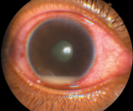
in a teaching hospital in Finland. Arch Intern Med 160:2781, 2000

21. Ramonas KM, Freilich BD: Iris abscess as an unusual presentation of endogenous endophthalmitis in 
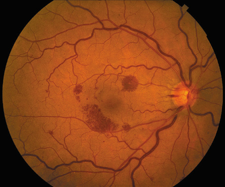
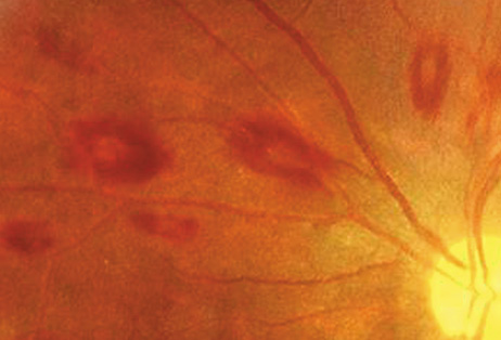
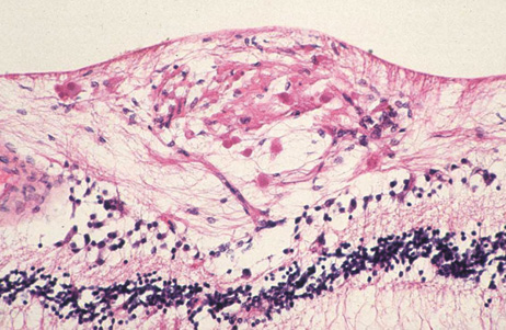
a patient with bacterial endocarditis. Am J Ophthalmol 135:228, 2003

22. Kennedy JE, Wise GN: Clinicopathological correlation of retinal lesions. Subacute bacterial endocarditis. Arch Ophthalmol 74:658, 1965

23. Burns CL: Bilateral endophthalmitis in acute bacterial endocarditis. Am J Ophthalmol 88:909, 1979

24. Treister G, Rothkoff L, Yalon M, et al: Bilateral blindness following panophthalmitis in a case of bacterial endocarditis. Ann Ophthalmol 14:663, 1982

25. Yanoff M, Fine BS: Neural (sensory) retina. In Ocular Pathology. Philadelphia: Mosby-Wolfe, 1996:376

26. Hermans PE: The clinical manifestations of infective endocarditis. Mayo Clin Proc 57:15, 1982

27. Brown GC, Magargal LE, Shields JA, et al: Retinal arterial obstruction in children and young adults. Ophthalmology 88:18, 1981

28. Munier F, Othenin-Girard P: Subretinal neovascularization secondary to choroidal septic metastasis from acute bacterial endocarditis. Retina 12:108, 1992

29. Jackson TL, Eykyn SJ, Graham EM, Stanford MR: Endogenous bacterial endophthalmitis: A 17-year prospective series and review of 267 reported cases. Surv Ophthalmol 48:403, 2003

30. Okada AA, Johnson RP, Liles WC, et al: Endogenous bacterial endophthalmitis. Report of a ten-year retrospective study. Ophthalmology 101:832, 1994

31. Christensen SR, Hansen AB, La CM, Fledelius HC: Bilateral endogenous bacterial endophthalmitis: A report of four cases. Acta Ophthalmol Scand 82:306, 2004

32. Lopez JA, Ross RS, Fishbein MC, Siegel RJ: Nonbacterial thrombotic endocarditis: A review. Am Heart J 113:773, 1987

33. Levine JS, Branch DW, Rauch J: The antiphospholipid syndrome. N Engl J Med 346:752, 2002

34. Dickens P, Chan AC: Nonbacterial thrombotic endocarditis in Hong Kong Chinese. Arch Pathol Lab Med 115:359, 1991

35. Schoen F: The heart. In Robbins and Cotran Pathologic Basis of Disease. Philadelphia: Saunders, 2005:598

36. Eiken PW, Edwards WD, Tazelaar HD, et al: Surgical pathology of nonbacterial thrombotic endocarditis in 30 patients, 1985–2000. Mayo Clin Proc 76:1204, 2001

37. Anderson D, Bell D, Lodge R, Grant E: Recurrent cerebral ischemia and mitral valve vegetation in a patient with antiphospholipid antibodies. J Rheumatol 14:839, 1987

38. Geyer SJ, Franzini DA: Myxomatous degeneration of the mitral valve complicated by nonbacterial thrombotic endocarditis with systemic embolization. Am J Clin Pathol 72:489, 1979

39. Pierrot-Deseilligny C, Gray F, Brunet P: Infarcts of both inferior parietal lobules with impairment of visually guided eye movements, peripheral visual inattention and optic ataxia. Brain 109:81, 1986

40. Jeresaty RM: Sudden death in the mitral valve prolapse-click syndrome. Am J Cardiol 37:317, 1976

41. Jeresaty RM: Mitral valve prolapse. An update. JAMA 254:793, 1985

42. Devereux RB: Mitral valve prolapse. J Am Med Women's Assoc 49:192, 1994

43. Procacci PM, Savran SV, Schreiter SL, Bryson AL: Prevalence of clinical mitral-valve prolapse in 1169 young women. N Engl J Med 294:1086, 1976

44. Markiewicz W, Stoner J, London E, et al: Mitral valve prolapse in one hundred presumably healthy young females. Circulation 53:464, 1976

45. Freed LA, Levy D, Levine RA, et al: Prevalence and clinical outcome of mitral-valve prolapse. N Engl J Med 341:1, 1999

46. Devereux RB, Hawkins I, Kramer-Fox R, et al: Complications of mitral valve prolapse. Disproportionate occurrence in men and older patients. Am J Med 81:751, 1986

47. O'Rourke RA, Crawford MH: The systolic click-murmur syndrome: Clinical recognition and management. Curr Probl Cardiol 1:1, 1976

48. Pyeritz RE, Wappel MA: Mitral valve dysfunction in the Marfan syndrome. Clinical and echocardiographic study of prevalence and natural history. Am J Med 74:797, 1983

49. Olsen EG, Al-Rufaie HK: The floppy mitral valve. Study on pathogenesis. Br Heart J 44:674, 1980

50. Whittaker P, Boughner DR, Perkins DG, Canham PB: Quantitative structural analysis of collagen in chordae tendineae and its relation to floppy mitral valves and proteoglycan infiltration. Br Heart J 57:264, 1987

51. Davies MJ, Moore BP, Braimbridge MV: The floppy mitral valve. Study of incidence, pathology, and complications in surgical, necropsy, and forensic material. Br Heart J 40:468, 1978

52. Braunwald E: Valvular heart disease. In Heart Disease: A textbook of cardiovascular medicine. Philadelphia: Saunders, 2001:1665

53. Freed LA, Benjamin EJ, Levy D, et al: Mitral valve prolapse in the general population: The benign nature of echocardiographic features in the Framingham Heart Study. J Am Coll Cardiol 40:1298, 2002

54. Langholz D, Mackin WJ, Wallis DE, et al: Transesophageal echocardiographic assessment of systolic mitral leaflet displacement among patients with mitral valve prolapse. Am Heart J 135:197, 1998

55. Lesser RL, Heinemann MH, Borkowski H Jr, Cohen LS: Mitral valve prolapse and amaurosis fugax. J Clin Neuroophthalmol 1:153, 1981

56. Seelenfreund MH, Silverstone BZ, Hirsch I, Rosenmann D: Mitral valve prolapse (Barlow's syndrome) and retinal emboli. Metab Pediatr Syst Ophthalmol 11:119, 1988

57. Hopkins A, Yiannikas J, Francis IC: Mitral valve prolapse and retinal infarction. Aust NZ J Ophthalmol 15:79, 1987

58. Lichter H, Loya N, Sagie A, et al: Keratoconus and mitral valve prolapse. Am J Ophthalmol 129:667, 2000

59. Street DA, Vinokur ET, Waring GO III, et al: Lack of association between keratoconus, mitral valve prolapse, and joint hypermobility. Ophthalmology 98:170, 1991

60. Traboulsi EI, Aswad MI, Jalkh AE, Malouf JF: Ocular findings in mitral valve prolapse syndrome. Ann Ophthalmol 19:354, 1987

61. Ross RS, McKusick VA: Aortic arch syndromes; diminished or absent pulses in arteries arising from arch of aorta. AMA Arch Intern Med 92:701, 1953

62. Kahn M, Knox DL, Green WR: Clinicopathologic studies of a case of aortic arch syndrome. Retina 6:228, 1986

63. Tour RL, Hoyt WF: The syndrome of the aortic arch: ocular manifestations of pulseless disease and a report of a surgically treated case. Am J Ophthalmol 47:35, 1959

64. Schmidt MH, Fox AJ, Nicolle DA: Bilateral anterior ischemic optic neuropathy as a presentation of Takayasu's disease. J Neuroophthalmol 17:156, 1997

65. Chun YS, Park SJ, Park IK, et al: The clinical and ocular manifestations of Takayasu arteritis. Retina 21:132, 2001

Back to Top