Chapter 68
Tumors of the Uveal Tract
JERRY A. SHIELDS and CAROL L. SHIELDS
Main Menu   Table Of Contents

Search

MELANOCYTIC TUMORS OF THE IRIS
MELANOCYTIC TUMORS OF THE POSTERIOR UVEA
UVEAL METASTASIS
CHOROIDAL HEMANGIOMA
CHOROIDAL OSTEOMA
LEIOMYOMA
LYMPHOID TUMORS
LEUKEMIA
TUMORS OF THE NONPIGMENTED CILIARY EPITHELIUM
REFERENCES

This chapter addresses some of the more common and more important neoplasms that arise in the uveal tract. Space limitations will not permit a detailed account of all the clinical variations, differential diagnosis, diagnostic techniques, pathology, management, and prognosis of all of the tumors that can develop in the iris, ciliary body, and choroid. Less common uveal tumors, such as neurilemoma and neurofibroma, are not discussed. The reader who is interested in further information should consult a recent textbook that covers these subjects in more detail.1
Back to Top
MELANOCYTIC TUMORS OF THE IRIS
Tumors of the iris can arise from the iris melanocytes, iris smooth muscle cells, and iris pigment epithelium. Systemic neoplasms such as lymphoma and metastatic tumors can also affect the iris. Nevus, malignant melanoma, and pigment epithelial tumors are discussed in this section.

NEVUS

An iris nevus is a benign tumor that arises in the melanocytes in the iris stroma.2 Although most iris nevi remain clinically stationary, they can occasionally give rise to malignant melanoma. Histopathologically, an iris nevus is usually composed of low-grade spindle-type cells. Occasionally it is composed of deeply pigmented, round cells similar to those seen in the melanocytoma of the optic disc.

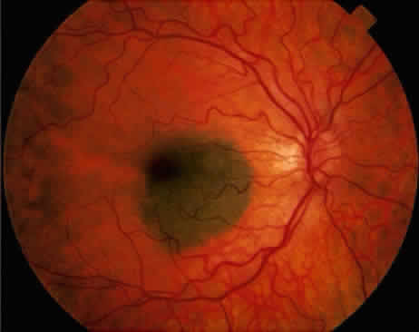
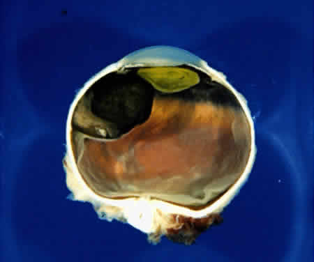
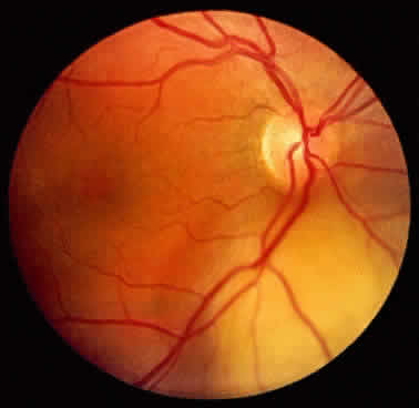
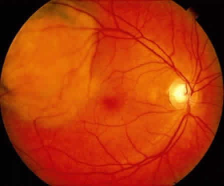
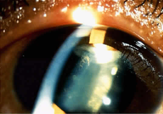
An iris nevus classically appears as a variably pigmented, well-circumscribed lesion in the iris stroma (Fig. 1). It may involve any portion of the iris from the pupillary border to the iris root and may be flat or minimally elevated. Occasionally, an iris nevus may occupy an entire sector of the iris from the pupillary border to the anterior chamber angle. In some cases, an iris nevus may be clinically amelanotic. An iris nevus can induce an irregular pupil or sector cortical cataract.

Fig. 1. Typical iris nevus.

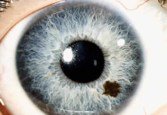
Iris nevus is best diagnosed by recognition of the typical lesion with slit-lamp biomicroscopy. Ancillary diagnostic studies, such as fluorescein angiography and ultrasonography, have little additional diagnostic value. Management includes documentation with accurate drawings or photographs and examination on a yearly basis, looking for evidence of enlargement of the lesion. If growth is detected, malignant transformation into melanoma should be suspected.

MELANOMA

An iris melanoma is a malignant melanocytic tumor that arises from the melanocytes of the iris stroma.2 Histopathologically, it is composed of spindle cells, epithelioid cells, or a combination of the two (mixed-cell type).

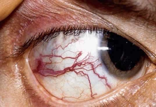
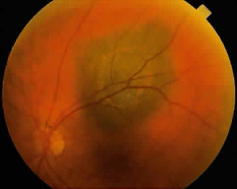
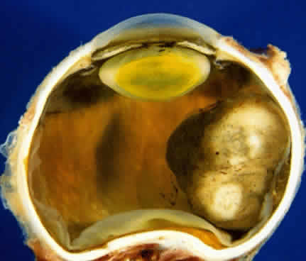
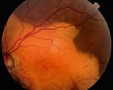
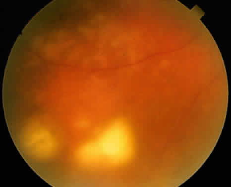
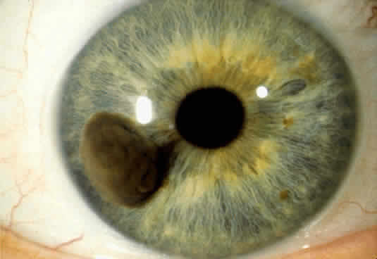
An iris melanoma is characterized clinically as a variably pigmented, elevated, circumscribed or diffuse, melanocytic neoplasm that affects the iris stroma. It is typically larger than an iris nevus. The lesion most often is deeply pigmented and elevated (Fig. 2). In can be amelanotic, or it can be partially pigmented and partially nonpigmented.

Fig. 2. Nodular iris melanoma.

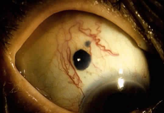
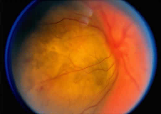
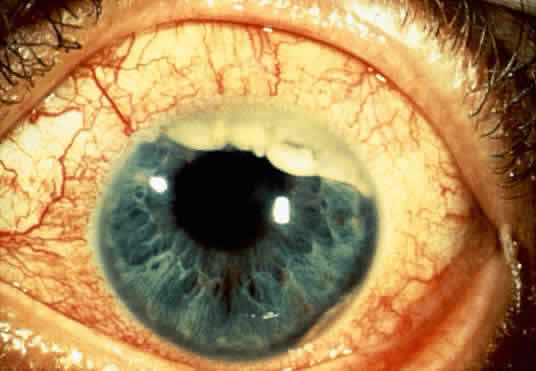
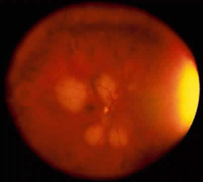
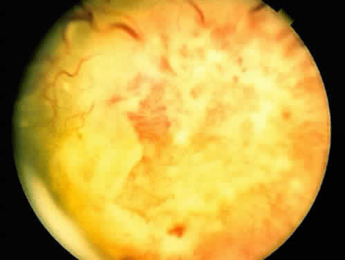
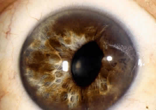
An important variant is the diffuse iris melanoma. It grows as a flat, diffuse, often multifocal mass that covers a large area of the iris surface (Fig. 3). The patient with a diffuse iris melanoma typically presents with a clinical syndrome of progressive acquired hyperchromic heterochromia and ipsilateral secondary glaucoma.

Fig. 3. Diffuse iris melanoma.

Iris melanoma has the capacity to exhibit distant metastasis to the liver and other organs. Lesions that are diffuse and produce secondary glaucoma tend to have a greater tendency to spawn metastatic disease.3

The diagnosis of iris melanoma is best made by an experienced observer who can recognize its typical features on slit-lamp examination. Although fluorescein angiography and ultrasonography have been employed, they add little useful diagnostic information. Fine-needle aspiration biopsy can be employed to confirm the diagnosis of diffuse iris melanoma in cases where enucleation is being considered.4

Once the diagnosis of iris melanoma is clearly established, usually by documentation of progressive growth, the best management is surgical excision of the lesion. Lesions confined to the iris can be managed by removal by partial sector or peripheral iridectomy. Those that extend into the ciliary body require iridocyclectomy or iridogoniocylectomy. The diffuse iris melanoma is often too large to resect locally and may require enucleation or plaque radiotherapy in selected cases.5

ADENOMA OF IRIS PIGMENT EPITHELIUM

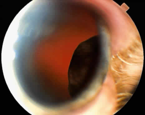
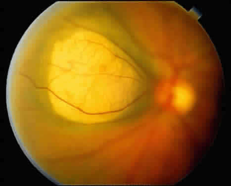
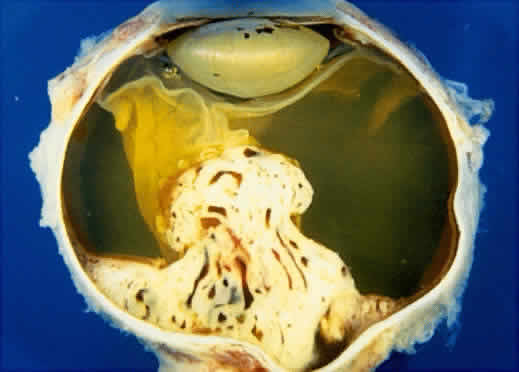
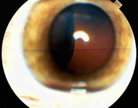
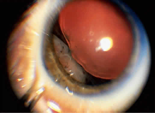
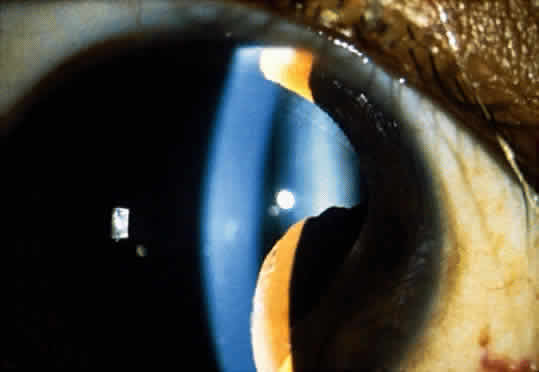
Adenoma of the iris pigment epithelium is a benign neoplasm that arises from the posterior pigment epithelium of the iris.6 Histopathologically, it is composed of columns or acini of pigmented epithelial cells. Clinically, it appears as a dark black lesion, seen in the angle as a rounded mass or as an irregular, multinodular mass (Fig. 4). The lesion typically remains stable or progresses very slowly. Transformation into adenocarcinoma is exceedingly rare.

Fig. 4. Adenoma of iris pigment epithelium.

The diagnosis of adenoma of the iris pigment epithelium is best made by clinical recognition of the characteristic features. Small, asymptomatic tumors can be managed by simple observation. If the lesion shows progressive enlargement or early secondary glaucoma, removal by iridectomy or iridocyclectomy is warranted.

Back to Top
MELANOCYTIC TUMORS OF THE POSTERIOR UVEA
Melanocytic tumors of the posterior uvea (ciliary body and choroid) include nevus and malignant melanoma.7 These tumors arise from the uveal melanocytes that are distributed throughout the uveal tract.

CHOROIDAL NEVUS

A choroidal nevus is a benign melanocytic tumor that arises in the choroid. Although most choroidal nevi remain clinically stationary for many years, they can occasionally give rise to malignant melanoma. Histopathologically, a choroidal nevus is usually composed of low-grade spindle-type cells. Occasionally it is composed of deeply pigmented, round cells similar to those seen in the melanocytoma of the optic disc. Such choroidal melanocytomas are very deeply pigmented.

Ophthalmoscopically, a choroidal nevus is a variably pigmented, fairly well-circumscribed lesion that usually occurs in the more posterior portions of the choroid. It usually has a gray to black color and is often rather homogeneous (Fig. 5). Small drusen may be present on the surface of the lesion. Occasionally an iris nevus may be clinically amelanotic. Choroidal nevi are usually less than 2 mm thick. Accumulation of a small amount of subretinal fluid can occasionally occur. The subretinal fluid may leak into the macular area, causing visual impairment.

Fig. 5. Homogeneous choroidal nevus in macular area.

Choroidal nevus is best diagnosed by an experienced clinician who can recognize the typical clinical features on careful ophthalmoscopic examination. Fluorescein angiography may help to differentiate the lesion from subretinal hemorrhage or from lesions of the retinal pigment epithelium. The best management of a choroidal nevus is to perform periodic ophthalmoscopic examinations, looking closely for enlargement of the lesion. Serial fundus photography is highly desirable for documenting any growth. If growth is documented, then the lesion should be treated as though it were a small melanoma. When a nevus produces a secondary retinal detachment that causes visual impairment, then laser photocoagulation to cause resolution of the subretinal fluid is often indicated.8

CILIARY BODY MELANOMA

A ciliary body melanoma is a malignant neoplasm that arises from the melanocytes of the ciliary body.9 Like iris melanoma, it may contain spindle or epithelioid cells. Because of the hidden location behind the iris, ciliary body melanoma usually attains a relatively large size before it is detected clinically. External ocular signs, however, such as dilated episcleral “sentinel” vessels (Fig. 6) or a focus of transscleral extension of the tumor (Fig. 7) can alert the clinician to a possible underlying neoplasm. Frequently, the tumor grows anteriorly through the iris root and appears in the anterior chamber angle. More typically, however, it is confined to the ciliary body and can be detected only by slit-lamp or fundus examination after wide dilation of the pupil (Fig. 8). Ciliary body melanoma can be uniformly pigmented, partially pigmented (Fig. 9), or amelanotic.

Fig. 6. Episcleral dilated vessels over a ciliary body melanoma.

Fig. 7. Extrascleral extension of ciliary body melanoma.

Fig. 8. Ciliary body melanoma seen through dilated pupil.

Fig. 9. Gross pathology of pigmented ciliary body melanoma.

Ciliary body melanoma is best diagnosed by recognition of the typical features of the tumor with slit-lamp biomicroscopy or indirect ophthalmoscopy through the widely dilated pupil. If the tumor is of sufficient size, ultrasonography may show the typical features of a uveal melanoma. In cases where the diagnosis is quite difficult, fine-needle aspiration biopsy can be used4; however, this is a difficult diagnostic technique that requires considerable experience to perform.

Ciliary body melanoma is most often managed surgically by local removal of the tumor via partial lamellar iridocyclectomy or some variation thereof.10 This technique is described in the literature and is beyond the scope of this discussion. Tumors that are too large to resect locally may require treatment with either a radioactive plaque or enucleation.

CHOROIDAL MELANOMA

A choroidal melanoma is a malignant melanocytic tumor that arises from the melanocytes within the choroid.7 Like ciliary body melanoma, it contains spindle cells, epithelioid cells, or a combination of the two (mixed-cell type).

In its earliest stages, a malignant choroidal melanoma may be clinically indistinguishable from a large benign choroidal nevus. A small choroidal melanoma characteristically occurs as a black or gray choroidal mass with fairly well-defined borders. A typical early feature is the orange pigment on the surface of the tumor (Fig. 10). This correlates with clumps of macrophages at the level of the retinal pigment epithelium that contain abundant lipofuscin pigment. A larger melanoma usually assumes a dome shape (Fig. 11). A melanoma that is clinically amelanotic is characterized by large, visible blood vessels in the substance of the tumor. A choroidal melanoma can eventually break through Bruch's membrane, assuming a mushroom-shaped configuration (Fig. 12).

Fig. 10. Small choroid melanoma with surface orange pigment.

Fig. 11. Dome-shaped choroidal melanoma adjacent to the optic disc.

Fig. 12. Juxtapapillary choroidal melanoma with a break through Bruch's membrane.

The growth patterns of choroidal melanoma can be best appreciated by examining gross sections of eyes enucleated for this disease. The tumors can either be dome shaped and deeply pigmented (Fig. 13) or amelanotic. The mushroom-shaped tumor typically produces a secondary nonrhegmatogenous retinal detachment (Fig. 14).

Fig. 13. Gross pathology of a dome-shaped choroidal melanoma.

Fig. 14. Gross pathology of a mushroom-shaped choroidal melanoma. (Courtesy of Ralph C. Eagle Jr, MD, Philadelphia, PA)

An experienced observer can diagnose choroidal melanoma most reliably by using indirect ophthalmoscopy.11 In atypical cases, the following ancillary procedures may be helpful: ultrasonography, fluorescein angiography, indocyanine green angiography, radioactive phosphorus uptake (32P) test, and fine-needle aspiration biopsy.

The management of choroidal melanoma is controversial.12 The details of this controversy are discussed in the literature.13–15 Enucleation in generally recommended for large tumors with little hope of salvageable vision. Radiotherapy, either with radioactive plaque12,16,17 or charged particles,18 is recommended for medium-sized and large melanomas when there is a possibility of salvaging some vision. Smaller tumors that are not near the foveal area can be treated by heavy laser application.12,19 More recently, the use of transpupillary thermotherapy either alone or combined with plaque radiotherapy has been advocated.20,21 Treatment of uveal melanoma varies from case to case, and the patient should be thoroughly counseled about all therapeutic options.12,22

Back to Top
UVEAL METASTASIS
Metastatic cancer to the uveal tract is the most common form of intraocular malignancy.23,24 Many patients with small, metastatic foci to the uveal tract, however, have advanced systemic metastasis and little or no visual symptoms, and thus do not come to medical attention. Therefore, in a clinical practice, uveal metastasis is not as common as primary uveal melanoma.

Most metastatic cancer to the intraocular structures resides in the uveal tract. Metastasis to the retina is exceedingly rare. Most are carcinomas, and metastatic melanoma and sarcoma to the uvea are much less common. Metastatic tumors to the uvea usually reach the choroid; metastasis to the iris and ciliary body are less common. All metastasis to the uvea comes through hematogenous routes, because there are no intraocular lymphatic channels. The primary malignancies that account for most uveal metastasis are breast cancer in women and lung cancer in men, although a number of other tumors can also spawn uveal metastasis.

The ocular manifestations vary depending on whether the metastasis is in the iris, ciliary body, or choroid. Iris metastasis can have several clinical variations.23–25 In general, it appears as a solitary, fleshy mass or as a white, fluffy, loosely cohesive mass (Fig. 15). These friable tumors can seed tumor cells into the anterior chamber, producing a tumor-induced pseudohypopyon. In other cases, an iris metastasis can bleed, causing a spontaneous hyphema. Ciliary body metastasis is less commonly recognized. It can attain a larger size than iris melanoma before it is recognized clinically. It often masquerades as a granulomatous iridocyclitis or endophthalmitis.

Fig. 15. Iris metastasis from breast cancer.

A choroidal metastasis is usually different from a metastasis to the iris or ciliary body. It is confined by Bruch's membrane and does not generally exhibit the tumor seeding that often characterizes anterior uveal metastasis. It appears as a creamyellow-colored sessile or minimally elevated mass (Fig. 16). Unlike an amelanotic choroidal melanoma, it is often multifocal (Fig. 17) and bilateral. Choroidal metastasis located near the optic disc has a propensity to infiltrate the nerve and produce disc edema and superficial hemorrhage.

Fig. 16. Solitary choroidal metastasis.

Fig. 17. Multifocal choroidal metastasis.

Uveal metastasis is best diagnosed by clinical recognition of the typical iris or fundus findings described earlier. In cases where the choroidal lesion is atypical and metastasis is a diagnostic consideration, fluorescein angiography and ultrasonography can provide diagnostic help. In cases where the diagnosis of an iris, ciliary body, or choroidal lesion remains uncertain despite noninvasive diagnostic studies, fine-needle aspiration biopsy can be used to make a definitive diagnosis.4

The management of uveal metastasis usually involves chemotherapy or irradiation. If the patient is receiving chemotherapy for systemic disease, then the choroidal metastasis can be followed without direct ocular treatment. If systemic chemotherapy does not control the uveal metastasis, then ocular irradiation (approximately 3500 cGy over a 4- to 5-week period) is advisable. About half of patients with choroidal metastasis require ocular irradiation, and the other half can be successfully controlled with chemotherapy or hormonal therapy.23,24

Back to Top
CHOROIDAL HEMANGIOMA
Choroidal hemangioma is the most prevalent vascular tumor of the uveal tract. It can occur as a single circumscribed lesion or as a diffuse type that is usually associated with facial nevus flammeus or variations of the Sturge-Weber syndrome.26 Unlike uveal melanoma, uveal hemangioma occurs only in the choroid.

Circumscribed choroidal hemangioma usually appears as a sessile or minimally elevated, redorange mass similar in color to the surrounding normal fundus. Smaller lesions (Fig. 18) are rather homogeneous and do not usually produce a secondary retinal detachment. Larger lesions can produce overlying proliferation or metaplasia of the retinal pigment epithelium and secondary retinal detachment.

Fig. 18. Circumscribed choroidal hemangioma.

The diagnosis of circumscribed choroidal hemangioma is best made by recognition of the typical red-orange color of the choroidal mass. Such coloration is not usually seen with amelanotic choroidal melanoma, choroidal metastasis, and other choroidal masses. Fluorescein angiography and ultrasonography can be used to help confirm the diagnosis. Fluorescein angiography usually shows linear hyperfluorescence of the blood vessels in the tumor in the prearterial phase and rather intense late staining of the mass. A-scan ultrasonography shows high internal reflectivity, and B-scan shows acoustic solidity without the choroidal excavation that characterizes melanoma.26

Since circumscribed choroidal hemangioma is a benign lesion that usually remains stationary or demonstrates only minimal enlargement, asymptomatic tumors generally require no treatment. If the tumor produces a secondary retinal detachment that causes visual impairment, then laser photocoagulation to control the detachment is often warranted.26 In cases where the retinal detachment cannot be controlled with laser therapy and there is a threat of neovascular glaucoma, plaque brachytherapy can be successfully used. After approximately 2000 cGy, the tumor generally becomes smaller and the retinal detachment resolves.

Diffuse choroidal hemangioma occurs ipsilateral to a facial nevus flammeus, sometimes in association with the Sturge-Weber syndrome.27 Clinically, it appears as a broad red-range thickening of the posterior choroid. The tumor appears more red than the background fundus, a finding sometimes called the “tomato ketchup” fundus. With time, the choroidal hemangioma produces cystic retinal degeneration and eventually an extensive secondary retinal detachment.

The presence of an ipsilateral facial nevus flammeus in a patient with a nonrhegmatogenous retinal detachment should suggest the diagnosis.27 Ultrasonography and fluorescein angiography can provide diagnostic assistance. Cranial computed tomography or magnetic resonance imaging to detect the meningeal calcification of the Sturge-Weber syndrome may also provide diagnostic help.

Diffuse choroidal hemangioma of the SturgeWeber syndrome can be difficult to manage. In its earliest stages, a refraction and treatment for hyperopic amblyopia is the best approach. Later, the secondary retinal detachment is best managed by laser photocoagulation or retinal detachment surgery, depending on the extent of the disease. External beam irradiation may bring about reduction of the retinal detachment.

Back to Top
CHOROIDAL OSTEOMA
A choroidal osteoma is a benign choroidal tumor composed of mature bone that occupies a full-thickness area of the posterior choroid.28,29 Clinically, it characteristically occurs as a well-delineated, sessile, yellow-orange tumor in the posterior choroid (Fig. 19), usually in a juxtapapillary or circumpapillary location. Occasionally, a choroidal osteoma is located in the macular area and does not touch the optic disc. The well-defined border is sometimes slightly irregular and occasionally has small pseudopodia-like projections. With time, a choroidal osteoma can cause subretinal neovascularization of choroidal origin and subretinal hemorrhage.

Fig. 19. Choroidal osteoma.

As with other lesions, the typical ophthalmoscopic features should be helpful when the diagnosis of choroidal osteoma is suspected. Ultrasonography and computed tomography can be used to demonstrate a bony plaque at the level of the choroid, providing further support for the diagnosis. There is no known treatment for a choroidal osteoma. When the tumor induces subretinal neovascularization of choroidal origin, then laser treatment to the neovascular membrane may be warranted.

Back to Top
LEIOMYOMA
Leiomyoma is a benign tumor of smooth muscle origin that usually occurs in the uterus; it tends to affect young women particularly.30 Rarely, it can arise in the uveal tract, most often in the ciliary body. The most important aspect in the diagnostic evaluation is that the ophthalmologist be familiar with the clinical features and clinical variations of leiomyoma. Leiomyoma appears clinically as a smooth-surfaced, amelanotic mass (Fig. 20). Like a comparable-sized melanoma, it can produce subluxation of the lens, secondary cataract, and retinal detachment.

Fig. 20. Leiomyoma of the ciliary body and peripheral choroid.

Unlike melanoma, which arises in the uveal stroma, uveal leiomyoma frequently grows in the suprauveal space between the uveal stroma and the sclera. Therefore, fluorescein angiography may show a relatively normal choroidal vascular pattern over the tumor, and ultrasonography may suggest an intact uveal tract over the tumor surface. However, these features are subtle, and most cases are somewhat difficult to differentiate clinically from melanoma. It appears that the best management of uveal leiomyoma is local resection by partial lamellar sclerouvectomy. The results of local resection have generally been favorable.30

Back to Top
LYMPHOID TUMORS
Benign and malignant tumors of lymphoid origin can also affect the uveal tract.31 A uveal lymphoid lesion can occur with only ocular involvement, or it can be a part of a systemic lymphoma. It may be impossible clinically to distinguish a benign lymphoma (reactive lymphoid hyperplasia) from a malignant lymphoma on the basis of ocular examination. Lymphoma can occur as a retinovitreal infiltrate that can simulate intraocular inflammation, or it can occur as a uveal infiltration. Only the uveal variant will be considered here.

A lymphoid tumor of the iris appears as an amelanotic diffuse or circumscribed mass in the iris. In the choroid it appears as one or more yellow choroidal lesions that is indistinguishable from a choroidal metastasis (Fig. 21).

Fig. 21. Choroidal lymphoma.

The management of uveal lymphoma is the same as for uveal metastasis. Systemic chemotherapy should be employed for systemic disease, with the addition of ocular radiotherapy if it fails to achieve tumor control.

Back to Top
LEUKEMIA
Leukemia most often affects the ocular fundus by causing retinal hemorrhages secondary to the hematologic alterations associated with the disease.31 However, leukemic cells occasionally infiltrate the uveal tract and retina directly.

Leukemic infiltration of the iris usually appears as a diffuse, friable mass in the inferior iris, often associated with a hyphema. Early leukemic involvement of the posterior segment appears as a gray thickening in the juxtapapillary area (Fig. 22). More advanced involvement appears as a massive yellow thickening of the optic nerve and adjacent retina and choroid.

Fig. 22. Massive optic nerve head and choroidal involvement with leukemia.

The management of leukemic infiltration of the intraocular structures is similar to the management of uveal metastasis. If chemotherapy that is employed for the systemic disease does not control the ocular disease, then ocular irradiation should be initiated. In cases of infiltration of the optic nerve, irradiation should be promptly considered because of the poor visual prognosis.

Back to Top
TUMORS OF THE NONPIGMENTED CILIARY EPITHELIUM
Tumors that arise from the nonpigmented epithelium of the ciliary body can be divided into congenital and acquired types.32–35

The most important congenital tumor of the nonpigmented ciliary epithelium is medulloepithelioma. Intraocular medulloepithelioma most often occurs unilaterally in young children in the first decade of life. It is an embryonic neoplasm that arises from the primitive medullary epithelium that is destined to form the nonpigmented ciliary epithelium in the adult. Cytological, it can be benign or malignant, but it rarely exhibits distant metastasis. Both the benign and malignant variants can occur as either a nonteratoid or a teratoid type. The nonteratoid type consists of a proliferation of fairly well-differentiated cells of the nonpigmented ciliary epithelium. The teratoid type contains heterologous elements, such as cartilage and skeletal muscle.

In the early stages, medulloepithelioma appears as a gray-white tumor of the ciliary body (Fig. 23). As it slowly enlarges, it can cause a subluxation of the lens, secondary cataract, secondary glaucoma, retinal detachment and a white pupillary reflex (leukocoria) A characteristic feature is the development of clear cysts that are visible near the surface of the lesion. These cysts frequently break away from the main lesion and float freely in the vitreous or aqueous cavity. The main lesion continues to slowly enlarge and can eventually fill the entire globe and rarely extend through the sclera into the orbit.

Fig. 23. Circumscribed medulloepithelioma of ciliary body.

The management of intraocular medulloepithelioma is removal of the tumor by partial lamellar sclerouvectomy when it is small and circumscribed. However, there is tendency for recurrence, and enucleation may ultimately be necessary in many cases.34 The systemic prognosis is generally quite favorable.

Acquired neoplasms can develop from the fully formed nonpigmented ciliary epithelium. Unlike the embryonic medulloepithelioma of childhood, acquired tumors of the nonpigmented ciliary epithelium do not contain teratoid elements. From a cytologic standpoint, they can be benign (adenoma) or malignant (adenocarcinoma). Even the malignant variant has little or no tendency to exhibit metastatic disease. Like the embryonic variant, these acquired tumors begin as an amelanotic mass of the pars plicata. They can have a smooth or irregular surface, and they occasionally contain clear cysts. They can also produce a secondary cataract (Fig. 24).

Fig. 24. Adenoma of nonpigmented ciliary epithelium with secondary cataract.

The management of tumors of the nonpigmented ciliary epithelium involves removal of the mass by partial lamellar sclerouvectomy. Although this surgery is quite difficult, in many cases the tumor can be removed successfully. Enucleation is necessary for larger or recurrent tumors.

Back to Top
REFERENCES

1. Shields JA, Shields CL (eds): Intraocular Tumors: A Text and Atlas. Philadelphia, WB Saunders, 1992

2. Shields JA, Shields CL (eds): Melanocytic tumors of the iris stroma. In: Intraocular Tumors: A Text and Atlas, pp 61–83. Philadelphia, WB Saunders, 1992

3. Shields JA, Shields CL: Hepatic metastases of diffuse iris melanoma 17 years after enucleation. Am J Ophthalmol 106:749, 1988

4. Shields JA, Shields CL, Ehya H et al: Fine needle aspiration biopsy of suspected intraocular tumors: The 1992 Urwick Lecture. Ophthalmology 100:1677, 1993

5. Shields CL, Shields JA, DePotter P et al: Treatment of nonresectable malignant iris tumors with custom designed plaque radiotherapy. Br J Ophthalmol 79:306, 1995

6. Shields JA, Shields CL (eds): Tumors and related lesions of the pigment epithelium. In: Intraocular Tumors: A Text and Atlas, pp 437–460. Philadelphia, WB Saunders, 1992

7. Shields JA, Shields CL (eds): Introduction to melanocytic tumors of the uvea. In: Intraocular Tumors: A Text and Atlas, pp 45–59. Philadelphia, WB Saunders, 1992

8. Shields JA: The expanding role of laser photocoagulation for intraocular tumors: The 1993 H. Christian Zweng Memorial Lecture. Retina 14:310, 1994

9. Shields JA, Shields CL (eds): Posterior uveal melanoma: Clinical and pathologic features, pp 117–136. In: Intraocular Tumors: A Text and Atlas. Philadelphia, WB Saunders, 1992

10. Shields JA, Shields CL, Shah P, Sivalingam V: Partial lamellar sclerouvectomy for ciliary body and choroidal tumors. Ophthalmology 98:971, 1991

11. Shields JA, Shields CL (eds): Diagnostic approaches to posterior uveal melanoma: Clinical and pathologic features. In: Intraocular Tumors: A Text and Atlas, pp 155–169. Philadelphia, WB Saunders, 1992

12. Shields JA, Shields CL (eds): Management of posterior uveal melanoma. In: Intraocular Tumors: A Text and Atlas, pp 171–205. Philadelphia, WB Saunders, 1992

13. Zimmerman LE, McLean IW, Foster WD: Does enucleation of the eye containing a malignant melanoma prevent or accelerate the dissemination of tumour cells? Br J Ophthalmol 62:420, 1978

14. Manschot WA, Van Peperzeel HA: Choroidal melanoma—Enucleation or observation?: a new approach. Arch Ophthalmol 98:71, 1980

15. Seigel D, Myers M, Ferris F, Steinhorn SC: Survival rates after enucleation of eyes with malignant melanoma. Am J Ophthalmol 87:751, 1979

16. Shields JA, Augsburger JJ, Brady LW, Day JL: Cobalt plaque therapy for posterior uveal melanoma. Ophthalmology 89:1201, 1982

17. Shields JA, Shields CL, DePotter P et al: Plaque radiotherapy for uveal melanoma. Int Ophthalmol Clin 33:129, 1993

18. Gragoudas ES, Seddon J, Egan K et al: Long-term results of proton irradiated choroidal melanomas. Ophthalmology 94:349, 1987

19. Shields JA, Glazer LC, Mieler WF, Shields CL: Comparison of xenon arc and argon laser photocoagulation in the treatment of choroidal melanomas. Am J Ophthalmol 109:647, 1990

20. Oosterhuis JA, Journee-de Korver HG, Kakebeeke-Kemme HM, Bleeker JC: Transpupillary thermotherapy in choroidal melanomas. Arch Ophthalmol 113:315, 1995

21. Shields CL, Shields JA, DePotter P, Kheterpel S: Transpupillary thermotherapy in the management of choroidal melanoma. Ophthalmology 103:1642, 1996

22. Shields JA: Counseling the patient with a posterior uveal melanoma [editorial]. Am J Ophthalmol 106:88, 1988

23. Shields JA, Shields CL (eds): Metastatic tumors to the intraocular structures. In: Intraocular Tumors: A Text and Atlas, pp 207–238. Philadelphia, WB Saunders, 1992

24. Shields CL, Shields JA, DePotter P et al: Survey of 520 uveal metastases. Ophthalmology (in press) 1997

25. Shields JA, Shields CL, Kiratli H, De Potter P: Metastatic tumors to the iris in 40 patients. Am J Ophthalmol 119: 422, 1995

26. Shields JA, Shields CL (eds): Vascular tumors of the uvea. In: Intraocular Tumors: A Text and Atlas, pp 239–259. Philadelphia, WB Saunders, 1992

27. Shields JA, Shields CL (eds): The systemic hamartomatoses (“phakomatoses”). In: Intraocular Tumors: A Text and Atlas, pp 513–539. Philadelphia, WB Saunders, 1992

28. Shields JA, Shields CL (eds): Osseous tumors of the uvea. In: Intraocular Tumors: A Text and Atlas, pp 261–271. Philadelphia, WB Saunders, 1992

29. Shields CL, Shields JA, Augsburger JJ: Review: Choroidal osteoma. Surv Ophthalmol 33:17, 1988

30. Shields JA, Shields CL, Eagle RC Jr, De Potter P: Observations on seven cases of intraocular leiomyoma: The 1993 Byron Demorest Lecture. Arch Ophthalmol 112:521, 1994

31. Shields JA, Shields CL (eds): Intraocular lymphoid tumors and leukemias. In: Intraocular Tumors: A Text and Atlas, pp 489–512. Philadelphia, WB Saunders, 1992

32. Zimmerman LE: The remarkable polymorphism of tumors of the ciliary epithelium: The Norman McAlister Gregg Lecture. Trans Aust Coll Ophthalmol 2:114, 1970

33. Shields JA, Shields CL (eds): Tumors of the nonpigmented ciliary epithelium. In: Intraocular Tumors: A Text and Atlas, pp 461–487. Philadelphia, WB Saunders, 1992

34. Shields JA, Eagle RC Jr, Shields CL, De Potter P: Congenital neoplasms of the nonpigmented ciliary epithelium. (medulloepithelioma). Ophthalmology 103:1998, 1996

35. Shields JA, Eagle RC Jr, Shields CL, De Potter P: Acquired neoplasms of the nonpigmented ciliary epithelium (adenoma and adenocarcinoma). Ophthalmology 103:2007, 1996

Back to Top