Chapter 62
Trauma: Inflammation
CONRAD L. GILES and T.F. SCHLAEGEL JR
Main Menu   Table Of Contents

Search

TRAUMATIC IRITIS
TRAUMATIC CHOROIDITIS
TRAUMATIC UVEITIS
REFERENCES

Whether it is the speed of a struck tennis or golf ball or the velocity of an automobile, we have witnessed a quantum leap in the speed of the environment over the past several years. No proportionate increase in the anatomic orbital protection or blink reflex has accompanied this however. The result, not surprisingly, is a significant increase in ocular and orbital trauma. This chapter deals with the inflammatory implications of trauma to the uveal tract.
Back to Top
TRAUMATIC IRITIS

CONTUSION

Mild contusions can occur without producing permanent structural change, whereas severe contusions result in permanent structural alterations. Flare and cells are seen in the anterior chamber, with the grade for flare usually being greater than the grade for cells. The intraocular pressure usually is low; however, because of a large number of factors, it may rise above normal later. In 15 of 27 cases of traumatic iritis, the intraocular pressure was less than 4 mmHg.1 The contusion disrupts the ciliary body and the secretion of the ciliary epithelium, but the anterior vitreous usually is free of cells. Treatment of the traumatic iritis is by the corticosteroid drops, mydriatics, and cycloplegics tailored to the severity of the process. It frequently is possible to use short-acting cycloplegic agents (tropicamide, cyclopentolate hydrochloride) in this clinical setting. Milder iritis frequently can be managed without treatment. It is possible for trauma to the cornea (from foreign bodies, fingernails, or contact lenses) to break the epithelial barrier and allow ingress of an agent such as the URI virus.2 Whether trauma or some other factor is responsible in the case of soft contact lens-induced hypopyon is unknown.3 Rarely bilateral uveitis has been reported to be precipitated in a patient with unilateral nonpenetrating trauma.4

FOREIGN BODIES

Persistent unilateral iridocyclitis after penetrating injury of the cornea with no radiographic or orbital computer tomography evidence of a foreign body should arouse suspension of foreign material in the angle of the anterior chamber.5 Intralenticular metallic foreign bodies may be well tolerated for years without causing anterior uveitis. This suggests that the management of nonferrous-containing (see siderosis bulbi) intralenticular metallic foreign bodies may be conservative until inflammation or significant cataract formation develops.6 Vegetable matter, oil, glass, stone, and metal may enter the anterior chamber at the time of injury; talcum and lint may be introduced during surgery; and ophthalmic ointments may enter afterward. If the foreign body lodges in the angle, it may not be obvious. Foreign bodies with a specific gravity greater than aqueous sinks to the lower recess, whereas oil floats and is obscured by the superior angle shelf. Thus, machine oil may be introduced into the anterior chamber at the time of injury and float to the upper angle. With the patient supine, the globules may float to the back of the center of the cornea. Irrigation with Ringer's solution through a limbal incision should clear the iridocyclitis.7 Foreign material can be in a liquid form, such as the juice from a plant that may pass through the cornea and set up an iridocyclitis.8

Tattooing with cobaltous aluminate9 in three young men resulted in a purulent discharge from the light-blue areas of the tattoo. The skin granulomas persisted, and approximately 6 months later, a uveitis appeared. Treatment consisted of the usual measures plus excision of the draining tattooed areas.

Some postoperative iridocyclitis probably is because of retained lens cortex (even in the case of an apparent intracapsular extraction). Occasionally, a little cortex may be left under the iris, where it is not visible. Although local administration of corticosteroids and cycloplegics affects cure in the majority of these patients, surgical removal of this hidden retained cortex has been helpful in at least one case. Other instances of postoperative iridocyclitis may be caused by irritation from other foreign bodies. (Cotton usually is well tolerated.) Because flare and cells are routine after cataract extraction, one must rely on other signs such as lid swelling, eye redness, and the development of keratic precipitates.

Ophthalmia Nodosa

Ophthalmia nodosa is an inflammation precipitated by hairs of certain insects or vegetable material. It derives its name from the nodular conjunctival reaction that results. These hairs in the cornea may be seen surrounded by nummular opacities. There is a latent period of a few days to a few months, presumably the period of migration into the eye.Caterpillar hairs have even been reported on top of10and underneath the retina.11 The recent increasing popularity of tarantulas as pets has resulted in a number of reported cases of ophthalmia nodosa.12 The iris reflects an intense inflammation, often with hypopyon and nodules on the iris. A granulomatous reaction, consisting of histiocytes, epithelioid cells, and macrophages, develops around the hair, but this reaction may be suppressed by local administration of steroids. Keratectomy, keratoplasty, iridectomy, or occasionally vitrectomy may be indicated to eliminate the offending hairs.13

Siderosis Bulbi

Iron in the eye, either from blood or an iron-containing foreign body, can cause inflammation thatsimulates other types of iritis. An inflamed blue iris assumes a greenish hue, but the green iris of siderosis bulbi that develops after weeks or years typically is a darker green. The iris also may become fixed and dilated.14 Other irides become browner with time. For example, after prolonged hyphema, chromatophores may migrate and hypertrophy. After severe contusions, darkening of the involved iris may occur because of hypertrophy of the superficial layers of the iris stroma. A radiograph of the eye for retained iron may be negative. If the particles are not small enough or have been absorbed, they do not show on radiograph. Rust spots beneath the anterior capsule of the lens is a typical feature of siderosis. On superficial examination, these rust spots resemble pigment clumps on the surface of the lens capsule from broken posterior synechiae. Siderosis bulbi may be caused by small intralenticular foreign bodies.15 These can be managed successfully with clear lens aspiration and foreign body removal. Intraocular foreign body removed with sclerotomy and external magnet, pars plana vitrectomy intraocular magnet, and forceps and posterior vitrectomy has resulted in a high degree of preservation of visual function without further visual deterioration after removal of the iron-containing object.16

Back to Top
TRAUMATIC CHOROIDITIS
Chorioretinitis sclopedaria refers to direct choroidal and retinal trauma from a bullet wound in the orbital area. The bullet does not penetrate the globe or the orbital wall; it lodges adjacent to the globe. Two areas of the eye usually are injured by concussion: (1) the area adjacent to the path of the bullet and (2) the macular area indirectly.17 One patient, observed during World War II, had a moderately large area of chorioretinitis in the temporal equator from a bullet that had passed between the sclera and the lateral orbital wall. Less often, an area directly opposite the concussion site also can exhibit evidence of chorioretinitis as a result of the contre-coup phenomenon.

A peculiar syndrome secondary to trauma, usually of the perforating type that involves the lens, is pseudoretinitis pigmentosa. There is a selective loss of the photoreceptor layer,18 extinguished electroretinogram, and migration of pigment into the retina. These features may develop within a few days after the injury.

FOREIGN BODIES

Rarely, localized choroiditis is associated with penetrating intraocular foreign bodies as an isolated feature. The condition of the host, the manner of penetration, and the character of the foreign body must have been so correlated as to avoid the more devastating endophthalmitis. Relatively inert foreign material (e.g., glass, stone, quartz) may lodge in the retina and choroid and cause a low-grade inflammatory response that walls off the foreign substance. Such an inflammation may simulate other types of uveitis such as pars planitis and a Toxocara granuloma. A long quiescent period or intermittent recurrences of inflammatory signs may ensue. When a foreign body is nonmagnetic, systemic and periocular administration of steroids should be initiated and used in the follow-up treatment. Removal of a magnetic foreign body may be well tolerated in the choroid; its path through the eye often provokes vitreous damage and secondary retinal complications.

RETINAL PERFORATION DURING SURGICAL MANAGEMENT OF STRABISMUS

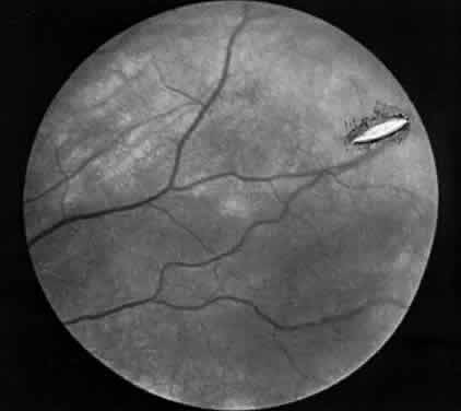
Hemorrhage initially covers the perforation site. Some of this hemorrhage may break through into the vitreous cavity but usually remains confined to the choroid and retina. In a day or two, bare sclera becomes visible, usually surrounded by residual hemorrhage and a rim of light pigmentation.19 It appears as if the choroid and the retina were scooped out, leaving bare sclera behind (Fig. 1). Inverted cutting needles seem responsible since this is a complication almost unheard of before or after their common use. Prophylaxis indicates the use of a needle that is not sharp on the convex curve. The management of these perforations remains controversial.20 In our experience, widely dilating the pupil permits nearly immediate diagnosis as the area can be clearly seen through the usually clear media. Depending on the characteristics of the perforation, transpupillary laser coagulation or direct cryotherapy can be administered easily. Some suggest, however, that observation without treatment is all that is required since the perforations are almost always self-sealing and result in no significant sequelae. Nevertheless, in a series of 728 patients with known scleral perforations, 14 (2%) of the cases had a retinal detachment and eight resolved without complications.21

Fig. 1. Scleral perforation several weeks after muscle surgery. (Schlaegel TF Jr: Essentials of Uveitis. Boston: Little, Brown, 1969:290.)

Back to Top
TRAUMATIC UVEITIS
The inclusive term uveitis is applied to uveal inflammation that develops after a traumatic incident in which both the anterior and posterior portions of the uveal tract participate in the inflammatory response.

Intraocular surgery, blunt trauma, retained intraocular foreign bodies, and incarceration of the lens capsule or the iris in penetrating wounds of the eye can evoke uveitis. The reaction probably occurs secondary to a hypersensitivity phenomenon to uveal or lenticular material, or the uveitis may result from necrosis of the damaged tissue and may be worsened by intraocular hemorrhage.

The anterior chamber may show various degrees of inflammatory response up to a dense flare with large “mutton-fat” keratic precipitates. Although this lesion may resolve spontaneously, secondary glaucoma or a plastic iridocyclitis progressing to occlusion pupillae and finally a shrunken blind globe often is the result.

The posterior pole manifestations of this uveal inflammation are retinal edema with serous detachment of the pigment epithelium and retinal exudates associated with vitreous debris and papillitis. Left untreated, this form of uveitis follows a course of profound and rapid deterioration in vision.

The offending traumatic vector (incarcerated iris, lens) should be eliminated and the inflammatory response minimized. Topical, periocular, and systemic administration of corticosteroids must be combined with cycloplegic-mydriatic treatment and other measures, such as antiglaucoma ones, to salvage the globe in the patient with uveitis secondary to trauma.

Back to Top
REFERENCES

1. Aronson SB, Elliott JH: Ocular Inflammation. St Louis: Mosby, 1972:226

2. Aronson SB, Moore TE, O'Day DM: The effect of structural alteration of anterior ocular inflammation. Am J Ophthalmol 70:886, 1970

3. Hull D, Hyndiuk RA, Dhin J: Clinical experience with the therapeutic hydrophilic contact lens. Presented at Central Section, ARVO Meeting, Milwaukee, March 23, 1973

4. Yamada K, Hayasaka S, Setagawa T: Non-penetrating trauma in the right eye induces anterior uveitis and secondary glaucoma in the contralateral eye. Ann Ophthalmol 25:277, 1993

5. Grayson MG, Teixler VA: Persistent unilateral iridocyclitis. Am J Ophthalmol 56:653, 1963

6. Lee LR, Briner AM: Intralenticular metallic foreign body. Aust NZ J Ophthalmol 24:361, 1996

7. Schlaegel TF Jr: Essentials of Uveitis. Boston: Little, Brown, 1969

8. Tomar VPS, Agarwal PK, Agarwal BL: Toxic iridocyclitis caused by calotropis. J All India Ophthalmol Soc 18:15, 1970

9. Rorsman H, Brehmer-Andersson E, Dahlquist I et al: Tattoo granuloma and uveitis. Lancet 2:27, 1969

10. Daman F: Raupenhaar-Uveitis durch Rauphenhaare des Brombeerspinners (Bombyx rubi bzw, Macrothylacia rubi). Klin Monatsbl Augenheilkd 153:643, 1968; abstract in Survey Ophthalmol 4:351, 1970

11. Velicky J: Damage to the eye due to caterpillar. Cesk Oftalmol 12:271, 1966

12. Lasudry JG, Brightbill FS: Ophthalmia nodosa caused by tarantula hairs. J Pediatr Ophthalmol Strabismus 34:197, 1997

13. Belyea DA, Tuman DC, Ward TP et al: The red eye revisited: Ophthalmia nodosa due to tarantula hairs. South Med J 91:565, June 1998

14. Scotcher SM, Canning CR, Dorrell D: Siderosis bulbi: An unusual cause of a unilaterally dilated pupil. Br J Hosp Med 54:110, 1995

15. Duffy D, Salmon JF: Siderosis bulbi resulting from an intralenticular foreign body. Am J Ophthalmol 127:218, 1999

16. Sneed SR, Weingeist TA: Management of siderosis bulbi due to a retained iron-containing intraocular foreign body. Ophthalmology 97:375, 1990

17. Richards RD, West CE, Meisels A: Chorioretinitis sclopetaria. Am J Ophthalmol 66:852, 1968

18. Cogan DG: Pseudoretinitis pigmentosa: Report of two traumatic cases of recent origin. Arch Ophthalmol 81:45, 1969

19. McLean JM, Galin MA, Baras I: Retinal perforation during strabismus surgery. Am J Ophthalmol 50:1167, 1960

20. Mittelman D, Bakos IM: The role of retinal cryopexy in the management of experimental perforation strabismus surgery. J Pediatr Ophthalmol Strabismus 21:186, 1984

21. Simon JW, Linninger LL, Scherage JL: Recognized scleral perforation during eye muscle surgery: Incidence and sequelae. J Pediatr Ophthalmol Strabismus 29:273, 1992

Back to Top