Chapter 54
Hypersensitivity Uveitis
M. Levent Civelek, Debra A. Goldstein and Howard H. Tessler
Main Menu   Table Of Contents

Search

HYPERSENSITIVITY REACTIONS
EXOGENOUS ANTIGENS
ENDOGENOUS ANTIGENS
LEUKOCYTOCLASTIC VASCULITIS (HYPERSENSITIVITY VASCULITIS)
REFERENCES

HYPERSENSITIVITY REACTIONS
When an immune response occurs and causes tissue damage, it is referred to as a hypersensitivity reaction. In the 1960s, British immunologists Coombs and Gell originally classified hypersensitivity reactions into four types, and, recently, a fifth and sometimes a sixth are also added. Although our understanding of the immune system has dramatically changed since the 1960s, the classification of Coombs and Gell remains almost unchanged; it is still used and valid.1

TYPE I: IMMEDIATE HYPERSENSITIVITY

Type I hypersensitivity is mediated by IgE antibodies. Antigen binds to two molecules of IgE or IgG attached to a receptor site (Fc) on the surface of a basophil or a mast cell and results in the degranulation of these cells. Type I ocular hypersensitivity usually results in primarily external disease, but “seasonal iritis” and retinal edema have been described in severe cases of hay fever.2,3,4

IgE antibodies specific for retinal S antigen (S-Ag) are detected early in experimental S-Ag induced uveitis in rats. The role of mast cells in the induction of experimental autoimmune uveoretinitis (EAU) is also supported by several studies: (1) mast cell degranulation occurs before the onset of EAU,5 (2) EAU is inhibited by drugs that affect mast cells,6,7 and (3) the number of mast cells in rat choroid,8 iris, and ciliary body9 is correlated with the susceptibility of rats to EAU. But according to a study in 2002, instillations of antiallergic agents, such as cromolyn sodium, tranilast, levocobastine hydrochloride, pemirolast potassium, and ibudilast, did not inhibit flare elevation in a rabbit study, whereas single instillation of betamethasone inhibited 88% of aqueous flare elevation. It is unlikely that antiallergic agents inhibit flare elevation and affect disruption of the blood–aqueous barrier.10

TYPE II: CYTOTOXIC ANTIBODY

Cytotoxic antibodies mediate type II reactions. IgG or IgM bind to antigen on the surface or basement membranes of target cells, resulting in cell death (e.g., ocular cicatricial pemphigoid and Mooren's ulcer).

Extraocular manifestations of sympathetic ophthalmia (SO) and Vogt-Koyanagi-Harada (VKH), such as vitiligo, poliosis, alopecia, dysacusis, and central nervous system symptoms consistent with irritation of the meninges (which contains melanin), appear to be caused by a reaction to melanin.11,12 Sugita et al.13 suggested that SO and VKH may be autoimmune diseases directed toward the MART-1 peptide of melanocytes. In SO and VKH disease, anti-melanin autoantibodies and melanin-sensitized lymphocytes have been demonstrated in the peripheral blood.14 Serum antibodies to retinal antigens have been detected15 to be consistent with this disease being mediated, at least in part, by a type II hypersensitivity.

TYPE III: IMMUNE COMPLEXES

Type III reactions cause tissue injury by precipitation and deposition of immune complexes. Antigen-antibody complexes can initiate the complement cascade, which attracts macrophages, neutrophils, and platelets, and causes tissue damage. The uvea is highly vascularized, and its blood supply has been said to resemble that of the renal cortex. Because immune complexes can be deposited in the renal glomerulus, they may also accumulate in the uvea. Systemic lupus erythematosus (SLE) retinopathy, rheumatoid sclerouveitis, sarcoidosis, retinal vasculitis, chronic idiopathic uveitis, Behçet's syndrome, rheumatoid arthritis, Wegener's granulomatosis, and lens induced uveitis may be due, in part, to type III hypersensitivity reactions; immune complexes have been observed in patients having these diseases.16,17,18,19

The formation of antigen-antibody complexes may also be a mechanism for removing antigens from the circulation or suppressing disease. In mild retinal vasculitis patients, it has been reported that the titer of immune complexes is high, whereas in severe inflammation the serum titers are low.4,20

TYPE IV: DELAYED-TYPE (CELL MEDIATED) HYPERSENSITIVITY

In contrast to the previous three reactions, no immunoglobulins are involved in type IV reactions, and the hypersensitivity is mediated directly through T cells. This reaction is triggered when antigen is presented to T lymphocytes by antigen presenting cells (APCs), which results in cytokine (lymphokine) release or lymphocyte stimulation. Examples of type IV reactions include contact dermatitis (eczema) and corneal graft rejection. Type IV reactions may play a role in ocular toxoplasmosis, herpetic uveitides, sympathetic ophthalmia, pars planitis, and birdshot retinochoroidopathy.21

TYPE V: STIMULATORY ANTIBODY

In type V hypersensitivity, IgG antibodies are directed toward cell-surface antigens and have a stimulatory effect on their target. An example is long-acting thyroid stimulator (LATS) antibody. LATS is directed toward a portion of the hormone receptor and mimics the function of thyroid-stimulating hormone. Maternal stimulating antithyroid IgG antibodies are able to cross the placenta and may cause neonatal hyperthyroidism.4,22

TYPE VI: ANTIBODY DEPENDENT CELL-MEDIATED CYTOTOXICITY (ADCC)

Type VI reactions are sometimes classified as a subgroup of type II hypersensitivity reactions. Target cells coated with antibody are destroyed by specialized killer cells (NK cells, killer T cells, macrophages), which bear receptors for the Fc portion of the coating antibody (Fc receptors). These receptors allow the killer cells to bind to the antibody-coated target (tumor rejection and defense against parasites). This cytotoxic reaction is complement independent.

For most of the uveitic entities, it remains uncertain which type of hypersensitivity reaction is involved. Uveitic disorders associated with hypersensitivity can be categorized into those associated with exogenous antigens (i.e., those found in the environment) and those associated with endogenous antigens (i.e., those found in the eye).

Back to Top
EXOGENOUS ANTIGENS

ENVIRONMENTAL ANTIGENS

Association between hypersensitivity to environmental antigens (e.g., allergies) and uveitis has been documented. Cases of uveitis have been reported in association with allergies to food,23,24 dust,23 ragweed,24 trees and grasses,25 cat, caterpillar, and tarantula hair.26–30

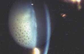
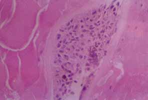
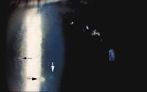
In 1904, Saemisch31 first used the term ophthalmia nodosa to describe the granulomatous nodules formed on the conjunctiva and iris in response to caterpillar hairs or sensory setae. Iritis, occasionally with formation of iris nodules and vitritis, is also described after ocular reaction to caterpillar hairs.32 Ophthalmia nodosa can also be caused by tarantula hairs. Tarantulas are occasionally kept as pets and are usually harmless, but, if threatened, they eject their dorsal barbed hairs with the hair of their hind legs. These hairs are capable of embedding into cornea and skin and inciting an inflammatory reaction. It is usually hard to detect those hairs in the cornea. Even with hairs in the anterior corneal stroma only, anterior uveitis with mutton fat deposits on endothelium may be seen.29 One patient is reported to have developed small peripheral choroidal lesions after 6 months.30 Extensive dissection of the cornea or sclera to remove the hairs is not recommended because successful management is obtainable with topical steroids (Fig. 1).

Fig. 1 Ophthalmia nodosa. Slit lamp photograph of left eye of a 13-year-old boy who developed a red and painful eye after playing with his pet tarantula. Note the granulomatous keratic precipitates (black arrows) and subepithelial opacity (white arrow). There was also a mild anterior chamber reaction.

An asthmatic patient has been described who developed a well-demarcated area of choroiditis after several bouts of urticaria. The authors felt that this was due to IgE-mediated release of vasoactive amines, which have been reported to trigger vasculitic syndromes.32

Bee and wasp stings of the cornea have been associated with significant ocular pathology. Uveitis has been observed after corneal bee sting33 and wasp bite.34 Bee venom is a complex toxin composed of several compounds with different actions. Toxicity is related to nonenzymatic polypeptide toxins (mellitin, apamin, iminimine) and enzymes (phospholipase A and B, hyaluronidase). Mellitin can cause depigmentation of the iris (heterochromia iridis), noted in cases of bee sting.35 Mellitin also causes the release of serotonin, histamine, and other chemical mediators of inflammation. Apamin is a neurotoxin that blocks neurotransmission, and internal ophthalmoplegia and sector iridoplegia have been reported as neurotoxic effects of apamin after corneal bee sting. Optic neuritis, papilledema, and optic atrophy have occurred following bee stings to the other parts of the body.36 The mechanism is thought to be focal demyelination of the optic nerve caused by an acute allergic reaction to the bee venom. Enzymes in the venom have high molecular weight and are highly antigenic, accounting for the immunologic injury to the eye following stings. Type I hypersensitive reaction takes place, with release of chemical mediators of inflammation and manifested by such findings as conjunctival injection, chemosis, and corneal edema.33 Corticosteroids alone, or in conjunction with cycloplegics and antibiotics, are used in corneal bee stings; antihistamines can be included in case of chemosis and conjunctival injection.

The cases above are unusual. In general, the association between allergies and uveitis is rare. Van Metre37 reported no cases due to allergy among 556 cases of uveitis studied. In a series of 1500 cases, Kimura26 did not find any statistically significant association between contact allergy and uveitis. These results also reflect our clinical experience.

INFECTIOUS AGENTS

Infectious causes (e.g., tuberculosis, syphilis, and toxoplasmosis) of uveitis are well known; however, uveitis associated with hypersensitivity to infectious agents is seldom recognized clinically.

Although the specific cause of uveitis remains unknown in many patients, the initiating stimuli for intraocular inflammation can be divided into two major pathways: an antigen specific (infectious agent) immune-mediated inflammatory response and a nonspecific inflammatory response (which is nonantigen specific).

Endotoxin-induced uveitis (EIU) is an animal model for nonantigen-specific stimulus induced ocular inflammation. In Lewis rats, intravenous, intraperitoneal, and intrafootpad endotoxin injection38 or intraocular endotoxin injection39 are demonstrated to induce EIU. According to kinetic studies, inflammatory cells migrate first into the eye about 6 hours after endotoxin injection, and ocular inflammation peaks approximately 18 hours later. Inflammation is related to the release of cytokines from activated cells. Tumor necrosis factor α (TNF-α), interleukin-1 (IL-1), IL-6, and IL-8 are inflammatory mediators that appear to be stimulated by endotoxin. Those cytokines and other inflammatory molecules can start the inflammatory cascade with breakdown of the blood-aqueous and blood-retinal barriers, leading to additional cellular infiltration of the eye.40

Antigen-specific ocular immune responses may also take place and are divided into cell-mediated and humoral responses. Both of these require processing of the antigen by specialized antigen-presenting cells (APC). It has been postulated that this results from molecular mimicry between part of the DNA of the various organisms and a portion of HLA-B27, although this is still controversial.

Immune responses against infectious agents may cross-react against ocular antigens and induce uveitis. For example, researchers noted homology between yeast histone and S-Ag.41 A number of forms of uveitis follow an infectious disease but do not seem to be caused by direct infection. Reiter's syndrome is associated with HLA-B27 haplotype. Uveitis in these patients may occur after gram-negative dysentery or after nongonococcal urethritis, as a result of chlamydia trachomatis and ureaplasma urealyticum.40

Post-streptococcal syndrome is an autoimmune disorder precipitated by infection with group A streptococci. The pathologic process is thought to stem from a cross-reaction between antibodies, sensitized lymphocytes, or both, generated against streptococcal antigens with the tissues of the host.42 Manifestations include acute rheumatic fever (ARF), post streptococcal reactive arthritis (PSRA), and acute glomerulonephritis. Recurrent anterior uveitis can occur in patients with a history of post-streptococcal syndrome who have repeated group A streptococcal infection. The intraocular inflammation follows a time course similar to that of the other manifestations of the syndrome, such as recurrent rheumatic fever.43,44 PSRA differs from ARF, in which evidence of carditis is not usually seen and the response of arthritis to aspirin is poor. Two adult patients with PSRA, both of whom developed uveitis, were recently described.45

There are probably multiple initiating infectious stimuli for inflammation in the uvea. Uveitis may result from the replication of the microbes, the host's hypersensitivity to the components of the microbe, or both. Unfortunately, the exact mechanism of uveitis in humans is still unknown.40

DRUG-INDUCED UVEITIS

Drug-induced uveitis is a relatively rare occurrence (reported to be less than 0.5 % in a tertiary referral uveitis clinic).46 Drug-induced uveitis is almost always reversible within weeks of discontinuation of the causative agent and treatment of the inflammation.

Naranjo et al.47 proposed the following seven criteria to establish causality of adverse events by drugs:

  1. The reaction is a frequently described event that is well documented.
  2. Recovery occurs upon withdrawal of the drug.
  3. Other possible causes for the event have been excluded.
  4. The reaction becomes more severe when the dose of drug is increased.
  5. The adverse event is documented by objective evidence.
  6. Similar effects can occur in a given patient with similar drugs.
  7. The event recurs on rechallenge with the suspected drug.

Several drugs have anecdotally been noted to cause uveitis in single case reports. Very few drugs that have been reported to cause uveitis have had causality confirmed by elimination of confounding variables, double-blind challenge, or rechallenge testing.

Systemic Drug-Induced Uveitis

Rifabutin, a derivate of rifampin, is used to treat or to prevent atypical mycobacterial infections in the immunocompromised host. It has been associated with anterior and posterior nongranulomatous uveitis, with or without hypopyon, which may be extremely severe.48,49,50 Rifabutin-associated uveitis, characterized by white-yellow inflammatory opacities in the inferior and posterior vitreous, has also been described by Chaknis et al.51 They described those lesions in three acquired immunodeficiency syndrome (AIDS) patients who were receiving 300 mg of rifabutin daily for 6 or more months for mycobacterium avium complex (MAC) prophylaxis. Three cases of acute uveitis without hypopyon were reported in patients with AIDS who did not have MAC bacteremia and who were taking prophylactic rifabutin.49 Rifabutin-associated uveitis may be an immune reaction to dead mycobacteria, but MAC associated uveitis (without rifabutin) is very rare, and anterior chamber paracentesis of the hypopyon, in these cases, failed to show any organisms on either aqueous cultures or microscopic examinations.52 Rifabutin reaction is probably not T cell-mediated because a paucity of these lymphocytes is one of the hallmarks of AIDS.48 Rifampin is known to be antigenic itself and after binding with serum and tissue proteins. Antibodies against rifampin can circulate or adhere to cellular surfaces and the antigen-antibody complexes induce an inflammatory reaction.53 The high prevalence, incidence of bilaterality, recurrence of uveitis with rechallenges, increasing severity of inflammation with dose escalation, improvement upon withdrawal, and exclusion of other possible causes of uveitis strongly implicate rifabutin as a cause of uveitis.54

Pamidronate sodium (Aredia), an intravenous bisphosphate, inhibits bone resorption and is used in the management of hypercalcemia associated with malignancy, osteolytic bone metastases, paget disease of the bone, and osteoporosis. Seventeen cases of unilateral scleritis and one case of bilateral scleritis have been reported within 6 hours to 2 days after intravenous pamidronate sodium injection, with positive dechallenge and rechallenge data. In 16 cases this occurred anteriorly and in one case, posteriorly.55 The most frequent ocular side effect of serious clinical importance associated with pamidronate is anterior uveitis; both eyes are affected in most patients and onset is within the first 48 hours of drug exposure. In some patients, the drug had to be discontinued, and the outcome was favorable within a few days after topical corticosteroid therapy.55,56 Pamidronate stimulates the production of a distinct group of T cells, which inhibit bone resorption. The activation of T cells releases cytokines, and this may contribute to an immunologic or toxic reaction in patients who develop uveitis or scleritis.57

A more recently developed oral bisphosphate, alendronate sodium (Fosamax), is 100 to 500 times more potent than amino-bisphosphonates and is being used successfully to prevent and to treat osteoporosis in postmenopausal women. Alendronate has also been associated with bilateral anterior uveitis,58 posterior scleritis, anterior scleritis, and orbital myositis that resolves with anti-inflammatory therapy and discontinuation of alendronate.59 There is no rechallenge data for alendronate. But, because this agent is in the same class as pamidronate, has the same pattern of onset, and requires discontinuation of the drug for the scleritis to resolve, a cause-and-effect relationship seems to be almost certain for alendronate.

A bilateral sudden-onset iritis in association with the use of trimethoprim-sulfamethoxazole has been reported by Tilden et al.60 The bilaterality and the recurrence of inflammation with rechallenge are strong evidence that systemic sulfonamides are a cause, albeit rare, of anterior uveitis. The intraocular inflammation may be the result of direct immunogenicity of sulfonamides or, as in the case of Stevens-Johnson syndrome, the result of a systemic, necrotizing vasculitis.54

Diethylcarbamazine (Ivermectin) is an antifilarial agent effective against Oncocerca volvulus, one of the leading causes of blindness in the world. Diethylcarbamazine rapidly and effectively kills microfilaria. Death of the microfilaria that are present in the cornea and anterior chamber liberates an antigenic load that may result in devastating intraocular inflammation. It is Jarisch-Herxheimer reaction, which is a hypersensitivity response, attributed to liberation of endotoxin-like substances or of flarial antigens from the killed or dying microorganisms. This reaction may exacerbate preexisting ocular inflammation, and prophylaxis with corticosteroids may be helpful.54,61,62

There have been old isolated reports of uveitis associated with oral contraceptives.63 There is one case of bilateral anterior uveitis64 and three cases of bilateral posterior uveitis and vasculitis in patients taking norethynodrel and mestranol. The evidence for causality is extremely weak, and there is no rechallenge data. Given the huge number of women using oral contraceptives, these rare cases may have occurred by chance alone. The pathogenesis of uveitis associated with oral contraceptives, if indeed this is a true entity, is unclear.54 Anterior granulomatous uveitis65 and acute nongranulomatous anterior uveitis66 have been reported in patients with hypersensitivity to quinidine.

Cidofovir (Vistide), a DNA polymerase inhibitor (HPMPC), has been successfully used for the treatment of cytomegalovirus retinitis and acyclovir-resistant herpes virus infections in patients with AIDS. Cidofovir-associated uveitis (CAU) has been described in 25% to 59% of patients receiving intravenous cidofovir.67,68 The uveitis is usually anterior, associated with posterior synechiae and accompanied by hypotony. It may be unilateral or bilateral, is dose related, and the risk is increased with highly active antiretroviral therapy (HAART) and with rising CD4+ cell counts.69 While CAU occurs mostly in eyes with inactive CMV retinitis, bilateral anterior uveitis was reported in an AIDS patient taking cidofovir because of presumed recurrence of CMV encephalitis. The patient was not on

HAART and had no evidence of CMV retinitis or any other abnormality on fundoscopy.70 Recently, several reports have proposed the use of cidofovir as a treatment for infections with the virus known as JC virus (JC are the initials of the first person diagnosed with this virus) that causes progressive multifocal leukoencephalopathy (PML) in patients with AIDS. Tacconelli et al.,71 in 2003, treated AIDS patients receiving HAART with cidofovir for CMV retinitis or PML. Sixty percent of CMV patients had CAU on the same side as the retinitis, whereas no cases were detected among the patients with PML. It is hypothesized that the retinal action of cidofovir is increased by concomitant retinal alteration caused by retinitis, previous mycobacterial disease, or toxoplasmosis. It is also possible that an increase in a patient's HIV-viremia level (viral load) may result in HIV-associated retinal alteration, which facilitates an increase in the penetration of cidofovir. CAU seems to occur more frequently in patients with retinitis, on HAART, in whom HAART has failed to restore immunity. The concomitant use of probenecid decreases the incidence of uveitis associated with intravitreal and also intravenous cidofovir.72 Probenecid inhibits renal tubular secretion of cidofovir73 and may inhibit secretion from the ciliary body, which shares many of the transport mechanisms in the kidney, resulting in decreased intraocular drug levels.67

Etanercept (Enbrel) inhibits the action of both TNF-α and TNF-γ. It is increasingly being used in the management of rheumatoid arthritis, psoriatic arthritis, and ankylosing spondylitis. The development of rheumatoid nodules and leukocytoclastic vasculitis is reported with etanercept.74,75 A case of anterior nongranulomatous uveitis was reported in 2003 following subcutaneous etanercept treatment for ankylosing spondylitis.76 The close temporal association of the start of etanercept and the uveitis and challenge-rechallenge data suggests that etanercept might have provoked the anterior uveitis.

Ibuprofen, a noncorticosteroidal anti-inflammatory drug (NSAID), can cause aseptic meningitis. A patient with aseptic meningitis and bilateral nongranulomatous iridocyclitis related to ibuprofen has been reported. And it has been suggested that a hypersensitivity reaction to NSAID should be considered when a patient has neurologic abnormalities after initiation of NSAID therapy in the presence or absence of iridocyclitis.77

Topiramate (Topamax) is an oral sulfamate medication used primarily as an antiepileptic. It is also used in the treatment of bipolar disorders and pain control of migraine. Cases of acute myopia and bilateral angle-closure glaucoma with ultrasound biomicroscopic signs of ciliochoroidal effusion have been reported associated with topiramate use.78,79 Fluid accumulated in the supraciliary space with ciliary body detachment is the main factor producing anterior rotation of the ciliary body. This rotation pushes the iris anteriorly and closes the angle.80 Acute myopia is explained by the forward displacement of the lens caused by supraciliary effusion, although some authors suggest that ciliary body swelling and lens thickening may also play a role.81

Topical Drug-Induced Uveitis

The nonselective β-adrenergic blocking agent metipranolol (Optipranolol) appears to cause granulomatous anterior uveitis with prominent mutton fat82 or medium-sized keratic precipitates83 and can also cause nongranulomatous anterior uveitis.84 In 1991 Akingbehin et al.85 presented 15 patients who developed granulomatous anterior uveitis (GAU) with characteristic mutton-fat keratic precipitates after using metipranolol. Intraocular pressure elevations more than 5 mm Hg above their baseline levels developed in over half of the eyes with GAU. This rise in intraocular pressure with metipranolol-associated uveitis may be due to inflammatory debris blocking the trabecular meshwork. The authors rechallenged 7 of the original 26 patients using 0.3% metipranolol in one eye and 0.5% timolol in the fellow eye for up to 14 days.86 In all of the eyes that were rechallenged with metipranolol, GAU, marked intraocular pressure increase, blepharoconjunctivitis, or periorbital dermatitis developed within 14 days. Two weeks after discontinuing the medication, all the patients had complete resolution of symptoms. Uveitis did not develop in the eyes treated with timolol. Because of the relatively large number of cases, time of onset, corresponding laterality of occurrence to drug treatment, and recurrence upon rechallenge, this report strongly suggests that metipranolol was the cause of uveitis.54 The etiology of intraocular inflammation from metipranolol remains unclear.

In 2000, Byles et al.87 suggested that highly selective a-adrenoreceptor agonist brimonidine tartrate (Alphagan) caused granulomatous anterior uveitis in four patients, after 12 months of brimonidine use. In all cases, uveitis settled rapidly after cessation of the drug and recurred on rechallenge testing. Two more cases of granulomatous anterior uveitis as a suspected adverse reaction to topical brimonidine are also reported.88,89

Latanoprost (Xalatan), a prostaglandin analog, has also been reported to cause anterior uveitis. Uveitis improves after cessation of latanoprost with or without corticosteroids,90 and it recurs after rechallenge.91

Anticholinesterase drugs have been used to reduce elevated intraocular pressures and to control accommodative esotropia. Diisopropyl fluorophosphate and phospholine iodide are the most commonly used agents. Vascular congestion secondary to these agents is believed to result in the breakdown of the blood-aqueous barrier, causing mild iritis.54 These drugs may also enhance existing iritis and cause formation of anterior and posterior synechiae and fine keratic precipitates.92

Betaxolol is a cardio-selective β-1 blocker. A 78-year-old female patient with rheumatoid arthritis developed bilateral nongranulomatous anterior uveitis 3 weeks after starting betaxolol. The anterior uveitis in both eyes resolved promptly on stopping betaxolol in conjunction with weak steroid drops and did not reappear on stopping steroids in the follow-up period of 6 months. Rechallenge was not performed, but it was suggested that betaxolol might be responsible for the anterior uveitis in this case.93

Iritis can occur with the use of topical epinephrine, and the presence of antiepinephrine antibody has been documented in patients treated with topical epinephrine.94 Experimentally induced uveitis with the use of topical epinephrine has also been reported.95 Furthermore, topical epinephrine has been known to induce and exacerbate postoperative cystoid macular edema, supporting the possibility that topical epinephrine may induce inflammation in the eye.

Corticosteroid withdrawal may be associated with the development of ocular inflammation.Two patients with ocular hypertension developed unilateral ocular hypotony during a period of provocative testing with topical dexamethasone and a unilateral anterior uveitis 3 to 5 days after discontinuation of dexamethasone.96 In another series involving topical dexamethasone testing, 7 of 401 glaucoma patients and 9 of the 220 apparently healthy individuals developed acute anterior uveitis within a few days of discontinuation of corticosteroid drops. The incidence of uveitis was significantly higher in blacks than in whites; 5.4% compared to 0.5%. In two cases symptoms began while the patients were still using the dexamethasone drops. In the majority of cases, a sharp drop in intraocular pressure to a level lower than the baseline pressure was also seen. Treatment, when necessary, consisted of mild cycloplegia. Almost all patients recovered completely within a period of 3 to 10 days. Since a decrease in intraocular pressure occurs simultaneously with the development of uveitis, and the uveitis occurs after termination of the corticosteroid treatment, the drop in the intraocular pressure may be attributed to the decrease in aqueous humor production caused by the uveitis.97 In another prospective study, Mindel et al.98 compared the intraocular pressure response to four different topically applied corticosteroid esters. They found that acute iritis developed within 72 hours of discontinuation of topical corticosteroids in three eyes of 2 volunteers among 54 treated for 6 weeks. Both patients were black. Each eye had received a different corticosteroid ester. This report confirmed that withdrawal of different topical corticosteroids may be associated with intraocular inflammation. The explanation for this corticosteroid-induced uveitis is not clear; a toxic effect of corticosteroids may weakly explain uveitis.97 Because the incidence of corticosteroid-induced uveitis appears to be much higher in blacks than in whites, melanin may play a role.54 Reactivation of a latent ocular infection is one other possible explanation; that is, corticosteroids might have increased ocular susceptibility to an already existing subclinical intraocular infection.96,97

Intraocular Drug-Induced Uveitis

Drugs administered directly into the anterior chamber or vitreous cavity can cause inflammation, with breakdown of the blood-aqueous or blood-retinal barrier. However, the act of simply placing a sterile needle inside the anterior chamber, that is, without injecting any drug, may also produce iritis. Nearly all antibiotics injected intracamerally have been reported to produce inflammation. This anterior segment inflammation is usually transient and mild.54 Aminoglycosides have a particularly toxic effect on ganglion and other neural cells of the retina.99

Urokinase, a plasminogen activator, is a clot-specific fibrinolytic agent that has been successfully used to treat fibrin membrane formation. Tissue plasminogen activator has been used to treat impending pupillary block glaucoma in patients with acute fibrinous HLA-B27 positive iridocyclitis.100 Intravitreal injection of urokinase has been reported to produce a sterile hypopyon-like reaction in up to 50% of patients with vitreous hemorrhages. The hypopyon was probably not due to inflammation because it was not associated with conjunctival injection, chemosis, or ocular pain. Resolution occurred within 6 days, regardless of whether or not topical corticosteroid therapy was administered.101

Cidofovir, previously described in the section on systemic agents, may also cause uveitis after intravitreal injection. Chavez de la Paz et al.,72 in 1997, reported the incidence and findings of an anterior nongranulomatous uveitis that occurred after intravitreal injection of 20 μg of cidofovir in 46 patients with AIDS and cytomegalovirus retinitis. Of the 130 injections in 69 eyes, 30 cases (23%) developed iritis after a median post injection period of 4 days. Posterior synechiae occurred in 37% of eyes. Oral probenecid treatment before cidofovir injection was associated with a statistically significantly lower incidence of iritis. It is hypothesized that oral probenecid might decrease or suppress the absorption of cidofovir into the ciliary body and reduce the incidence of iritis.73

Sterile endophthalmitis after intravitreal injection of triamcinolone diacetate (Kenolog) has been reported. This may be a toxic reaction to preservatives in the diluents or possibly a poorly understood immune reaction.102

Vaccination-Induced Uveitis

Bacilli Calmette-Guérin (BCG) vaccine is used to immunize against tuberculosis and to treat carcinoma in-situ of the bladder. Donaldson et al.103 reported two cases of bilateral uveitis associated with vitiligo in patients injected subcutaneously with multiple 0.1 mL aliquots of BCG for metastatic malignant melanoma. There was associated severe atrophy of both irides of the two patients. Arthritis and unilateral iritis developed within 4 weeks in three patients who were treated with intravesical BCG therapy for superficial bladder carcinoma.104,105,106

One patient was HLA-B27 positive.105 The BCG vaccine appeared to be an adjuvant for melanin-induced uveitis.105,107 The second possible mechanism is a direct toxic effect of melanin on the uvea. Melanin is abundant in patients with metastatic malignant melanoma. Therapies that destroy melanoma tissue will release this pigment. Melanin toxicity has been implicated in the development of intraocular inflammation.107 Melanin may also act as a depot for antigens and thus hold antigens and promote inflammation in the eye.108

Panuveitis has been reported in patients after tuberculin challenge. Lish and Berman109 reported development of bilateral anterior segment inflammation and vitritis 2 weeks after a tuberculin (purified protein derivative) skin test. The time course suggested a cell-mediated immune response (type IV delayed-type hypersensitivity reaction) to the skin test. The ocular inflammation improved after 4 weeks of treatment with systemic antimycobacterial medications and 8 weeks of treatment with prednisone. Nussenblatt and Palestine110 described a multifocal choroiditis resembling Vogt-Koyanagi-Harada syndrome 5 days after tuberculin challenge in a woman who was known to be tuberculin positive in the past. The uveitis was characterized by serous retinal detachments and choroidal Dalen-Fuchs-like nodules throughout the fundus. This patient also improved with systemic corticosteroids. In these patients the uveal tract may have been sensitized by hematogenous spread of mycobacteria or tuberculous proteins during the initial clinical phase of the pulmonary tuberculosis.109

A 77-year-old woman developed recrudescence of iritis, vitreous haze, and clinical cystoid macular edema within 2 weeks after receiving influenza vaccine.111 Whether the effect was the result of circulating immune complexes, some antigenic reaction with inflamed tissue or other mechanisms is unknown at this time. There have been three reports of uveitis after influenza vaccination involving a total of five patients. Blumberg et al.112 reported development of optic neuritis in one eye and iritis in the fellow eye after vaccination with a split product of A-Victoria influenza. This patient did not have a previous history of uveitis in either eye. No rechallenge was given. The authors suggested that small-vessel vasculitis may have been the mechanism for the iritis and optic neuritis. Knopf111 reported one case of recurrent uveitis after influenza vaccination, and this was followed by a report of three additional cases.113 All four had a history of unilateral recurrent intraocular inflammation prior to vaccination. One patient had had recurrent iritis and scleritis for 25 years prior to vaccination. Another had had a single episode of iritis 5 months earlier. Two other patients had had previous cataract surgery complicated by vitreous loss, recurrent iritis, and vitritis from 7 to 12 months prior to influenza vaccination. Patients ranged in age from 51 to 77 years. Systemic and laboratory evaluations were performed in two patients and were negative. Three patients developed iritis and one developed iritis and vitritis in the previously inflamed eye within 2 weeks of vaccination. In none of the patients did iritis develop in the fellow eye after vaccination. In three patients the iritis responded to topical corticosteroids and nonsteroidal anti-inflammatory agents. One patient required both topical and systemic corticosteroids. No rechallenge was administered to any of the four patients.113 Because four of these five patients had a history of prior uveitis, the vaccination may have been coincidental and unrelated to the uveitis. Alternatively, the vaccination may have been a nonspecific stimulant to a preexisting intraocular inflammation. As the fellow eye was not involved in any of the patients, the authors speculated that a low-grade inflammatory response produced by the vaccine was additive to an already compromised blood-ocular barrier, leading to the reactivation of uveitis.111,113

Uveitis secondary to live attenuated varicella virus vaccine was reported in a 16-year-old girl 7 days after receiving vaccine. The incidence of vaccine-induced chickenpox is estimated at about 3% to 5%. Cutaneous eruptions may occur after varicella vaccine immunization, including vaccine-strain varicella, which occurs within 30 days of vaccination; wild strain varicella, which occurs more than 30 days after vaccination because of incomplete immunity after vaccination; and zoster caused by reactivation of the vaccine or wild-strain varicella.114 In 2003, a 9-year-old boy presented with herpes zoster ophthalmicus 3 years following vaccination with live attenuated varicella vaccine (Oka strain). Amplified DNA from fluid taken from the base of a cutaneous vesicle produced wild-type varicella zoster virus (VZV) DNA not Oka strain. HZV infection therefore needs to be considered in all patients who present with scleritis, keratitis, or anterior uveitis, regardless of their varicella vaccine status.115

Two children developed anterior uveitis 4 to 6 weeks after having received the combined vaccination for measles, mumps, and rubella (MMR). During immunization viral antigen or tissue culture products may have initiated the early events in the immune activation pathway to induce anterior uveitis by antigen mimicry.116

Iridocyclitis, chorioretinitis, and peripheral periphlebitis have occurred after antityphoid vaccination.117,118 Nongranulomatous iritis after administration of diphtheria antitoxin and antitetanus vaccination has also been described.119

An acute bilateral posterior uveitis, which occurred after first and second boost of a hepatitis B vaccination, has been reported. It was suggested that the surface antigen of the vaccine and HBs antibodies of the immune response after vaccination might have formed immune complexes, which initiated bilateral uveitis.120

Although vaccine induced uveitis is rare, parients with inflammatory eye disease should be questioned carefully about recent immunizations.

Back to Top
ENDOGENOUS ANTIGENS
Intraocular inflammation can also be triggered or augmented by materials that are endogenous to the eye.

PHACOANTIGENIC UVEITIS

Phacoantigenic uveitis is a form of granulomatous uveitis that follows the disruption of the lens capsule, which is usually caused by intraocular surgery or trauma, although phacoantigenic uveitis associated with spontaneous rupture of the lens capsule has been reported.121 Phacoantigenic uveitis has been described after extracapsular cataract extraction, phacoemulsification, and trabeculectomy.122–125 In 1965, Riise124 reported the incidence of phacoantigenic uveitis to be 5% to 20% after extracapsular cataract extraction. In a study about 9 years later, of 2875 cases of phacoemulsification no case of phacoantigenic uveitis was reported.125

According to Thach et al.,126 in 20% of cases of phacoantigenic uveitis, there is no history of trauma or surgery. These cases appeared due to spontaneous rupture of the lens capsule in cases of hypermature cataract.

Microphthalmos is an associated feature in eyes with nontraumatic phacoantigenic uveitis.31 Phacoanaphylactoid reaction has been reported in cases of persistent hyperplastic primary vitreous without any history of ocular trauma or surgery.127,128 It is of interest that eyes with persistent hyperplastic primary vitreous are usually microphthalmic.

Phacoanaphylactic endophthalmitis (endophthalmitis phacoanaphylactica) is another term that has been used to describe this entity. We prefer the term phacoantigenic uveitis, which was coined by Witmer,129 because it implies that certain lens antigens, yet to be characterized, play a role in the pathogenesis of this disorder.

Clinical Manifestations

Phacoantigenic uveitis may occur from 1 day to 59 years after the disruption of the lens capsule.126,130,131,132 In most cases, however, the inflammation begins within the first 2 weeks.133,134 There are no pathognomonic signs of phacoantigenic uveitis. The clinical presentation of phacoantigenic uveitis resembles that of granulomatous iridocyclitis and vitritis.The uveitis is usually unilateral but may be bilateral in some cases.135,136 The signs of phacoantigenic uveitis include ciliary injection, anterior chamber cells and flare, vitreous cells, and mutton-fat keratic precipitates. Lens cortical and nuclear material may be present in the anterior chamber or in the vitreous cavity (Fig. 2). The intraocular pressure may be low or elevated. Intravitreal lens fragments may be demonstrated on ultrasonography.137

Fig. 2 Phacoantigenic uveitis. Note mutton-fat keratic precipitates.

Pathologic Features

The classic histopathologic description of phacoantigenic uveitis is zonal granulomatous inflammation centered on lens or lens material.126,138,139

Polymorphonuclear leukocytes make up the inner layer, surrounded by an intermediate mantle layer of epithelioid cells, macrophages, and giant cells. The outer layer contains variable amounts of fibrous connective tissue and collagen infiltrated by lymphocytes126,138,139,140 (Fig. 3).

Fig. 3 Zonal granulomatous inflammation centered on lens material (H.E.). This specimen was obtained from the patient depicted in Figure 2.

Vitreous biopsy and anterior chamber paracentesis may show the presence of polymorphonuclear leukocytes, histiocytes, and plasma cells surrounding amorphous lens material.141 Concomitant inflammation of the retina and choroid has been reported in 76% and 51% of cases, respectively.126

Diagnosis

The diagnosis of phacoantigenic uveitis can be suspected or made presumptively in cases of postoperative or posttraumatic granulomatous inflammation with concomitant disruption of the lens capsule. A definitive diagnosis of phacoantigenic uveitis requires vitreal biopsy or anterior chamber paracentesis, which may yield macrophages that show engorged cytoplasm, presumably filled with lens material. Of 144 histopathologically reviewed cases of phacoantigenic uveitis that Marak141 reported, only six (5%) were correctly diagnosed clinically. Since clinical diagnosis of phacoantigenic uveitis is difficult, in suspected cases anterior chamber fine needle biopsy and its cytopathology is reported to be a valuable adjunct to clinical examination.

Differential Diagnosis

Phacoantigenic uveitis should be differentiated from other forms of postoperative uveitides, including sympathetic ophthalmia, delayed infectious endophthalmitis, toxic reactions due to foreign material introduced at surgery, and those associated with intraocular lens (such as mechanical disturbances, manufacturing problems, etc.).138

Sympathetic ophthalmia (SO) is, by definition, bilateral and, in one-third of cases, characterized by the presence of Dalen-Fuchs nodules, whereas phacoantigenic uveitis is usually unilateral and is not characterized by fundus lesions. Although phacoantigenic uveitis may be bilateral at times, the inflammation is usually simultaneously active in sympathetic ophthalmia. Choroidal thickening is observed in SO on ultrasound, and there is minimal choroidal inflammation in phacoantigenic uveitis. Sympathetic ophthalmia can, however, coexist with phacoantigenic uveitis.

Delayed infectious endophthalmitis after intraocular surgery and trauma may mimic phacoantigenic uveitis. One of the primary examples is Propionibacterium acnes endophthalmitis, which usually presents as chronic granulomatous uveitis and may be difficult to distinguish from phacoantigenic uveitis. Like phacoantigenic uveitis, P. acnes endophthalmitis typically occurs after uneventful extracapsular cataract extraction and is characterized by mutton-fat keratic precipitates, anterior chamber reaction, and vitreous cells. The presence of plaque-like material on the capsular bag may be a helpful feature in distinguishing P. acnes endophthalmitis from phacoantigenic uveitis (Fig. 4). The diagnosis of P. acnes endophthalmitis is usually confirmed by positive cultures of the aqueous humor or the vitreous. Like sympathetic ophthalmia, P. acnes endophthalmitis may be associated with phacoantigenic uveitis.142 Other causes of delayed infectious endophthalmitis include coagulase-negative Staphylococcus and corynebacterium.143–146 Various fungi, particularly Candida species, can produce a similar picture.143,144,146,147

Fig. 4 Propionibacterium acnes endophthalmitis. Left: Mutton-fat keratic precipitates. Right: Plaque-like material on the capsular bag, a helpful feature in distinguishing P. acnes endophthalmitis from phacoantigenic uveitis.

In cases of phacoantigenic uveitis with elevated intraocular pressure, phacolytic glaucomas should be considered in the differential diagnosis. In phacolytic glaucoma, the lens is usually hypermature and the elevation of intraocular pressure is the result of blockage of trabecular meshwork by macrophages with ingested lens material.148 The treatment of phacolytic glaucoma is removal of the cataract.

Pathogenesis

The exact pathogenesis of phacoantigenic uveitis is unknown. It has been suggested for years that phacoantigenic uveitis is an immune rejection of a sequestered foreign material that is released as a result of lens injury.149 However, embryonic lens material is surrounded by the vascular plexus-tunica vasculosa lentis, and lens tissue is well recognized by the immune system. The large number of extracapsular cataract extractions performed annually and the rarity of phacoantigenic uveitis also do not favor this hypothesis. An alternative hypothesis suggests that phacoantigenic uveitis represents a loss of tolerance of the immune system to lens antigens. T-cell tolerance is normally maintained by small amounts of circulating lens protein. This tolerance may be altered by trauma and infection.138 Genetic predisposition may also play a role.

Phacoantigenic uveitis seems to be an immune complex disease (type III hypersensitivity reaction). The amount of inflammation in phacoantigenic uveitis does not depend upon the amount of lens material that remains in the eye.150 The degree of inflammation has, however, been correlated with antibody titer.151 De novo production of antilens antibodies has been reported after extracapsular cataract extraction, and these antilens antibodies can cross-react with uveal antigen.151 In experimental phacoantigenic uveitis induced in rats, Rao et al.152 showed that oxygen radicals, such as hydrogen peroxide and hydroxyl radicals, play an important role.

Treatment

The mainstay of treatment of phacoantigenic uveitis is removal of all the remnant lens material (including cortical material and nuclear fragments) in the eye. Removal of the capsular remnants and the intraocular lens is also recommended.122,124 Use of chymotrypsin, which breaks down zonules, may facilitate the complete removal of the residual capsular remnant.

Topical, periocular, and systemic corticosteroids may be used as adjuncts to surgical intervention to minimize the intraocular inflammation. Antiglaucomatous medications may be necessary in cases of phacoantigenic uveitis with concomitant elevation of intraocular pressure.

Some mild cases of suspected phacoantigenic uveitis occurring after cataract surgery seem to go into remission spontaneously after several weeks. It is possible that exposed antigen is eliminated or sequestered by the body such that inflammation stops. In many of these cases, retained lens material is still visible on slit lamp examination.

SYMPATHETIC OPHTHALMIA

Sympathetic ophthalmia (SO) is a bilateral granulomatous panuveitis occurring after penetrating injury to one eye. The inciting injury may also result from nonpenetrating ocular surgery. The incidence of SO after intraocular surgery has been reported to be 0.01% to 0.05% (type of surgery not specified) and 0.28% to 1.9% for nonsurgical penetrating injuries.12,153,154 However, a more recent prospective survey on SO performed in England and Ireland in 1997 and 1998 for 15 months, found the estimated incidence to be 0.03 in 100,000 people (23 cases/estimated 59 million populations). Baseline data were available on 18 patients, in whom SO occurred after surgery in 11 patients, after retinal surgery alone in 6 patients, and after accidental trauma in 7 patients.155 According to this study, the most frequent cause of SO was not penetrating trauma but retinal surgery, no sex predilection was identified, and 75% of the patients had a 20/40 or better vision at 1 year.

Onset of clinical inflammation has been reported from 5 days to 66 years after ocular injury, 90% present within 1 year.156,157 Possible etiologies are autoimmunity against uveal melanin, uveal melanocytes, retinal pigment epithelium, or retinal antigens.158

Extraocular findings of vitiligo, poliosis, dysacusis, alopecia, and irritation of the meninges (which contains melanin) suggest a generalized sensitization against melanin pigment-containing tissues in at least some cases.12 Antimelanin autoantibodies and melanin- sensitized lymphocytes have been demonstrated, and serum antibodies to retinal antigens have been detected in SO.14,15 Immunohistopathologic studies of eyes with SO reveal that the choroidal infiltrates are preponderantly T-lymphocytes, with B-lymphocytes comprising less than 5% to 15% of the infiltrate.12,158 These findings suggest that type II or type IV hypersensitivity may be involved in the pathogenesis of SO.

In one-third of SO patients, white-yellowish lesions can be seen in the peripheral fundus, similar to those seen in VKH. These may or may not represent Dallen-Fuchs nodules, which are histopathologically seen lesions between Bruch's membrane and RPE. Recent evidence suggests that Dallen-Fuchs nodules are not the cause of the white lesions seen clinically in VKH,159 and it may be that these two entities are not correlated in SO.

The injured eyes of patients with SO have generally been enucleated early in the course of the disease, whereas enucleation in VKH occurs late, after medical and surgical treatment. Early phases of both SO and VKH syndrome include a granulomatous inflammation, which primarily involves the uveal tissue, except for the choriocapillaris.160 In early phases, the RPE may play a crucial role by producing factors, such as transforming growth factor-β (TGF-β) and “retinal pigmented epithelial protective protein” that provide relative protection for the choriocapillaris, the RPE, and the retina.160–162 In late phases of both SO and VKH cases, the choriocapillaris seems to be involved with diffuse lymphatic infiltration and histopathologically, the enucleated globes revealed identical features.163

The only way to prevent SO is the enucleation of the injured eye before sensitization occurs. Enucleation of a traumatized eye with no potential for vision is recommended before 5 to 10 days after the injury. After this time enucleation may not be protective. Enucleation after development of SO is controversial. There is no good evidence that enucleation after clinical SO is present and makes the disease less severe. The exciting eye may become the eye with better vision and so, if potentially useful vision is present, enucleation should be avoided.

Corticosteroids are the mainstay of systemic treatment. Steroid–sparing immunosuppressive or immunomodulatory drugs may be the choice of therapy in patients experiencing intolerance or unacceptable side effects from corticosteroid medication. We prefer to not use long-term high-dose systemic steroids (i.e., >10 mg/day for more than 6 months). Immunosuppressive drugs, such as chlorambucil, may place patients in long-term sustained remission so that the disease no longer requires therapy.164 This is in contradistinction to corticosteroid therapy that may be required for years.

UVEITOGENIC ANTIGENS OF THE EYE

Retinal-S antigen (arrestin) is a soluble protein found in the outer segments of rod photoreceptors and, in some animals, the pineal gland. Experimental uveitis resembling sympathetic ophthalmia has been induced in animals using S-antigen.165,166 It has been shown that induction of uveitis in animals with S-antigen also requires complete Freund's adjuvant.166 In addition, Rao et al.165 demonstrated that bilateral uveitis can be induced in rabbits by subconjunctival injection of S-antigen in one eye, whereas intraocular injection of S-antigen fails to induce sympathetic uveitis. This suggests that induction of sympathetic uveitis may require exposure of S-antigen to the lymphatic system. Interphotoreceptor retinoid-binding protein (IRBP), another protein found in outer segments, can also induce ocular inflammation in animals.167 IRBP-induced uveitis is of shorter duration than that seen with S-Ag. Unlike retinal-S antigen, IRBP also immunize the meninges surrounding the pineal glands of animals and causes inflammation.22

Recoverin is a calcium binding protein that induces both uveitis and pinealitis after immunization of rats.168 Kaya et al.108 reported that melanin is able to augment inflammation in experimental uveitis. Melanin appears to bind antigens and possibly immune complexes and increases the concentration of antigens in the eye, similar to the binding of certain drugs to melanin, such as pilocarpine, chloroquine, and phenothiazine. This finding supports the clinical impression that uveitis tends to be more severe in deeply pigmented patients than in whites. It is also of interest that epinephrine and melanin are similar in terms of their molecular structure, and both can contribute to intraocular inflammation. The extraocular manifestations of SO and VKH seem to be also related to melanin.12,13

Rhodopsin, an intrinsic membrane glycoprotein of rod photoreceptor cells, induces S-Ag-like experimental autoimmune uveitis (EAU) and pinealitis in high concentrations.169 Opsin (rhodopsin's illuminated form) seems to be less uveitogenic than rhodopsin. It induces EAU in rats after foot pad injection.170,171

Phosducin is a retinal protein that plays a role in the phototransduction of rods.172 After foot pad injection, half of the Lewis rats had patchy focal chorioretinal lesions with vitritis and retinal vascular involvement. In contrast to the severe EAU induced by other retinal antigens, the clinical disease was late in onset, low grade in severity.173

Back to Top
LEUKOCYTOCLASTIC VASCULITIS (HYPERSENSITIVITY VASCULITIS)
Leukocytoclastic vasculitis (LCCV) is an immune complex-mediated, small vessel disease that is clinically characterized by the presence of palpable purpuric lesions, most often in association with rheumatic diseases. Ocular manifestations of LCCV are rare.

The kidneys, joints, lungs, brain, and gastrointestinal tract may be involved, resulting in gastrointestinal symptoms (e.g., nausea, vomiting, diarrhea, and constipation), arthralgia, hematuria, and glomerulonephritis. Pathologically, the vasculitis involves the postcapillary venules in a majority of cases; however, the arterioles may also be involved. Leukocytoclastic vasculitis often occurs in patients with connective tissue diseases (e.g., systemic lupus erythematosus and rheumatoid arthritis). Circulating immune complexes are believed to be important in the pathogenesis of leukocytoclastic vasculitis. Tsai et al.174 described a case of panuveitis and multifocal retinitis in a patient with LCCV.

Autoimmune keratolysis secondary to cutaneous LCCV has also been described.175

The development of LCCV has been reported after the use of the TNF antagonist etanercept.75

We, too, have seen a patient with LCCV who developed chronic iridocyclitis and retinal vasculitis (Fig. 5). As in the case reported by Tsai et al.,174 the diagnosis in our patient was confirmed through skin biopsy; and in both cases the patients were treated with systemic corticosteroids.

Fig. 5 Left: Purpura in a patient diagnosed with leukocytoclastic vasculitis secondary to systemic lupus. Right: Choroidal lesions in the same patient.

Back to Top
REFERENCES

1. Descotes J, Choquet-Kastylevsky G: Gell and Coombs's classification: Is it still valid? Toxicology 158:43–49, 2001

2. Allansmith MR, O'Connor GR: Immunoglobulins: Structure, function and relation to the eye. Surv Ophthalmol 14:367, 1970

3. Ruedemann AD: Ocular manifestations of allergy. In Thomas JW (ed): Allergy in Clinical Practice. Philadelphia: JB Lippincott, 1961:256–274

4. Maybodi M, Guler ML, Kaplan HJ: Immunology of uveitis. In Tasman W, Jaeger EA (eds): Duane's Clinical Ophthalmology. Philadelphia: JB Lippincott, 2002

5. De Kozak Y: Antibody response in uveitis. Eye 11:194, 1997

6. De Kozak Y, Sainte-Laudy J, Benveniste J, Faure JP: Pharmological modulation of IgE-dependent mast cell degranulation in experimental autoimmune uveoretinitis. Jpn J Ophthalmol 27:598–608, 1980

7. Watanabe K, Hayasaka S, Hiraki S, et al: Effects of topical clonidine on prostaglandin-E(2)-induced aqueous flare elevation in pigmented rabbits. Ophthalmic Res 32:210–214, 2000

8. Mochizuki M, Kuwabara T, Chan CC, et al: An association between susceptibility to EAU and choroidal mast cell numbers. J Immunol 133:1699–1701, 1984

9. Li Q, Caspi RR, Najafian F, et al: Association between mast cells and the development of experimental autoimmune uveitis in different rat strains. Clin Immunol Immunopathol 65:294–299, 1992

10. Hayasaka Y, Hayasaka S, Zhang XY, Nagaki Y: Effects of topical anti-inflammatory and antiallergic eye drops on prostaglandin E2-induced aqueous flare elevation in pigmented rabbits. Arch Ophthalmol 120:950–953, 2002

11. Jurij RB: Enucleation, evisceration and sympathetic ophthalmia. Curr Opin Ophthalmol 11:372–385, 2000

12. Gasch AT, Foster CS, Cynthia LG, Pasquale LR: Postoperative sympathetic ophthalmia. Int Ophthalmol Clin 40(1):69–84, 2000

13. Sugita S, Sagava K, Mochizuki M, et al: Melanocyte lysis by cyotoxic T lymphocytes recognizing the MART-1 melanoma antigen in HLA-A2 patients with Vogt-Koyanagi-Harada disease. Int Immunol 8:799–803, 1996

14. Hammer H: Lymphocyte transformation test in sympathetic ophthalmitis and the Vogt-Koyanagi-Harada syndrome. Br J Ophthalmol 55:850, 1971

15. Chan CC, Palestine AG, Nussenblatt RB, et al: Anti-retinal autoantibodies in Vogt-Koyanagi-Harada syndrome, Behçet's disease and sympathetic ophthalmia. Ophthalmol 92:1025, 1985

16. James DG, Graham E, Hamblin A: Immunology of multisystem ocular disease. Surv Ophthalmol 30:155, 1985

17. James DG, Neville E, Langley DA: Ocular sarcoidosis. Trans Ophthalmol Soc UK 96:133, 1976

18. Levinsky RJ: Circulating soluble immune complexes in recurrent oral ulceration and Behçet's syndrome. Clin Exp Immunol 32:193, 1978

19. O'Connor GR: Factors related to the initiation and recurrences of uveitis. Am J Ophthalmol 96:577, 1983

20. Stanford MR: A longitudinal study of clinical and immunological findings in two patients with retinal vasculitis. Br J Ophthalmol 72:442, 1988

21. Whitcup SM, Nussenblatt RB: Immunologic mechanisms of uveitis: New targets for immunomodulation. Arch Ophthalmol 115:520, 1997

22. Nussenblatt RB, Whitcup SM, Palestine GP: Fundamentals and Clinical Practice, 2nd ed., 1996

23. Callaghan WC: Allergic uveitis. Trans Indiana Acad Ophthalmol Otolaryngol 38:35, 1955

24. Ruedemann AD: Ocular manifestations of allergy. In Thomas JW (ed): Allergy in Clinical Practice. Philadelphia: JB Lippincott, 1941:256–274

25. Peshkin M: Personal communication. In Theodore FH, Schlossman A (eds): Ocular Allergy. Baltimore: Williams & Wilkins, 1958:38

26. Kimura SJ: Relationship of allergy to uveitis. Presented at the Uveitis Symposium, San Francisco, September 17–18, 1957

27. Walker V: Discussion on allergy in ophthalmology. Proc R Soc Med 40:582, 1947

28. Cadera W, Pachtman MA, Fountain JA, et al: Ocular lesions caused by caterpillar hairs (ophthalmia nodosa). Can J Ophthalmol 19:40–44, 1984

29. Watts P, Mcpherson R, Hawksworth NR: Tarantula keratouveitis. Cornea 19(3):393–394, 2000

30. Hered RW, Spaulding GA, Sanitato JJ, Wander AH: Ophthalmia nodosa caused by tarantula hairs. Ophthalmol 95:166–169, 1988

31. Saemisch T: Ophthalmia nodosa. Graefe-Saemisch Handbuch der gesamten Augenheilkunde, 2nd ed. Leipzig: W. Engelmann, vol. 5, 1904:548–564

32. Bielory L, Noble KG, Frohman LP: Urticarial vasculitis and visual loss. J Allergy Clin Immunol 88:819–821, 1991

33. Douglas GS, Raymond JR: Corneal bee sting with retained stinger. J Emerg Med 20(2):125–128, 2001

34. Babushkin AE: Uveitis, caused by a wasp bite. Vestn Oftalmolm 118(2):43–44, 2002

35. Singh G: Bee sting of the cornea. Ann Ophthalmol 16:320–322, 1984

36. Berrios RR, Serrano LA: Bilateral optic neuritis after a bee sting. Am J Ophthalmol 117(5):677–678, 1994

37. Van Metre TEJr : Role of the allergist in diagnosis and management of patients with uveitis. JAMA 195:167–172, 1966

38. Rosenbaum JT, McDevitt HO, Guss RB, Egbert PR: Endotoxin-induced uveitis in rats as a model for human disease. Nature 286:611–613, 1980

39. Forrester JV, Worgul BV, Merriam GRJr : Endotoxin-induced uveitis in the rat. Albrecht Von Graefes Arch Klin Exp Ophthalmol 213(4):221–233, 1980

40. Whitcup SM: The initiating stimuli for uveitis. Eye 11:167–170, 1997

41. Singh VK, Yamaki K, Donosa LA, Shinohara P: Molecular mimicry: Yeast histone H3-induced experimental autoimmune uveitis. J Immunol 142:1512–1517, 1989

42. Amigo MC, Martinez-Lavin M, Reyes PA: Acute rheumatic fever. Rheum Dis Clin North Am 19:333–350, 1993

43. Leiba H, Barash J, Pollack A: Post streptococcal uveitis. Am J Ophthalmol 126(2):317–318, 1998

44. Holland GN: Recurrent anterior uveitis associated with streptococcal pharyngitis in a patient with a streptococcal pharyngitis in a patient with a history of poststreptococcal syndrome. Am J Ophthalmol 127(3):346–347, 1999

45. Kobayashi S, Tamura N, Ikeda M, et al: Uveitis in adult patients with poststreptococcal reactive arthritis: The first two cases reported with uveitis. Clin Rheumatol 21:533–535, 2002

46. Fraunfelder FW, Rosenbaum JT: Drug-induced uveitis. Incidence, prevention and treatment. Drug Saf 17(3):197–207, 1997

47. Naranjo CA, Busto U, Sellers EM, et al: A method for estimating the probability of adverse drug reactions. Clin Pharmacol Ther 30:239–245, 1981

48. Saran BR, Maguire AM, Nichols C, et al: Hypopyon uveitis in patients with acquired immunodeficiency syndrome treated for systemic mycobacterium avium complex infection with rifabutin. Arch Ophthalmol 112:1159–1165, 1994

49. Karbassi M, Nikou S: Acute uveitis in patients with acquired immunodeficiency syndrome receiving prophylactic rifabutin. Arch Ophthalmol 113:699–701, 1995

50. Shafran SD, Deschenes J, Miller M, et al: Uveitis and pseudojaundice during a regimen of clarithromycin, rifabutin, and ethambutol. MAC Study Group of the Canadian HIV Trials Network. N Engl J Med 330:438–439, 1994

51. Chaknis MJ, Brooks SE, Mitchell KT, Marcus DM: Inflammatory opacities of the vitreous in rifabutin-associated uveitis. Am J Ophthalmol 122:580–582, 1996

52. Jacobs DS, Piliero PJ, Kuperwaser MG, et al: Acute uveitis associated with rifabutin use in patients with human immunodeficiency virus infection. Am J Ophthalmol 118:716–722, 1994

53. Worlledge S: The detection of rifampin dependent antibodies. Scand J Resp Dis Suppl 84:60–63, 1973

54. Moorthy RS, Valluri S, Jampol LM: Drug-induced uveitis. Surv Ophthalmol 42(6):557–570, 1998

55. Fraunfelder FW, Fraunfelder FT, Jensvold B: Scleritis and other ocular side effects associated with pamidronate disodium. Am J Ophthalmol 135:219–222, 2003

56. Rey J, Daumen-Legre V, Pham T, et al: Uveitis, an under-recognized adverse effect of pamidronate. Case report and literature review. Jt Bone Spine 67(4):337–340, 2000

57. Macarol V, Fraunfelder FT: Pamidronate disodium and possible ocular adverse drug reactions. Am J Ophthalmol 15; 118(2):220–224, 1994

58. Malik AR, Campbell SH, Toma NM: Bilateral acute anterior uveitis after alendronate. Br J Ophthalmol 86(12):1443, 2002

59. Mbekeani JN, Slamovits TL, Schwartz BH, Sauer HL: Ocular inflammation associated with alendronate therapy. Arch Ophthalmol 117(6):837–838, 1999

60. Tilden ME, Rosenbaum JT, Fraunfelder FT: Systemic sulfonamides as a cause of bilateral, anterior uveitis. Arch Ophthalmol 109:67–69, 1991

61. Anderson J, Fuglsang H: Topical diethylcarbamazine in ocular onchocerciasis. Trans R Soc Trop Med Hyg 67(5):710–717, 1973

62. Jones BR, Anderson J, Fuglsang H: Effects of various concentrations of diethylcarbamazine citrate applied as eye drops in ocular onchocerciasis, and the possibilities of improved therapy from continuous non-pulsed delivery. Br J Ophthalmol 62(7):428–439, 1978

63. Radnot M, Follmann P: Ocular side effects of oral contraceptives. Ann Clin Res 5:197–204, 1973

64. Davidson S: Reported adverse effects of oral contraceptives on the eye. Trans Ophthalmol Soc UK 91:561–574, 1971

65. Hustead JD: Granulomatous uveitis and quinidine hypersensitivity (Letter). Am J Ophthalmol 112(4):461–462, 1991

66. Spitzberg DH: Acute anterior uveitis secondary to quinidine sensitivity (Letter). Arch Ophthalmol 97:1993, 1979

67. Ambati J, Wynne KB, Angerame MC, Robinson MR: Anterior uveitis associated with intravenous cidofovir use in patients with cytomegalovirus retinitis. Br J Ophthalmol 83(10):1153–1158, 1999

68. Davis JL, Taskintuna I, Freeman WR, et al: Iritis and hypotony after treatment with intravenous cidofovir for cytomegalovirus retinitis. Arch Ophthalmol 115(6):733–737, 1997

69. Cunningham ETJr : Uveitis in HIV positive patients. Br J Ophthalmol 84:233–235, 2000

70. Scott RA, Pavesio C: Ocular side effects from systemic HPMPC (Cidofovir) for a non-ocular cytomegalovirus infection. Am J Ophthalmol 130(1):126–127, 2000

71. Tacconelli E, Tumbarello M, Rabagliati R, et al: Correlation between cidofovir-associated uveitis and failing immunorestoration during HAART. Eur J Clin Microbiol Infect Dis 22:114–117, 2003

72. Chavez de la Paz E, Arevalo JF, Kirsch LS, et al: Anterior nongranulomatous uveitis after intravitreal HPMPC (cidofovir) for the treatment of peripheral cytomegalovirus retinitis. Ophthalmology 104:539–544, 1997

73. Cundy KC, Li ZH, Lee WA: Effect of probenecid on the distribution, metabolism, and excretion of cidofovir in rabbits. Drug Metab Dispos 24:315–321, 1996

74. Kekow J, Welte T, Kellner U, Pap T: Development of rheumatoid nodules during anti-TNFα therapy with etanercept. Arthritis Rheum 46:843–844, 2002

75. Galaria NA, Werth VP, Schumacher HR: Leukocytoclastic vasculitis due to etanercept. J Rheumatol 27:2041–2044, 2000

76. Reddy AR, Backhouse OC: Does etanercept induce uveitis? Br J Ophthalmol 87(7):925, 2003

77. Kaplan BH, Nevitt MP, Pach JM, Herman DC: Aseptic meningitis and iridocyclitis related to ibuprofen. Am J Ophthalmol 117(1):119–120, 1994

78. Sankar PS, Paquale LR, Grosskreutz CL: Uveal effusion and secondary angle-closure glaucoma associated with topiramate use. Arch Ophthalmol 119(8):1210–1211, 2001

79. Rhee DJ, Goldberg MJ, Parrish RK: Bilateral angle-closure glaucoma and ciliary body swelling from topiramate. Arch Ophthalmol 119(11):1721–1723, 2001

80. Medeiros FA, Zhang XY, Bernd AS, Weinreb RN: Angle-closure glaucoma associated with ciliary body detachment in patients using topiramate. Arch Ophthalmol 121(2):282–285, 2003

81. Krieg PH, Schipper I: Drug induced ciliary body edema: A new theory. Eye 10:121–126, 1996

82. Melles RB, Wong IG: Metipranolol-associated granulomatous iritis. Am J Ophthalmol 118:712–715, 1994

83. Watanabe TM, Hodes B: Bilateral anterior uveitis associated with brand of metipranolol. Arch Ophthalmol 115(3):422–423, 1997

84. Patel NP, Patel KH, Moster MR, Spaeth GL: Metipranolol- associated nongranulomatous anterior uveitis. Am J Ophthalmol 123(6):843–844, 1997

85. Akingbehin T, Villada JR: Metipranolol associated granulomatous uveitis. Br J Ophthalmol 75:519–523, 1991

86. Akingbehin T, Villada JR, Walley T: Metipranolol-induced adverse reactions: I. The rechallenge study. Eye 6:277–279, 1992

87. Byles DB, Frith P, Salmon JF: Anterior uveitis as a side effect of topical brimonidine. Am J Ophthalmol 130(3):287–291, 2000

88. Cates CA, Jeffrey MN: Granulomatous anterior uveitis associated with 0.2% topical brimonidine. Eye 17(5):670–671, 2003

89. Goyal R, Ram AR: Brimonidine tartrate 0.2% (Alphagan) associated granulomatous anterior uveitis. Eye 14:908–910, 2000

90. Warwar RE, Bullock JD, Ballal D: Cystoid macular edema and anterior uveitis associated with latanoprost use. Experience and incidence in a retrospective review of 94 patients. Ophthalmol 105:263–268, 1998

91. Fechtner RD, Khouri AS, Zimmerman TJ, et al: Anterior uveitis associated with latanoprost. Am J Ophthalmol 126:37–41, 1998

92. Eggers HM: Toxicity of drugs in diagnosis and treatment of strabismus. In Srinvasan D (ed): Ocular Therapeutics Masson. New York: , 1980:115–122

93. Jain S: Betaxolol-associated anterior uveitis. Eye 8:708–709, 1994

94. Aronson SB, Yamamoto EA: Ocular hypersensitivity to epinephrine. Invest Ophthalmol 5:75, 1966

95. Aronson SB, Sassetti R: Experimental ocular hypersensitivity to polyepinephrine and its analogues. Invest Ophthalmol 9:12, 1970

96. Krupin T, LeBlanc RP, Becker B, et al: Uveitis in association with topically administered corticosteroid. Am J Ophthalmol 70:883–885, 1970

97. Martins JC, Wilensky JT, Asseff CF, et al: Corticosteroid-induced uveitis. Am J Ophthalmol 77:433–437, 1974

98. Mindel JS, Goldberg J, Tavitian HO: Similarity of the intraocular pressure response to different corticosteroid esters when compliance is controlled. Trans Am Acad Ophthalmol 86:99–107, 1979

99. Fraunfelder FT: Drug Induced Ocular Side Effects, 5th ed. , 2001:17–18

100. Skolnick CA, Fiscella RG, Tessler HH, Goldstein DA: Tissue plasminogen activator to treat impending pupillary block glaucoma in patients with acute fibrinous HLA-B27 positive iridocyclitis. Am J Ophthalmol 129(3):363–366, 2000

101. Chapman-Smith JS, Crock GW: Urokinase in the management of vitreous hemorrhage. Br J Ophthalmol 61(8):500–505, 1977

102. Sutter FK, Gillies MC: Pseudo-endophthalmitis after intravitreal injection of triamcinolone. Br J Ophthalmol 87(8):972–974, 2003

103. Donaldson RC, Canaan SAJr , McLean RB, Ackerman LV: Uveitis and vitiligo associated with BCG treatment for malignant melanoma. Surgery 76:771–778, 1974

104. Faus S, Martinez Montauti JM, Puig L: Reiter's syndrome after administration of intravesical bacille Calmette-Guérin (Letter). Clin Infect Dis 17:526–527, 1993

105. Pancaldi P, Van Linthoudt D, Alborino D, et al: Reiter's syndrome after intravesical Bacillus Calmette-Guérin treatment for superficial bladder carcinoma. Br J Rheumatol 32:1096–1098, 1993

106. Price GE: Arthritis and iritis after BCG therapy for bladder cancer. J Rheumatol 21:564–565, 1994

107. Broekhuyse RM, Kuhlmann ED, Winkens HJ: Experimental autoimmune anterior uveitis (EAAU): III. Induction by immunization with purified uveal and skin melanins. Exp Eye Res 56:575–573, 1993

108. Kaya M, Edward DP, Tessler H, Hendricks RL: Augmentation of intraocular inflammation by melanin. Invest Ophthalmol Vis Sci 33(3):522–531, 1992

109. Lish A, Berman DH: Tuberculin-triggered panuveitis in a patient recently treated for active pulmonary tuberculosis. Am J Ophthalmol 116:771–773, 1993

110. Nussenblatt RB, Palestine AG: Uveitis Fundamentals and Practice. Chicago: Yearbook Medical Publishers, 1989

111. Knopf HL: Recurrent uveitis after influenza vaccination. Ann Ophthalmol 23:213–214, 1991

112. Blumberg S, Bienfang D, Kantrowitz FG: A possible association between influenza vaccination and small vessel vasculitis. Arch Intern Med 140:847–848, 1980

113. Knopf HLS, Wexler SA: Recurrent uveitis following influenza vaccination. Ophthalmic Practice 14:21–24, 1999

114. Esmaeli-Gutstein B, Winkelman JZ: Uveitis associated with varicella virus vaccine. Am J Ophthalmol 127(6):733–734, 1999

115. Naseri A, Good WV, Cunningham ETJr : Herpes zoster virus sclerokeratitis and anterior uveitis in a child following varicella vaccination. Am J Ophthalmol 135:415–417, 2003

116. Islam SM, El-Sheikh HF, Tabbara KF: Anterior uveitis following combined vaccination for measles, mumps and rubella (MMR): A report of two cases. Acta Ophthalmol Scand 78:590–592, 2000

117. Prelat J: Un cas d'iridocyclité bilaterale au cours de la vaccination antilymphoidique (TAB). Arch Ophthalmol 35:742, 1917

118. Scialdone D, Lampis R: Corioretinite central e periflebite retinca consequenti a vaccinazione poliovalente. Ann Ottalmol Clin Ocul 89:931, 1963

119. Hoover RE: Nongranulomatous uveitis: A complication of serum sickness. Am J Ophthalmol 41:534, 1956

120. Fried M, Conen D, Conzelmann M, Steinemann E: Uveitis after hepatitis B vaccination. Lancet 2(8559):631–632, 1987

121. Chishti M, Henkind P: Spontaneous rupture of anterior lens capsule (phacoanaphylactic endophthalmitis). Am J Ophthalmol 69:264–270, 1970

122. Abrahams IW: Phacoanaphylaxis as a cause of granulomatous uveitis following extracapsular cataract surgery. Ann Ophthalmol 19:211–214, 1987

123. Margo CE, McKnight GT: Lens-induced granulomatous uveitis following trabeculectomy. Case report. Arch Ophthalmol 106:1035, 1988

124. Riise P: Endophthalmitis phacoanaphylactica: In clinical ophthalmology. Am J Ophthalmol 60:911–915, 1965

125. Emery JM, Paton D: Phacoemulsification: A survey of 2,875 cases. Trans Am Acad Ophthalmol Otolaryngol 78:31, 1974

126. Thach AB, Marak GEJr , McLean IW, Green W: Phacoanaphylactic endophthalmitis: A clinicopathological review. Int Ophthalmol 15:271–279, 1991

127. Caudill JW, Streeten BW, Tso MO: Phacoanaphylactoid reaction in persistent hyperplastic primary vitreous. Ophthalmology 92:1153–1158, 1985

128. Haddad R, Font RL, Reeser F: Persistent hyperplastic primary vitreous: A clinicopathologic study of 62 cases and review of the literature. Surv Ophthalmol 23:123–134, 1978

129. Witmer R: Phaco-antigenic uveitis. Doc Ophthalmol 16:271, 1962

130. Apple DJ, Mamalis N, Steinmetz RL, et al: Phacoanaphylactic endophthalmitis following ECCE and IOL implantation. J Am Intraocul Implant Soc 10:423, 1984

131. McMahon MS, Weiss JS, Riedel KG, et al: Clinically unsuspected phacoanaphylaxis after extracapsular cataract extraction with intraocular lens implantation. Br J Ophthalmol 69:836–840, 1985

132. Carrasquillo AM, Goldstein DA: Postoperative uveitis. In Tasman W, Jeager EA (eds): Duane's Clinical Ophthalmology. Philadelphia: JB Lippincott, 2002

133. Schlaegel TFJr : Uveitis following cataract surgery. In Bellows JG (ed): Cataract and Abnormalities of the Lens. New York: Grune & Stratton, 1975:429–434

134. Rahi AHS, Garner A: Immunopathology of the Eye. Oxford: Blackwell Scientific, 1976

135. Easom HA, Zimmerman LE: Sympathetic ophthalmia and bilateral phacoanaphylaxis. Arch Ophthalmol 72:9, 1964

136. Courtney RH: Endophthalmitis with secondary glaucoma accompanying absorption of the crystalline lens. Trans Am Ophthalmol Soc 40:355, 1942

137. Hodes BL, Stern G: Phacoanaphylactic endophthalmitis: Echographic diagnosis of phacoanaphylactic endophthalmitis. Ophthalmic Surg 7(3):60, 1976

138. Marak GEJr : Phacoanaphylactic endophthalmitis. Surv Ophthalmol 36:325–339, 1992

139. Yanoff M, Fine BS: Ocular Pathology: A Text and Atlas, 2nd ed. Philadelphia: Harper & Row, 1982:916

140. Rao NA: Lens-induced uveitis. In Tasman W, Jeager EA (eds): Duane's Clinical Ophthalmology. Philadelphia: JB Lippincott, 1992

141. Hochman M, Sugino IK, Lesko C, et al: Diagnosis of phacoanaphylactic endophthalmitis by fine needle aspiration biopsy. Ophthalmic Surg Lasers 30:152–154, 1999

142. Meisler DM, Mandelbaum S: Propionibacterium-associated endophthalmitis after extracapsular cataract extraction: Review of report cases. Ophthalmology 96:54–61, 1989

143. Fox GM, Joondeph BC, Flynn Jr HW, et al: Delayed-onset pseudophakic endophthalmitis. Am J Ophthalmol 111:163–173, 1991

144. Rogers NK, Fox PD, Noble BA, et al: Aggressive management of an epidemic of chronic pseudophakic endophthalmitis: Results and literature survey. Br J Ophthalmol 78:115–119, 1994

145. Manners RM, Canning CR: Posterior lens capsule abscess due to propionibacterium acnes and staphylococcus epidermidis following extracapsular cataract extraction. Br J Ophthalmol 75:710–712, 1991

146. Pflugfelder SC, Flynn HWJr , Zwickey TA, et al: Exogenous fungal endophthalmitis. Ophthalmology 95:19–30, 1988

147. Pettit TH, Olson RJ, Foos RY, Martin WJ: Fungal endophthalmitis following intraocular lens implantation: A surgical epidemic. Arch Ophthalmol 98:1025–1039, 1980

148. Forstot SL, Price PK, Hovland KR, Keyser RB: Phacolytic glaucoma after extracapsular cataract extraction. Glaucoma 5:206, 1983

149. Blodi FC: Sympathetic uveitis as an allergic phenomenon: With a study of its association with phacoanaphylactic uveitis and a report on the pathologic findings in sympathizing eyes. Trans Am Acad Ophthalmol Otolaryngol 63:642, 1959

150. Bloch-Michel E: Autoimmunity to the lens. In Campinchi R, Faure JP, Bloch-Michel E, Haut J(eds): Uveitis: Immunologic and Allergic Phenomena. Golden B, Givoiset MM (transls). Springfield, IL: Charles C Thomas, 1973:114

151. Wirostko E, Spalter HF: Lens-induced uveitis. Arch Ophthalmol 78:1, 1967

152. Rao NA, Fernandez MA, Sevanian A, et al: Antiphlogistic effect of catalase on experimental phacoanaphylactic endophthalmitis. Ophthalmic Res 18:185–191, 1986

153. Liddy L, Stuart J: Sympathetic ophthalmia in Canada. Can J Ophthalmol 7:157–159, 1972

154. Allen JC: Sympathetic uveitis and phacoanaphylaxis. Am J Ophthalmol 63:280–283, 1967

155. Kilmartin DJ, Dick AD, Forrester JV: Prospective surveillance of sympathetic ophthalmia in the UK and Republic of Ireland. Br J Ophthalmol 84(3):259–263, 2000

156. Lubin JR, Albert DM, Weinstein M: Sixty-five years of sympathetic ophthalmia: A clinicopathologic review of 105 cases (1913–1978). Ophthalmology 87:109–21, 1980

157. Zaharia MA, Lamarche J, Laurin M: Sympathetic uveitis 66 years after injury. Can J Ophthalmol 19:240–243, 1984

158. Chan CC, Whitcup SM, Nussenblatt RB: Sympathetic ophthalmia and Vogt–Koyanagi–Harada syndrome. In Tasman W, Jeager EA (eds): Duane's Clinical Ophthalmology. Philadelphia: JB Lippincott, 2002

159. Inomata H, Rao NA: Depigmented atrophic lesions in sunset glow fundi of Vogt-Koyanagi-Harada disease. Am J Ophthalmol 131(15):607–614, 2001

160. Rao NA: Mechanisms of inflammatory response in sympathetic ophthalmia. Eye 11:213–216, 1997

161. Chu DS, Foster CS: Sympathetic ophthalmia. Int Ophthalmol Clin 42(3):179–185, 2002

162. Wu GS, Swiderek KM, Rao NA: A novel retinal pigment epithelial protein suppresses neutrophil superoxide generation: II. Purification and microsequencing analysis. Exp Eye Res 63(6):727–737, 1996

163. Rao NA, Marak GE: Sympathetic ophthalmia simulating Vogt-Koyanagi-Harada's Disease: A clinico-pathologic study of four cases. Jpn J Ophthalmol 27:506–511, 1983

164. Goldstein DA, Fontanilla FA, Kaul S, et al: Long-term follow-up of patients treated with short-term high-dose chlorambucil for sight-threatening ocular inflammation. Ophthalmology 109(2):370–377, 2002

165. Rao NA, Robin J, Hartmann D, et al: The role of the penetrating wound in the development of sympathetic ophthalmia: Experimental observations. Arch Ophthalmol 101:102–104, 1983

166. Wacker WB, Rao NA, Marak GEJr : Experimental sympathetic ophthalmia. In Silverstein AM, O'Connor GR (eds): Immunology and Immunopathology of the Eye. New York: Masson, 1979:121–126

167. Hirose S, Kuwabara T, Nussenblatt RB, et al: Uveitis induced in primates by interphotoreceptor retinoid-binding protein. Arch Ophthalmol 104:1698–1702, 1986

168. Gery I, Chanaud NP III , Anglade E: Recoverin is highly uveitogenic in Lewis rats. Invest Ophthalmol Vis Sci 35:3342–3345, 1994

169. Schalken JJ, Winkens HJ, Van Vugt AH, et al: Rhodopsin-induced experimental autoimmune uveoretinitis in monkeys. Br J Ophthalmol 73:168–172, 1989

170. Schalken JJ, van Vugt AH, Winkens HJ, et al: Experimental autoimmune uveoretinitis in rats induced by rod visual pigment: Rhodopsin is more pathogenic than opsin. Graefes Arch Clin Exp Ophthalmol 226(3):255–261, 1988

171. Broekhuyse RM, Kuhlmann ED, van Vugt AH, Winkens HJ: Immunological and immunopathological aspects of opsin-induced uveoretinitis. Graefes Arch Clin Exp Ophthalmol 225(1):45–49, 1987

172. Lee RH, Fowler A, McGinnis JF, et al: Amino acid and cDNA sequence of bovine phosducin, a soluble phosphoprotein from photoreceptor cells. J Biol Chem 265:15867–15873, 1990

173. Dua HS, Lee RH, Lolley RN, et al: Induction of experimental autoimmune uveitis by the retinal photoreceptor cell protein, phosducin. Curr Eye Res 11:107–111, 1992

174. Tsai JC, Forster DJ, Ober RR, Rao NA: Panuveitis and multifocal retinitis in a patient with leukocytoclastic vasculitis. Br J Ophthalmol 77:318–320, 1993

175. Casanova FH, Meirelles RL, Tojar M, et al: Autoimmune keratolysis in a patient with leukocytoclastic vasculitis: Unusual erythema elevatum diutinum with granulomatous pattern. Cornea 20(3):329–332, 2001

Back to Top