Chapter 53
Intraocular Masquerade Syndromes
DEVRON H. CHAR
Main Menu   Table Of Contents

Search

ADULT NEOPLASTIC PROCESSES
ADULT NON-NEOPLASTIC–SIMULATING LESIONS
CHILDHOOD MALIGNANT PROCESSES
NON-NEOPLASTIC CHILDHOOD DISEASES
REFERENCES

When Duke-Elder's System of Ophthalmology was published in 1966, the term ocular masquerade syndromes was not included.1 The term was first used in the ophthalmic literature in 1967 to describe conjunctival malignancies that simulate chronic conjunctivitis.2,3 Most ophthalmologists now use the term for any malignant process that simulates benign disease.

This chapter discusses only intraocular masquerade lesions. In previous editions of this chapter, I considered any noninflammatory condition that simulated uveitis to be a masquerade syndrome. In this edition, although I am using this broad definition, I have emphasized neoplastic-simulating lesions because more harm occurs when a malignant process is mistaken for a benign lesion.

Increased awareness that malignant intraocular processes can simulate benign lesions (most commonly in adults as a unilateral media opacity or in children less than 2 years of age with intraocular inflammation), together with the development of better diagnostic techniques, has decreased the frequency of incorrect diagnoses. However, diagnostic problems still occur even with the use of ultrasonography, computed tomography (CT), and magnetic resonance imaging (MRI). Occasionally, a correct diagnosis is not established until a cytologic, histopathologic, or newer, molecular-based laboratory examination is performed.

Back to Top
ADULT NEOPLASTIC PROCESSES
In adults, the three most common intraocular malignancies that can simulate intraocular inflammation are uveal melanoma, metastatic tumors, and intraocular lymphoma.4

UVEAL MELANOMA

Any elderly patient with a unilateral media opacity and inflammation has a uveal melanoma until proven otherwise with appropriate imaging studies and, when necessary, invasive diagnostic techniques.4 Although unapparent intraocular foreign bodies are a much more common cause of unilateral cataract, in as many as 10% of blind eyes submitted to eye pathology laboratories, an undiagnosed uveal melanoma is found. All adults with unexplained unilateral media opacity require a thorough evaluation, including a diagnostic ultrasound examination.5,6 We have managed many very large uveal melanomas that were discovered only after cataract extraction and were not detected at the time of ultrasonic biometry to measure axial length.

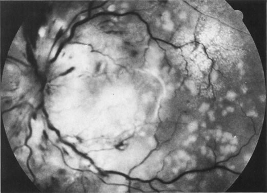
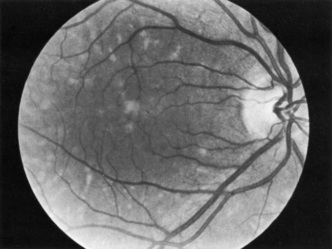
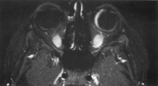
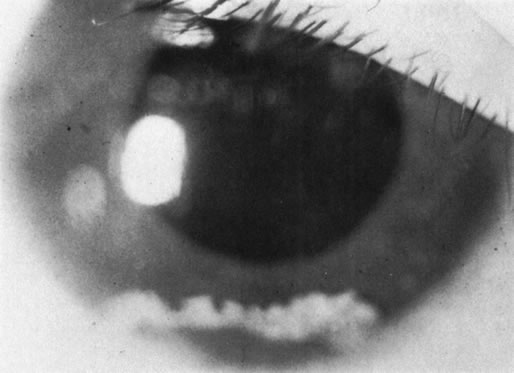
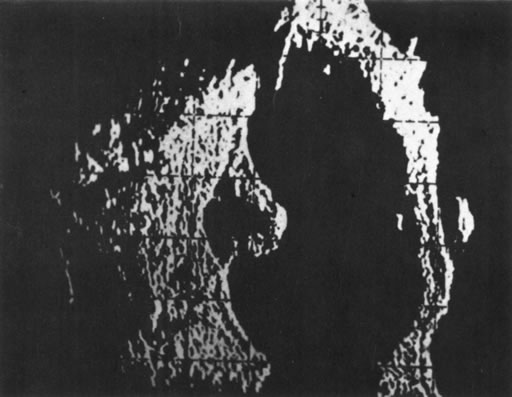
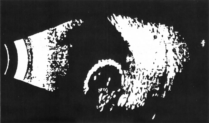
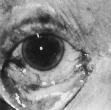
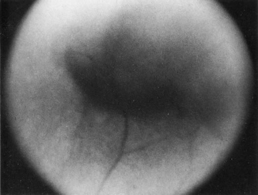
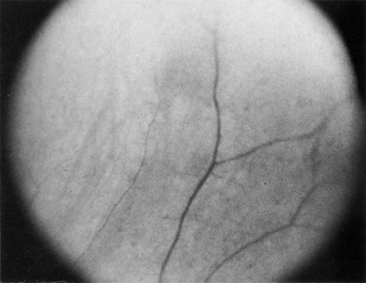
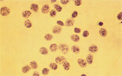
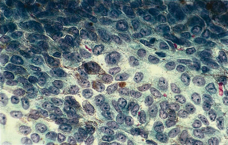
Necrotic melanomas account for approximately 5% of uveal melanomas; in the majority of cases, there is intraocular inflammation accompanying the cataract.7–9Figures 1 and 2 demonstrate a typical case. The patient had a long history of unilateral decreased vision. The eye had become painful 1 month before admission, and he was referred for evaluation of uveitis. Clinically, there was a dense, unilateral cataract with significant intraocular inflammation, which was manifested as a ciliary flush with 2+ cells and flare. Media opacity obscured all fundus detail. An immersion B-scan demonstrated a large intraocular tumor that was most consistent with a uveal melanoma. The eye was removed, and the diagnosis was confirmed histologically.10

Fig. 1. A unilateral media opacity in an adult, requiring that either a foreign body or an intraocular tumor be ruled out. The finding of a large episcleral (“sentinel”) vessel was consistent with either an intraocular tumor or an inflammation. An immersion B-scan helped determine the diagnosis (see Fig. 2).

Fig. 2. Immersion B-scan of the patient in Figure 1 demonstrates a uveal melanoma.

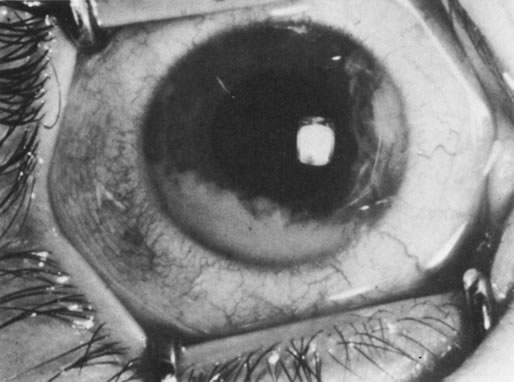
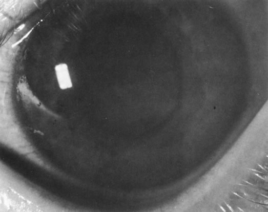
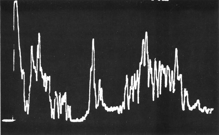
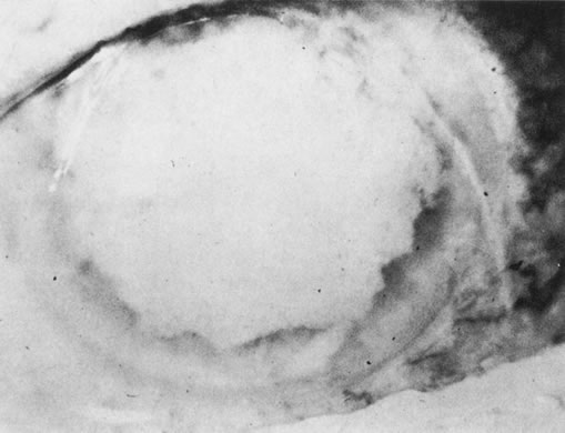
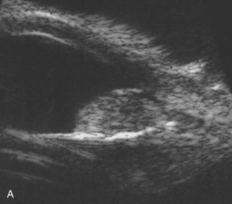
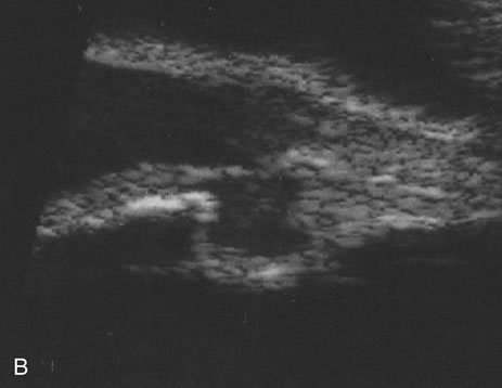
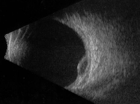
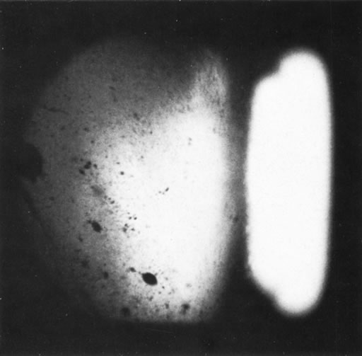
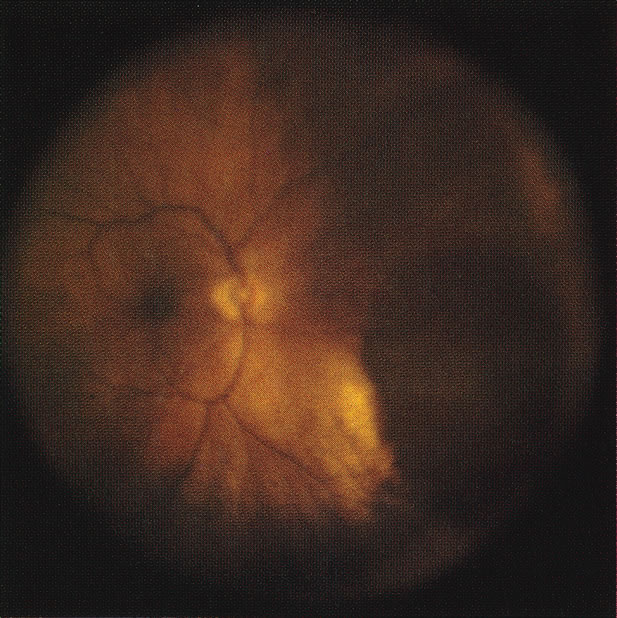
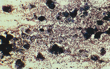
Unfortunately, imaging studies are not always diagnostic. The A- and B-scans shown in Figures 3 and 4 were from a patient with unilateral media opacity referred for evaluation and treatment of a presumed uveal melanoma. Occasionally, if the cataractous lens is scanned tangentially, artifact can occur. A repeat ultrasound at our institution was negative for tumor, and the referral ophthalmologist who performed a cataract extraction noted that no tumor was present and that the patient has an excellent visual outcome. In contrast, the patient shown in Figure 5 was referred with a presumed uveal melanoma with secondary inflammation and a history similar to that described for the patient shown in Figure 111 Ultrasound and CT examinations were not diagnostic. Because the eye was blind and painful, it was enucleated, and an extremely necrotic uveal melanoma was noted histologically. In some necrotic melanomas, the fundus cannot be visualized and the diagnosis cannot be established with imaging studies. Very rarely, a necrotic uveal melanoma can produce sufficient pigment dispersion into the vitreous to obscure the correct diagnosis.12

Fig. 3. B-scan in a patient with dense cataract, showing a possible tumor. Tangential cuts through a cataractous lens can produce a melanoma-like pattern (pseudomelanoma).

Fig. 4. A-scan through the area shown in Figure 3 shows a pseudomelanoma pattern. What distinguished this from a melanoma was a positive-angle kappa (climbing posterior spike). This patient did not have an eye tumor and had 20/20 vision after cataract removal.

Fig. 5. Clinical appearance of an eye with inflammation and pain. Ultrasound and CT were not diagnostic. Histologic examination revealed a necrotic melanoma.

The role of MRI in the evaluation of intraocular masquerade syndromes is unclear. There was initial enthusiasm about the possibility of differentiating atypical uveal melanomas from hemorrhagic-simulating lesions based on the unique paramagnetic properties of melanin.13 In relation to vitreous, a uveal melanoma typically has a bright signal on a T1 image and a dark signal on a T2 image. In contrast, hemorrhagic processes tend to be either bright or dark on a T1 image and bright on a T2 image.14

Unfortunately, there are several problems with MRI for uveal melanoma diagnosis. First, several investigators have shown that unless high field-strength, thin-section scans with surface coils are used, the quality of data obtained is suboptimal.15 Second, even in a state-of-the-art center, we have had a number of cases in which we were unable to differentiate between a uveal melanoma and a simulating lesion on the basis of MRI data. We obtain MRI studies in older patients with opaque media, equivocal ultrasonographic data, and large masses. In some of these cases, the MRI is diagnostic of either a melanoma or an extramacular disciform (hemorrhagic) process. It is also a useful adjunctive study if localized extraocular extension is suspected. In equivocal cases, however, we proceed to using invasive diagnostic techniques, such as fine-needle aspiration biopsy (FNAB) (see Metastatic Tumors).

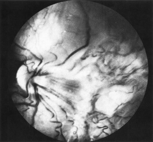
In addition to producing media opacity, inflammation, and cataract, uveal melanomas can also produce other signs that may lead to the incorrect diagnosis of uveitis. Anterior melanomas, especially those that involve the ciliary body, often produce dilated episcleral vessels (sentinel vessels) that occasionally have been misdiagnosed as scleritis (Fig. 6).

Fig. 6. Sentinel vessels present over a ciliary body melanoma. (Char DH: Clinical Ocular Oncology. 2nd ed. Philadelphia, Lippincott-Raven, 1996.)

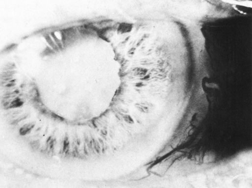
Iris melanomas can produce anterior chamber cell and flare, and if the tumor is located in the extreme iris periphery, it may be missed during a cursory examination. Iris melanomas, especially those in a circumferential or ring configuration, are often difficult to diagnose and may simulate either an anterior uveitis or glaucoma.16 Newer, higher-megahertz (high-frequency) ultrasound equipment is better for imaging anterior uveal tumors (Fig. 7).

Fig. 7. A. A typical iris tumor is easily visualized. Unfortunately, with atypical iris-ciliary body lesions, it may be difficult sometimes to differentiate different processes. B.A cystic change in a melanoma. On ultrasound, it is uncertain whether this is a benign or malignant lesion. Similarly, we are unable differentiate different types of neoplasms with this technique, and we have seen a few other anterior segment malignancies simulate melanoma on high-frequency ultrasound. Even with high-frequency ultrasound, we are unable to diagnose a “ring” melanoma.

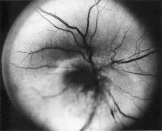
If direct ophthalmoscopy is used, a circumferential peripapillary melanoma can simulate optic neuritis (Fig. 8).

Fig. 8. A peripapillary melanoma simulating an optic neuritis. (Char DH: Clinical Ocular Oncology. 2nd ed. Philadelphia, Lippincott-Raven, 1996.)

Pseudomelanoma

Melanomas can masquerade as uveitis, and some inflammations or hemorrhagic processes can simulate melanoma. These processes include posterior scleritis, focal choroidal hemorrhage or detachments, and focal scleral thinning. Figures 9 and 10 demonstrate focal scleral thinning in the area of the superior rectus insertion in a patient with rheumatoid scleritis that produced a pseudomelanoma. An apparent elevated pigmented mass was noted on upgaze, but no tumor was observed when the eye was in primary position. Posterior scleritis is often not painful, although some of these patients have deep, piercing pain and increased discomfort during eye movement. Ultrasound usually is diagnostic, demonstrating fluid inside of Tenon's capsule (Fig. 11). A focal hemorrhagic choroidal detachment after cataract surgery can simulate a melanoma. These lesions usually have a bright area of hemorrhage at their base, whereas melanomas typically do not have associated hemorrhage until they break through Bruch's membrane (most commonly, such tumors are more than 5 mm thick). The differential diagnosis of uveal melanoma is discussed elsewhere in these volumes.

Fig. 9. A patient with rheumatoid arthritis and weakened sclera with a pseudomelanoma on upgaze.

Fig. 10. Lesion shown in Figure 8 disappeared when the patient's eye was in the primary position. (Note vessel pattern in Figs. 9 and 10.)

Fig. 11. A B-scan showing scleritis adjacent to a proton-irradiated choroidal melanoma.

METASTATIC TUMORS

Choroidal metastases can also produce signs that mimic uveitis. As many as 50% of patients with metastases to the eye may present to the ophthalmologist before the primary malignancy is discovered.17 Patients with renal and lung carcinomas have the highest frequency of presenting with eye symptoms as the initial complaint associated with the neoplasm; in contrast, more than 90% of breast carcinomas have a known primary at the time ocular metastases are noted. Usually, intraocular metastases are solid, posterior, amelanotic, choroidal tumors; however, in less than 10% of cases, they can simulate a benign intraocular inflammation. At least five patterns of inflammatory masquerade syndromes can occur with intraocular metastases.

  1. Tumors may produce a pseudoscleritis.
  2. Patients with metastatic carcinoma may present with a diffuse infiltration of the iris (Fig. 12).

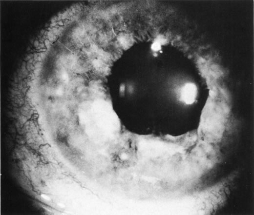
    Fig. 12. Breast carcinoma diffusely infiltrating the iris.

  3. In rare cases, an optic nerve metastasis can simulate either an optic neuritis or an acute anterior ischemic neuropathy (Fig. 13).

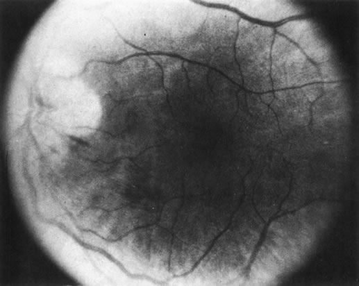
    Fig. 13. Bronchiogenic carcinoma metastatic to the optic nerve. (Char DH: Clinical Ocular Oncology. 2nd ed. Philadelphia, Lippincott-Raven, 1996.)

  4. Isolated metastatic cells in the vitreous or anterior chamber may occur as the only manifestation of an ocular metastasis.18
  5. Figure 14 shows a patient with bilateral pseudohypopyon as the first stigmata of widespread disease.

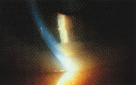
    Fig. 14. Bilateral pseudohypopyon as the initial manifestation of widespread metastases.

Most commonly, pseudohypopyons in adults develop in association with either a systemic lymphoma or leukemic relapse. Rarely, metastatic cutaneous melanoma can produce this pattern; however, usually these later cells are pigmented (Fig. 15). Much more frequently, vitreous cells in patients with known metastases are benign, originating from either infectious or endogenous uveitis.

Fig. 15. Vitreous cavity filled with cells and strands. (Char DH, Schwartz A, Miller TR, Abele JS: Ocular metastases from systemic melanoma. Am J Ophthalmol 90:702, 1980. Published with permission from the American Journal of Ophthalmology. Copyright by the Ophthalmic Publishing Co.)

A masquerade syndrome secondary to a distant malignancy can take several forms. In some metastatic tumor patients, multiple areas of increased choroidal pigmentation develop. Although these paraneoplastic syndromes are rare, a more common form is autoimmune destruction of retinal photoreceptor or ganglion on cells that more commonly occur either with small cell carcinomas of the lung or metastatic cutaneous melanoma.19–23

The evaluation of patients with metastatic tumors to the eye may be difficult. Approximately 10% of uveal melanoma patients have a history of a systemic malignancy. Approximately 50% of patients with uveal metastases are found to have an elevated plasma carcinoembryonic antigen or cancer antigen 125 level; in contrast, uveal melanoma patients do not have significantly elevated levels.24 Most metastases to the uveal tract can be correctly diagnosed with ultrasonography. In atypical presentations of metastatic tumors in which there is diagnostic uncertainty, we have confirmed the diagnosis with an intraocular FNAB.18

In several ocular oncology units, FNAB has become an important diagnostic adjuvant in difficult cases.25–27 We use FNAB in intraocular oncologic cases that cannot be diagnosed with noninvasive techniques.4 We have observed no false-positives in uveal melanoma diagnosis, and in that malignancy, we can accurately predict prognosis on the basis of the cellular composition (percentage of epithelioid cells) of the aspirate.27 Rarely in a necrotic melanoma, insufficient viable tumor cells are present to establish a diagnosis with this technique. In several lesions that simulate either retinoblastoma or melanoma, cytologic pattern on FNAB is diagnostic. We have found this technique especially useful in atypical metastatic uveal tumors. Often such patients are incorrectly diagnosed as having either a uveal melanoma or a benign lesion. Establishing the correct diagnosis results in optimum treatment. A misdiagnosis of a uveal melanoma would require a subsequently higher radiation dose with a higher likelihood of radiation vasculopathy. Conversely, a misdiagnosis of a benign process would result in ineffective therapy.

Figure 16 shows a case of what clinically and ultrasonographically appeared to be a uveal melanoma. The FNAB specimen revealed a cellular pattern that was diagnostic of a primary carcinoid (Fig. 17), and the patient responded dramatically with 40 gray of photon radiation. Figure 18 shows the FNAB cytopathology of a typical epithelioid choroidal melanoma; in contrast, a smear of a spindle cell tumor has spindle-shaped cytoplasm and smaller nuclei. Figure 19 shows a benign pigmented mass, such as a retinal pigment epithelial proliferation or a melanocytoma. In contrast to a melanoma, this type of mass has much larger pigment granules and benign cytomorphologic detail. In addition to standard cytopathologic FNAB evaluation, aspirated material may be used for several molecular biology studies (e.g., fluorescence in situ hybridization, comparative genomic hybridization), flow cytometry, special stains, tissue culture, and ultrastructural analyses.

Fig. 16. Lesion in the posterior pole suggestive of a uveal melanoma.

Fig. 17. Fine-needle aspiration biopsy of the patient in Figure 16 shows that the lesion is a primary carcinoid.

Fig. 18. Cytopathology from a fine-needle aspiration biopsy demonstrates an epithelioid melanoma.

Fig. 19. Fine-needle biopsy from a benign melanocytoma. In contrast to a uveal melanoma, a benign melanocytoma has pigment granules that are much larger. Its nuclear detail is also bland and lacking nucleoli.

A number of nonuveitis entities can produce vitreous cells, and some of these conditions are listed in Table 1.


Table 1. Vitreous Cells Not Associated With Uveitis

Primary and secondary intraocular lymphomas
Metastatic tumors
Retinoblastoma
Old vitreous hemorrhage (diabetes, retinal detachment)
“Tobacco dust” from retinal detachment
Battered baby syndrome
Retinitis pigmentosa
Asteroid hyalosis
Synchysis scintillans
Familial amyloidosis


INTRAOCULAR LYMPHOMA

Intraocular lymphoma is usually initially misdiagnosed in older patients as a chronic diffuse uveitis.28 In a series of 828 consecutive patients at the uveitis center, approximately 1.5% of cases (13), had intraocular lymphomas.29 In any patient more than 50 years of age with an onset of a diffuse uveitis or vitritis, the diagnosis of intraocular lymphoma must be excluded. The classification and nomenclature of intraocular lymphomas is in flux.28,30,31 Erroneously termed ocular reticulum cell sarcoma, this neoplastic disease is usually limited to the eye and central nervous system (CNS). This process was initially thought to be due to a proliferation of malignant reticulum cells, but more recent studies have shown that this is usually a malignant B-cell lymphoma.30 Most investigators have adopted the term primary central nervous system lymphoma (PCNSL). The incidence of this disease has increased, although reasons for the rise are unclear.32 Ocular involvement occurs between 12% and 25% of patients. There are three types of intraocular lymphomas.

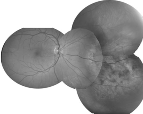
  1. The intraocular findings in primary intraocular–CNS lymphoma may simulate a diffuse uveitis, usually with a plethora of vitreous cells and often with virtually pathognomonic yellowish white chorioretinal infiltrates (Fig. 20). Less common clinical presentations can mimic toxoplasmosis, acute retinal necrosis, a branch vein occlusion, or various forms of retinal vasculitis.

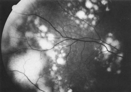
    Fig. 20. Yellowish white chorioretinal infiltrate typical of primary intraocular lymphoma (erroneously termed ocular reticulum cell sarcoma). (Char DH: Clinical Ocular Oncology. 2nd ed. Philadelphia, Lippincott-Raven, 1996.)

  2. A less common form of primary ocular lymphoma consists of lesions in the choroid with fewer vitreous cells. Systemic, non-CNS lymphoma usually develops in these patients.33 Rarely, we have observed a variant of this second group, in which a patient has a solid intraocular lymphoma that involves the choroid as the first manifestation of widespread disease.34
  3. Intraocular involvement may occur as a sequela of systemic lymphoma, often as the first sign of reactivation after therapy. Such patients usually present with a malignant hypopyon (Fig. 21). In very rare cases, a patient with mycosis fungoides, a cutaneous T cell lymphoma, is found to have an associated anterior or posterior uveal involvement.

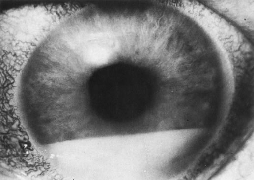
    Fig. 21. Malignant hypopyon in a patient with systemic lymphoma.

In all three forms of intraocular malignant lymphoma, the diagnosis is made with FNAB. In rare instances, a chorioretinal biopsy is necessary.35,36 In 15% of cases, a single vitreous biopsy may not demonstrate malignant cells, and multiple biopsies may be required.28

All patients with intraocular lymphoma require a thorough systemic evaluation, including examination of cerebral spinal fluid cytology, gadolinium-enhanced brain MRI, abdominal-chest CT, routine blood studies, and bone marrow biopsies. Probably, positron emission tomography scans will supplant some of these studies, but at present, data from this entity are insufficient. Historically, primary intraocular–CNS lymphomas were almost uniformly fatal, but newer, aggressive therapies with either intrathecal or systemic chemotherapy plus eye and CNS irradiation have produced long-term survival in some patients. Early diagnosis and treatment are associated with better prognosis, so patients should be evaluated promptly.28

The diagnostic evaluation of these patients continues to evolve. Superb cytologic analysis remains the central diagnostic test. Several approaches can be used to assess monoclonality (usually associated with lymphoma) versus polyclonality (usually associated with benign processes). The major problem with all of these techniques is that the vitreous sample is limited, and all of these ancillary assays may have false-positive and false-negative results. Flow cytometric features of lymphoma include an abnormal kappa-lambda ratio of either greater than 3 or less than 0.33, and monoclonal expansion of either B cells (more common) or T cells.37,38 A more elegant technique is to adapt a variation of polymerase chain reaction assay to identify a clonal B or T cell expansion, gene rearrangements, or oncogenes. However, errors do occur even with these sensitive assays.39,40 A few vitrectomy specimens that were not diagnosed cytologically were correctly diagnosed with these techniques.41,42 In addition, often the vitreous ratio of two cytokines, interleukin (IL)-10 and IL-6, is increased in lymphoma; however, exceptions occasionally have been reported.43,44

Pseudolymphoma

A benign variant of intraocular lymphoid tumors may occur in either an isolated form or, more commonly, a diffuse form involving both the intraocular contents and contiguous sclera and orbit.45 Usually these lesions simulate a solid intraocular neoplasm (e.g., melanoma), and they may occur with or without pain (Fig. 22). Most of these patients have an ultrasound pattern consistent with a melanoma, and the correct diagnosis is established with FNAB. As Cockerham and colleagues46 noted, the episcleral lesion may appear benign, whereas the intraocular component demonstrates lymphoma.

Fig. 22. A benign lymphoid intraocular lesion diagnosed with fine-needle aspiration biopsy.

Rarely, after liver transplantation, patients develop a spectrum of post-transplantation lymphoid proliferative disorders that may range from benign to malignant lymphomas and have involved the eye.47–49

Back to Top
ADULT NON-NEOPLASTIC–SIMULATING LESIONS
A number of nonendogenous, noninflammatory processes can simulate uveitis. Undiagnosed intraocular foreign bodies and endophthalmitis due to replicating organisms are discussed elsewhere in these volumes. These entities can mimic anterior, posterior, or diffuse uveitis.

Medications can alter the normal clarity of the anterior chamber and produce a pattern that may be mistaken for an anterior uveitis. For example, patients, especially those with a dark iris on dilation with phenylephrine hydrochloride, may have a transient appearance of anterior chamber pigmented cells that may last for up to 24 hours. Administering drops before performing an anterior segment examination can impair the clinician's ability to grade aqueous cell and flare.50,51 Similarly, occasionally Krukenberg spindles with pigment dispersion may also mimic uveitis. Anterior uveitis has developed in several acquired immunodeficiency syndrome patients receiving rifabutin therapy.52

A number of adult cardiovascular diseases can produce ophthalmic manifestations that simulate uveitis. In severe hypertension, retinal detachment, small choroidal vascular insults, cotton wool spots, and hemorrhages may occur, simulating posterior uveitis (Fig. 23). In patients with carotid artery disease, anterior segment ischemia similar to mild anterior uveitis may be observed. This pattern may also occur after either extensive ocular irradiation or disinsertion of more than two extraocular muscles during retinal or strabismus surgery.

Fig. 23. Atypical severe hypertension with exudative detachment and choroidal lesions.

A number of retinal lesions can either produce or simulate uveitis. Retinal detachments can occasionally produce enough inflammation for anterior uveitis to obscure both fundus detail and the correct diagnosis. In a patient with markedly decreased vision and media opacification, an ultrasonographic evaluation is mandatory to rule out a neoplastic or foreign body process. Cases of inapparent retinal detachment also can be correctly diagnosed with ultrasonography. Uncommonly in central serous choroidopathy, protein deposition at the leakage point can simulate a focal choroiditis. Medullated nerve fibers can simulate retinochoroiditis, but the shape, corresponding to the nerve fiber layer with the absence of other signs of inflammation, usually allows a clear-cut differentiation.53

Some retinal dystrophies and degenerative processes can simulate a retinochoroiditis. Vitreous cells may occur in cases of retinitis pigmentosa. Pigmented paravenous retinochoroidal atrophy has a distinctive pattern of pigmentary deposits surrounding retinal veins; it may occur as an idiopathic degenerative disorder or sometimes as a sequela of ocular sarcoid. Fundus flavimaculatus is a rare autosomal recessive dystrophy that is usually detected in the third decade of life. It produces typical comma-shaped, yellowish retinal pigment epithelial opacities in the posterior pole, but it does not involve the macula. It can simulate a diffuse choroiditis (Fig. 24). Myopic degeneration can produce small, white, posterior pole spots that can simulate a posterior choroiditis. Peripheral fundus “paving stone” degeneration is rarely confused with an inflammatory process. These lesions show no evidence of retinal or vitreal inflammation and are quite common.54

Fig. 24. Fundus flavimaculatus. Note comma- or fish-shaped yellowish Figures at the level of pigment epithelium.

Back to Top
CHILDHOOD MALIGNANT PROCESSES
The differential diagnoses of uveitis in young children are relatively limited. In patients less than 2 years of age, uveitis is uncommon. Therefore, if a child of this age presents with signs of uveitis, a number of tumors (e.g., retinoblastoma, leukemia, medulloepithelioma) must be considered as differential diagnoses.

RETINOBLASTOMA

Retinoblastoma is the most common childhood intraocular malignancy. More than 98% of patients present before the age of 5 years, and any child less than 3 years of age presenting with leukocoria, strabismus, or intraocular inflammation must have retinoblastoma excluded from the differential diagnosis by a thorough fundus evaluation. At least three patterns of retinoblastoma can simulate a benign inflammatory process.

  1. Inflammatory signs occur in as many as 10% of retinoblastomas; the child presents with redness and ciliary flush. In any child less than 2 years of age who has evidence of intraocular inflammation, either a tumor or a trauma must be ruled out.
  2. Very large retinoblastomas can contiguously involve the anterior segment and produce a pseudohypopyon (Fig. 25). Unlike a benign collection of white cells, a pseudohypopyon is made up of malignant cells, and clinically it is not as mobile as an inflammatory hypopyon. Usually the diagnosis in these cases is relatively straightforward because the large retinoblastoma can be either seen ophthalmoscopically or imaged with CT or ultrasound.

    Fig. 25. Large retinoblastoma with “spillover” pseudohypopyon.

  3. Diffuse retinoblastoma, which accounts for less than 2% of cases, often does not produce a discrete tumor (Fig. 26). The diffuse cases I have managed have had atypical retinal detachments without clinical, CT, or ultrasonographic evidence of a calcified tumor mass. Diagnosis has been established with an intraocular FNAB.55

    Fig. 26. Diffuse retinoblastoma without an obvious tumor mass.

Pseudoretinoblastoma

The diagnosis and classification of pseudoretinoblastoma lesions are evolving. Genetic tests will more precisely differentiate among several entities grouped under the rubrics of either retinal dysplasia or atypical persistent hyperplastic primary vitreous. In children with lesions that produce leukocoria, such molecular biologic testing for several diseases is often useful (see http://www.genetests.org). A recent example in which such tests altered our clinical impression is the case of a young female patient referred to us to rule out retinoblastoma. Our findings on clinical examination were consistent with an atypical retinal dysplasia. There was no family history, but genetic testing demonstrated that she was a mosaic for Norrie's disease.56 It is likely that such types of studies will help to clarify the nature of many of the pseudoretinoblastomas that have been classified clinically as variants of either persistent hyperplastic primary vitreous or retinal dysplasia.

LEUKEMIA

Ocular involvement is a common presentation of acute myelomonocytic leukemia in children from the Mediterranean area.57 These children often present with simultaneous ocular and orbital tumor.58,59 In a few cases, we have established the correct diagnosis with an anterior chamber paracentesis. Most American children managed for leukemia-associated ocular problems, however, have the acute form of lymphocytic disease, and ocular problems usually develop as a sign of systemic or CNS relapse. In rare cases, an anterior chamber collection of malignant cells (similar in appearance to Fig. 21) or tumor infiltrating the optic nerve head can be the presenting sign of this disease.58,59 Rarely, where the possibility of intraocular recurrence of acute lymphocytic leukemia is not recognized and topical corticosteroids are given for presumed anterior uveitis, the anterior chamber clears and the patient presents with a ciliary body mass (Fig. 27).

Fig. 27. A patient who had an ocular relapse of acute lymphocytic leukemia treated with topical corticosteroids because the symptoms were presumed to be due to anterior uveitis. As a result, the anterior chamber cleared and the patient presented with a ciliary body mass. The magnetic resonance image demonstrates a pseudoscleritis in that eye.

RARE PEDIATRIC INTRAOCULAR NEOPLASMS

Less common neoplasms in children can also simulate benign forms of inflammation. Medulloepithelioma is a rare tumor that can affect different intraocular structures but most commonly involves the iris-ciliary body. This tumor has a typically velvety surface60 and may present with intraocular inflammatory signs (Fig. 28).

Fig. 28. A patient with a medulloepithelioma presented with white, flocculent anterior chamber debris.

Juvenile xanthogranuloma, a benign lesion, usually presents in children less than 1 year of age; it is characterized by a spontaneous hemorrhage and a poorly demarcated yellowish iris tumor.61–64 Only about-one third of children have the complete form of this disease.64

Combined retinal and retinal pigment epithelial hamartomas can also produce a pattern that simulates retinitis (Fig. 29). The pigment at the level of the retinal pigment epithelium, the surface fibrosis, and the vascular changes are typical features of this entity.

Fig. 29. Combined retinal pigment epithelial-retinal hamartoma. (Char DH: Clinical Ocular Oncology. 2nd ed. Philadelphia, Lippincott-Raven, 1996.)

Back to Top
NON-NEOPLASTIC CHILDHOOD DISEASES
A long list of non-neoplastic conditions can either produce or simulate inflammation in children. Patients with shaken baby syndrome may have findings suggestive of intraocular inflammation with both anterior chamber and vitreous cells, and occasionally with swelling of the optic nerve head in conjunction with vitritis. Foreign bodies in children may be missed as a result of an inaccurate history.

A number of macular dystrophies can be mistaken for a central choroiditis. Best's vitelliform macular dystrophy is often referred to as a focal macular choroiditis. This dystrophy is autosomal dominantly inherited and has variable penetrance and expressivity. An “egg yolk” lesion in the fovea is a classic sign, but this characteristic is not always observed. In difficult cases of Best's vitelliform macular dystrophy, an electro-oculogram may be performed and will be abnormal, even in normal-appearing disease carriers.

Back to Top
REFERENCES

1. Duke-Elder S, Perkins ES: Diseases of the uveal tract. In Duke-Elder S (ed): System of Ophthalmology, pp 35–594. Vol 9. St. Louis, CV Mosby, 1966

2. Theodore FH: Conjunctival carcinoma masquerading as chronic conjunctivitis. Eye Ear Nose Throat Mon 46:1419, 1967

3. Irvine AR: Diffuse epibulbar squamous cell epithelioma. Am J Ophthalmol 64:550, 1968

4. Char DH: Tumors of the Eye and Adnexa. Toronto, BC Decker, 2001

5. Makley TA Jr, Teed RW: Unsuspected intraocular malignant melanomas. Arch Ophthalmol 60:475, 1958

6. Zimmerman LE: Problems in the diagnosis of malignant melanomas of the choroid and ciliary body. Am J Ophthalmol 75:917, 1973

7. Kirk HQ, Petty RW: Malignant melanoma of the choroid: A correlation of clinical and histological findings. Arch Ophthalmol 56:843, 1956

8. Litricin O: Unsuspected uveal melanomas. Am J Ophthalmol 76:734, 1973

9. Fraser DJ, Font RL: Ocular inflammation and hemorrhage as initial manifestations of uveal malignant melanoma: Incidence and prognosis. Arch Ophthalmol 97:1311, 1979

10. Shields JA, Augsburger JJ: Cataract surgery and intraocular lenses in patients with unsuspected malignant melanoma of the ciliary body and choroid. Ophthalmology 92:823, 1985

11. Char DH, Howes EL Jr, Fries PD, et al: Uveal melanoma with opaque media: Absence of definitive diagnosis prior to enucleation. Can J Ophthalmol 23:2226, 1988

12. El Baba F, Hagler WS, de la Cruz A, Green WR: Choroidal melanoma with pigment dispersion in vitreous and melanomalytic glaucoma. Ophthalmology 95:370, 1988

13. Raymond WR, Char DH, Norman D, Protzko EE: Magnetic resonance imaging evaluation of uveal tumors. Am J Ophthalmol 111:633, 1991

14. De Potter T, Flanders AE, Shields JA, et al: The role of fat-suppression technique and gadopentetate dimeglumine in magnetic resonance imaging evaluation of intraocular tumors and simulating lesions. Arch Ophthalmol 112:340, 1994

15. deKeizer RJ, Vielvoye GJ, deWolff-Rouendaal D: Nuclear magnetic resonance imaging of intraocular tumors. Am J Ophthalmol 102:438, 1986

16. Litricin O: Diffuse malignant ring melanoma of the iris and ciliary body. Ophthalmologica 178:235, 1979

17. Ferry AP, Font RL: Carcinoma metastatic to the eye and orbit: I. A clinicopathologic study of 227 cases. Arch Ophthalmol 92:276, 1974

18. Char DH, Schwartz A, Miller TR, Abele JS: Ocular metastases from systemic melanoma. Am J Ophthalmol 90:702, 1980

19. Buchanan TAS, Gardiner TA, Archer DB: An ultrastructural study of retinal photoreceptor degeneration associated with bronchial carcinoma. Am J Ophthalmol 97:277, 1984

20. Ryll DL, Campbell JR, Robertson BM, Brubaker SJ: Pseudometastatic lesions of the choroid. Ophthalmology 87:1181, 1980

21. Borruat FX, Othenin-Girard P, Uffer S, et al: Natural history of diffuse uveal melanocytic proliferation: Case report. Ophthalmology 99:1698, 1992

22. Thirkill CE, Tait RC, Tyler NK, et al: The cancer-associated retinopathy antigen is a recoverin-like protein. Invest Ophthalmol Vis Sci 33:2768, 1992

23. Campo E, Brunier MN, Merino MJ: Small cell carcinoma of the endometrium with associated ocular paraneoplastic syndrome. Cancer 69:2283, 1992

24. Michelson JB, Felberg NT, Shields JA: Evaluation of metastatic cancer to the eye: Carcinoembryonic antigen and gamma glutamyl transpeptidase. Arch Ophthalmol 95:692, 1977

25. Augsburger JJ: Fine needle aspiration biopsy of suspected metastatic cancer to the posterior uvea. Trans Am Ophthalmol Soc 86:500, 1988

26. Leff SR, Yarian DL, Shields JA, et al: Tumor-associated retinal pigment epithelial proliferation simulating retinal pigment epithelial tear. Retina 9:267, 1989

27. Char DH, Miller TR, Ljung BM, et al: Fine needle aspiration biopsy in uveal melanoma. Acta Cytol 33:599, 1989

28. Char DH: Primary intraocular lymphoma (ocular reticulum cell sarcoma): Diagnosis and management. Ophthalmology 95:625, 1988

29. Rothova A, Ooijman F, Kerkhoff F, et al: Uveitis masquerade syndromes. Ophthalmology 108:386, 2001

30. Char DH, Ljung BM, Deschenes J, Miller TR: Intraocular lymphoma: Immunologic and cytologic analysis. Br J Ophthalmol 72:905, 1988

31. Harbour JW, Char DH: Intraocular lymphoid tumors. In Chang S, Shields J, Greed WR, Guyer DR (eds): Retina, Vitreous, Macula. Philadelphia, WB Saunders, 1999

32. O'Leary TJ: Flow cytometry in diagnostic cytology. Diagn Cytopathol 18:41, 1998

33. Klingele TG, Hogan MJ: Ocular reticulum cell sarcoma. Am J Ophthalmol 79:39, 1975

34. Frederick D, Char DH, Ljung BM, Brinton G: Solitary intraocular lymphoma as an initial presentation of widespread disease. Arch Ophthalmol 107:395, 1989

35. Martin DF, Chan CC, de Smet MD, et al: The role of chorioretinal biopsy in the management of posterior uveitis. Ophthalmology 100:705, 1993

36. Lafaut BA, Hanssens M, Verbraeken H, De Laey JJ: Chorioretinal biopsy in the diagnosis of intraocular lymphoma: A case report. Bull Soc Belge Ophtalmol 252:67, 1994

37. Wilson DJ, Braziel R, Rosenbaum JT: Intraocular lymphoma. Immunopathologic analysis of vitreous biopsy specimens. Arch Ophthalmol 110:1455, 1992

38. Davis JL, Viciana AL, Ruiz P: Diagnosis of intraocular lymphoma by flow cytometry. Am J Ophthalmol 124:362, 1997

39. Abdel-Reheim FA, Edwards E, Arber DA: Utility of a rapid polymerase chain reaction panel for the detection of molecular changes in B-cell lymphoma. Arch Pathol Lab Med 120:357, 1996

40. Weston AP, Banerjee SK, Horvat RT, et al: Specificity of polymerase chain reaction monoclonality for diagnosis of gastric mucosa-associated lymphoid tissue (MALT) lymphoma: Direct comparison to Southern blot gene rearrangement. Dig Dis Sci 43:290, 1998

41. Katai N, Kuroiwa S, Fujimori K, Yoshimura N: Diagnosis of intraocular lymphoma by polymerase chain reaction. Graefes Arch Clin Exp Ophthalmol 235:431, 1997

42. White VA, Gascoyne RD, Paton KE: Use of the polymerase chain reaction to detect B and T-cell gene rearrangements in vitreous specimens from patients with intraocular lymphoma. Arch Ophthalmol 117:761, 1999

43. Whitcup SM, Stark-Vancs V, Wittes RE, et al: Association of interleukin 10 in the vitreous and cerebrospinal fluid and primary central nervous system lymphoma. Arch Ophthalmol 115:1157, 1997

44. Akpek EK, Maca SM, Christen WG, Foster CS: Elevated vitreous interleukin-10 level is not diagnostic of intraocular central nervous system lymphoma. Ophthalmology 106:2291, 1999

45. Ryan SJ, Zimmerman LE, King FM: Reactive lymphoid hyperplasia: An unusual form of intraocular pseudotumor. Trans Am Acad Ophthalmol Otolaryngol 76:652, 1972

46. Cockerham GC, Hidayat AA, Bijwaard KE, Sheng ZM: Re-evaluation of reactive lymphoid hyperplasia of the uvea: An immunohistochemical and molecular analysis of 10 cases. Ophthalmology 107:151, 2000

47. Brodsky MC, Casteel H, Barber LD, et al: Bilateral iris tumors in an immunosuppressed child. Surv Ophthalmol 36:217, 1991

48. Clark WL, Scott IU, Murray TG, et al: Primary intraocular post-transplantation lymphoproliferative disorder. Arch Ophthalmol 116:1667, 1998

49. Robinson R, Murray PI, Willshaw HE, et al: Primary ocular post transplant lymphoproliferative disease. J Pediatr Ophthalmol Strabismus 32:393, 1995

50. Mitsui Y, Takagi Y: Nature of aqueous floaters due to sympathomimetic mydriatics. Arch Ophthalmol 65:626, 1961

51. Haddad NJ, Moyer NJ, Riley FC Jr: Mydriatic effect of phenylephrine hydrochloride. Am J Ophthalmol 70:729, 1970

52. Jacobs DS, Piliero PJ, Kuperwaser MG, et al: Acute uveitis associated with rifabutin use in patients with human immunodeficiency virus infection. Am J Ophthalmol 118:716, 1994

53. Baarsma CS: Acquired medullated nerve fibers. Br J Ophthalmol 64:651, 1980

54. Okum E: Gross and microscopic pathology in autopsy eyes: Part II. Peripheral chorioretinal atrophy. Am J Ophthalmol 50:574, 1960

55. Char DH, Miller TR: Fine needle biopsy in retinoblastoma. Am J Ophthalmol 97:686, 1984

56. Black G, Redmond RM: The molecular biology of Norrie's disease. Eye 8:491, 1994

57. Cavdar AO, Arcasoy A, Babacan E, et al: Ocular granulocytic sarcoma (chloroma) with acute myelomonocytic leukemia in Turkish children. Cancer 41:1606, 1978

58. Kincaid MC, Green WR: Ocular and orbital involvement in leukemia. Surv Ophthalmol 27:211, 1983

59. Rosenthal AR: Ocular manifestations of leukemia: A review. Ophthalmology 90:899, 1983

60. Broughton WL, Zimmerman LE: A clinicopathologic study of 56 cases of intraocular medulloepitheliomas. Am J Ophthalmol 85:407, 1978

61. Smith ME, Sanders TW, Bresnick GH: Juvenile xanthogranuloma of the ciliary body in an adult. Arch Ophthalmol 81:813, 1969

62. Zimmerman LE: Ocular lesions of juvenile xanthogranuloma: Nevoxanthoendothelioma. Am J Ophthalmol 60:1011, 1965

63. Smith JLS, Ingram RM: Juvenile oculodermal xanthogranuloma. Br J Ophthalmol 52:696, 1968

64. Treacy KW, Letson RD, Summers CG: Subconjunctival steroid in the management of uveal juvenile xanthogranuloma: A case report. Pediatr Ophthalmol Strabismus 27:126, 1990

Back to Top