Chapter 50
Syphilis
ROBERT S. WEINBERG
Main Menu   Table Of Contents

Search

HISTORY
PATHOGENESIS
INCIDENCE
OCULAR SYPHILIS
SYPHILITIC UVEITIS
LABORATORY TESTS
TREATMENT
SUMMARY AND RECOMMENDATIONS
REFERENCES

Despite a decline in the incidence of syphilis in the United States during the 1990s, the increased incidence in the United States during the 1980s is being mirrored now in other parts of the world. It is likely that more and more physicians will be treating patients with the disease. The ophthalmologist may be the first physician to examine a patient with syphilis, and the increased number of patients with systemic syphilis means a greater likelihood of seeing patients with ocular syphilis. Familiarity with the disease, the laboratory tests available for diagnosis, and effective therapeutic regimens used to treat syphilis are important in managing a curable form of uveitis.
Back to Top
HISTORY
The earliest epidemic of syphilis was described in the late 1400s and early 1500s.1 Syphilis may have been introduced into western Europe in 1493 after Columbus's initial voyage to the Western Hemisphere.2 Syphilitic involvement of the nervous system was first described in the 1800s.3 In 1903, syphilis was first transmitted to experimental animals, and in 1905 Schaudinn and Hoffman discovered Treponema pallidum to be the causative agent.4 The Wassermann blood test for syphilis was developed in 1907.4 In 1918 Igesheimer wrote a book on syphilis that covered syphilitic ocular disease.5 In 1943 the introduction of penicillin revolutionized the treatment of the disease.3 Antibiotics were found in the 1950s to treat syphilis in penicillin-allergic patients.3 In the 1980s there was an increase in the incidence of syphilis associated with the increased incidence of intravenous drug abuse and AIDS.6 Although in the 1990s the incidence of new cases of syphilis in the United States declined, syphilis still remains a significant health problem, not only in the United States, but in many developing countries and in Eastern Europe.7,8
Back to Top
PATHOGENESIS
Syphilis is an infectious disease caused by the spiral motile flagellated bacteria T. pallidum. The organism is a fastidious, microaerophilic obligate parasite of humans and has not been cultured in vitro.9 However, the genome sequence of T. pallidum is available and may provide information about virulence.9

Inoculation occurs through the epithelium and is followed by a generalized transient infection (spirochetemia). A primary chancre develops at the inoculation site, generally genital or oral, and regional lymphadenopathy follows. In the secondary stage of syphilis, there is hematogenous spread to local foci in the skin and mucous membranes. During the tertiary stage of syphilis, there is diffuse low-grade inflammation with degeneration and necrosis in the cardiovascular and central nervous systems. Ocular involvement is present in congenital syphilis and may occur in primary, secondary, or tertiary syphilis. In latent syphilis, patients are seroreactive but have no other disease activity.

Back to Top
INCIDENCE
The incidence of syphilis in the United States, measured by the number of cases reported to the Centers for Disease Control (CDC), began to decline during the 1990s.10 Elsewhere in the world, reporting mechanisms are less well established, but the incidence of syphilis still appears to be increasing.11,12 With an increased rate of primary and secondary syphilis, there is an increased likelihood of congenital syphilis. Because syphilitic genital ulceration may increase the transmission of the AIDS virus, there is an increased risk of AIDS associated with an increase in the incidence of syphilis.13
Back to Top
OCULAR SYPHILIS
Ocular involvement may occur at any stage of syphilis. In congenital syphilis, acute iridocyclitis may be present from birth to age 6 months. Chorioretinitis with the “salt and pepper” fundus, foci of old choroiditis, areas of retinal pigment epithelial hypertrophy, and atrophy may occur at any time after 6 months. Vitritis may be present. Interstitial keratitis may be present at birth but is more commonly a late manifestation, occurring bilaterally and between ages 5 and 20.14

In acquired syphilis, chancre of the conjunctiva or lids may be seen in the primary stage. In secondary syphilis, nonspecific conjunctivitis, scleroconjunctivitis, and syphilides (painless white nodules on the conjunctiva) have all been described.5 Iris roseola, early diffuse hyperemia of the iris vasculature, and late localized iris inflammation have been reported.15 Iritis and iridocyclitis are common. Syphilitic uveitis in tertiary syphilis may occur up to 20 years after the primary infection. Any part of the eye can be affected by syphilis (Table 1).


TABLE 1. Ocular Involvement in Syphilis


LocationManifestation
EyelidsChancre
ConjunctivaConjunctivitis, nodules
CorneaInterstitial keratitis
IrisPatchy iris atrophy, Argyll Robertson pupil
LensDislocation
UveaIridocyclitis, chorioretinitis
Optic NerveOptic atrophy


Back to Top
SYPHILITIC UVEITIS
Syphilitic uveitis is a curable form of uveitis, albeit one that can lead to permanent and profound loss of vision if not diagnosed and treated promptly. Reports of the incidence of syphilitic uveitis in patients with syphilis vary, and there are no recent studies. The relative incidence of syphilitic uveitis compared with all uveitis cases reported ranges from 0.8% to 4.3%.16,17 Syphilis is an increasing cause of new uveitis in the elderly.18

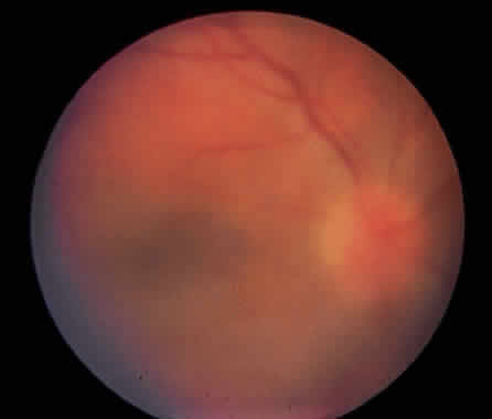
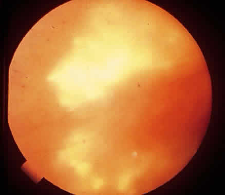
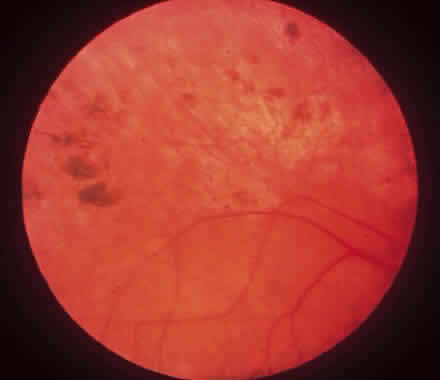
There are no pathognomonic features of syphilitic uveitis. Iridocyclitis in early disease may be unilateral and severe. In late tertiary syphilis, iridocyclitis may also be unilateral and appear 4 to 6 months after inadequate treatment of tertiary syphilis. Syphilitic chorioretinitis also has no pathognomonic features. There may be multifocal chorioretinal infiltrates, overlying vitritis, and papillitis.19 Early findings include vitritis (Fig. 1), multifocal chorioretinitis (Fig. 2), optic neuritis, retinal vasculitis, retinal hemorrhages, and retinal edema. Pigment proliferation is present late in the course of the disease (Fig. 3). Presenting complaints in patients with acute syphilitic uveitis include blurred vision, pain, redness, photophobia, and floating spots. Ocular involvement may be unilateral or bilateral, and acute or chronic. Syphilitic uveitis may be described as a panuveitis, but frequently the anatomic terms iritis, iridocyclitis, vitritis, chorioretinitis, and retinal vasculitis are appropriate, depending on which part of the eye appears to be involved primarily.

Fig. 1. Diffuse vitritis without focal retinitis in a 23-year-old white heterosexual man with abnormal cerebrospinal fluid (elevated protein and cell count), a positive serum Venereal Disease Research Laboratory test result, and a positive fluorescent treponemal antibody absorption test result. Visual acuity was 20/400.

Fig. 2. Acute retinitis in a 36-year-old white homosexual HIV-negative man with elevated cerebrospinal fluid protein and cell count, a positive serum Venereal Disease Research Laboratory test result, and a positive fluorescent treponemal antibody absorption test result.

Fig. 3. Resolving multifocal retinitis in a 44-year-old white homosexual HIV-negative man treated for syphilitic uveitis. Areas of retinal pigment epithelial hypertrophy have appeared in locations of resolved acute retinitis.

The differential diagnosis of syphilitic uveitis when iritis is the primary presentation includes the HLA-B27 positive syndromes. When more posterior involvement is present, with vitreous cells and active retinitis, several entities must be considered (Table 2). History and laboratory studies are helpful in the differentiation. Cytomegalovirus infection may coexist with syphilitic infection in the patient with AIDS and must be considered as a cause of inflammation in any patient immunosuppressed for whatever reason. Syphilis is more rapidly progressive in HIV-infected patients.20 In these patients, dense vitritis may be the presenting ocular abnormality.21 Toxoplasmosis can be differentiated by the presence of a compatible lesion and a positive Toxoplasma titer. Candida endophthalmitis may coexist with syphilis in an intravenous drug abuser. Behçet's disease may be difficult to rule out clinically, especially when there is oral or genital ulceration, but HLA typing may be helpful. Sarcoidosis must be considered in any differential diagnosis of posterior uveitis, but it is less likely if levels of angiotensin converting enzyme and serum lysozyme and the chest radiograph are normal. Intraocular lym phoma (reticulum cell sarcoma) should be considered in the elderly patient with vitreous cell. Intraocular lymphoma may be diagnosed by cytologic examination of the vitreous. The problem is that syphilis may be present even when there are other causes of uveitis. If tests for syphilis are positive, the patient should be treated for syphilis, even when the patient has other likely sources of inflammation.


TABLE 2 Differential Diagnosis of Posterior Syphilitic Uveitis

  Cytomegalovirus retinitis
  Toxoplasmosis
  Candida endophthalmitis
  Behcet's disease
  Sarcoidosis
  Intraocular lymphoma (reticulum cell sarcoma)


Back to Top
LABORATORY TESTS
Ophthalmologists, who may be the first physicians to see a patient with syphilis, should be familiar with the various tests available to confirm a diagnosis of syphilis. There are three types of tests for syphilis: direct identification of treponemes, nontreponemal serologic tests, and treponemal serologic tests. Direct identification of treponemes is possible in primary syphilis by examining exudate from a primary lesion or chancre with a compound microscope with a dark-field condenser (dark-field examination). Also available is the direct fluorescent antibody T. pallidum test, which entails fluorescein-labeled anti-T. pallidum globulin incubation and fluorescence microscopy. The disadvantage of this test is that incubation requires a delay of 24 to 48 hours; the dark-field examination can be done immediately. The nontreponemal tests for syphilis detect antibodies against lipoidal antigen. The Venereal Disease Research Laboratory (VDRL) test uses cardiolipin-lecithin-cholesterol antigen mixed with heated serum on a glass slide. Flocculation is read microscopically and reported as reactive, nonreactive, or weakly reactive. The reactivity can be quantitated by serial dilution of serum, and cerebrospinal fluid (CSF) can be tested. The rapid plasma reagent (RPR) test is similar but does not require the use of heated serum.22 These tests become reactive during primary syphilis. The titer rises initially and then decreases with time (even in untreated cases), and it is estimated that 25% of untreated patients become nonreactive after 5 years.22 The titer of the VDRL or RPR falls after treatment, and the rate of fall appears dependent on the duration of infection before treatment. Biologic false-positive reactions are uncommon but may occur during pregnancy, mononucleosis, leprosy, with aging, or in systemic lupus erythematosus. Lyme disease, caused by Borrelia burdorferi, can cause a positive VDRL or RPR result.

Treponemal tests detect antibodies against T. pallidum. The fluorescent treponemal antibody absorption (FTA-ABS) test uses heated test serum mixed with sorbent to remove nonspecific antibody, incubation of slides containing T. pallidum antigen, and fluorescein-labeled antihuman globulin applied to the slides. Examination is by fluorescence microscopy, and the test is reported as either reactive or nonreactive. The microhemagglutination (MHA-TP) test for T. pallidum uses sensitized sheep erythrocytes coated with lysed T. pallidum. Agglutination occurs when antitreponemal antibody is present in serum. Again, the result is reported as reactive or nonreactive. These tests have a lower incidence of false positivity (rarely being positive in connective tissue diseases) and are more specific and sensitive than the nontreponemal tests.22 Reactive treponemal tests tend to remain reactive regardless of treatment or disease activity. However, approximately 20% of patients treated during the primary stage have negative treponemal tests after several years.23 The FTA-ABS appears to be more sensitive than the MHA-TP for identifying primary syphilis.24 Additional diagnostic tests using polymerase chain reaction techniques or enzyme immunoassay, either with pooled recombinant T. pallidum antigens or T. pallidum whole antigen, appear to be both sensitive and specific but are not as commonly used as the FTA-ABS and MHA-TP.22,25,26

Examination of CSF is helpful both diagnostically and therapeutically in patients with syphilitic uveitis. CSF examination is recommended in any patient with ophthalmic involvement, because central nervous system disease may occur during any stage of syphilis.23 Inflammation in the central nervous system is manifest by CSF protein elevation and pleocytosis, and both abnormalities tend to revert to normal with treatment. However, there are no uniformly accepted criteria for the diagnosis of asymptomatic neurosyphilis. The CSF VDRL test is helpful if positive, but the test is only 27% sensitive for diagnosing neurosyphilis. CSF MHA-TP or CSF FTA-ABS may help to diagnose neurosyphilis in patients with a negative CSF VDRL but minimal CSF pleocytosis or protein elevation.27

Back to Top
TREATMENT
Multiple treatment regimens are available for treating syphilis, and there are no controlled studies demonstrating that one regimen is significantly better than another. However, the CDC has released guidelines for the therapy of syphilis, and these are generally accepted as being correct and appropriate for treatment of the disease. At present, there are no specific CDC guidelines for the treatment of syphilitic uveitis. Because uveitis can occur at any stage of the disease, and because ocular disease implies neurologic involvement and is often associated with neurosyphilis, treatment of syphilitic uveitis with a neurosyphilis regimen is advised. Effectiveness of therapy in patients with CSF pleocytosis can be monitored by semiannual CSF cell counts until the cell count returns to normal.23

CDC guidelines for the treatment of syphilis are shown in Table 3. Penicillin remains the drug of choice for treatment of syphilitic uveitis.23 Oral azithromycin may be an alternative choice in patients allergic to penicillin, but its efficacy in neurosyphilis has not been determined.28 Desensitization to penicillin may be necessary in penicillin-allergic patients.23


TABLE 3 Treatment of Neurosyphilis and Syphilitic Uveitis

  Aqueous crystalline penicillin G 18–24 million units a day, administered as 3–4 million units IV q4h for 10–14 days (may be followed by benzathine penicillin 2.4 million units IM)

  or


  Procaine penicillin 2,4 million units IM daily, plus probenecid 500 mg PO QID, both for 10–14 days (may be followed by benzathine penicillin 2.4 million units IM)


Back to Top
SUMMARY AND RECOMMENDATIONS
Syphilis must be considered as a cause of ocular inflammation in any adult, especially one with multiple sexual partners. Ocular inflammation caused by syphilis may occur as anterior, posterior, or diffuse uveitis. Ocular inflammation caused by syphilis may be unilateral or bilateral. Laboratory examination should include both nontreponemal and treponemal tests. The workup of syphilitic uveitis should include CSF examination. Therapy of syphilitic uveitis should be a neurosyphilis regimen. Syphilis is a treatable cause of uveitis in patients with AIDS and is a curable form of uveitis.
Back to Top
REFERENCES

1. Turner TB: The spirochetes. In Dubos RJ, Hirsch JG: Bacterial and Mycotic Infections of Man, p 574. Philadelphia: JB Lippincott, 1965

2. Grieco MH: The voyage of Columbus led to the spread of syphilis to Europe. Allergy Proc 13:233, 1992

3. Loewenfeld IE: The Argyll Robertson pupil, 1869-1969: A critical survey of the literature. Surv Ophthalmol 14:199, 1969

4. Rasmussen DH: The treatment of syphilis. Surv Ophthalmol 14:184, 1969

5. Duke-Elder S, Perkins ES: Disease of the uveal tract. In Duke-Elder S (ed): System of Ophthalmology, p 292. Vol 9. London: Henry Klimpton, 1966

6. Tramont EC: Syphilis in the AIDS era. N Engl J Med 316:1600, 1987

7. Riser JH, Hwang LY, Risser WL et al: The epidemiology of syphilis in the waning years of an epidemic: Houston, Texas, 1991-1997. Sex Transm Dis 26:121, 1999

8. VanVoorst Vader PC. Syphilis management and treatment. Dermatologic Clin 16:699, 1998

9. Weinstock GM, Hardham J, McLeod MP et al: The genome of Treponema pallidum: New light on the agent of syphilis. FEMS Microbiol Rev 22:323, 1998

10. CDC: Primary and secondary syphilis—United States, 1998. MMWR 488:873, 1999

11. Tichonova L, Borisenko K, Ward H et al: Epidemics of syphilis in the Russian Federation: Trends, origins, and priorities for control. Lancet 350:210, 1997

12. Rathore AS, Ray K, Ramesh V, Mukherjee A: Periodic syphilis profile in a New Delhi hospital. J Communicable Dis 30:153, 1998

13. Chesson HW, Pinkerton SD, Irwin KL et al: New HIV cases attributable to syphilis in the USA: Estimates from a simplified transmission model. AIDS 13:1387, 1999

14. Fogel ES, Yee RW: Interstitial keratitis. In Tabbara KF, Hyndiuk RA (eds): Infections of the Eye, pp 603–614. 2nd ed. Boston: Little, Brown, 1996

15. Schwartz LK, O'Connor GR: Secondary syphilis with iris papules. Am J Ophthalmol 90:380, 1980

16. Henderly DE, Gentsler AJ, Smith RE et al: Changing patterns of uveitis. Am J Ophthalmol 103:131, 1987

17. Tamesis RR, Foster CS: Ocular syphilis. Ophthalmology 97:1281, 1990

18. Chatzistefanou K, Markomichelakis NN, Christen W et al: Characteristics of uveitis presenting for the first time in the elderly. Ophthalmology 105:347, 1998

19. Weinberg RS: Endogenous bacterial and fungal infections of the retina and choroid. In Tabbara KF, Hyndiuk RA (eds): Infections of the Eye, pp 511–523. 2nd ed. Boston: Little, Brown, 1996

20. Shalaby IA, Dunn JP, Semba RD et al: Syphilitis uveitis in human immunodeficiency virus-infected patients. Arch Ophthalmol 115:469, 1997

21. Kuo IC, Kapusta MA, Rao NA: Vitritis as the primary manifestation of ocular syphilis in patients with HIV infection. Am J Ophthalmol 125:306, 1998

22. Larsen SA, Steiner BM, Rudolph AH: Laboratory diagnosis and interpretation of tests for syphilis. Clin Microbiol Rev 8:1, 1995

23. CDC: 1998 guidelines for treatment of sexually transmitted diseases. MMWR 47(No. RR-1):1, 1998

24. Augenbraun M, Rolfs R, Johnson R et al: Treponemal specific tests for the serodiagnosis of syphilis. Syphilis and HIV Study Group. Sex Transm Dis 25:549, 1998

25. Ebel A, Bachelart L, Alonso JM: Evaluation of a new competitive immunoassay (BioElisa Syphilis) for screening for Treponema pallidum antibodies at various stages of syphilis. J Clin Microbiol 36:358, 1998

26. Young H, Moyes A, de Ste Croix I, McMillan A: A new recombinant antigen latex agglutination test (Syphilis Fast) for the rapid serological diagnosis of syphilis. Intl J STD AIDS 9:196, 1998

27. MacLean S, Luger A: Finding neurosyphilis without the Venereal Disease Research Laboratory test. Sex Transm Dis 23:392, 1996

28. Mashkilleyson AL, Gomberg MA, Mashkilleyson N, Kutin SA: Treatment of syphilis with azithromycin. Intl J STD AIDS 7(Suppl 1):13, 1996

Back to Top