Chapter 48
Ocular Histoplasmosis Syndrome
NORBERT BECKER and HOWARD H. TESSLER
Main Menu   Table Of Contents

Search

OCULAR INVOLVEMENT
HISTORICAL VIEWPOINT
CLINICAL MANIFESTATIONS
EXACERBATION AND RECURRENCES
GEOGRAPHIC DISTRIBUTION AND EPIDEMIOLOGY
HUMAN LEUKOCYTE ANTIGENSIN PRESUMED OCULAR HISTOPLASMOSIS SYNDROME
HISTOPATHOLOGY
EXPERIMENTAL DATA
DIAGNOSIS
DIFFERENTIAL DIAGNOSIS
TREATMENT
SPONTANEOUS RECOVERY
REFERENCES

The ocular histoplasmosis syndrome, often termed the presumed ocular histoplasmosis syndrome (POHS), refers to a choroidopathy believed to be caused by the fungus Histoplasma capsulatum. Koch's postulates have not proved Histoplasma to be the cause of this eye disease; instead, epidemiologic evidence has been used to implicate this fungus as the cause of the syndrome. POHS clinically presents as multiple depigmented or pigmented atrophic choroidal spots, peripapillary atrophy, subretinal hemorrhagic disciform maculopathy, and clear uninflamed media.

H. capsulatum is a dimorphic fungus that grows in nature or on Sabouraud's agar at room temperature as a mold. It grows as small budding yeast, however, in host tissue. Despite its name, H. capsulatum is an unencapsulated organism. The fungus is encountered in many areas of the world but is most common in the southeastern, Middle Atlantic, and central United States. The endemic regions are probably determined by availability of the conditions necessary for the growth of the fungus. H. capsulatum is most commonly found in surface soil, particularly when enriched by the droppings of certain birds and bats. Exposure to such dust, which may occur by raking, cleaning dirt floor chicken coops, bulldozing, or spelunking, may stir up the organism and cause aerosol formation.1 Microconidia of H. capsulatum are most frequently infective by way of the respiratory route and are small enough to reach alveoli on inhalation. These microconidia are often smaller than 5 μm.

An intense granulomatous reaction may occur and may lead to caseation necrosis or calcification, which may mimic tuberculosis. The primary infection usually heals completely, but calcific granulomas may be left in the hilar nodes or lungs. Transient dissemination of the disease may lead to calcific granulomas in the spleen. In a few patients, histoplasmosis becomes a progressive and even potentially fatal disease. A patient predisposed to progressive disease usually has a history of cigarette use and associated chronic obstructive pulmonary disease.2 A rapidly fatal form of the disease exists and is usually encountered in young children and immunosuppressed individuals.3 Systemic histoplasmosis is an uncommon manifestation of AIDS.4

Most infections are either asymptomatic or very mild. They are usually manifested by cough, fever, and malaise, and chest radiograph findings show hilar adenopathy. Definitive diagnosis requires demonstration of the organism by culture or histology. Culture of H. capsulatum is difficult but is the procedure of choice with pulmonary involvement. Acute pulmonary histoplasmosis requires no therapy, but chronic fibronodular pulmonary histoplasmosis and disseminated histoplasmosis are treated with systemic ketoconazole and amphotericin B.5

Back to Top
OCULAR INVOLVEMENT
Three types of proven and presumed ocular involvement by H. capsulatum have been described in humans: histoplasmic endophthalmitis, solitary histoplasmic chorioretinal granuloma, and POHS. Histoplasmic endophthalmitis is usually recognized in patients with disseminated histoplasmosis.6 None of the characteristic lesions of POHS are seen in eyes with histoplasmic endophthalmitis. Ocular involvement usually begins as a peripheral focal retinitis associated with vitritis and iridocyclitis. The diagnosis is made by cultures of the aqueous and vitreous. Treatment is with systemic amphotericin B and ketoconazole. Intravitreal injections are used in severe cases that are unresponsive to systemic treatment. Solitary histoplasmic chorioretinal granulomas (histoplasmoma) are a rare presentation of ocular involvement with the H. capsulatum organism.7 They present as a solitary granuloma and may mimic ocular toxocariasis. This type of granuloma and histoplasmic endophthalmitis are often seen in immunocompromised individuals and are becoming even more common as a result of the widespread use of chemotherapeutic agents.3,4
Back to Top
HISTORICAL VIEWPOINT
Krause and Hopkins8 in 1951 reported a case of exudative chorioretinitis that flared up after skin testing with the histoplasmin antigen. Schlaegel9 hypothesized that H. capsulatum may be a cause of choroiditis without anterior segment inflammation. In 1960, Woods and Wahlen10 described POHS in detail and postulated that histoplasmosis was the probable cause of a mild disseminated chorioretinitis and was also a cause of disciform detachment of the macula. The H. capsulatum organism has never been cultured from an individual with POHS. A report by Roth11 in 1977 showed histopathologic findings suggestive of Histoplasma organisms. Gass and Zimmerman disputed this finding.12 Khalil13 and others14–16 have demonstrated organisms suggestive of H. capsulatum in clinically inactive lesions in an eye that was enucleated because of melanoma.
Back to Top
CLINICAL MANIFESTATIONS
In the United States, POHS is an important cause of loss of central acuity.17 This syndrome affects patients during their most productive years, in the third, fourth, and fifth decades of life.18 POHS is characterized by four clinical manifestations: a disseminated choroiditis producing the so-called histo spots; subretinal choroidal neovascularization, usually in or adjacent to the macula; peripapillary chorioretinal atrophy; and clear ocular media.

HISTO SPOTS

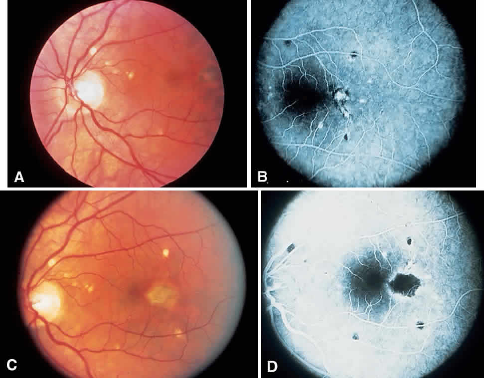
Histo spots (Fig. 1) represent a multifocal or a disseminated choroiditis characterized by atrophic or yellowish white scars in the posterior pole and the periphery. In 1972, Smith and colleagues18 showed the number of disseminated scars to range from 0 to 102. They occurred bilaterally in 62% of the patients, with the majority of the scars found posterior to the equator. The random distribution of the scars throughout the midperipheral areas and posterior pole of the fundus suggests a hematogenous dissemination of H. capsulatum to the eye. Histo spots seem to have a predilection for areas of greater blood supply. Skin testing in 59% of the general population in an endemic area was positive for the histoplasmin antigen; however, only 4.4% of this group was found to have histo scars on clinical examination.19

Fig. 1. Histo spots. Note the punched-out appearance. Some spots contain pigment and others are depigmented. Choroidal vessels often can be seen, indicating these scars are old and not actively inflamed.

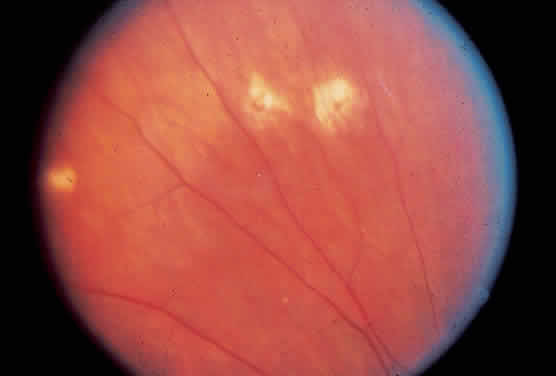
About 5% of patients with POHS demonstrate what have been called peripheral streak lesions of the fundus. These streaks are of variable length and pigmentation (Fig. 2).20 The streak lesions are most likely to be the result of a linear aggregation of histo spots. The histo spots are normally stable but may undergo changes in shape, size, and intensity. They have been shown to become larger over the course of time. The mechanism of change in size and shape of these lesions is unknown. Long-term follow-up has shown that 9% to 16% of patients will develop new histo spots.21–23 It has also been shown that 50% of eyes will have changes in atrophic scarring manifested by increased size or by formation of new scars in the peripapillary, macular, and peripheral regions. In 1969, Krill and associates24 described the evolution of these choroidal infiltrates as being hypofluorescent in the early stages of the disease. During the course of time, they assume a hyperfluorescent appearance, which is more characteristic of the mature lesions. Therefore, the late atrophic histo spot has the fluorescein angiographic picture of a pigment epithelial window defect.

Fig. 2. Linear streaks are sometimes seen in ocular histoplasmosis. These streaks may represent coalescent histo spots. There are some nearby typical histo spots.

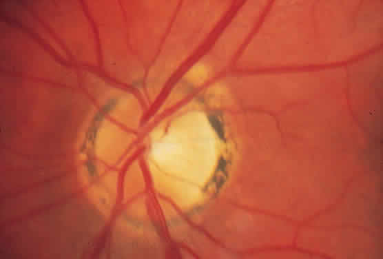
PERIPAPILLARY ATROPHY

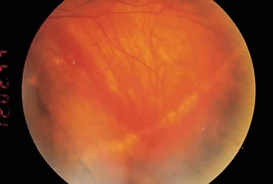
A ring of peripapillary atrophy (Fig. 3) occurs in the majority of cases of POHS. This ring is characteristic, with a narrow inner pigment zone adjacent to the disc edge and a white depigmented zone away from the disc. It is hypothesized that a circle of granulomas forms during the active infective stage of the disease and that these granulomas atrophy, leaving the ring of peripapillary atrophy. Neovascular membranes can occur in these rings of atrophy and sometimes involve the macula.25
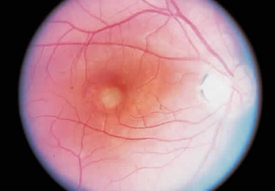
Fig. 3. Peripapillary atrophy. Note the characteristic ring of pigment adjacent to the disc, with the depigmented ring more distal.

MACULAR LESIONS

It is the macular lesions that bring patients to an ophthalmologist with symptoms of metamorphopsia. Subsequently, patients may develop profound loss of central visual acuity.

Histo spots located in the macular area tendto develop subretinal neovascular membranes (SRNVMs) (Fig. 4). These SRNVMs are similar to those seen with age-related macular degeneration, angioid streaks, and choroidal ruptures.26 Histologically, there is an egress of capillaries from the choroid to the subretinal space between Bruch's membrane and the neurosensory retina.

Fig. 4. Signet ring (gray-green lesion) in macula indicating a probable subretinal neovascular net. There is characteristic peripapillary atrophy of presumed ocular histoplasmosis syndrome.

The reason for the formation of SRNVMs in the macular area in POHS, as in age-related macular degeneration, is not understood. Some investigators have postulated that an immune reaction against H. capsulatum may play a part. Others believe that the SRNVMs are just a deterioration that accompanies any break in Bruch's membrane.26 Ophthalmoscopically, SRNVMs may first appear as dirty gray-green subretinal lesions. This formation has been likened to a signet ring. The initial lesions may pro-gress to actual subretinal hemorrhage. Deposition of lipid and serous elevation of the retina may also occur as a result of increased permeability in the SRNVMs. In time, this condition progresses to an atrophic or gliotic scar, which may assume various forms. Most of the scars are white because of fibrous tissue that develops secondary to the hemorrhage. Microcystic degeneration of the sensory retina may also occur as a result of chronic serous detachment of the neurosensory retina. The visual acuity in patients who have had SRNVMs is seldom better than 20/200, unless the location of the scars is eccentric to the fovea. In one retrospective study, only 14% of untreated eyes with subfoveal neovascular networks ended with a 20/40 acuity.27 If the scar is greater than several disc diameters, the vision may fall to the counting fingers level. Just as in age-related macular degeneration, choroidal neovascular membranes may progress to massive vitreous hemorrhage. This infrequent circumstance is due to an actual hemorrhage through a break in the neurosensory retina.28,29

OCULAR MEDIA

The ocular media in patients with POHS are absolutely clear of cells. No inflammatory cells can be found in either the aqueous or vitreous. Part of the reason for this lack of cells may be because the inflammatory stage has ended by the time patients are seen clinically. Another possibility is that inflammatory cells deep in the choroid have difficulty penetrating into the vitreous. This finding is in contrast to toxoplasmosis, a retinochoroiditis, in which the primary inflammation lies in the retina. Subsequently, in toxoplasmosis, cells spill over into the vitreous and aqueous. Patients who have vitreous or aqueous cells and present with peripheral punched-out lesions, peripapillary atrophy, and macular SRNVMs should be diagnosed as having multifocal choroiditis (also called pseudohistoplasmosis).30,31 The cause of the pseudohistoplasmosis syndrome has been described to include presumed sarcoidosis, possibly tuberculosis or syphilis. In about 30% of patients, no cause of multifocal choroiditis can be determined.

Back to Top
EXACERBATION AND RECURRENCES
A significant reduction in central visual acuity is caused by POHS in at least 2000 young and middle-aged adults every year in the United States. About 0.5% of cases of legal blindness in Tennessee are due to histoplasmosis.7 Most exacerbations occur at the site of previous histo scars. The chance of macular involvement primarily depends on whether histo spots are present in the macula at the time of the recurrence. Lewis and colleagues32 noted a 23% risk of developing asymptomatic macular lesions in a contralateral eye with asymptomatic macular scars. Their finding agrees with the 20% rate of onset in the second eye in 3 years reported by Elliot and Jackson33 and by Sawelson and associates.34 Lewis and colleagues found that 58% of untreated eyes with macular SRNVMs eventually deteriorate to a visual acuity of 20/200 or worse.

In a study by Watzke and Claussen,23 16.6% of eyes developed new infiltrates in the choroid and retina in areas that were clinically and angiographically normal 10 years previously. The basic lesion was found to be a nodular choroidal infiltrate that evolved clinically and angiographically from normal retina. Angiographically, the lesion was initially hypofluorescent with the later hyperfluorescence of a choroidal scar.

Back to Top
GEOGRAPHIC DISTRIBUTION AND EPIDEMIOLOGY
A positive response to the histoplasmin skin test has for many years been associated with POHS. A geographic relationship exists between clinically diagnosed ocular histoplasmosis and the sensitivity patterns of the histoplasmin skin test. This relationship was first demonstrated by Ellis and Schlaegel35 in 1973 (Fig. 5). Perhaps the highest prevalence of this association is found in the United States along the Ohio and Mississippi River valleys. In Missouri, Illinois, Indiana, Kentucky, Tennessee, and Mississippi, more than 80% of the population in certain localities become positive reactors to the histoplasmin test by adulthood. Areas in South America, Africa, and Southeast Asia are also known to have a high prevalence of systemic histoplasmosis.

Fig. 5. Worldwide pattern of histoplasmin sensitivity in human populations. (Edwards PQ, Billings EL: Worldwide pattern of skin sensitivity to histoplasmin. Am J Trop Med Hyg 20:288, 1971.)

In a community study of 842 people in Walkersville, Maryland, 59% of the general population had positive histoplasmin skin test results, and 100% of 22 patients with POHS had positive responses.14 Among all patients with positive skin test reactions, 4.4% had evidence of POHS. Other studies report a similar correlation between POHS and positive responses to the histoplasmin skin test.24,36 The complement fixation test does not correlate highly with POHS. This observation implies that by the time an ophthalmologist examines a patient, the initial acute infection has long since resolved. Only residual cellular immunity remains, which is manifested by a positive skin test response. Eleven percent of patients with this syndrome have negative results of skin tests.16 Waning skin test immunity could explain some of these negative reactions. The results of chest radiography do not correlate with ocular histoplasmosis.37 One interesting study found that only two of six patients with POHS showed in vitro lymphocyte stimulation to commercial histoplasmin38; however, all six patients had lymphocyte stimulation to a sonicate of H. capsulatum. Histoplasmin skin test material is a culture titrate of the mycelial phase of the fungus. The yeast phase antigens are missing from histoplasmin, and it is possible the yeast form of the organism is important in POHS.38 Gass and Wilkinson15 have postulated that Histoplasma organisms arrive at the choroid by hematogenous spread. Focal granulomas develop and rapidly destroy the organism. Small focal atrophic scars (histo spots) occur in the choroid and pigment epithelium as these granulomas resolve. Patients are asymptomatic at this time. When an ophthalmologist subsequently examines them, only the scars are seen. There is no cellular reaction in the aqueous or vitreous. Synechiae never form.

Scattered reports of POHS have come from Europe; however, histoplasmosis is not endemic there. European patients have negative reactions to skin tests.39,40 Thus, other possible causes of this syndrome besides histoplasmosis must be considered.

It is also interesting that only 0.7% of patients with POHS are black. The occurrence of subretinal neovascularization in blacks with POHS appears to be unusual.41 This observation correlates well with the low incidence of subretinal neovascularization seen with age-related macular degeneration in blacks.

Back to Top
HUMAN LEUKOCYTE ANTIGENSIN PRESUMED OCULAR HISTOPLASMOSIS SYNDROME
Braley and colleagues42 were among the first to observe an association between HLA-B7 and POHS. Meredith and colleagues43 demonstrated an increased frequency of HLA-B7 in patients with disciform scarring. They did not find this association in patients with peripheral atrophic spots. Further HLA analysis demonstrated that the HLA-DRw2 locus was found in 81% of patients with disciform scars and 62% of patients with peripheral atrophic scars.31,44 Only 28% of controls tested positive for the DRw2 antigen. POHS, thus, has two markers for increased frequency of both peripheral atrophic scarring and disciform scarring.
Back to Top
HISTOPATHOLOGY
Clinically, three characteristic lesions can be identified in POHS: the small, oval to round punched-out scars in the midperiphery or posterior pole; the macular SRNVMs; and peripapillary lesions.

Histologically, the peripheral punched-out lesions show various degrees of inflammatory activity. Focal aggregations of lymphocytes in the choroid without damage to Bruch's membrane may be seen. In contrast, discrete choroidal lesions with or without overlying retinal pigment epithelium (RPE) or Bruch's membrane damage can also be seen. The choriocapillaris, rods, and cones in this area may be destroyed. The variability of the histologic appearance of the peripheral lesions may account for the occasional clinical observation that these small lesions can change. No organisms are seen in these spots except for the rare cases mentioned earlier. The presence of these lymphocytes beneath clinically inactive lesions has been hypothesized torepresent sensitized cells that might respond to antigenic exposure and lead to subretinal neovascularization.

The macular SRNVMs of POHS occur in the areas of pre-existing asymptomatic macular histo spots, as previously noted. Histo spots may contain breaks or cracks in Bruch's membrane with damage to the RPE and choroidal capillaries. Inflammatory cells and even blood from a hemorrhage may enter the subretinal space, causing a disciform detachment of the retina. In time, proliferation of the RPE is noted. Along with fibrovascular tissue organization, this scar tissue may subsequently become hyalinized.45

The lesions surrounding the optic nerve in POHS are usually quiet and asymptomatic. They may take on either the histopathologic appearance of the peripheral atrophic spots or rarely may demonstrate SRNVM formation, which is similar to age-related idiopathic peripapillary SRNVM.

Back to Top
EXPERIMENTAL DATA
Animal models can produce a picture similar to POHS. Intracarotid injection of yeast phase organisms in rabbits resulted in multiple patches of choroiditis within 24 to 48 hours on the injected side and 7 to 20 days later in the contralateral eye. The lesions in the fellow eye resembled those seen in humans. The aqueous and vitreous were free of inflammatory cells. The lesions resolved in 1 to 2 weeks. Both histopathology and immunofluorescence failed to demonstrate H. capsulatum organisms in the healed granulomas at 6 to 9 weeks.46,47

A primate model of ocular histoplasmosis has been described and a picture similar to the human model can be produced.48,49 However, macular lesions have not been produced in the animal model. Immunogenic stimulation with killed organisms can cause an increase in lymphocytes at the base of old choroidal scars, but subretinal neovascularization is not regularly seen.

These experimental studies help confirm that H. capsulatum is one cause of POHS; that is, organisms are seldom or never identified in human eyes by histopathology. One would expect this because the organisms disappear from the experimental model after several weeks. Also, the lack of clinical inflammatory signs, such as cells in the vitreous, in experimental animals mirrors the lack of inflammatory signs in humans.

Back to Top
DIAGNOSIS
A diagnosis of POHS is made by clinical examination. Ocular findings include histo spots, peripapillary atrophy, subretinal hemorrhage disciform maculopathy, and clear uninflamed media.

In cases of hemorrhagic disciform maculopathy, fluorescein angiography should be performed to delineate the exact location of the SRNVMs. This is necessary before laser photocoagulation can be performed.

The histoplasmin skin test is the most valuable laboratory test in the diagnosis of ocular histoplasmosis. Although 80% of patients with POHS have positive reactions to skin tests, they are not routinely used in clinical practice because in endemic regions up to two thirds of the population may be positive responders. There is also the anecdotal danger of causing a macular hemorrhage via the skin test.

The complement fixation test is not useful in ocular histoplasmosis because results are positive only during the acute phase of the systemic disease.

Probably the most important measure in monitoring the clinical course of a patient with POHS is daily testing with an Amsler grid. Metamorphopsia noted on Amsler grid testing is the most sensitive indicator of SRNVM formation.

Back to Top
DIFFERENTIAL DIAGNOSIS
The differential diagnosis of POHS includes any inflammatory or noninflammatory process that may cause small areas of chorioretinal scarring to form (Table 1). Multifocal choroiditis and toxoplasmosis are frequently mistaken for ocular histoplasmosis. The primary differentiating feature is the presence of vitreous inflammatory cells and debris in both toxoplasmosis and multifocal choroiditis. The hallmark of POHS is a clear vitreous.


Back to Top
TREATMENT

LASER

Treatment with amphotericin B is of no benefit in POHS. There is no evidence of active fungal replication. The mainstay of therapy is laser photocoagulation of the SRNVMs. Research done by the Macular Photocoagulation Study Group in the 1980s50,51 showed conclusively that laser photocoagulation was beneficial in the treatment of SRNVMs associated with POHS.

The initial study50 dealt with choroidal neovascular membranes 200 to 2500 μm (one-fourth discdiameter) from the center of the foveal avascular zone (FAZ). The study showed that 34.2% of untreated eyes versus 9.4% of treated eyes had a decrease of six or more lines of visual acuity from baseline. All cases were documented by fluorescein angiography and were treated with argon (blue-green) laser photocoagulation to obliterate totally the corresponding area of subretinal neovascularization.

The subsequent study by the same group51 dealt with choroidal neovascularization 1 to 199 μm from the center of the FAZ or with choroidal neovascularization 200 μm or farther from the FAZ center, with blood or pigment extending within 200 μm of the FAZ center. Krypton laser photocoagulation was used to obliterate the SRNVMs in this study, which showed that 24.8% of untreated eyes, in contrast to 6.6% of treated eyes, had lost six or more lines of visual acuity. The goal of laser therapy was to cover completely the area of hyperfluorescence with a uniform whitening of the overlying retina (Fig. 6). It was not necessary to treat areas of blocked fluorescence secondary to blood or pigment. Treatment beyond the hyperfluorescent area also was not required when the SRNVMs were determined to be 100 μm or less from the center of the FAZ.

Fig. 6. A. Signet ring lesion temporal to macula. B. Fluorescein angiogram showing subretinal neovascular membrane with dark periphery indicating subretinal blood. This lesion meets the Macular Photocoagulation Study criteria for laser treatment. C. Same lesion after laser therapy. Note multiple histo spots.D. Fluorescein angiogram of lesion after laser therapy.

Further studies52,53 comparing krypton red and argon green laser photocoagulation for the treatment of SRNVM failed to show any clinical or statistical differences in outcome.

Subsequent studies54 determined that laser photocoagulation of extrafoveal or juxtafoveal SRNVMs nasal to the fovea were consistent with the beneficial results of treatment observed in the entire group of eyes studied by the Macular Photocoagulation Study Group.

Photocoagulation is not without risk. Some reports have described an 18.8% loss of two lines or more of central visual acuity after obliteration of subretinal neovascularization.55 Thirty percent of patients in the Macular Photocoagulation Study had recurrent extrafoveal subretinal membranes after laser treatment. Recurrences tended to be toward the fovea.56

Prophylactic laser therapy for atrophic spots near the macula to prevent neovascularization from forming in the second macula of patients in whom one macula has been damaged is not recommended. In fact, neovascularization has occurred in the laser scars from such a prophylactic attempt.57

When SRNVMs extend beneath the center of the fovea, conventional thermal (argon and krypton) laser treatment may limit scotoma size; however, central visual function may be reduced immediately as a result of secondary damage to the overlying neurosensory retina. The extent of visual loss due to treatment of SRNVMs in non-age-related macular degeneration cases may be greater than that resulting from the natural history of the disease itself.58 Because of the limitations of treating subfoveal SRNVMs, new therapies are being developed.

Photodynamic therapy is a treatment modality involving the intravenous injection of a photosensitzer followed by irradiation of the neovascular tissue by nonthermal light at the absorption peak of the dye. Verteporfin is a second-generation lipophilic/ampiphilic photosensitizer (absorption peak 689 nm) that was first demonstrated to achieve vascular occlusion in the rabbit choroid without damage to the neurosensory retina and Bruch's membrane.59 A preliminary study60 of photodynamic therapy using verteporfin for SRNVM associated with non-age-related macular degeneration cases including POHS showed short-term cessation of fluorescein leakage from SRNVM in a few patients without loss of vision. Improvement in vision after verteporfin therapy was greatest (six to nine lines) in three patients with relatively poor baseline visual acutiy (20/200 to 20/800). Further studies need to be done to determine whether photodynamic therapy is indeed beneficial for the long-term closure of SRNVMs in POHS.

SUBRETINAL SURGERY

Recent advances in subretinal surgical techniques and instrumentation have shown the potential for successful treatment of subfoveal SRNVMs and preservation of neurosensory retina function. Thomas and Kaplan61 first demonstrated subretinal surgical techniques used to remove SRNVM in POHS. A small retinotomy followed by the creation of a serous neurosensory detachment of the retina allows access to the fibrovascular membrane with minimal trauma to the overlying neurosensory retina. Dramatic improvement in visual acuity from 20/400 to 20/20 and 20/40 was seen in the initial cases, with no evidence of persistent or recurrent SRNVM with less than 1 year of follow-up. Subsequent studies62–64 with larger numbers of patients and longer follow-up yielded less dramatic results. Berger and colleagues62 demonstrated a 35% substantial improvement (defined as two lines or more) in Snellen acuity, whereas 79% improved or stabilized. A final visual acuity of 20/50 or better was achieved in 16% of eyes. Additionally, a recurrence of the SRNVM occurred in 38% of the eyes in the postoperative period and was associated with a poor visual prognosis. Previous laser photocoagulation was also associated with a worse visual outcome.

Subretinal surgery gives better results in patients with POHS than in patients with age-related macular degeneration. This may be because patients with POHS are younger. Gass65 has shown that SRNVMs in POHS may be above the RPE and mixed with RPE cells. Thus, removal of the membranes may allow the RPE to proliferate and recover. The SRNVMs in age-related macular degeneration are deeper, and surgical excision leaves no RPE to recover.

Further studies need to be done to look at the long-term efficacy of photodynamic therapy and to compare the results with submacular surgery for the treatment of subfoveal SRNVMs.

PERIPAPILLARY NEOVASCULARIZATION

Peripapillary neovascularization also occurs in ocular histoplasmosis. Many of these neovascular membranes regress without loss of vision or therapy.64 One study recommended therapy for peripapillary neovascularization only when a prolonged serous or hemorrhagic detachment of the fovea occurs. Documented progression of a network of subretinal vessels or loss of central vision in the other eye may be considered reasons for treatment. Residual and recurrent neovascularization are complications of treating peripapillary membranes.66

Further studies need to be done to compare the results of argon blue-green, argon green, krypton red, and dye laser wavelengths in treating choroidal neovascular lesions within the FAZ. Surgical excision of extensive peripapillary membranes has been performed with success.67

CORTICOSTEROIDS

Schlaegel has advocated the use of high-dose prednisone for acute exacerbations of macular disease.68 Therapy may be needed for 2 weeks to 2 years, depending on the severity of the disease. Although few hard data are available to substantiate the use of corticosteroids, evidence favoring their use is based on reports of improved vision in patients treated with steroids and worsening vision when the drugs were discontinued.

Back to Top
SPONTANEOUS RECOVERY
Finally, spontaneous recovery of visual acuity has been reported to occur in patients with severe loss of vision due to disciform macular scars. One retrospective study reported that 8 of 700 patients had improvement of two lines of vision after bilateral macular scarring.69 A hypothesized mechanism of this improvement is that the patients learned to use eccentric fixation.70,71
Back to Top
REFERENCES

1. Parker JD, Sarosi GA, Doto IL et al: Treatment of chronic pulmonary histoplasmosis. N Engl J Med 283:225, 1970

2. Smith JW, Utz JP: Progressive disseminated histoplasmosis. Ann Intern Med 76:557, 1972

3. Scholz R, Green R, Kutys R et al: Histoplasma capsulatum in the eye. Ophthalmology 91:1100, 1984

4. Mother A, Rodriques MM, Kaplan W et al: Disseminated bilateral chorioretinitis due to Histoplasrna capsulatum in a patient with the acquired immunodeficiency syndrome. Ophthalmology 92:1159, 1985

5. Sutliff WD: Histoplasmosis cooperative study. V. Amphotericin B dosage for chronic pulmonary histoplasmosis. Am Rev Respir Dis 105:60, 1972

6. Goldstein BG, Buettuer H: Histoplasmic endophthalmitis: A clinical correlation. Arch Ophthalmol 101:774, 1983

7. Craig EL, Suie T: Histoplasma capsulatum and ocular tissue. Arch Ophthalmol 91:285, 1974

8. Krause AC, Hopkins WG: Ocular manifestation of histoplasmosis. Am J Ophthalmol 34:564, 1951

9. Schlaegel TF Jr: Granulomatous uveitis: Etiologic survey of 100 cases. Trans Am Acad Ophthalmol Otolaryngol 62:813, 1958

10. Woods AD, Wahlen HE: The probable role of benign histoplasmosis in the etiology of granulomatous uveitis. Am J Ophthalmol 49:205, 1960

11. Roth A: Histoplasma capsulatum in the presumed ocular histoplasmosis syndrome. Am J Ophthalmol 84:293, 1977

12. Gass JD, Zimmerman LE: Histopathologic demonstration of Histoplasma capsulatum. Am J Ophthalmol 85:725, 1978

13. Khalil MK: Histopathology of presumed ocular histoplasmosis. Am J Ophthalmol 94:369, 1982

14. Smith RE, Canicy JP: Presumed ocular histoplasmosis. I. Histoplasmin skin test sensitivity in cases identified during a community survey. Arch Ophthalmol 87:245, 1972

15. Gass JD, Wilkinson CP: Follow-up study of presumed ocular histoplasmosis. Trans Am Acad Ophthalmol Otolaryngol 76:672, 1972

16. Schlaegel TF: Histoplasmic choroiditis. Ann Ophthalmol 6:237, 1974

17. Feman SS, Podgorski SF, Penn MK: Blindness from presumed ocular histuptasmosis in Tennessee. Ophthalmology 89:1295, 1982

18. Smith RE, Ganley JP, Knox DL: Presumed ocular histoplasmosis: Patterns of peripheral and peripapillary scarring in persons with nonmacular disease. Arch Ophthalmol 87:251, 1972

19. Smith RE, Ganley JP: An epidemiologic study of presumed ocular histoplasmosis. Trans Am Acad Ophthalmol Oto-laryngol 75:995, 1971

20. Fountain JA, Schlaegel TF Jr: Linear streaks of the equator in presumed ocular histoplasmosis syndrome. Arch Ophthalmol 99:246, 1981

21. Schlaegel TF: The natural history of histo spots in the disc and macular area. Int Ophthalmol Clin 15:19, 1975

22. Lewis ML, Van Newkirk MR, Gass JD: Follow-up study of presumed ocular histoplasmosis syndrome. Ophthalmology 87:390, 1980

23. Watzke RC, Claussen R: The long-term course of multifocal choroiditis (presumed ocular histoplasmosis). Am J Ophthalmol 91:750, 1981

24. Krill AE, Christi MI, Klien BA et al: Multifocal inner chorniditis. Trans Am Acad Ophthalmol Otolaryngol 73:222, 1969

25. Schlaegel TF, Kenney D: Changes around the optic nervehead in presumed ocular histoplasmosis. Am J Ophthalmol 62:454, 1966

26. Meredith TA, Green WR, Key SN et al: Ocular histoplasmosis: Clinicopathologic correlation of 3 cases. Surv Ophthalmol 22:189, 1977

27. Kleiner RC, Rather C, Enger C: Subfoveal neovascularization in the ocular histoplasmosis syndrome: A natural history study. Retina 8:225, 1988

28. Kranias G: Vitreous hemorrhage secondary to presumed ocular histoplasmosis syndrome. Ann Ophthalmol 17:295, 1985

29. Schlaegel TF, Weber JC, Helvestun E et al: Presumed histoplasmic choroiditis. Am J Ophthalmol 63:919, 1967

30. Deutsch T, Tessler H: Inflammatory pseudo histoplasmosis. Ann Ophthalmol 17:461, 1985

31. Dreyer RF, Gass JDM: Multifocal choroiditis and panuveitis: A syndrome that mimics ocular histoplasmosis. Arch Ophthalmol 102:1776, 1984

32. Lewis ML, Van Newkirk M, Gass JD: Follow-up study of presumed ocular histoplasmosis syndrome. Ophthalmology 87:390, 1980

33. Elliot JH, Jackson DJ: Presumed histoplasmic maculopathy: Clinical course and prognosis in nonphotocoagulated eyes. Int Ophthalmol Clin 15:29, 1975

34. Sawelson H, Goldberg RE, Annelyse WH et al: Presumed ocular histoplasmosis syndrome: The fellow eye. Arch Ophthalmol 94:221, 1976

35. Ellis FD, Schlaegel TF: The geographic localization of presumed ocular histoplasmic choroiditis. Am J Ophthalmol 75:953, 1973

36. VanMetr TE, Maumenee AE: Specific ocular uveal lesions in patients with histoplasmosis. Arch Ophthalmol 71:314, 1964

37. Ganley JP: Epidemiology of presumed ocular histoplasmosis. Arch Ophthalmol 102:1754, 1984

38. Check I J, Diddle KR, Jay WM et al: Lymphocyte stimulation by yeast phase Histoplasma capsulatum in presumed ocular histoplasmosis syndrome. Am J Ophthalmol 87:311, 1979

39. Braunstein RA, Rusen DA, Bird AC: Ocular histoplasmosis syndrome in the United Kingdom. Br J Ophthalmol 58:893, 1974

40. Segato T, Piermarocchi S, Midena E: Presumed ocular histoplasmosis in Europe: A case report. Ann Ophthalmol 15:354, 1983

41. Baskin MA, Jampol L, Huamonte F et al: Macular lesions in blacks with presumed ocular histoplasmosis syndrome. Am J Ophthalmol 89:77, 1980

42. Braley RE, Meredith TA, Aaberg TM et al: The prevalence of HLA-B7 in presumed ocular histoplasmosis. Am J Ophthalmol 85:859, 1978

43. Meredith TA, Smith RP, Braley RE et al: The prevalence of HLA-B7 and presumed ocular histoplasmosis in patients with peripheral atrophic scars. Am J Ophthalmol 86:325, 1978

44. Meredith TA, Smith RE, Dueqesnoy RJ: Association of HLA-DR2 antigen with presumed ocular histoplasmosis. Am J Ophthalmol 89:70, 1980

45. Ryan SJ: Histopathologic correlates of presumed ocular histoplasmosis. Int Ophthalmol Clin 15: 125, 1975

46. Smith RE, O'Connor GR, Halde CJ et al: Clinical course in rabbits after experimental induction of ocular histoplasmosis. Am J Ophthalmol 76:284, 1973

47. Smith RE, Scalarone M, O'Connor GR et al: Detection of Histoplasma capsulatum by fluorescent antibody techniques in experimental ocular histoplasmosis. Am J Ophthalmol 76:375, 1973

48. Smith RE, Dunn S, Jester JV: Natural history of experimental choroiditis in the primates. I. Clinical features. Invest Ophthalmol Vis Sci 25:801, 1984

49. Smith Re, Dunn S, Jester JV: Natural history of experimental histoplasmic choroiditis in the primate. II. Histopathologic features. Invest Ophthalmol Vis Sci 25:810, 1984

50. Macular Photocoagulation Study Group: Argon laser photocoagulation for ocular histoplasmosis. Arch Ophthalmol 10l:1347, 1983

51. Macular Photocoagulation Study Group: Krypton laser photocoagulation for neovascular lesions of ocular histoplasmosis. Arch Ophthalmol 105:1499, 1987

52. Macular Photocoagulation Study Group: Evaluation of argon green vs krypton red laser for photocoagulation of subfoveal choroidal neovascularization in the macular photocoagulation study. Arch Ophthalmol 112:1176, 1994

53. The Canadian Ophthalmology Study Group: Argon green vs krypton red laser photocoagulation for extrafoveal choroidal neovascularization. Arch Ophthalmol 112:1166, 1994

54. Macular Photocoagulation Study Group: Laser photocoagulation for neovascular lesions nasal to the fovea. Arch Ophthalmol 113:56, 1995

55. Han DP, Folk JC, Bratton AR: Visual loss after successful photocoagulation of choroidal neovascularization. Ophthalmology 95: 1380, 1988

56. Macular Photocoagulation Study Group: Recurrent choroidal neovascularization after argon laser photocoagulation for neovascular maculopathy. Arch Ophthalmol 104:503, 1986

57. Fine SL, Patz A, Orth DH et al: Subretinal neovascularizalion developing after prophylactic laser photocoagulation of atrophic macular scars. Am J Ophthalmol 82:352, 1976

58. Freund KB, Yannuzzi LA, Sorenson JA: Age-related macular degeneration and choroidal neovascularization. Am J Ophthalmol 115:786, 1993

59. Schmidt-Erfurth U, Hasan, Gragoudas E et al: Vascular targeting in photodynamic occlusion of subretinal vessels. Ophthalmology 101:1953, 1994

60. Sickenberg M, Schmidt-Erfurth U, et al: A preliminary study of photodynamic therapy using verteprorfin for choroidal neovascularization in pathologic myopia, ocular histoplasmosis syndrome, angioid streaks, and idiopathic causes. Arch Ophthalmol 118:327, 2000

61. Thomas M, Kaplan H: Surgical removal of subfoveal neovascularization in the presumed ocular histoplasmosis syndrome. Am J Ophthalmol 111:1, 1991

62. Berger A, Conway M, et al: Submacular surgery for subfoveal choroidal neovascular membranes in patients with presumed ocular histoplasmosis. Arch Ophthalmol 115:991, 1997

63. Holekamp N, Thomas M, Dickinson J, Valluri S: Surgical removal of choroidal neovascularization in presumed ocular histoplasmosis. Ophthalmology 104:22, 1997

64. Meredith TA, Aaberg TM: Hemorrhagic peripapillary lesions in presumed ocular histoplasmosis. Am J Ophthalmol 84:160, 1977

65. Gass JD: Biomicroscopic and histopathologic considerations regarding the feasibility of surgical excision of subfoveal neovascular membranes. Am J Ophthalmol 118:285, 1994

66. Cantrill HL, Burgess D: Peripapillary neovascular membranes in presumed ocular histoplasmosis. Am J Ophthalmol 89:192, 1980

67. Atebara NH, Thomas MA, Holekamp NM et al: Surgical removal of extensive peripapillary choroidal neovascularization with presumed ocular histoplasmosis syndrome. Ophthalmology 98:1598, 1997

68. Schlaegel TF: Treatment of the POHS. In Schlaegel TF (ed): Ocular Histoplasmosis. New York: Grune & Stratton, 1977:209–259

69. Jost BF, Olk RJ, Burgess DB: Factors related to spontaneous visual recovery in the ocular histoplasmosis syndrome. Retina 7:1, 1987

70. Singerman LJ, Wong BA, Smith S: Spontaneous visual improvement in the first affected eye of patients with bilateral disciform scars. Retina 5:135, 1985

71. Harris MJ, Robins D, Dieter JM: Eccentric visual acuity in patients with macular disease. Ophthalmology 92:1550, 1985

Back to Top